2021
[1]\fnmSho C. \surTakatori
1]\orgdivDepartment of Chemical Engineering, \orgnameUniversity of California, Santa Barbara, \orgaddress\citySanta Barbara, \stateCA, \postcode93106
Antibody binding reports spatial heterogeneities in cell membrane organization
Abstract
The spatial organization of cell membrane glycoproteins and glycolipids is critical for mediating the binding of ligands, receptors, and macromolecules on the plasma membrane. However, we currently do not have the methods to quantify the spatial heterogeneities of macromolecular crowding on live cell surfaces. In this work, we combine experiment and simulation to report crowding heterogeneities on reconstituted membranes and live cell membranes with nanometer spatial resolution. By quantifying the effective binding affinity of IgG monoclonal antibodies to engineered antigen sensors, we discovered sharp gradients in crowding within a few nanometers of the crowded membrane surface. Our measurements on human cancer cells support the hypothesis that raft-like membrane domains exclude bulky membrane proteins and glycoproteins. Our facile and high-throughput method to quantify spatial crowding heterogeneities on live cell membranes may facilitate monoclonal antibody design and provide a mechanistic understanding of plasma membrane biophysical organization.
keywords:
Plasma membrane, Glycocalyx, Ligand binding, Antibodies, Cell surface crowding, Proteomics, Molecular dynamicsIntroduction
Physical crowding of the cell surface glycocalyx has been shown recently to alter the biophysical properties of membranes in a manner that significantly impacts cell function. These alterations include membrane bending, stretching, and fission on reconstituted lipid bilayers Snead2017 ; Stachowiak2012 ; Busch2015 ; Chen2016 ; Zeno2016 , as well as tubulation and fission in the plasma membranes of cultured cells Shurer2019 ; Snead2019 . In addition to inducing membrane deformation, cell surface crowding also modulates the physical accessibility of surface receptors to large soluble ligands and macromolecules Honigfort2021 . Experiments on reconstituted membranes with grafted synthetic polymers or purified proteins further confirm a decrease in protein binding affinity with increasing grafting density Rex1998 ; Du1997 ; Leventis2010 ; Jung2008 . Most clinical monoclonal antibody drugs that rely on direct effector-cell activity are known to target antigen receptors that are buried deep inside the glycocalyx, often within 10 nm from the membrane surface Bakalar2018 ; Gul2015 , suggesting that their effectiveness may be highly dependent upon crowding near the receptor. However, there are currently no methods to characterize the piconewton-scale forces generated by the crowding of 10 nm cell surface proteins Kuo2021 .
In addition to surface-orthogonal variations, the mammalian plasma membrane composition is also laterally-heterogeneous, with nanometer-scale protein and lipid clusters forming and dissipating on sub-second timescales Lowe2020 ; Kuo2021 . In giant plasma membrane vesicles (GPMVs) isolated from cells, Levental et al. Levental2011 ; Levental2015 ; Levental2020 showed that raft-like membrane microdomains exclude most transmembrane proteins, suggesting that raft-like domains on cells are lipid-rich and protein-poor relative to the bulk regions of the membrane. Given the lateral and membrane-orthogonal heterogeneity of the glycocalyx, a complete picture of the crowding profile requires three-dimensional (3D) characterization.
While techniques like electron microscopy enable nanometer-scale characterization of the plasma membrane, the preparation process is destructive Noble2020 , leaving a need for appropriate molecular probes to study these complex, dynamic systems in-vivo Kuo2021 . Recently, Houser et al. Houser2020 quantified the surface pressures on reconstituted crowded membranes by measuring the separation distance between FRET fluorophores that stretch due to steric interactions within the brush. The stretching distance in polymer brushes depends weakly on surface density, as height scales with chain density according to in the brush regime DeGennes1987 ; Milner1991 and follows even weaker scaling in the mushroom regime Rubinstein2003 . Therefore, the technique may lose accuracy at the crowding densities observed in physiological surface densities on live cells.
In this work, we develop synthetic antigen sensors with precise spatial localization and measure the binding affinity of complimentary immunoglobulin G (IgG) monoclonal antibodies in these local crowding environments. We leverage a technique developed recently by Takatori and Son et al. Takatori2022 , in which a macromolecular probe is introduced to the extracellular side of a plasma membrane to quantify the local osmotic pressure posed by the crowded cell surface via a reduction in effective binding affinity. We advance this technique by enabling spatial localization of the binding site to measure the membrane-orthogonal crowding heterogeneity on both reconstituted membranes and red blood cell (RBC) surfaces. We then reconstruct these systems in-silico, combining proteomics with molecular dynamics (MD) simulations and experiments to map RBC glycocalyx crowding with nanometer-scale spatial precision. Using targeted antigen probes, we expand our spatial resolution laterally, in the plane of the membrane, measuring differences in crowding between plasma membrane domains on live tumor cells. Our findings support the hypothesis that raft-like domains of native membranes exclude bulky proteins, consistent with the findings of Levental et al. on GPMVs Levental2011 ; Levental2015 ; Levental2020 . Our simple IgG binding assay to probe spatial heterogeneities on native cell membranes suggests an important role of structural complexities on glycocalyx organization.
Results
Synthetic antigen sensors report crowding heterogeneities with nanometer height resolution
The glycocalyx is heterogeneous in both composition and density, which vary as a function of distance from the membrane surface (henceforth “height”). Height heterogeneities in crowding can arise from variations in protein sizes Bausch-Fluck2018 ; Klijn2015 ; Pollock2018 ; Stoeckius2017 and also from polymer brush dynamics of disordered glycoproteins like mucins in the glycocalyx Milner1988 ; Tom2021 ; Delaveris2020 ; Shurer2019 ; Honigfort2021 . To characterize the cell surface height heterogeneity, we developed a noninvasive synthetic antigen sensor that inserts into the lipid membrane using a cholesterol tag conjugated to a polyethylene glycol (PEG) linker and a fluorescein isothiocyanate (FITC) fluorophore (Fig. 1A). We developed a family of cholesterol-PEG-FITC sensors with varying PEG linker lengths to adjust the height of the FITC antigen presented above the membrane. After presenting the antigen sensors on the cell surface, we obtain the effective binding avidity of anti-FITC (\textalphaFITC) IgG antibody as a function of antigen height.
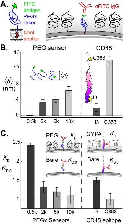
The PEG linker enables the FITC antigen to sample a distribution of heights above the membrane, while the mean height, , increases with the molecular weight of PEG. We used cell surface optical profilometry (CSOP) Son2020 to measure of the FITC antigen for sensor linker lengths of 0.5 kDa PEG (PEG0.5k), 2k, 5k, and 10k using silica beads coated with a 1,2-dioleoyl-sn-glycero-3-phosphocholine (DOPC) supported lipid bilayer (SLB). We recovered the predicted increase in with molecular weight (Fig. 1B), suggesting that the antigen is probing different crowding microenvironments as a function of linker length.
To validate that our different sensors are probing the height heterogeneities of a crowded membrane surface, we measured \textalphaFITC IgG binding to our antigen sensors on a reconstituted glycocalyx-mimetic PEG brush. Our reconstituted SLB on 4 µm silica beads included 3% 1,2-dioleoyl-sn-glycero-3-phosphoethanolamine-N-[methoxy(polyethylene glycol)-2000] (DOPE-PEG2k) to act as a repulsive brush, with synthetic antigen sensors of a single type inserted into the outer membrane leaflet (see Materials and Methods). Beads were incubated in varying concentrations of fluorescently-labeled \textalphaFITC antibodies and allowed to reach equilibrium before fluorescence intensities of beads were collected via fluorescence microscopy. Intensities were fit to a Hill binding isotherm to calculate the dissociation constant (see Supplementary Information). The ratio of on the PEG-crowded SLB to that on a bare SLB with no PEG crowders, , decreases toward unity as the average FITC height increases (Fig. 1C). The FITC antigen on our 10k antigen sensor samples the majority of its height distribution above the DOPE-PEG2k steric brush and has a \textalphaFITC binding avidity that is essentially unchanged from the bare membrane value. In contrast, the 0.5k antigen sensor is buried deep inside the PEG brush and the accessibility of the FITC antigen is hindered by a factor of six (Fig. 1C). Our results are consistent with classical polymer brush theory, which predicts a monotonic decrease in brush monomer density with height Milner1988 and a reduction in the effective adsorption energy of a globular protein onto a brush-coated surface Halperin1999 .
Based on our results for synthetic sensors on a PEG brush surface, we hypothesized that the height-dependent avidity of IgG would also apply to protein antigens buried within a crowded surface of other membrane proteins. To investigate, we reconstituted an SLB containing 5% 1,2-dioleoyl-sn-glycero-3-[(N-(5-amino-1- carboxypentyl)iminodiacetic acid)succinyl] (DGS-NTA) and created a crowded surface of poly-histidine tagged glycoprotein, Glycophorin A (GYPA). Instead of synthetic antigen sensors, we tethered a dilute surface density of tyrosine phosphatase CD45 on the SLB among the crowded excess of GYPA. As a readout of GYPA crowding, we used \textalphaCD45 antibodies that target two different epitope sites: pan-CD45 I3 epitope on the first FN3 domain ( nm), and RB isoform epitope C363 on the upper mucin-like domain ( nm). Using scaling arguments and the known glycosylation sequence of GYPA Aoki2017 ; Paturej2016 we estimate an average GYPA height of nm, so we expected the C363 epitope to explore uncrowded regions above the GYPA brush, while the I3 epitope to remain buried within the brush. Indeed, the relative avidities of \textalphaC363 and \textalphaI3 agree with this hypothesis, as is for \textalphaC363 while it is for \textalphaI3 (Fig. 1C). The consistent correlation between increasing antigen height, , and decreasing dissociation constant, , on both PEG and protein brushes confirms that antibody avidity is a robust metric of local crowding.
Macromolecular binding is a direct reporter of steric energies on crowded surfaces
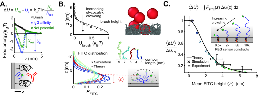
In this section, we aim to obtain a direct relation between the antibody binding avidity and the local steric free energy penalty of a crowded surface. We combine polymer brush theories with coarse-grained MD simulations to obtain a mechanistic understanding of our synthetic antigen sensors and their applicability on crowded membrane surfaces.
To characterize the energy profile on the membrane, we separately simulated free antibody insertion into a surface-tethered PEG2k brush and antibody binding to surface-tethered sensors to obtain the repulsive penalty associated with crowding, , (Fig. 2A), and the attractive binding potential, , respectively (see Materials and Methods). We invoke the theory of Halperin Halperin1999 and hypothesize that the effective antibody binding potential on a crowded interface, , is a superposition of and . The bare membrane binding avidity reports the attractive enthalpic term , so that the repulsive entropic energy penalty posed by the brush is given by
| (1) |
The repulsive energy barrier of the brush, , is proportional to the osmotic pressure, , which scales with monomer volume fraction as Halperin1999 ; Rubinstein2003 . The Milner, Witten, and Cates Milner1988 self-consistent field description of a polymer brush predicts a parabolic monomer distribution, so the potential follows a stretched parabolic profile (Fig. 2B, see Supporting Information for analytical form). Kenworthy et al. showed experimentally that the pressure between apposite membrane-tethered PEG brushes under compression varies with distance according to a profile derived from Milner theory Kenworthy1995 . We therefore invoke this theory to describe the form of our PEG2k brush potential, which we verify using MD simulations (see Materials and Methods).
The flexibility of the PEG linker in our synthetic antigen sensors causes the antibody to bind across a distribution of FITC heights for any given sensor. Thus, we define our experimentally-measured crowding potential for a given sensor as a mean potential , which can be predicted by weighting the FITC 1-D probability density by the potential profile in and integrating across all space:
| (2) |
To describe , we invoke the continuous Gaussian chain model of a surface-tethered polymer of mean height in an ideal solvent, calculating the chain-end distribution (see Supporting Information for calculations) Russel1989 . We verified with coarse-grained MD simulations of dilute surface-tethered PEG polymers, finding that the end-monomer distribution closely agrees with theory (Fig. 2B). Numerically evaluating the integral in Eq. 2 for a set of PEG-FITC sensors with mean heights yields matching theoretical and computational predictions for the observed crowding profile as a function of mean sensor height (Fig. 2C).
Recasting the data from Fig. 1C in the form given by Eq. 1 and plotting as a function of the mean sensor heights reported in Fig. 1B, shows quantitative agreement with the theoretical and MD profiles developed in Eq. 2 (Fig. 2C). Our experimental and simulation data support a mechanism by which the brush sterically excludes the antibody, suggesting that our synthetic antigen sensors act as direct reporters of crowding heterogeneities with nanometer resolution.
Synthetic sensors validate crowding predictions based on red blood cell proteomics
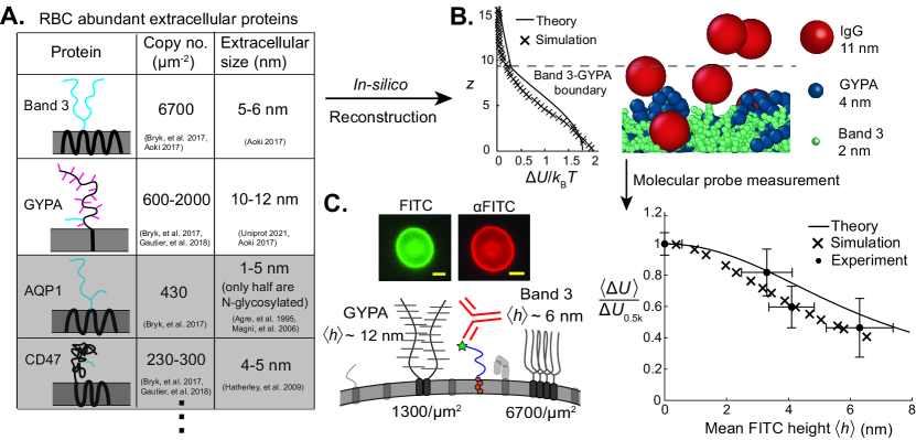
After validating our experimental antigen sensors on reconstituted membranes with analytical theory and coarse-grained simulations, we sought to use theorertical and computational methods synergystically with experiments to map the extracellular crowding landscape of the human red blood cell (RBC). Since the RBC surface proteome is fully-characterized Bryk2017 ; Aoki2017 , we identified the most abundant extracellular proteins, and estimated extracellular domain sizes (Fig. 3A). In particular, we identified two abundant proteins with bulky extracellular domains Aoki2017 ; Bryk2017 ; Gautier2018 : anion transporter Band 3 and mucin-like sialoglycoprotein GYPA.
Using both analytical theory and coarse-grained MD simulations, we modeled the RBC glycocalyx as a bidisperse polymer brush whose extracellular crowding profile opposes the adsorption of colloids like IgG (Fig. 3B). We used the lengths of extracellular peptides and glycans to estimate both the statistical monomer size and chain height of GYPA and Band 3 (see Supporting Information). We input predicted chain height and known chain grafting densities into a model that superimposed two parabolic polymer brush density profiles Milner1988 , and applied the scaling to model the repulsive potential Rubinstein2003 ; Halperin1999 . We also developed an in-silico model of a bidisperse brush, with each protein modeled as a bead-spring polymer (see Supporting Information for coarse-graining details). Fig. 3B shows close agreement between the analytical and MD descriptions of the glycocalyx, with the MD potential decaying faster because it relaxes the assumption of a strongly-stretched brush.
To verify our predicted -direction crowding profile, we incubated human RBCs in our synthetic antigen sensors and observed strong membrane incorporation (Fig. 3C). We measured the dissociation constant of anti-FITC binding to PEG0.5k, 2k, 5k, and 10k sensors, normalizing by the uncrowded on beads to find (Eq. 1). Antibody binding increased 5x from the most surface-proximal (PEG0.5k) to the most membrane-distal probe (PEG10k), corresponding to the crowding free energy penalty doubling from to (Fig. 3C). The experimental crowding landscape closely tracks the theoretical and simulated potentials, weighted by the FITC distributions in Fig. 2B.
These data demonstrate that for the relatively simple RBC plasma membrane, detailed proteomics data including copy number, structure, and glycosylation of surface proteins provide a robust approximation of membrane-orthogonal crowding heterogeneity. Computational techniques like machine learning are rapidly accelerating the identification of surface proteins and glycosylation sites Bausch-Fluck2015 ; Bausch-Fluck2015 ; Zhou2020 , and with more detailed characterization of glycan sequences and surface protein densities on the horizon, we expect that the in-silico reconstruction of more complex mammalian cells will become feasible. Mapping crowding heterogeneities on these nanometer length scales with simulations and molecular probes may reveal the accessibility of receptors based on height, improving our understanding of signaling and optimizing drug delivery target selection.
Development of phase-partitioning antigen sensors to measure lateral heterogeneities in surface crowding
The existence of raft-like microdomains of lipids, cholesterol, and proteins on plasma membranes has been hypothesized to govern various physiological processes, like signal transduction, endocytosis, and membrane reorganization Simons2011 ; Carquin2016 ; Brown1998 ; K.1997 ; Lingwood2010 ; Brown2000 ; Silvius2005 ; Klymchenko2014a ; Lorent2015 . Levental et al. Levental2011 ; Levental2015 ; Levental2020 showed that ordered raft-like domains on giant plasma membrane vesicles (GPMVs) isolated from cells are depleted of transmembrane proteins, suggesting that crowding may vary between these domains and the bulk. However, while plasma membrane vesicles form macroscopic equilibrium domains, lipids and proteins on live cells form transient 10-200 nm domains, which fluctuate on sub-second time scales Sezgin2012 ; Pike2003 ; Lingwood2010 ; Jacobson2019 . As a result, the optical characterization of raft-like domains on live cells is challenging Lillemeier2006 ; Skotland2019 ; Raghupathy2015 . To probe these lateral crowding heterogeneities, we used different antigen sensors that preferentially localize into ordered or disordered membrane domains to measure spatial variations in IgG binding on both reconstituted and live plasma membranes.
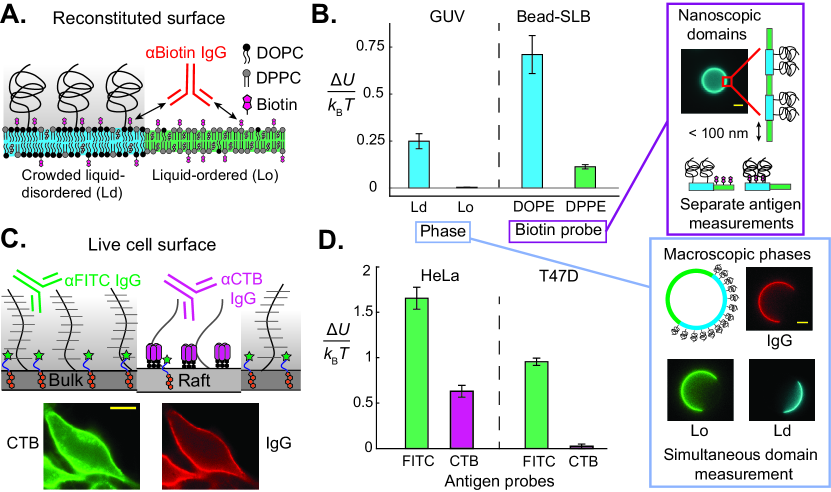
In this section, we present crowding measurements on phase-separated giant unilamellar vesicles (GUVs) with crowding heterogeneities, where macroscopic phase domains are easily visualized. We produced GUVs containing the ternary lipid mixture 2:2:1 1,2-dipalmitoyl-sn-glycero-3-phosphocholine (DPPC):DOPC:cholesterol that phase separates into macroscopic liquid-ordered (Lo) and liquid-disordered (Ld) domains Veatch2003 . We preferentially crowded the Ld phase with 2% DOPE-PEG2k (Fig. 4A) and added DOPE-biotin and 1,2-dipalmitoyl-sn-glycero-3-phosphoethanolamine-N-(biotinyl) (DPPE-biotin) to present the biotin antigens in each phase. The biotin was located at the membrane surface, with zero height. We measured the crowding free energy penalty for \textalphaBiotin IgG binding to each domain (Fig. 4A). Consistent with the experiments in Figs. 1-2, the PEG brush inhibited antibody binding on the crowded Ld domain and increased the normalized effective by 60% compared to the bare surface (Fig. 4B). In contrast, \textalphabiotin binding in the less crowded Lo domain did not change.
Although macroscopic phase domains on GUVs enable a simple measurement of lateral crowding heterogeneity, this approach is not possible on live cell surfaces which do not contain macroscopic phase domains. To overcome this challenge, we performed crowding measurements on SLB-coated beads with the same ternary lipid mixture, where the underlying substrate friction arrests phase domains into nm nanoscopic features, similar to the size of raft-like domains Honigmann2012 ; Lingwood2010 . Since the individual phase domains cannot be identified, we measured the crowding on each phase by quantifying the \textalphabiotin IgG binding on the beads containing only one type of antigen, either DPPE-biotin or DOPE-biotin.
As shown in Fig. 4B, we found that the Ld antigen (DOPE-biotin) reported a crowding penalty 7x higher than that reported by the Lo antigen (DPPE-biotin). While the absolute magnitudes of observed were higher on beads than GUVs, we attribute this difference to the lower membrane friction on GUVs enabling IgG to more easily exclude PEG2k when binding. Given the strong qualitative difference in crowding reported by antibody binding to antigens partitioning into Lo or Ld domains, we conclude that probes that preferentially associate with one phase over another are suitable reporters of lateral crowding heterogeneities in diffraction-limited domains like lipid rafts on live cells.
Antibody binding to raft-associated antigens reports lateral crowding heterogeneities on live cell surfaces
Motivated by our ability to measure crowding heterogeneities on nanoscopic phase domains on reconstituted membranes, we used laterally-segregating antigen probes to measure in-plane crowding heterogeneities on live mammalian cells. The ganglioside GM1 is known to form clusters on live cell membranes Shi2007 ; Skotland2019 and to associate with raft-like domains, particularly when bound to the pentavalent cholera toxin B (CTB) Silvius2005 ; Klymchenko2014a ; Lorent2015 . We capitalized on the raft-association of CTB by applying it to cells in culture and measuring the of \textalphaCTB IgG, which is a direct reporter of crowding above raft-like domains. We compared the of \textalphaCTB to that of \textalphaFITC binding to cholesterol-PEG0.5k-FITC sensors and normalized by bare values on beads, to differentiate raft-specific crowding from that of the bulk cell membrane (Fig. 4C). GM1 protrudes only 1-2 nm above the bilayer surface Shi2007 and CTB is only about 2-3 nm in size Ohtomo1976 , so we assume that crowding at the CTB epitope is similar to that at the bilayer surface. We observed nearly uniform partitioning of cholesterol-PEG0.5k-FITC sensors between Lo and Ld phases on GUVs (see Supporting Information), consistent with prior work showing that functionalized cholesterol tends to favor the disordered bulk due to its reduced packing efficiency Baumgart2007 ; Klymchenko2014a , despite native cholesterol favoring the ordered and raft-like domains Veatch2003 ; Brown2000 ; Ostermeyer1999 ; Baumgart2007 .
We compared raft versus bulk crowding on HeLa cells and T47D breast cancer cells, both of which have surface proteomes rich in bulky proteins Nagaraj2011 ; Shurer2019 . HeLa and T47D, like many cancer cells, both express MUC1, a tall (200-500 nm), heavily glycosylated (50-90% carbohydrate by mass) mucin that can protect against destruction by the immune system Hattrup2008 ; Brayman2004 ; Shurer2019 ; Zhang1997 . MUC1 is transcribed at copies/HeLa cell on average, about twice the average for T47D, although the distribution of MUC1 expression in T47D is broader by several orders of magnitude Nagaraj2011 ; Shurer2019 . Paszek et al. demonstrated a direct correlation between MUC1 expression and membrane tubule formation on both T47D and HeLa cells, suggesting MUC1 is a major contributor to surface crowding and membrane tension Shurer2019 . The crowded surfaceomes of HeLa and T47D cells make these cells rich models for studying lateral heterogeneities. Bulk crowding for both cell lines as measured by our cholesterol-PEG0.5k-FITC sensors was on the order of 1-1.5 , consistent with the brush exclusion energy on the surface of RBCs (Fig 4D). The greater bulk crowding on HeLa cells over T47D is consistent with the greater expression of MUC1 on HeLa, although it is possible that a high density of short proteins and glycolipids is the source of the crowding differences as measured by our probes.
Surprisingly, CTB antigens reported significantly less crowding than the FITC antigen on both cells, suggesting that the extracellular space above the raft-like domains is not heavily crowded with proteins and sugars. In particular, T47D exhibited a complete elimination of its crowding free energy penalty in raft-like domains compared to the bulk membrane surface (Fig. 4D). These data are consistent with the results of Levental et al. on GPMVs Levental2011 ; Levental2015 ; Levental2020 , and support the hypothesis that raft-like domains of native membranes exclude proteins that contribute to extracellular crowding. This is a significant result because GPMVs exclude membrane proteins that are bound to the actin cytoskeleton, and it was unclear whether actin, myosin, and other structural features affect the surface crowding on live cell membranes. While Mayor et al. identified the coupling between cortical actin and GPI-anchored protein clusters, our results suggest that cytoskeletal involvement does not dramatically change the concentration of bulky, disordered proteins in rafts relative to the bulk Raghupathy2015 ; Skotland2019 .
Viral particles like simian virus (SV) 40 and other polyomaviruses Qian2009 ; Engel2011 , and toxins like Shiga and cholera toxin Sandvig2010 ; Fujinaga2003 , bind to gangliosides that enrich in raft-like domains. SV40 virus is in size VanRosmalen2020 , about three times larger than an IgG. Since the mechanical work required to insert a particle into a crowded space scales approximately as the particle volume (see Supporting Information), a viral particle would be posed with an energy barrier of if it tried to penetrate the glycocalyx above the bulk membrane of T47D cells. In contrast, in the raft-like regions where the gangliosides enrich, the binding penalty is merely on T47D cells, suggesting that viral particles may experience a thirty-fold larger effective affinity towards the less-crowded, ganglioside-rich domains.
Between the two cell lines, the ratio of bulk to raft crowding free energy is also 18x greater in T47D than in HeLa, suggesting that rafts play a cell-specific role in organizing surface proteins. Our unique ability to probe native cell membranes will advance further mechanistic insight into the roles of the actin cytoskeleton and other structural complexities on glycocalyx organization.
Discussion
In this work, we developed a simple experimental technique to study the spatial heterogeneities of surface crowding on live cell membranes with exquisite spatial resolution. Alternative approaches like detergent resistant membranes (DRMs) and GPMVs Lorent2015 are invasive techniques that do not provide a description of surface organization in the native cell membrane environment. Prior to this work, existing techniques were capable of measuring spatial organization on cell surfaces with a very thick glycocalyx (0.2-1 µm), such as endothelial cells in the vasculature Weinbaum2007 ; Tarbell2016 ; Weinbaum2021 ; Michel1997 . However, studying the spatial organization of live cell surfaces with glycocalyx thicknesses of nm was a challenge because standard optical microscopy cannot resolve nanometer variations.
While not reporting spatial heterogeneity, recent measurements with membrane-binding macromolecular probes reported osmotic pressures of 1-4 kPa at the surface of mammalian cells Takatori2022 . These surface pressures are comparable to and in some cases larger than the stiffness of the cell cortex ( 1 kPa), providing new insight on the physical role of protein and glycan crowding on cell membranes. In this work, we demonstrated that these pressures are highly dependent on proximity to the membrane surface and that glycocalyx crowding decays rapidly away from the membrane. Our probes are physiologically relevant because many protein-protein interactions occur at a finite distance away from the membrane, like kinetic segregation in T-cell receptor triggering ( 10-15 nm) Davis2006 ; Shaw1997 .
We present our antigen sensors on live cell surfaces with nanometer precision and use antibody binding equilibria to directly report spatial variations in surface crowding. Our sensors achieve this nanometer spatial sensitivity by leveraging the exponential amplification of our readout () as a function of the crowding energy, (Eq. 1). This exponential amplification distinguishes our approach from previous techniques that rely on polymer brush height as the readout of crowding, which scales weakly with surface density, Houser2020 ; Halperin1999 ; Rubinstein2003 . Taking the energy barrier to be proportional to the osmotic pressure, and in turn surface density, Halperin1999 ; Rubinstein2003 , we obtain the scaling , which is significantly weaker than . The change in crowding we observe within 6 nm of the RBC surface confirms this spatial sensitivity, and demonstrates the power of our technique in characterizing the highly heterogeneous membrane-proximal surfaceome, in which surface signaling and viral entry occur Delaveris2020 ; Son2020 .
Monoclonal antibody (mAb) drug candidates are currently screened using surface-plasmon resonance (SPR), in which binding affinity and avidity are measured on a bare hydrogel chip without regard to multi-body interactions Lakayan2018 . With mAbs like the breast cancer treatment trastuzumab targeting a 4 nm tall epitope on human epidermal growth factor receptor 2 (HER2), probing local crowding variations may inform target selection and improve potency Son2020 . Indeed, Chung et al. found that trastuzumab and pertuzumab attenuate tubule structures enriched in HER2, suggesting that biophysical interactions like crowding may influence the potency of mAb therapies Chung2016 . Our crowding measurements may also help inform the biophysical mechanisms governing antibody-dependent phagocytosis, which have been recently shown to have a strong dependence on the relative heights of the macrophage Fc receptor and the target cell surface proteins Bakalar2018 ; Suter2021 . In conclusion, our sensors may be used to inform important physiological processes, like antibody binding to buried surface receptors, membrane organization of lipid raft-like domains, and cellular phagocytosis.
When characterizing the RBC surface, we also demonstrated the potential to augment experimental crowding measurements with an in-silico cell surface reconstruction based upon proteomics data. As recent advances in surface proteomics continue to better characterize glycocalyx components for a broad host of cell lines Bausch-Fluck2015 ; Bausch-Fluck2015 ; Zhou2020 , we expect that accurate in-silico models will become possible on more complex mammalian cell surfaceomes. A technology to describe the extracellular crowding landscape for any cell a priori, using only proteomics data, may advance advance our basic understanding of cell membrane biology.
Using laterally-segregating antigen probes that target different plasma membrane domains, we demonstrated reduced crowding on GM1-associated raft-like domains on T47D and HeLa cells. These findings are consistent with the known reduction in transmembrane protein density on ordered domains in GPMVs Levental2011 ; Levental2015 ; Levental2020 , but our non-invasive measurements on live cells provide further insight into the dynamic cell surface ecosystem, including the interplay between the actin cytoskeleton and the membrane. Indeed, there has been considerable interest in the connection between the cytoskeleton and transmembrane protein organization over the past few decades Kusumi2012 ; Kusumi1996 ; Ritchie2003 , as actin is known to redistribute lipid domains on the cell surface Honigmann2014 ; Raghupathy2015 ; Skotland2019 ; Gomez-Mouton2001 . By bridging this gap and characterizing raft-like domains on live cells, we speculate that on length scales of order 10 nm, the cytoskeleton may not dramatically change bulky protein composition beyond that of equilibrium domains, consistent with the hypothesis that GPI-anchored proteins dominate the raft proteome Levental2011 . However, the discrepancy between relative raft-to-bulk crowding on HeLa and T47D cells indicates that the role of GM1-enriched raft-like domains in organizing the glycocalyx varies considerably from cell to cell. Future direct comparisons between lateral heterogeneity on both live cells and their secreted membrane vesicles will provide a more thorough description of the fraction of extracellular bulk that remains anchored to the cytoskeleton in both disordered and raft-like membrane domains.
Methods
Antigen probe synthesis
Cholesterol-PEGx-NH2, where x represents PEG0.5k, 2k, 5k, or 10k, was reacted with a 10x excess of N-hydroxy-succinimidyl ester (NHS)-FITC, overnight at 50°C in dimethylsulfoxide (DMSO). Unreacted FITC was removed via a 7K MWCO Zeba spin desalting column. SLB-coated beads were incubated with 100 nM FITC antigen sensors for 15 minutes at room temperature.
Microscope for all imaging experiments
All imaging was carried out on an inverted Nikon Ti2-Eclipse microscope (Nikon Instruments) using an oil-immersion objective (Apo 60x, numerical aperture (NA) 1.4, oil; Apo 100x, NA 1.45, oil). Lumencor SpectraX Multi-Line LED Light Source was used for excitation (Lumencor, Inc). Fluorescent light was spectrally filtered with emission filters (432/36, 515/30, 595/31, and 680/42; Semrock, IDEX Health and Science) and imaged on a Photometrics Prime 95 CMOS Camera (Teledyne Photometrics).
Sensor height measurement
Small unilamellar vesicles (SUVs) were formed using an established sonication method Bakalar2018 . A lipid film containing 1,2-dioleoyl-sn-glycero-3-phos-phocholine (DOPC), 3% 1,2-dioleoyl-sn-glycero-3-phosphoethanolamine-N-[methoxy(polyethylene glycol)-2000] (DOPE-PEG2k), and DOPE-rhodamine was dried under nitrogen and then vacuum for 30 minutes. The film was rehydrated in Milli-Q (MQ) water to 0.2 mg/mL lipids, sonicated at low power using a tip sonicator (Branson SFX250 Sonifier) at 20% of maximum, 1s/2s on/off, for three minutes. We added MOPS buffer at a final concentration of 50 mM MOPS pH 7.4, 100 mM NaCl to the resulting SUV mixture. Then, 10 µL of 4 µm silica bead slurry (10% solids) was cleaned with piranha solution (3:2 H2SO4:H2O2) and washed three times with 1 mL MQ water before being suspended in 100 µL MQ water (1% solids). 3 µL of bead slurry was mixed with 30µL SUVs and incubated for ten minutes at room temperature before washing five times with HEPES buffer (50 mM HEPES pH 7.4, 100 mM NaCl).
FITC sensor heights were established using cell surface optical profilometry (CSOP) Son2020 . SLB-coated beads were incubated in 200 nM cholesterol-PEGx-FITC at room temperature for 15 minutes, where x represents PEG0.5k, 2k, 5k, and 10k. Unbound sensors were washed from the bulk and CSOP measurement used to find the difference in apparent bead radius on the 488 nm FITC channel and 555 nm rhodamine channel . To correct for chromatic aberration, a baseline difference in 488nm and 555 nm radii was measured on SLB-coated beads containing DOPC with 0.05% DOPE-rhodamine, and 0.05% DOPE-Atto 488 lipids. The FITC antigen height was obtained by subtracting this baseline from the observed height: .
Dissociation constant measurement for reconstituted PEG brushes
4 µm SLB-coated beads with PEG brushes were formed using a mixture of DOPC, 3% DSPE-PEG2k, and 0.05% DOPE-rhodamine. Bare beads for measuring were formed with only DOPC and 0.05% DOPE-rhodamine. Beads were incubated in 100nM cholesterol-PEGx-FITC antigen sensors for 15 minutes at room temperature, then washed with HEPES buffer.
Lysine residues of anti-FITC (\textalphaFITC) IgG antibodies were randomly labeled by reacting with 8x excess NHS-Alexa Fluor 647 for one hour at room temperature in 50 mM sodium bicarbonate solution. Unreacted dye was separated via a 7MWCO spin desalting column and the recovery and labeling ratio measured via Nanodrop UV-vis spectroscopy.
Coverslips were passivated with 1 mM bovine serum albumin (BSA) to prevent nonspecific antibody adsorption. Antigen-coated beads were added to coverslip wells containing \textalphaFITC-647, and allowed to sediment and equilibrate with IgG for 30 minutes at room temperature. At least 50 beads were imaged for each bulk IgG concentration, with an approximately equatorial focal plane. Images were subdivided into individual beads, and the edges identified by the brightest 5% of pixels, on the 555 nm (DOPE-rhodamine) channel, for each sub-image. The background intensity was taken to be the 30th percentile of \textalphaFITC intensities for each bead subimage, and the bead intensity signal was calculated by subtracting background from the \textalphaFITC signal associated with the brightest rhodamine pixels. The intensity signal for each bead was averaged to yield a mean bead signal, and the mean bead signals were then averaged for each \textalphaFITC bulk concentration, and fit to a Hill isotherm to find .
Dissociation constant measurement for CD45 antigens
A GYPA brush with dilute CD45 antigens was reconstituted by incubating beads in 10:1 GYPA:CD45. SLB-coated beads containing DOPC, 8% 1,2-dioleoyl-sn-glycero-3-[(N-(5-amino-1-carboxypentyl) iminodiacetic acid)succinyl] (DGS-Ni-NTA), and 0.2% DOPE-Atto 390 were incubated with 10 nM His-tagged mouse CD45 and 100 nM His-tagged glycophorin A (GYPA) for 15 minutes at 37°C. Unbound protein was washed five times from the bulk with HEPES buffer. GYPA was labeled with NHS-Alexa Fluor 555 and CD45 was labeled with NHS-Alexa Fluor 488, and we thus confirmed a qualitative excess of the GYPA blockers on the beads. Beads were incubated in either Alexa Fluor 647-labeled \textalphaC363 or \textalphaI3 on CD45 for 30 minutes, and the measured. Baseline for was measured for both CD45 epitopes on beads with no GYPA.
Red blood cell (RBC) dissociation constant measurement
Single-donor human whole blood in K2-EDTA was purchased from Innovative Research and used within three days of arrival. The researchers in this study had no contact with human subjects and vendor samples were de-identified, precluding a need for IRB clearance. Blood was centrifuged at 300g for 5 minutes to isolate RBCs. RBCs were incubated with 100 nM cholesterol-PEGx-FITC (x represents PEG0.5k, 2k, 5k, and 10k) antigen sensors for 15 minutes at 37°C. RBCs were added to \textalphaFITC-647, pipette mixed, and incubated for 30 minutes before imaging. RBC images were analyzed using the same methods as beads, with at least 50 cells per IgG concentration, to calculate . For calculations, was normalized against the bare-bead , for each antigen.
Lateral crowding heterogeneity measurements on macroscopic membrane domains
Antibody dissociation constants were measured on crowded and uncrowded coexisting domains in liquid-liquid phase-separated giant unilamellar vesicles (GUVs). DOPC, 1,2-dipalmitoyl-sn-glycero-3-phosphocholine (DPPC), and cholesterol were combined in a 2:2:1 ratio, which phase separates at room temperature Veatch2003 . 0.3% DOPE-biotin and 0.05% 1,2-dipalmitoyl-sn-glycero-3-phosphoethanolamine-N-(biotinyl) (DPPE-biotin) were added as liquid-disordered and liquid-ordered antigen probes, respectively. We set the relative amounts of DOPE- and DPPE-biotin such that the antigen density in each phase was approximately equivalent, as reported by \textalphaBiotin binding. GUVs were formed either with or without 2% DOPE-PEG2k, which formed a crowding brush in only the liquid-disordered (Ld) phase. 0.05% each of DOPE-rhodamine and 1,2-distearoyl-sn-glycero-3-phosphoethanolamine-N-[poly(ethylene glycol)2000-N’-carboxyfluorescein] (DSPE-PEG2k-FITC) were also added to label the Ld and liquid-ordered (Lo) phases, respectively.
GUVs were produced via a modified electroformation protocol Angelova1986 ; Schmid2015 . Lipids dissolved in chloroform were spread onto an indium tin oxide (ITO)-coated slide and the resulting film dried under vacuum for ¿30 minutes. The lipid film was rehydrated in a 300 mM sucrose solution, placed in contact with a second ITO-coated slide, and an AC sinusoidal voltage applied across the two slides: 10 Hz/1.0 V for two hours then 0.4V/2 Hz for 20 minutes. GUVs were electroformed at 50°C to ensure phase mixing, then cooled below the melting point to room temperature once electroformation was stopped.
Phase-separated GUVs were incubated in Alexa Fluor 647-labeled \textalphaBiotin antibody for at least 30 minutes. The GUV size distribution spanned tens of microns, requiring that each vesicle be imaged individually to preserve a consistent equatorial focus. For each vesicle, Lo and Ld domains were identified by selecting the brightest 2% of pixels on the 488 (DSPE-PEG2k-FITC) and 555 nm (DOPE-rhodamine) channels, respectively, for each GUV image. The corresponding 647 nm intensities for these pixels were averaged and subtracted from the bottom 30th percentile of 647 intensities across the entire image, yielding a mean intensity for each phase. Lo and Ld intensities were averaged across all GUVs for each bulk IgG concentration and fit to a Hill isotherm to find (with DOPE-PEG2k) and (without DOPE-PEG2k).
Lateral crowding heterogeneity measurements on diffraction-limited domains
Antibody dissociation constants were measured for Lo- and Ld-favoring antigens on phase-separated SLBs, with kinetically-arrested nanoscopic domains. Beads were coated with SLBs containing 2:2:1 DOPC:DPPC:cholesterol, with 0.05% DOPE-rhodamine Ld label and 1% DOPE-PEG2k Ld crowder. Only one of either DOPE-biotin or DPPE-biotin was also included, localizing the antigens primarily on either the Ld or Lo domains, respectively. Beads were incubated in \textalphaBiotin-647, imaged, and fit for each antigen. was measured with SLBs containing no DOPE-PEG2k.
Lateral crowding heterogeneity measurements on human cancer cells
Human cervical cancer HeLa and breast cancer T47D cells were obtained from ATCC. HeLa cells were cultured in Dulbecco’s Modified Eagle Medium (DMEM) and T47D cells were cultured in RPMI 1640 Medium, both supplemented with 10% fetal bovine serum (FBS) and and 1% Pen-Strep. Cells were incubated at 37°C with 5% CO2.
Cells were plated approximately 24 hours before imaging. Immediately before imaging, media was exchanged with PBS containing either 100 nM cholesterol-PEG0.5k-FITC and cholesterol-PEG0.5k-Alexa Fluor 555, or 100 nM Alexa Fluor 488-labeled cholera toxin B (CTB), and incubated for 20 minutes at 37°C. Unbound antigen was washed from the well five times with PBS.
Cells were incubated in either \textalphaFITC-647 or \textalphaCTB-647 for 30 minutes. At least 15 cells were analyzed for each bulk concentration. Fluorescence images of individual cells were captured, focusing on the equatorial plane, so that the plasma membrane outline was clearly visible. To select pixels for analysis, we took the product of the antigen and antibody signal for each pixel, identifying the top 7% for analysis. We took the mean IgG signal for these pixels, for each cell, to obtain the peak signal for that cell. In HeLa and T47D cell measurements, we observed some sensor internalization into the cell interior, but the antibody largely remained on the exterior of the cell. Taking the product of IgG and antigen ensured that only plasma membrane signal was analyzed. For cells with cholesterol-PEG0.5k-FITC antigens, we used the co-incubated Alexa Fluor 555 constructs to identify the pixels of highest antigen density, because the \textalphaFITC IgG quenches FITC. The background signal was set to the bottom 30th percentile of pixels, and the mean of the differences between peak and baseline for each cell taken to represent the surface-bound antibody. Bound antibody fraction was plotted against bulk IgG concentration and fit using the Hill isotherm (n=2) to find .
The bare dissociation constant for \textalphaCTB was measured on SLB-coated beads containing DOPC and 0.05% ovine brain GM1. Beads were incubated with 100 nM CTB for 15 min, washed, incubated in \textalphaCTB for one hour, and then imaged. was fit to IgG intensity data according to the procedures in earlier bead experiments. For the cholesterol-PEG0.5k-FITC antigen, the value from earlier bead experiments was used. We calculated the free energies associated with CTB-reported raft-like domain crowding and FITC-reported bulk crowding using Eq. 1.
Molecular Dynamics Simulations
To validate theoretical predictions for the surface crowding profile, we performed coarse-grained molecular dynamics simulations using a graphics processing unit (GPU)-enabled HOOMD-Blue simulation package Anderson2008 ; Anderson2019 . We simulated membrane-bound PEG-conjugated FITC sensors using the Kremer-Grest bead-spring model Kremer1990 for polymers chains, with bead diameter to represent the ethylene glycol monomer. One polymer end was confined to the bottom of the simulation box using wall potentials but was allowed to diffuse laterally Son2020 . We imposed periodic boundary conditions along and while the boundaries were impenetrable. We used a system box size of where and was adjusted to achieve the specified surface density and number of chains. All particle pair interactions and wall potentials are modeled using the Weeks-Chandler-Anderson potential Weeks1971 . The bond potentials were modeled using the finite extensive nonlinear elastic (FENE) potential with spring constant . The semiflexibility of polymer chains was imposed through a harmonic angle potential , where is the bond angle between adjacent particles , is the resting angle, and is the bending energy, defined with persistence length and bond length . We first simulated the experimental surface density of 1000 chains/µm2 and averaged over 2000 polymers to verify that chains were dilute and non-interacting. We then simulated single chains and varied the degrees of polymerization to span PEG0.5k to PEG10k. Using simulation snapshots, we binned the spatial distribution of the FITC sensor normal to the surface, . Single-chain dynamics were averaged over 15 simulations of 1000 snapshots each.
To characterize surface crowding, we separately simulated spherical antibody particles in the presence of surface-confined polymers. PEG2k crowders on reconstituted beads were modeled as a monodisperse polymer brush with degree of polymerization and surface density of 30000/µm2 and averaged over 1000 chains. In separate simulations, we also modeled RBC cell surface proteins using a bidisperse polymer brush with the same coarse-graining as PEG. GYPA was coarse-grained into a 7-bead chain with a bead diameter of 4 nm, corresponding to the size of the sugar side chains along the backbone. Band 3 was coarse-grained into a 10-bead chain with 2 nm beads, representing the two large branches of the N-glycan. We chose surface coverages of 1300 and 6700 chains/µm2 to match reported copy numbers of GYPA and Band 3. The Fab region of IgG was coarse-grained into a single spherical bead of size 4 nm in simulations of the reconstituted PEG2k brush, while the full IgG antibody was coarse-grained into an 11 nm bead in simulations of the RBC surface. The 2-3 nm PEG2k brush is smaller than a nm IgG, so we assume only the Fab domain penetrates the reconstituted brush, while the full IgG penetrates the thicker RBC glycocalyx DeMichele2016 ; Tan2008 .
We calculated the probability distribution of antibodies on the cell surface in the presence of crowding polymers or proteins, and used the Boltzmann relation to compute the brush repulsive potential at equilibrium. We numerically integrated Eq. 2 to compute the mean crowding potential given height fluctuations in the FITC sensor (Figs. 2C, 3B).
Acknowledgements
This material is based upon work supported by the National Science Foundation under Grant No. 2150686. D.P.A. is supported by the National Science Foundation Graduate Research Fellowship under Grant No. 2139319. Y.X. is supported by the Dow Chemical Discovery Fellowship in the Department of Chemical Engineering at the University of California, Santa Barbara. S.C.T. is supported by the Packard Fellowship in Science and Engineering. The authors acknowledge the assistance of Dr. Jennifer Smith, manager of the Biological Nanostructures Laboratory within the California NanoSystems Institute, supported by the University of California, Santa Barbara. Use was made of computational facilities purchased with funds from the National Science Foundation (CNS-1725797) and administered by the Center for Scientific Computing (CSC). The CSC is supported by the California NanoSystems Institute and the Materials Research Science and Engineering Center (MRSEC; NSF DMR 1720256) at UC Santa Barbara.
Declarations
Author contributions
D.P.A. and S.C.T. conceived of the study; all authors designed research; D.P.A. performed experiments; Y.X. performed simulations; S.C.T. supervised the study; and all authors wrote the paper.
Competing interests
The authors declare no competing interests.
Data availability
All data generated or analyzed during this study are included in this published article (and its supplementary information files).
Materials and correspondence
All correspondence and materials requests should be directed to Sho Takatori: stakatori@ucsb.edu
References
- \bibcommenthead
- (1) Snead, W.T., Hayden, C.C., Gadok, A.K., Zhao, C., Lafer, E.M., Rangamani, P., Stachowiak, J.C.: Membrane fission by protein crowding. Proceedings of the National Academy of Sciences of the United States of America 114, 3258–3267 (2017). https://doi.org/10.1073/pnas.1616199114
- (2) Stachowiak, J.C., Schmid, E.M., Ryan, C.J., Ann, H.S., Sasaki, D.Y., Sherman, M.B., Geissler, P.L., Fletcher, D.A., Hayden, C.C.: Membrane bending by protein-protein crowding. Nature Cell Biology 14, 944–949 (2012). https://doi.org/10.1038/ncb2561
- (3) Busch, D.J., Houser, J.R., Hayden, C.C., Sherman, M.B., Lafer, E.M., Stachowiak, J.C.: Intrinsically disordered proteins drive membrane curvature. Nature Communications 6 (2015). https://doi.org/%****␣sn-article.bbl␣Line␣100␣****10.1038/ncomms8875
- (4) Chen, Z., Atefi, E., Baumgart, T.: Membrane shape instability induced by protein crowding. Biophysical Journal 111, 1823–1826 (2016). https://doi.org/10.1016/j.bpj.2016.09.039
- (5) Zeno, W.F., Johnson, K.E., Sasaki, D.Y., Risbud, S.H., Longo, M.L.: Dynamics of crowding-induced mixing in phase separated lipid bilayers. Journal of Physical Chemistry B 120, 11180–11190 (2016). https://doi.org/10.1021/acs.jpcb.6b07119
- (6) Shurer, C.R., Kuo, J.C.H., Roberts, L.D.M., Gandhi, J.G., Colville, M.J., Enoki, T.A., Pan, H., Su, J., Noble, J.M., Hollander, M.J., O’Donnell, J.P., Yin, R., Pedram, K., Möckl, L., Kourkoutis, L.F., Moerner, W.E., Bertozzi, C.R., Feigenson, G.W., Reesink, H.L., Paszek, M.J.: Physical principles of membrane shape regulation by the glycocalyx. Cell 177, 1757–177021 (2019). https://doi.org/10.1016/j.cell.2019.04.017
- (7) Snead, W.T., Zeno, W.F., Kago, G., Perkins, R.W., Richter, J.B., Zhao, C., Lafer, E.M., Stachowiak, J.C.: Bar scaffolds drive membrane fission by crowding disordered domains. Journal of Cell Biology 218, 664–682 (2019). https://doi.org/10.1083/jcb.201807119
- (8) Honigfort, D.J., Altman, M.O., Gagneux, P., Godula, K.: Glycocalyx crowding with mucin mimetics strengthens binding of soluble and virus-associated lectins to host cell glycan receptors. Proceedings of the National Academy of Sciences 118, 2107896118 (2021). https://doi.org/10.1073/pnas.2107896118
- (9) Rex, S., Zuckermann, M.J., Lafleur, M., Silvius, J.R.: Experimental and monte carlo simulation studies of the thermodynamics of polyethyleneglycol chains grafted to lipid bilayers. Biophysical Journal 75, 2900–2914 (1998). https://doi.org/10.1016/S0006-3495(98)77732-X
- (10) Du, H., Chandaroy, P., Hui, S.W.: Grafted poly-(ethylene glycol) on lipid surfaces inhibits protein adsorption and cell adhesion. Biochimica et Biophysica Acta (BBA) - Biomembranes 1326, 236–248 (1997). https://doi.org/10.1016/S0005-2736(97)00027-8
- (11) Leventis, R., Silvius, J.R.: Quantitative experimental assessment of macromolecular crowding effects at membrane surfaces. Biophysical Journal 99, 2125–2133 (2010). https://doi.org/%****␣sn-article.bbl␣Line␣250␣****10.1016/j.bpj.2010.07.047
- (12) Jung, H., Yang, T., Lasagna, M.D., Shi, J., Reinhart, G.D., Cremer, P.S.: Impact of hapten presentation on antibody binding at lipid membrane interfaces. Biophysical Journal 94, 3094–3103 (2008). https://doi.org/10.1529/biophysj.107.115519
- (13) Bakalar, M.H., Joffe, A.M., Schmid, E.M., Son, S., Podolski, M., Fletcher, D.A.: Size-dependent segregation controls macrophage phagocytosis of antibody-opsonized targets. Cell 174, 131–14213 (2018). https://doi.org/10.1016/j.cell.2018.05.059. HeLa surface area 1600um
- (14) Gül, N., Egmond, M.V.: Antibody-dependent phagocytosis of tumor cells by macrophages: A potent effector mechanism of monoclonal antibody therapy of cancer. Cancer Research 75, 5008–5013 (2015). https://doi.org/10.1158/0008-5472.CAN-15-1330
- (15) Kuo, J.C.H., Paszek, M.J.: Glycocalyx curving the membrane: Forces emerging from the cell exterior. Annual Review of Cell and Developmental Biology 37, 257–283 (2021). https://doi.org/10.1146/annurev-cellbio-120219-054401
- (16) Löwe, M., Kalacheva, M., Boersma, A.J., Kedrov, A.: The more the merrier: effects of macromolecular crowding on the structure and dynamics of biological membranes. FEBS Journal 287, 5039–5067 (2020). https://doi.org/10.1111/febs.15429
- (17) Levental, I., Grzybek, M., Simons, K.: Raft domains of variable properties and compositions in plasma membrane vesicles. Proceedings of the National Academy of Sciences of the United States of America 108, 11411–11416 (2011). https://doi.org/10.1073/pnas.1105996108
- (18) Levental, K.R., Levental, I.: Chapter Two - Giant Plasma Membrane Vesicles: Models for Understanding Membrane Organization. https://doi.org/10.1016/bs.ctm.2015.03.009. https://www.sciencedirect.com/science/article/pii/S1063582315000101
- (19) Levental, I., Levental, K.R., Heberle, F.A.: Lipid rafts: Controversies resolved, mysteries remain. Trends in Cell Biology 30, 341–353 (2020). https://doi.org/10.1016/j.tcb.2020.01.009
- (20) Noble, J.M., Roberts, L.D.M., Vidavsky, N., Chiou, A.E., Fischbach, C., Paszek, M.J., Estroff, L.A., Kourkoutis, L.F.: Direct comparison of optical and electron microscopy methods for structural characterization of extracellular vesicles. Journal of Structural Biology 210 (2020). https://doi.org/%****␣sn-article.bbl␣Line␣400␣****10.1016/j.jsb.2020.107474
- (21) Houser, J.R., Hayden, C.C., Thirumalai, D., Stachowiak, J.C.: A förster resonance energy transfer-based sensor of steric pressure on membrane surfaces. Journal of the American Chemical Society 142, 20796–20805 (2020). https://doi.org/10.1021/jacs.0c09802
- (22) de Gennes, P.G.: Polymers at an interface; a simplified view. Advances in Colloid and Interface Science 27, 189–209 (1987). https://doi.org/10.1016/0001-8686(87)85003-0
- (23) Milner, S.T.: Polymer brushes. Science 251, 905–914 (1991). https://doi.org/10.1126/science.251.4996.905
- (24) Rubinstein, M., Colby, R.: Polymer Physics, 1st edn. Oxford University Press, ??? (2003)
- (25) Takatori, S.C., Son, S., Lee, D., Fletcher, D.A.: Engineered molecular sensors of cell surface crowding. bioRxiv (2022) https://www.biorxiv.org/content/early/2022/11/20/2022.11.18.517164.full.pdf. https://doi.org/10.1101/2022.11.18.517164
- (26) Bausch-Fluck, D., Goldmann, U., Müller, S., van Oostrum, M., Müller, M., Schubert, O.T., Wollscheid, B.: The in silico human surfaceome. Proceedings of the National Academy of Sciences of the United States of America 115, 10988–10997 (2018). https://doi.org/10.1073/pnas.1808790115
- (27) Klijn, C., Durinck, S., Stawiski, E.W., Haverty, P.M., Jiang, Z., Liu, H., Degenhardt, J., Mayba, O., Gnad, F., Liu, J., Pau, G., Reeder, J., Cao, Y., Mukhyala, K., Selvaraj, S.K., Yu, M., Zynda, G.J., Brauer, M.J., Wu, T.D., Gentleman, R.C., Manning, G., Yauch, R.L., Bourgon, R., Stokoe, D., Modrusan, Z., Neve, R.M., Sauvage, F.J.D., Settleman, J., Seshagiri, S., Zhang, Z.: A comprehensive transcriptional portrait of human cancer cell lines. Nature Biotechnology 33, 306–312 (2015). https://doi.org/10.1038/nbt.3080
- (28) Pollock, S.B., Hu, A., Mou, Y., Martinko, A.J., Julien, O., Hornsby, M., Ploder, L., Adams, J.J., Geng, H., Müschen, M., Sidhu, S.S., Moffat, J., Wells, J.A.: Highly multiplexed and quantitative cell-surface protein profiling using genetically barcoded antibodies. Proceedings of the National Academy of Sciences of the United States of America 115, 2836–2841 (2018). https://doi.org/10.1073/pnas.1721899115
- (29) Stoeckius, M., Hafemeister, C., Stephenson, W., Houck-Loomis, B., Chattopadhyay, P.K., Swerdlow, H., Satija, R., Smibert, P.: Simultaneous epitope and transcriptome measurement in single cells. Nature Methods 14, 865–868 (2017). https://doi.org/10.1038/nmeth.4380
- (30) Milner, S.T., Witten, T.A., Cates, M.E.: Theory of the grafted polymer brush. Macromolecules 21, 2610–2619 (1988). https://doi.org/10.1021/ma00186a051
- (31) Tom, A.M., Kim, W.K., Hyeon, C.: Polymer brush-induced depletion interactions and clustering of membrane proteins. The Journal of Chemical Physics 154, 214901 (2021). https://doi.org/10.1063/5.0048554
- (32) Delaveris, C.S., Webster, E.R., Banik, S.M., Boxer, S.G., Bertozzi, C.R.: Membrane-tethered mucin-like polypeptides sterically inhibit binding and slow fusion kinetics of influenza a virus. Proceedings of the National Academy of Sciences of the United States of America 117, 12643–12650 (2020). https://doi.org/10.1073/pnas.1921962117
- (33) Son, S., Takatori, S.C., Belardi, B., Podolski, M., Bakalara, M.H., Fletcher, D.A., Fletcher, D.A., Fletcher, D.A.: Molecular height measurement by cell surface optical profilometry (csop). Proceedings of the National Academy of Sciences of the United States of America 117, 14209–14219 (2020). https://doi.org/10.1073/pnas.1922626117
- (34) Russel, W.B., Saville, D.A., Schowalter, W.R.: Colloidal Dispersions. Cambridge University Press, ??? (1989). https://doi.org/10.1017/CBO9780511608810. https://www.cambridge.org/core/product/identifier/9780511608810/type/book
- (35) Halperin, A.: Polymer brushes that resist adsorption of model proteins: Design parameters. Langmuir 15, 2525–2533 (1999). https://doi.org/10.1021/la981356f
- (36) Aoki, T.: A comprehensive review of our current understanding of red blood cell (rbc) glycoproteins. Membranes 7, 1–19 (2017). https://doi.org/10.3390/membranes7040056
- (37) Paturej, J., Sheiko, S.S., Panyukov, S., Rubinstein, M.: Molecular structure of bottlebrush polymers in melts. Science Advances 2 (2016). https://doi.org/10.1126/sciadv.1601478
- (38) Kenworthy, A.K., Hristova, K., Needham, D., McIntosh, T.J.: Range and magnitude of the steric pressure between bilayers containing phospholipids with covalently attached poly(ethylene glycol). Biophysical Journal 68, 1921–1936 (1995). https://doi.org/%****␣sn-article.bbl␣Line␣725␣****10.1016/S0006-3495(95)80369-3
- (39) Bryk, A.H., Wiśniewski, J.R.: Quantitative analysis of human red blood cell proteome. Journal of Proteome Research 16, 2752–2761 (2017). https://doi.org/10.1021/acs.jproteome.7b00025
- (40) Gautier, E.F., Leduc, M., Cochet, S., Bailly, K., Lacombe, C., Mohandas, N., Guillonneau, F., Nemer, W.E., Mayeux, P.: Absolute proteome quantification of highly purified populations of circulating reticulocytes and mature erythrocytes. Blood Advances 2, 2646–2657 (2018). https://doi.org/10.1182/bloodadvances.2018023515
- (41) Agre, P., Smith, B.L., Preston, G.M.: Abh and colton blood group antigens on aquaporin-1, the human red cell water channel protein. Transfusion clinique et biologique 2, 303–308 (1995). https://doi.org/10.1016/S1246-7820(05)80096-5
- (42) Bateman, A., Martin, M.-J., Orchard, S., Magrane, M., Agivetova, R., Ahmad, S., Alpi, E., Bowler-Barnett, E.H., Britto, R., Bursteinas, B., Bye-A-Jee, H., Coetzee, R., Cukura, A., Silva, A.D., Denny, P., Dogan, T., Ebenezer, T., Fan, J., Castro, L.G., Garmiri, P., Georghiou, G., Gonzales, L., Hatton-Ellis, E., Hussein, A., Ignatchenko, A., Insana, G., Ishtiaq, R., Jokinen, P., Joshi, V., Jyothi, D., Lock, A., Lopez, R., Luciani, A., Luo, J., Lussi, Y., MacDougall, A., Madeira, F., Mahmoudy, M., Menchi, M., Mishra, A., Moulang, K., Nightingale, A., Oliveira, C.S., Pundir, S., Qi, G., Raj, S., Rice, D., Lopez, M.R., Saidi, R., Sampson, J., Sawford, T., Speretta, E., Turner, E., Tyagi, N., Vasudev, P., Volynkin, V., Warner, K., Watkins, X., Zaru, R., Zellner, H., Bridge, A., Poux, S., Redaschi, N., Aimo, L., Argoud-Puy, G., Auchincloss, A., Axelsen, K., Bansal, P., Baratin, D., Blatter, M.-C., Bolleman, J., Boutet, E., Breuza, L., Casals-Casas, C., de Castro, E., Echioukh, K.C., Coudert, E., Cuche, B., Doche, M., Dornevil, D., Estreicher, A., Famiglietti, M.L., Feuermann, M., Gasteiger, E., Gehant, S., Gerritsen, V., Gos, A., Gruaz-Gumowski, N., Hinz, U., Hulo, C., Hyka-Nouspikel, N., Jungo, F., Keller, G., Kerhornou, A., Lara, V., Mercier, P.L., Lieberherr, D., Lombardot, T., Martin, X., Masson, P., Morgat, A., Neto, T.B., Paesano, S., Pedruzzi, I., Pilbout, S., Pourcel, L., Pozzato, M., Pruess, M., Rivoire, C., Sigrist, C., Sonesson, K., Stutz, A., Sundaram, S., Tognolli, M., Verbregue, L., Wu, C.H., Arighi, C.N., Arminski, L., Chen, C., Chen, Y., Garavelli, J.S., Huang, H., Laiho, K., McGarvey, P., Natale, D.A., Ross, K., Vinayaka, C.R., Wang, Q., Wang, Y., Yeh, L.-S., Zhang, J., Ruch, P., Teodoro, D.: Uniprot: the universal protein knowledgebase in 2021. Nucleic Acids Research 49, 480–489 (2021). https://doi.org/10.1093/nar/gkaa1100
- (43) Magni, F., Sarto, C., Ticozzi, D., Soldi, M., Bosso, N., Mocarelli, P., Kienle, M.G.: Proteomic knowledge of human aquaporins. PROTEOMICS 6, 5637–5649 (2006). https://doi.org/10.1002/pmic.200600212
- (44) Hatherley, D., Graham, S.C., Harlos, K., Stuart, D.I., Barclay, A.N.: Structure of signal-regulatory protein α. Journal of Biological Chemistry 284, 26613–26619 (2009). https://doi.org/10.1074/jbc.M109.017566
- (45) Bausch-Fluck, D., Hofmann, A., Bock, T., Frei, A.P., Cerciello, F., Jacobs, A., Moest, H., Omasits, U., Gundry, R.L., Yoon, C., Schiess, R., Schmidt, A., Mirkowska, P., Härtlová, A., Eyk, J.E.V., Bourquin, J.P., Aebersold, R., Boheler, K.R., Zandstra, P., Wollscheid, B.: A mass spectrometric-derived cell surface protein atlas. PLoS ONE 10, 1–22 (2015). https://doi.org/10.1371/journal.pone.0121314
- (46) Zhou, Z., Ye, C., Wang, J., Zhang, N.R.: Surface protein imputation from single cell transcriptomes by deep neural networks. Nature Communications 11, 1–10 (2020). https://doi.org/10.1038/s41467-020-14391-0
- (47) Simons, K., Sampaio, J.L.: Membrane organization and lipid rafts. Cold Spring Harbor Perspectives in Biology 3, 1–17 (2011). https://doi.org/10.1101/cshperspect.a004697
- (48) Carquin, M., D’Auria, L., Pollet, H., Bongarzone, E.R., Tyteca, D.: Recent progress on lipid lateral heterogeneity in plasma membranes: From rafts to submicrometric domains. Progress in Lipid Research 62, 1–24 (2016). https://doi.org/10.1016/j.plipres.2015.12.004
- (49) Brown, D.A., London, E.: Functions of lipid rafts in biological membranes. Annual Review of Cell and Developmental Biology 14, 111–136 (1998). https://doi.org/10.1146/annurev.cellbio.14.1.111
- (50) Simons, K., Ikonen, E.: Functional rafts in cell membranes. Nature 387, 569–572 (1997). https://doi.org/10.1038/42408
- (51) Lingwood, D., Simons, K.: Lipid rafts as a membrane-organizing principle. Science 327, 46–50 (2010). https://doi.org/10.1126/science.1174621
- (52) Brown, D.A., London, E.: Structure and function of sphingolipid- and cholesterol-rich membrane rafts. Journal of Biological Chemistry 275, 17221–17224 (2000). https://doi.org/10.1074/jbc.R000005200
- (53) Silvius, J.R.: Partitioning of membrane molecules between raft and non-raft domains: Insights from model-membrane studies. Biochimica et Biophysica Acta - Molecular Cell Research 1746, 193–202 (2005). https://doi.org/10.1016/j.bbamcr.2005.09.003
- (54) Klymchenko, A.S., Kreder, R.: Fluorescent probes for lipid rafts: From model membranes to living cells. Chemistry and Biology 21, 97–113 (2014). https://doi.org/%****␣sn-article.bbl␣Line␣1125␣****10.1016/j.chembiol.2013.11.009
- (55) Lorent, J.H., Levental, I.: Structural determinants of protein partitioning into ordered membrane domains and lipid rafts. Chemistry and Physics of Lipids 192, 23–32 (2015). https://doi.org/10.1016/j.chemphyslip.2015.07.022
- (56) Sezgin, E., Kaiser, H.J., Baumgart, T., Schwille, P., Simons, K., Levental, I.: Elucidating membrane structure and protein behavior using giant plasma membrane vesicles. Nature Protocols 7, 1042–1051 (2012). https://doi.org/10.1038/nprot.2012.059
- (57) Pike, L.J.: Lipid rafts: Bringing order to chaos. Journal of Lipid Research 44, 655–667 (2003). https://doi.org/10.1194/jlr.R200021-JLR200
- (58) Jacobson, K., Liu, P., Lagerholm, B.C.: The lateral organization and mobility of plasma membrane components. Cell 177, 806–819 (2019). https://doi.org/10.1016/j.cell.2019.04.018
- (59) Lillemeier, B.F., Pfeiffer, J.R., Surviladze, Z., Wilson, B.S., Davis, M.M.: Plasma membrane-associated proteins are clustered into islands attached to the cytoskeleton. Proceedings of the National Academy of Sciences of the United States of America 103, 18992–18997 (2006). https://doi.org/10.1073/pnas.0609009103
- (60) Skotland, T., Sandvig, K.: The role of ps 18:0/18:1 in membrane function. Nature Communications 10, 1–10 (2019). https://doi.org/10.1038/s41467-019-10711-1
- (61) Raghupathy, R., Anilkumar, A.A., Polley, A., Singh, P.P., Yadav, M., Johnson, C., Suryawanshi, S., Saikam, V., Sawant, S.D., Panda, A., Guo, Z., Vishwakarma, R.A., Rao, M., Mayor, S.: Transbilayer lipid interactions mediate nanoclustering of lipid-anchored proteins. Cell 161, 581–594 (2015). https://doi.org/10.1016/j.cell.2015.03.048
- (62) Veatch, S.L., Keller, S.L.: Separation of liquid phases in giant vesicles of ternary mixtures of phospholipids and cholesterol. Biophysical Journal 85, 3074–3083 (2003). https://doi.org/10.1016/S0006-3495(03)74726-2
- (63) Honigmann, A., Mueller, V., Hell, S.W., Eggeling, C.: Sted microscopy detects and quantifies liquid phase separation in lipid membranes using a new far-red emitting fluorescent phosphoglycerolipid analogue. Faraday Discussions 161, 77–89 (2012). https://doi.org/10.1039/c2fd20107k
- (64) Shi, J., Yang, T., Kataoka, S., Zhang, Y., Diaz, A.J., Cremer, P.S.: Gm1 clustering inhibits cholera toxin binding in supported phospholipid membranes. Journal of the American Chemical Society 129, 5954–5961 (2007). https://doi.org/10.1021/ja069375w
- (65) Ohtomo, N., Muraoka, T., Tashiro, A., Zinnaka, Y., Amako, K.: Size and structure of the cholera toxin molecule and its subunits. The Journal of infectious diseases 133 Suppl, 31–40 (1976). https://doi.org/10.1093/infdis/133.supplement_1.s31
- (66) Baumgart, T., Hunt, G., Farkas, E.R., Webb, W.W., Feigenson, G.W.: Fluorescence probe partitioning between lo/ld phases in lipid membranes. Biochimica et Biophysica Acta - Biomembranes 1768, 2182–2194 (2007). https://doi.org/10.1016/j.bbamem.2007.05.012
- (67) Ostermeyer, A.G., Beckrich, B.T., Ivarson, K.A., Grove, K.E., Brown, D.A.: Glycosphingolipids are not essential for formation of detergent- resistant membrane rafts in melanoma cells. methyl-β-cyclodextrin does not affect cell surface transport of a gpi-anchored protein. Journal of Biological Chemistry 274, 34459–34466 (1999). https://doi.org/10.1074/jbc.274.48.34459
- (68) Nagaraj, N., Wisniewski, J.R., Geiger, T., Cox, J., Kircher, M., Kelso, J., Pääbo, S., Mann, M.: Deep proteome and transcriptome mapping of a human cancer cell line. Molecular Systems Biology 7, 1–8 (2011). https://doi.org/10.1038/msb.2011.81
- (69) Hattrup, C.L., Gendler, S.J.: Structure and function of the cell surface (tethered) mucins. Annual Review of Physiology 70, 431–457 (2008). https://doi.org/10.1146/annurev.physiol.70.113006.100659
- (70) Brayman, M., Thathiah, A., Carson, D.D.: Muc1: A multifunctional cell surface component of reproductive tissue epithelia. Reproductive Biology and Endocrinology 2, 1–9 (2004). https://doi.org/10.1186/1477-7827-2-4
- (71) Zhang, K., Sikut, R., Hansson, G.C.: A muc1 mucin secreted from a colon carcinoma cell line inhibits target cell lysis by natural killer cells. Cellular Immunology 176, 158–165 (1997). https://doi.org/10.1006/cimm.1997.1085
- (72) Qian, M., Cai, D., Verhey, K.J., Tsai, B.: A lipid receptor sorts polyomavirus from the endolysosome to the endoplasmic reticulum to cause infection. PLoS Pathogens 5 (2009). https://doi.org/10.1371/journal.ppat.1000465
- (73) Engel, S., Heger, T., Mancini, R., Herzog, F., Kartenbeck, J., Hayer, A., Helenius, A.: Role of endosomes in simian virus 40 entry and infection. Journal of Virology 85, 4198–4211 (2011). https://doi.org/10.1128/jvi.02179-10
- (74) Sandvig, K., Bergan, J., Dyve, A.B., Skotland, T., Torgersen, M.L.: Endocytosis and retrograde transport of shiga toxin. Toxicon 56, 1181–1185 (2010). https://doi.org/10.1016/j.toxicon.2009.11.021
- (75) Fujinaga, Y., Wolf, A.A., Rodighiero, C., Wheeler, H., Tsai, B., Allen, L., Jobling, M.G., Rapoport, T., Holmes, R.K., Lencer, W.I.: Gangliosides that associate with lipid rafts mediate transport of cholera and related toxins from the plasma membrane to endoplasmic reticulm. Molecular Biology of the Cell 14, 4783–4793 (2003). https://doi.org/10.1091/mbc.e03-06-0354
- (76) Rosmalen, M.G.M.V., Kamsma, D., Biebricher, A.S., Li, C., Zlotnick, A., Roos, W.H., Wuite, G.J.L.: Revealing in real-time a multistep assembly mechanism for sv40 virus-like particles. Science Advances 6, 1–8 (2020). https://doi.org/10.1126/sciadv.aaz1639
- (77) Weinbaum, S., Tarbell, J.M., Damiano, E.R.: The structure and function of the endothelial glycocalyx layer. Annual Review of Biomedical Engineering 9, 121–167 (2007). https://doi.org/10.1146/annurev.bioeng.9.060906.151959
- (78) Tarbell, J.M., Cancel, L.M.: The glycocalyx and its significance in human medicine. Journal of Internal Medicine 280, 97–113 (2016). https://doi.org/10.1111/joim.12465
- (79) Weinbaum, S., Cancel, L.M., Fu, B.M., Tarbell, J.M.: The glycocalyx and its role in vascular physiology and vascular related diseases. Cardiovascular Engineering and Technology 12, 37–71 (2021). https://doi.org/10.1007/s13239-020-00485-9
- (80) Michel, C.C.: Starling: The formulation of his hypothesis of microvascular fluid exchange and its significance after 100 years. Experimental Physiology 82, 1–30 (1997). https://doi.org/10.1113/expphysiol.1997.sp004000
- (81) Davis, S.J., van der Merwe, P.A.: The kinetic-segregation model: TCR triggering and beyond. Nature Immunology 7(8), 803–809 (2006). https://doi.org/10.1038/ni1369
- (82) Shaw, A.S., Dustin, M.L.: Making the T cell receptor go the distance: A topological view of T cell activation. Immunity 6(4), 361–369 (1997). https://doi.org/10.1016/S1074-7613(00)80279-4
- (83) Lakayan, D., Haselberg, R., Gahoual, R., Somsen, G.W., Kool, J.: Affinity profiling of monoclonal antibody and antibody-drug-conjugate preparations by coupled liquid chromatography-surface plasmon resonance biosensing. Analytical and Bioanalytical Chemistry 410, 7837–7848 (2018). https://doi.org/10.1007/s00216-018-1414-y
- (84) Chung, I., Reichelt, M., Shao, L., Akita, R.W., Koeppen, H., Rangell, L., Schaefer, G., Mellman, I., Sliwkowski, M.X.: High cell-surface density of her2 deforms cell membranes. Nature Communications 7 (2016). https://doi.org/10.1038/ncomms12742
- (85) Suter, E.C., Schmid, E.M., Harris, A.R., Voets, E., Francica, B., Fletcher, D.A.: Antibody:cd47 ratio regulates macrophage phagocytosis through competitive receptor phosphorylation. Cell Reports 36, 109587 (2021). https://doi.org/10.1016/j.celrep.2021.109587
- (86) Kusumi, A., Fujiwara, T.K., Chadda, R., Xie, M., Tsunoyama, T.A., Kalay, Z., Kasai, R.S., Suzuki, K.G.N.: Dynamic organizing principles of the plasma membrane that regulate signal transduction: Commemorating the fortieth anniversary of singer and nicolson’s fluid-mosaic model. Annual Review of Cell and Developmental Biology 28, 215–250 (2012). https://doi.org/10.1146/annurev-cellbio-100809-151736
- (87) Kusumi, A., Sako, Y.: Cell surface organization by the membrane skeleton. Current Opinion in Cell Biology 8, 566–574 (1996). https://doi.org/10.1016/S0955-0674(96)80036-6
- (88) Ritchie, K., Iino, R., Fujiwara, T., Murase, K., Kusumi, A.: The fence and picket structure of the plasma membrane of live cells as revealed by single molecule techniques (review). Molecular Membrane Biology 20, 13–18 (2003). https://doi.org/10.1080/0968768021000055698
- (89) Honigmann, A., Sadeghi, S., Keller, J., Hell, S.W., Eggeling, C., Vink, R.: A lipid bound actin meshwork organizes liquid phase separation in model membranes. eLife 2014, 1–16 (2014). https://doi.org/10.7554/eLife.01671
- (90) Gómez-Moutón, C., Abad, J.L., Mira, E., Lacalle, R.A., Gallardo, E., Jiménez-Baranda, S., Illa, I., Bernad, A., Mañes, S., Martínez-A., C.: Segregation of leading-edge and uropod components into specific lipid rafts during t cell polarization. Proceedings of the National Academy of Sciences 98, 9642–9647 (2001). https://doi.org/10.1073/pnas.171160298
- (91) Angelova, M.I., Dimitrov, D.S.: Liposome electroformation. Faraday Discussions of the Chemical Society 81, 303 (1986). https://doi.org/10.1039/dc9868100303
- (92) Schmid, E.M., Richmond, D.L., Fletcher, D.A.: Reconstitution of Proteins on Electroformed Giant Unilamellar Vesicles. https://doi.org/10.1016/bs.mcb.2015.02.004. https://linkinghub.elsevier.com/retrieve/pii/S0091679X15000679
- (93) Anderson, J.A., Lorenz, C.D., Travesset, A., Anderson, J.A., Lorenz, C.D., Travesset, A.: General purpose molecular dynamics simulations fully implemented on graphics processing units. JCoPh 227(10), 5342–5359 (2008). https://doi.org/10.1016/J.JCP.2008.01.047
- (94) Anderson, J.A., Glaser, J., Glotzer, S.C.: HOOMD-blue: A Python package for high-performance molecular dynamics and hard particle Monte Carlo simulations. Computational Materials Science 173 (2020) arXiv:1308.5587. https://doi.org/10.1016/j.commatsci.2019.109363
- (95) Kremer, K., Grest, G.S.: Dynamics of entangled linear polymer melts: A molecular-dynamics simulation. The Journal of Chemical Physics 92(8), 5057–5086 (1990). https://doi.org/10.1063/1.458541
- (96) Weeks, J.D., Chandler, D., Andersen, H.C.: Role of Repulsive Forces in Determining the Equilibrium Structure of Simple Liquids. The Journal of Chemical Physics 54(12), 5237 (1971). https://doi.org/10.1063/1.1674820
- (97) Michele, C.D., Rios, P.D.L., Foffi, G., Piazza, F.: Simulation and theory of antibody binding to crowded antigen-covered surfaces. PLoS Computational Biology 12, 1–17 (2016). https://doi.org/10.1371/journal.pcbi.1004752
- (98) Tan, Y.H., Liu, M., Nolting, B., Go, J.G., Gervay-Hague, J., Liu, G.-y.: A nanoengineering approach for investigation and regulation of protein immobilization. ACS Nano 2, 2374–2384 (2008). https://doi.org/10.1021/nn800508f