Quantum paramagnetism in a non-Kramers rare-earth oxide: Monoclinic
Abstract
Little is so far known about the magnetism of the monoclinic layered perovskites that replace the spin-ice supporting pyrochlore structure for . We show that high quality monoclinic Pr2Ti2O7 single crystals with a three-dimensional network of non-Kramers Pr3+ ions that interact through edge-sharing super exchange interactions, form a singlet ground state quantum paramagnet that does not undergo any magnetic phase transitions down to at least 1.8 K. The chemical phase stability, structure, and magnetic properties of the layered perovskite Pr2Ti2O7 were investigated using x-ray diffraction, transmission electron microscopy, and magnetization measurements. Synthesis of polycrystalline samples with the nominal compositions of Pr2Ti2+xO7 () showed that deviations from the Pr2Ti2O7 stoichiometry lead to secondary phases of related structures including the perovskite phase Pr2/3TiO3 and the orthorhombic phases Pr4Ti9O24 and Pr2TiO5. No indications of site disordering (stuffing and antistuffing) or vacancy defects were observed in the Pr2Ti2O7 majority phase. A procedure for growth of high-structural-quality stoichiometric single crystals of Pr2Ti2O7 by the traveling solvent floating zone method is reported. Thermomagnetic measurements of single-crystalline Pr2Ti2O7 reveal an isolated singlet ground state that we associate with the low symmetry crystal electric field environments that split the -fold degenerate spin-orbital multiplets of the four differently coordinated Pr3+ ions into 36 isolated singlets resulting in an anisotropic temperature-independent van Vleck susceptibility at low . A small isotropic Curie term is associated with 0.96(2)% noninteracting Pr4+ impurities.
I Introduction
The ternary oxides of the family A2B2O7, predominantly with the face-centered cubic pyrochlore lattice in which A3+ is a trivalent cation and B4+ is a transition metal, have been extensively studied due to a wide variety of interesting physical properties Millican et al. (2007). These include the anomalous Hall effect in Nd2Mo2O7 Taguchi et al. (2001), giant magnetoresistance in Tl2Mn2O7 Subramanian et al. (1996, 1997); Ramirez and Subramanian (1997), superconductivity in Cd2Re2O7 Hanawa et al. (2001), a phase transition from a paramagnetic metal to an antiferromagnetic insulator in Eu2Ir2O7 Ishikawa et al. (2012), and classic spin ice in Dy2Ti2O7 and Ho2Ti2O7 Balents (2010); Morris et al. (2009); Fennell et al. (2009); Kimura et al. (2013). Pyrochlore magnets are also promising systems for realizing three-dimensional (3D) quantum spin liquids Kimura et al. (2013); Sibille et al. (2018) and other exotic states of matter Bojesen and Onoda (2017); Machida et al. (2010); Cheng et al. (2017); Ross et al. (2011); Säubert et al. (2020); Scheie et al. (2020); Hirschberger et al. (arXiv:1903.00595); Scheie et al. (2017); Wen et al. (2017); Gardner et al. (2010); Wang et al. (2021); Zhang et al. (2021); Scheie et al. (arXiv:2202.11085).
Among the extensively studied rare-earth titanates [RE2Ti2O7], those which adopt the layered perovskite monoclinic structure have been relatively less explored. The formation and stability of this layered structure depends on the ratio between the radii of the A3+ and B4+ cations (here Pr3+ and Ti4+). For , which occurs for La, Ce, Pr, and Nd, the low-symmetry layered-perovskite monoclinic structure (space-group P21) is preferred, whereas in the range of (from Sm to Lu and Y), the titanates form the cubic pyrochlore structure (space group Fdm) Shcherbakova et al. (1979); Kesari et al. (2016).
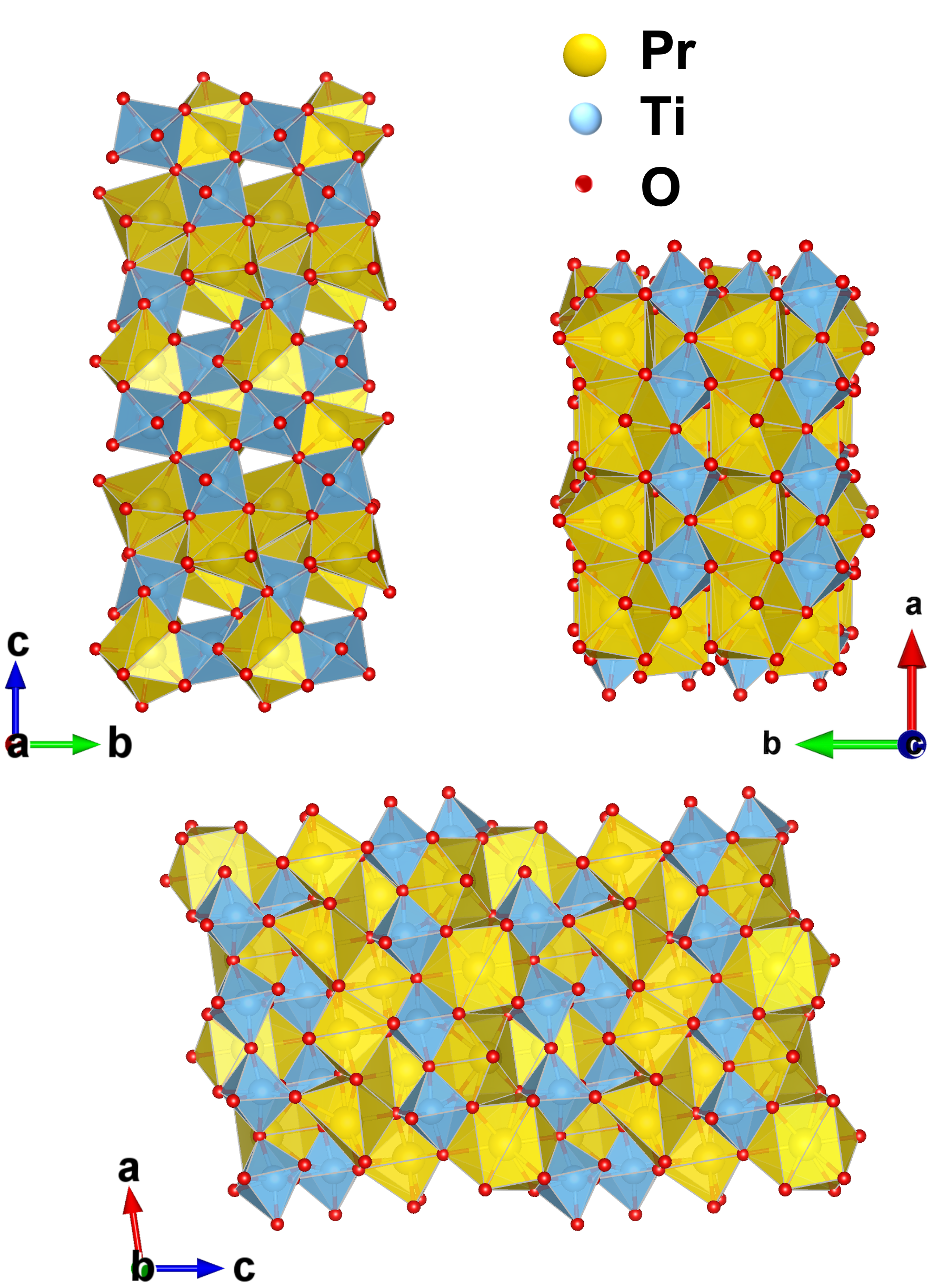
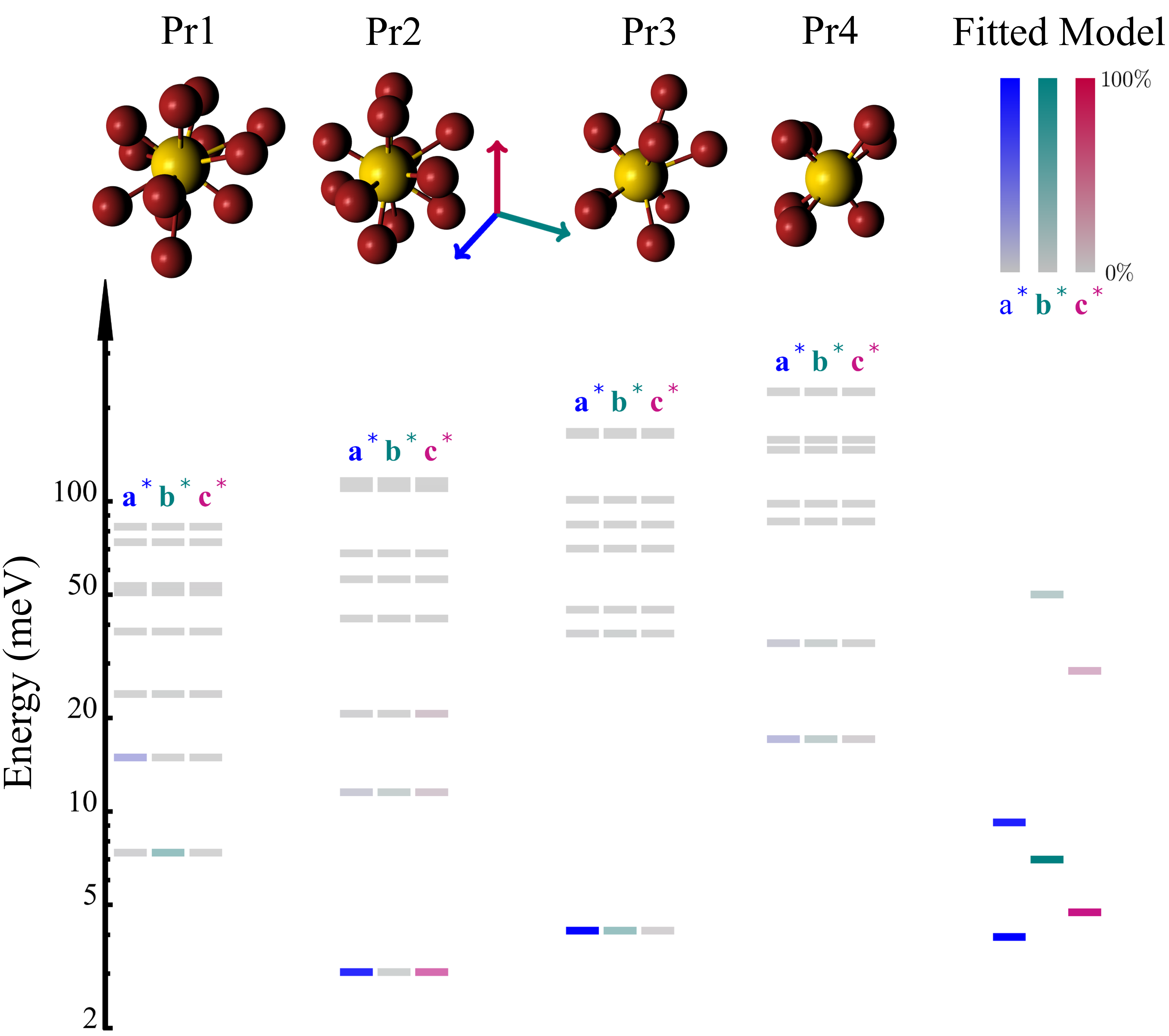
The monoclinic structure is described by four (001) layers () of corner-sharing TiO6 octahedra that form a perovskite like slab separated by two (001) layers of RE-site cations (Fig. 1). Praseodymium occupies four Wyckoff sites that fall in two groups. Located in the perovskite slab within interstices defined by the TiO6 octahedra, Pr1 and Pr4 are twelve-fold and seven-fold coordinated by oxygen, respectively. Pr2 and Pr3 with oxygen coordination number 10, bracket the perovskite slabs Granger et al. (Chapter 11, pp. 233-257, 2016). Since these four Pr sites carry the low- point-group symmetry, the spin-orbital multiplet of Pr3+ must be split into nine nonmagnetic singlet levels. In addition, as the four sites are different, a total of singlet-crystal-field levels should be anticipated. If the energy scale for interactions is less than the splitting between the singlet ground state and the first excited singlet, then we can expect to form a paramagnetic band insulator, which although magnetizable, should not, in general, be expected to undergo a magnetic phase transition. It should, however, be possible to drive the material to quantum criticality and an ordered state through the application of pressure or field at low temperatures.
The monoclinic layered perovskite titanates were previously studied for their interesting ferroelectric Sun et al. (2013); Bayart et al. (2014); Patwe et al. (2015); Atuchin et al. (2012); Gao et al. (2013); Nanamatsu et al. (1974); Yan et al. (2009); Valdez and Spaldin (2019), piezoelectric Bayart et al. (2014), nonlinear optical Zakharov et al. (1978), photocatalytic Hwang et al. (2003); Sayede et al. (2013), and dielectric Sun et al. (2013); Krishnankutty and Dayas (2008); Sayede et al. (2013) properties as well as their high ferroelectric Curie temperatureGao et al. (2013); Nanamatsu et al. (1974); Yan et al. (2009). Monoclinic Pr2Ti2O7, in particular, was grown and studied as powder through solid-state reaction Kesari et al. (2016), nanoparticles through the sol-gel method Sun et al. (2013) or the modified self-propagated high-temperature synthesis method Krishnankutty and Dayas (2008), and as epitaxial thin films grown by pulsed laser deposition Bayart et al. (2014). Single-crystal synthesis was also reported Zakharov et al. (1978); Saha et al. (2011). The powder samples of Pr2Ti2O7 showed a monoclinic structure in the noncentrosymmetric 21 space group and were investigated by x-ray diffraction and Raman spectroscopy Kesari et al. (2016); Patwe et al. (2015); Kozmin et al. (1997). The electronic structure of Pr2Ti2O7 has been studied with x-ray photoelectron spectroscopy Atuchin et al. (2012); Hwang et al. (2003), and the optical properties were investigated by first-principles density functional theory calculations Sayede et al. (2013). The photocatalytic activity of RE2Ti2O7 (RE = La, Pr, and Nd) is highly dependent on their electronic band structure Hwang et al. (2003). Ferroelectric measurements showed a Curie temperature beyond 1555(5) ∘CGao et al. (2013), meanwhil, similar properties were observed for monoclinic La2Ti2O7 and Nd2Ti2O7 with Curie temperatures of 1461(5) ∘CNanamatsu et al. (1974); Yan et al. (2009) and 1482(5) ∘CYan et al. (2009), respectively. The temperature-dependent Raman spectroscopy is very different for the pyrochlore “dynamic spin-ice” compound Pr2Sn2O7 and its non-pyrochlore (monoclinic) counterpart Pr2Ti2O7 Saha et al. (2011).
We conduct a comprehensive study of the phases, structures and disorders associated with the compositional deviations from the stoichiometric Pr2Ti2O7 here. As we will see, this reveals a distinct contrast in chemical solubility compared to pyrochlore titanates. An investigation of the low-symmetry lattice structure’s effect on the low-temperature magnetic properties of Pr2Ti2O7 is lacking. In this paper, we report a systematic investigation of the phase and structural stability of Pr2Ti2O7 based on synthesis of polycrystalline powders and single crystals. A process to develop a stoichiometric high-quality Pr2Ti2O7 single crystal by the traveling solvent floating zone (TSFZ) technique is reported. In order to understand the role which the lattice structure plays in determining the magnetic properties, single-crystalline Pr2Ti2O7 is characterized through magnetization measurements versus temperature, field, and crystalline directions. Our paper shows there is no magnetic ordering and the non-Kramers Pr3+ ions form singlet ground states with an energy gap to excited states. The presence of a three dimensional network of edge-sharing praseodymium oxide polyhedra suggest that it may be possible to induce a quantum phase transition to an ordered state through the application of pressure, strain, or high magnetic fields.
II Experimental details
II.1 Synthesis of powders
Powder samples of praseodymium titanates with nominal compositions Pr2Ti2+xO7, were synthesized by the solid-state reaction method. The Pr6O11 (99.99% Alfa Aesar) and TiO2 (99.99% Alfa Aesar) starting materials were dried at 1050 ∘C for 10 h. Pr6O11 powder was reduced to Pr2O3 in a hydrogen atmosphere. The resulting powders were then weighed, and the appropriate ratio was mixed, thoroughly ground, and heated to 1300 ∘C in air for 10 h. The process of mixing, grinding, and heating was repeated four times.
II.2 Single-crystal growth
Crystal growth of praseodymium titanates was carried out using the floating-zone melting technique Koohpayeh et al. (2008); Koohpayeh (2016) in a four-mirror image furnace equipped with four 1 kW halogen lamps (Crystal Systems, Inc., FZ-T-4000-H-VII-VPO-PC). Powder samples of stoichiometric Pr2Ti2O7 were first pressed into rods typically 5 mm in diameter and 8 cm in length and sintered in air at 1300 ∘C for 10 h. The feed rod and the seed rod were mounted on the upper and lower shafts of the furnace, respectively, with the mirror stage moving upward during the growth process. The rotation speeds of the upper and lower shafts were 3 and 6 rpm, respectively, during growth.
II.3 Characterization
The resulting phases, crystal structures, and lattice parameters were determined using powder x-ray diffraction (XRD) at room temperature. Data were acquired for 4 covering a scattering angle range of on a Bruker D8 focus diffractometer with monochromatic Cu Kα radiation and a LynxEye strip detector. Standard Si (silicon) powder was added as a reference into the samples. Rietveld refinement of the XRD patterns was carried out using TOPAS software (Bruker AXS).
All reflection intensities of single-crystalline Pr2Ti2O7 were measured at 213(2) K using a SuperNova diffractometer (equipped with an Atlas detector) with Mo radiation ( Å) under the program CrysAlisPro (Version CrysAlisPro 1.171.39.29c, Rigaku OD, 2017). The same program was used to refine the cell dimensions and for data reduction. The structure was solved with the program SHELXS-2018/3 and was refined against the squared structure factor with SHELXL-2018/3Sheldrick (2015). Empirical absorption correction using spherical harmonics was applied through CrysAlisPro. The sample temperature during data acquisition was controlled using a Cryojet system (manufactured by Oxford Instruments).
The single crystals were oriented using a tungsten anode back-reflection x-ray Laue diffractometer with a 1-mm diameter x-ray beam spot. A single crystal oriented with (100), (010), and (001) planes was cut using a diamond wire saw to the dimension of mm3, respectively, for a mass of 29.8 mg. For transmission electron microscopy (TEM), samples were prepared by crushing as-grown crystals in ethanol and placing a single drop of the solvent on a TEM carbon grid. We used a JEOL 2100 field emission TEM equipped with a high-resolution pole piece. Temperature-dependent susceptibility and isothermal magnetization measurements were performed using a Quantum Design superconducting quantum interference device magnetometer on the single crystal mentioned above in three different orientations and on a stoichiometric polycrystalline sample.
III Experimental Results
III.1 Polycrystalline Pr2Ti2O7
Contrary to the high-temperature dynamic melt/liquid-zoning crystal growth processes, solid-state powder synthesis at lower temperatures is a highly controllable process to explore both stoichiometric Pr2Ti2O7 and neighboring off-stoichiometric phase(s)/structures. Detailed in the Supplementary Material SM (See Supplemental Material at http://link.aps.org/supplemental/ 10.1103/PhysRevMaterials.7.063401 for solid state synthesis of polycrystalline samples, single crystal x-ray diffraction and the point charge model for the magnetic susceptibility) (see also references MacChesney and Sauer (1962); kapin et al. (2000); Par (2013); Sheldrik (2008); Gong et al. (2016); Kestigian and Ward (1955); Koohpayeh et al. (2014); Ghasemi et al. (2018); Wang et al. (2019); Yokoyama et al. (1989); Yoshii (2000); Zhang et al. (2007); Aughterson et al. (2008, 2015); Abe and Uchino (1974); Arpino et al. (2017); Bolz (Chapter 5, pp. 129-180, 1977); Hull (2004); Pussacq et al. (2017); Rig (2018); Sheldrik (2018) therein), our study of the effect of variations in the net reagent composition in Pr2Ti2+xO7 for () shows the monoclinic phase forms as a stoichiometric compound mixed with secondary phases to accommodate the reagent composition. This sets Pr2Ti2O7 apart from the cubic rare earth pyrochlores, which can deviate considerably from the ideal 2:2:7 stoichiometry. The stability of Pr2Ti2O7 as a stoichiometric solid, facilitates the process of growing high quality stoichiometric single crystals.
III.2 Single crystal of Pr2Ti2O7
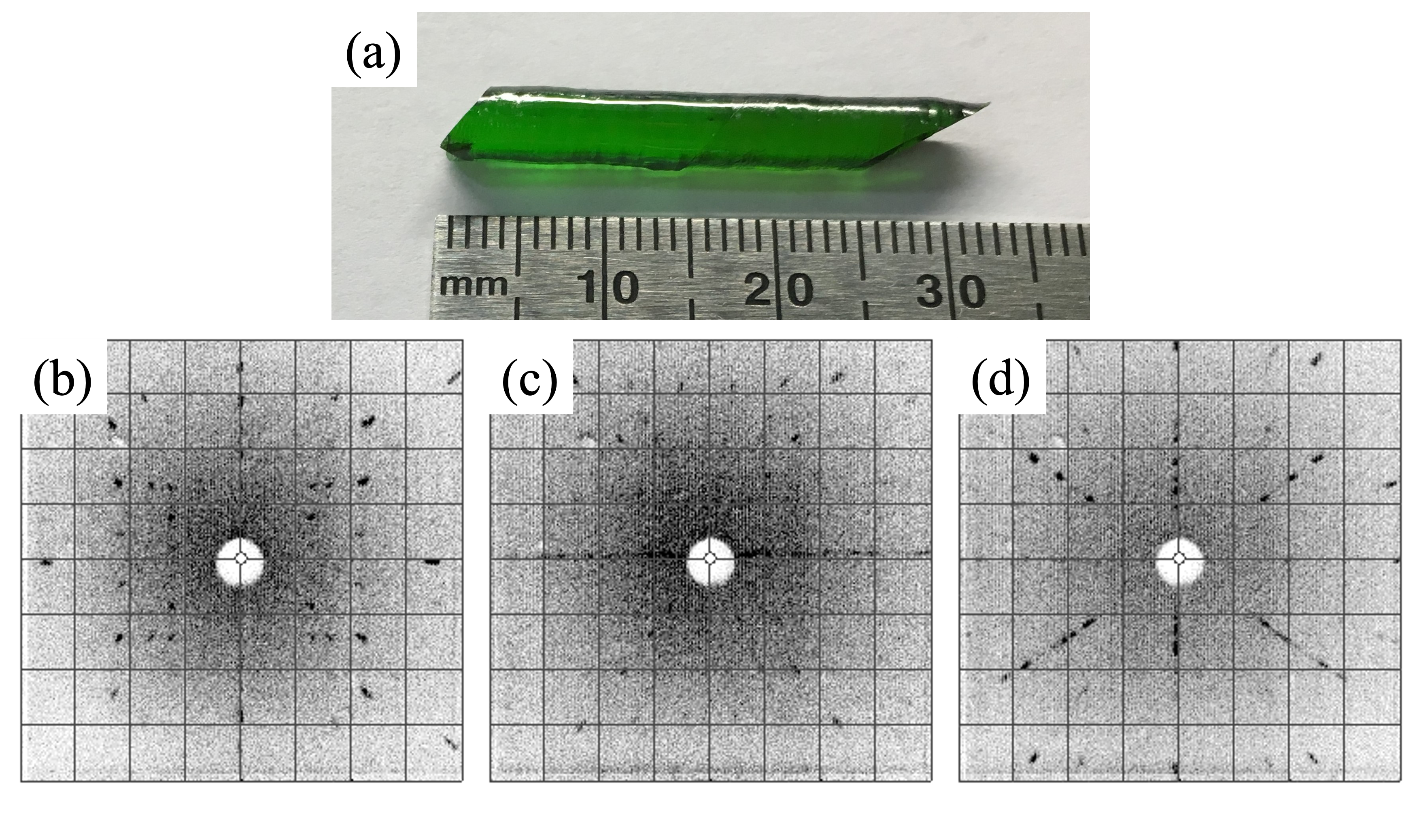
Although high-temperature phase instabilities and incongruent melting were encountered during the FZ melting process, crystal growths of Pr2Ti2O7 were successfully performed by the TSFZ technique under 1-bar static ultrahigh purity argon. The growth was stable during the entire process at the consistent lamp power of 56.5%, and no vaporization was observed. A typical transparent and green Pr2Ti2O7 crystal together with the Laue patterns along the main crystallographic planes are shown in Fig. 2. X-ray Laue patterns taken at regular intervals along the lengths and cross sections of the crystals indicated high crystalline quality with no detectable variation of orientation and no evidence of spot splitting or distortion. The crystal showed an easy cleavage along the (001) plane.
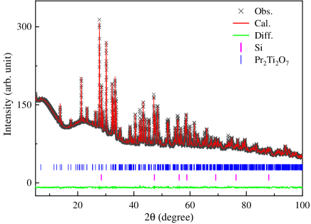
The Rietveld refinement of the XRD pattern (Fig. 3) taken from the crushed crystal at room temperature confirms that the crystal is single phase, the structure is monoclinic with space group 21, and the lattice constants were measured to be , , Å, and , similar to the stoichiometric powder sample.
The crystal data and structure refinement for Pr2Ti2O7 measured at 213(2) K is shown in Table S2 of the Supplemental Material SM (See Supplemental Material at http://link.aps.org/supplemental/ 10.1103/PhysRevMaterials.7.063401 for solid state synthesis of polycrystalline samples, single crystal x-ray diffraction and the point charge model for the magnetic susceptibility). The occupancy factors for PrX and TiX (X = 1-4) were all refined freely in Table S3 of the Supplemental Material SM (See Supplemental Material at http://link.aps.org/supplemental/ 10.1103/PhysRevMaterials.7.063401 for solid state synthesis of polycrystalline samples, single crystal x-ray diffraction and the point charge model for the magnetic susceptibility), and their final values are as follows: Pr1 0.970(5), Pr2 0.969(5), Pr3 0.959(5), Pr4 0.961(5), Ti1 0.966(6), Ti2 0.964(6), Ti3 0.963(6), and Ti4 0.955(6). The occupancy factors for all O atoms refine to 1 within their standard uncertainties, and were all constrained to be 1 in the final refinement. The absolute configuration has been established by anomalous dispersion effects in diffraction measurements on the crystal, and the Flack and Hooft parameters refine to -0.024(11) and -0.026(9), respectively.
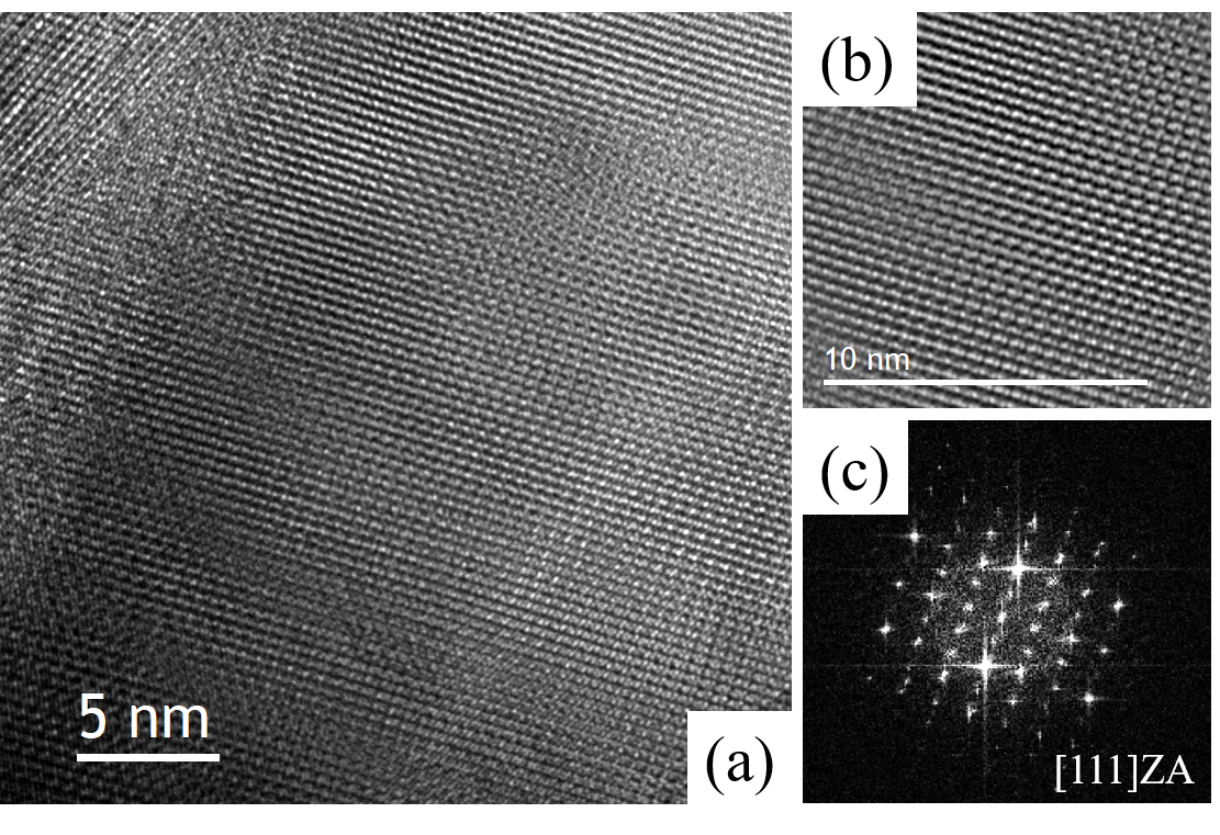
Single crystals of Pr2Ti2O7 were further examined by high-resolution transmission electron microscopy (HRTEM) from the [111] zone axis, indicating a crystal of high structural quality (Fig. 4).
III.3 Magnetization measurements
| Field direction () | () | () | (meV) | () | (meV) |
|---|---|---|---|---|---|
| 0.0054(2) | 0.50(2) | 3.93(7) | 1.03(2) | 9.2(1) | |
| 0.0046(1) | 0.789(3) | 6.99(2) | 0.84(1) | 50(1) | |
| 0.0050(2) | 0.324(3) | 4.72(4) | 0.805(5) | 28.4(4) | |
| Average | 0.0050(2) | 0.54(2) | 5.2(1) | 0.89(2) | 29.2(4) |
| Powder sample | 0.0086(3) | 0.711(9) | 5.55(5) | 0.918(8) | 28.3(7) |
The high quality single crystals described in the previous sections enabled us to explore the anisotropic magnetism of interacting non-Kramers rare-earth ions occupying four distinct low-symmetry sites. Prior experiments characterizing the physical properties of are quite limited. Room-temperature ferromagnetism was reported for polycrystalline nanoparticles grown through the sol-gel method. This effect is, however, unrelated to the rare-earth magnetism and appears to be connected with oxygen vacancies at the surfaces of nanoparticles Sun et al. (2013). A Raman study on a single crystal of synthesized by the float-zone method revealed structural stability up to 18 GPa and a complex spectrum as the low-symmetry monoclinic space group leads to 129 Raman active optical phonons Salke et al. (2015); Saha et al. (2011).
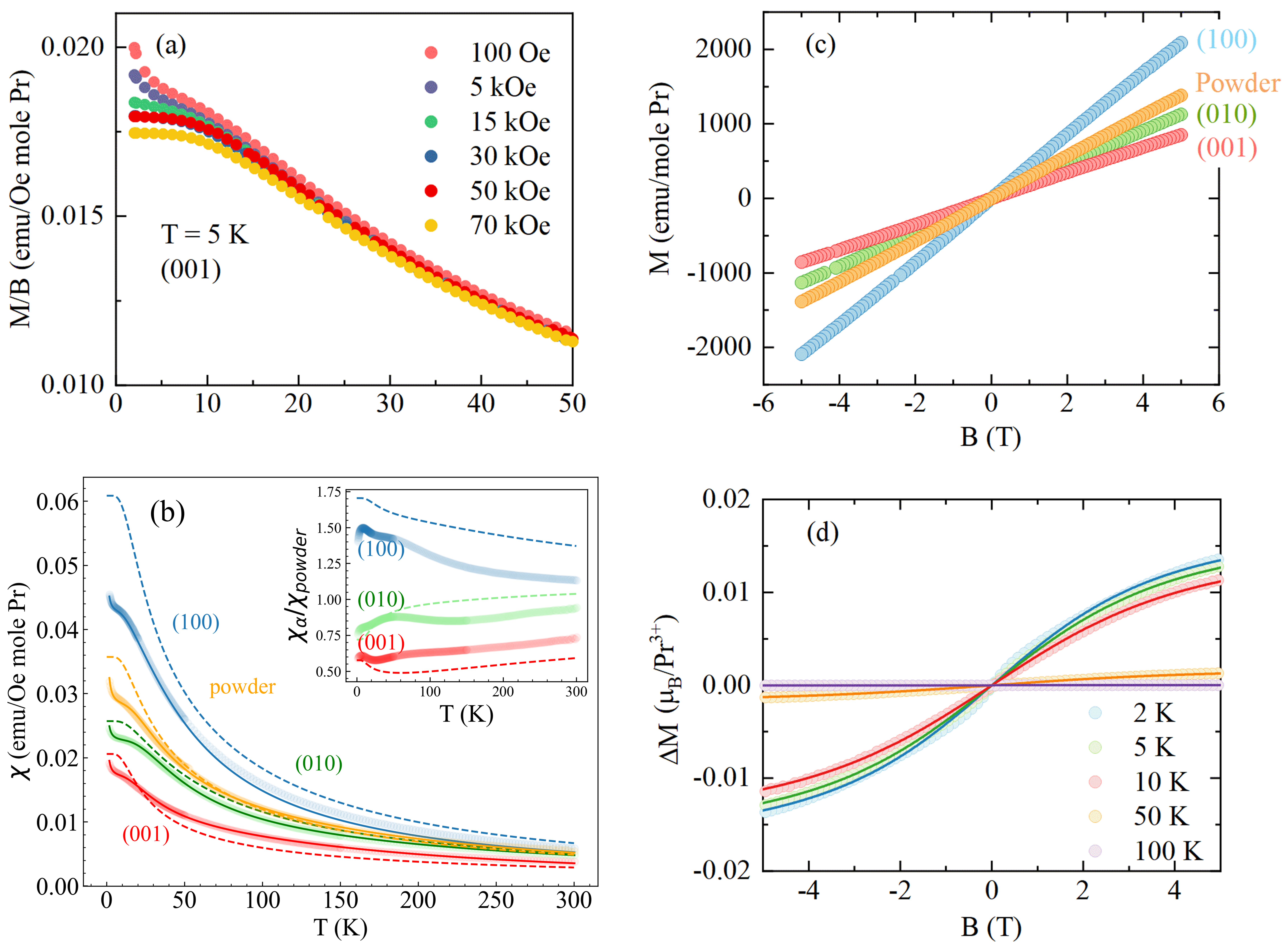
We measured the temperature-dependent magnetic susceptibility for both the single- and the polycrystalline Pr2Ti2O7 samples. Figure 5(a) shows as a function of temperature for applied fields normal to the (001) plane ranging from 100 Oe to 70 kOe. No anomalies or hysteresis is observed down to K. This indicates Pr2Ti2O7 has no magnetic phase transition in these temperature and field ranges. The field dependence of at low for kOe indicates the presence of paramagnetic impurities. We use measurements of at kOe to report the magnetic susceptibility versus temperature and field direction in Fig. 5(b). As should be anticipated for the non-Kramers ion Pr3+ in a monoclinic structure, there is considerable single-ion crystal electric-field-driven anisotropy with more than a factor 2 difference in low- susceptibility for the easy (100) versus the hard (001) directions. No field direction has indications of a magnetic phase transition. The inset shows the ratio of the single crystal to the powder sample magnetic susceptibility versus , which provides a dimensionless measure of the magnetic anisotropy. The anisotropy is reduced at higher , which indicates the population of excited crystal-field levels in that temperature range. An upturn in is apparent at the lowest temperatures for both the single-crystal and polycrystalline samples. As detailed later, we associate this “Curie tail” with paramagnetic Pr4+ impurities.
IV Analysis and Discussion
IV.1 Phenomenological crystal-field level scheme
With the non-Kramers Pr3+ ion in four different sites with symmetry there should be 36 crystal-field levels in . Neutron and/or Raman scatterings could, in principle, be used to determine these, but that is beyond the scope of this paper. By analyzing the dependence of the magnetic susceptibility, we can, however, obtain an estimate for the energy range associated with these crystal-field levels. For this purpose, we fit the data to the following three component phenomenological forms:
| (1) |
Here, , and is the Boltzmann constant. The first term is the Curie term, which we will associate with Pr4+impurities. The summations are over each of the four Pr3+ sites, which are approximated as two-level systems with a singlet ground state and excited levels at a characteristic energy . This form is obtained as a low- approximation to the general Van Vleck susceptibility in the Supplementary Material Eq.(7) SM (See Supplemental Material at http://link.aps.org/supplemental/ 10.1103/PhysRevMaterials.7.063401 for solid state synthesis of polycrystalline samples, single crystal x-ray diffraction and the point charge model for the magnetic susceptibility). The expression has been constructed so that the high limit takes the Curie-form as for the impurity term. We found that two terms are sufficient to obtain excellent fits for all field directions.
The best fit parameters are listed in Table 1. Consider first the fit to the poly-crystalline data. The sum of Curie constants (emuK/Oemole) agrees well with the Curie constant for Pr3+ (emuK/Oemole). The two polycrystal gap values provide a scale for the crystal-field levels from 5 to 28 meV. For comparison the crystal field levels for are at [0, 10, 57, 82, 93, 109] meVKimura et al. (2013). The ratio of the Curie constants indicates similar spectral weight at and . The ratio % provides the order of magnitude of the Kramers rare-earth impurity concentration.
The fits to single-crystal data yield a broader range of gap values with the smallest gap value meV associated with the easy (100) direction and the largest gap value meV for the (010) direction. The different energies obtained for different field directions indicate the energy range for crystal-field levels with dominant contributions to the susceptibility for each field direction . However, with a total of 32 crystal-field transitions available from the ground state of , these data can only provide a general sense of the energy scale for the crystal-field levels that contribute most to the Van Vleck susceptibility for each field direction. The Curie constants for each field direction reflect the site averaged dipolar matrix elements for crystal-field levels separated by energies for . The consistency between the directionally averaged single-crystal parameters and those obtained for polycrystalline samples supports the evidence from x-ray diffraction that the single crystal and powder samples are similar at the atomic scale despite the very different growth conditions. We note the Curie constant associated with impurities is more isotropic than for the intrinsic bulk terms . The impurity content as measured by is 40% less in the single crystal as compared to the powder samples.
In this insulating rare-earth compound with shared ligands, super exchange interactions between praseodymium are expected to be on the 0.5-meV scale. This is an order of magnitude smaller than needed to close the lowest crystal-field energy gap. The low-symmetry environment, thus, effectively renders the non-Kramers rare-earth ion compound non-magnetic and precludes a phase transition or indeed any truly collective physics. It might, however, be possible to induce a quantum phase transition through the application of pressure as in TlCuCl3Rüegg et al. (2004); Goto et al. (2004) and Tb2Ti2O7Mirebeau et al. (2002) before inducing a structural phase transition.
IV.2 Point charge model
The splitting of the multiplet and the associated magnetic single-ion anisotropy is due to the crystal electric field (CEF) associated with the ligands surrounding each rare-earth ion. As detailed in the Supplementary MaterialSM (See Supplemental Material at http://link.aps.org/supplemental/ 10.1103/PhysRevMaterials.7.063401 for solid state synthesis of polycrystalline samples, single crystal x-ray diffraction and the point charge model for the magnetic susceptibility), the corresponding single ion Hamiltonian can be expressed in the form
| (2) |
where O are Stevens operatorsM.T.Hutchings (1964); Stevens (1952) and parametrize the effects of the CEF on the multiplet. Starting from the crystal structure, the point charge model yields estimates for which are provided for each distinct Pr-site within in Table IV of the Supplementary MaterialSM (See Supplemental Material at http://link.aps.org/supplemental/ 10.1103/PhysRevMaterials.7.063401 for solid state synthesis of polycrystalline samples, single crystal x-ray diffraction and the point charge model for the magnetic susceptibility).
Diagonalizing Eq. (2) yields the CEF level scheme shown for each of the four Pr Wyckoff sites in Fig. 6. The predicted contribution of crystal field level to the low- van Vleck susceptibility for each of three field directions is listed in Table 5 of the Supplementary MaterialSM (See Supplemental Material at http://link.aps.org/supplemental/ 10.1103/PhysRevMaterials.7.063401 for solid state synthesis of polycrystalline samples, single crystal x-ray diffraction and the point charge model for the magnetic susceptibility) and indicated by the color scheme in Fig. 6. The point charge model predicts that the Pr2 and Pr3 sites, which are ten fold coordinated by oxygen and bracket the perovskite (001) slabs (Fig. 1), are the dominant contributors to the low- van Vleck susceptibility (see also Fig. S1 of the Supplementary MaterialSM (See Supplemental Material at http://link.aps.org/supplemental/ 10.1103/PhysRevMaterials.7.063401 for solid state synthesis of polycrystalline samples, single crystal x-ray diffraction and the point charge model for the magnetic susceptibility)).
The point charge model also indicates that the first excited CEF levels provide the largest contribution to the magnetic susceptibility. This support our use of the phenomenological form in Eq. (1) to fit the magnetic susceptibility data. Comparison of the point charge energy level scheme with that inferred from these fits shows a remarkable agreement in identifying the CEF levels that dominate the low- magnetic susceptibility for each of the three field directions. There is however, a consistent trend that the point charge model predicts lower-energy levels than inferred from the phenomenological fitting. This is also apparent in Fig. 5(b) where the point charge model (dashed lines) generally overestimates the low- magnetic susceptibility. Overall, although considering that there are no adjustable parameters, the point charge model does fairly well. For example, it correctly predicts that . Our analysis shows that Eq. (1) provides an excellent account of the anisotropic -dependent magnetic susceptibility of this singlet ground state system. The corresponding energy levels on the far right in Fig.6 represent our best experimental estimate of the crystal-field level scheme in .
IV.3 Curie tails and Pr4+ impurities
Examining the low field behavior in greater detail, Fig. 5(c) shows magnetization curves for various field directions at K. For all orientations, the data look linear on this scale. However, if we subtract a linear fit to the high-field regime from 6.5 to 7 T from the data, we obtain the non-linear component shown for the (001) direction in Fig. 5(d). Resembling the magnetization curve for a paramagnetic impurity, the nonlinear component becomes more prominent when the thermal energy scale falls below the Zeeman energy scale. Under the assumption of a single characteristic impurity species it is possible to extract both the saturation magnetization and the impurity concentration from such data. To do so we fit the Langevin magnetization curve to the data,
| (3) |
Here is the paramagnetic impurity fraction, is Avagadro’s number, and the Langevin function is given by . is the Brillouin function describing the magnetization curve for a paramagnetic impurity with spin-orbital angular momentum quantum number . Figure 5(d) shows that this functional form provides an excellent fit to the data with an impurity fraction of and a saturation moment of . The Curie constant corresponding to these parameters is approximately (emuK/Oemole), which is entirely consistent with in Table 1 derived from -dependent susceptibility data. Note that, although the majority component of the magnetization is excluded from this analysis, the Langevin fit is consistent with the concentration estimate obtained from the ratio of Curie constants. We consider the concentration from the Langevin analysis to be more accurate, though, because it does not rely on the impurities having the same effective moment as the majority phase.
The specification of the starting material was % rare-earth oxide with 99.99% of the rare earth oxide being . A non-Pr source for the paramagnetic impurity is, thus, unlikely. A possible explanation is that the rare-earth impurity is Pr4+, which has a single electron and the same magnetic properties as Ce3+. In particular, it is a Kramers ion with a saturation moment of 2.14 , consistent with the Langevin analysis of the magnetization data.
V Conclusion
We have successfully synthesized both polycrystalline samples and stoichiometric single crystals of Pr2Ti2O7. Small levels of compositional deviations from the stoichiometric Pr2Ti2O7 target compound lead to secondary phases of different, albeit related, structures. In contrast to the pyrochlore titanates, site disordering was not observed in the lower-symmetry monoclinic Pr2Ti2O7 structure.
Building upon the chemical and structural stabilities of monoclinic , we provide a process to grow large high-quality single crystals. Stoichiometric Pr2Ti2O7 single crystals were grown using the TSFZ method. The susceptibility and magnetization measurements show no indications of a phase transition down to K. Our analysis of the susceptibility data indicates a singlet ground state with excited CEF levels at energies ranging from 3.93(7) to 50(1) meV. This is as anticipated for the low-symmetry crystal electric-field environment associated with Pr3+ in monoclinic . At sufficiently low density, Kramers rare-earth impurities within behave as isolated paramagnetic impurities that dominate over the majority phase paramagnetic van Vleck susceptibility at the lowest temperatures and fields ( K, kOe). Analysis of the magnetization data in this regime yields a 0.96(2)% impurity content with a saturation moment of , which is consistent with a low concentration of Pr4+ impurities.
The monoclinic structure contains a 3D network of Pr3+ with edge-sharing ligand polyhedra assuring superexchange interactions on the 0.5 meV energy scale. If the gap between the two lowest-lying singlets can be closed through the application of hydrostatic of uniaxial pressure, or fields, a quantum phase transition into an ordered magnetic state induced by these interactions can be anticipated. , thus, may provide an interesting opportunity to explore universal aspects of quantum critical spin dynamics in a material where large high-quality crystals are attainable.
VI Acknowledgements
This work was supported as part of the Institute for Quantum Matter, an Energy Frontier Research Center funded by the U.S. Department of Energy, Office of Science, Basic Energy Sciences under Award No. DE-SC0019331. H.M., A.G. and C.L.B. were also supported by the Gordon and Betty Moore Foundation under Grant No. GBMF9456. The work at University of Houston (M.A. and C.W.C.) was supported by U. S. Air Force Office of Scientific Research Grants FA9550-15-1-0236 and FA9550-20-1-0068, the T. L. L. Temple Foundation, the John J. and Rebecca Moores Endowment, and the State of Texas through the Texas Center for Superconductivity at the University of Houston. Y.L. and C.L.C. were supported by U.S. Department of Energy, Basic Energy Science under Award Grant No. DESC0009390. M.L.T. and E.A. acknowledge funding, in part, from the U.S. Department of Energy, Office of Basic Energy Sciences through Contract No. DE-SC0020314. M.L.T. also acknowledges funding, in part, from the Office of Naval Research Multidisciplinary University Research Initiative (MURI) program through Contract No. N00014-20-1-2368.
References
- Millican et al. (2007) J. N. Millican, R. T. Macaluso, S. Nakatsuji, Y. Machida, Y. Maeno, and J. Y. Chan, Materials Research Bulletin 42, 928 (2007).
- Taguchi et al. (2001) Y. Taguchi, Y. Oohara, H. Yoshizawa, N. Nagaosa, and Y. Tokura, Science 291, 2573 (2001).
- Subramanian et al. (1996) M. A. Subramanian, B. H. Toby, A. P. Ramirez, W. J. Marshall, A. W. Sleight, and G. H. Kwei, Science 273, 81 (1996).
- Subramanian et al. (1997) M. A. Subramanian, J. E. Greedan, and N. P. Raju, J. Phys. IV France 7, 625 (1997).
- Ramirez and Subramanian (1997) A. P. Ramirez and M. A. Subramanian, Science 277, 546 (1997).
- Hanawa et al. (2001) M. Hanawa, Y. Muraoka, T. Tayama, T. Sakakibara, J. Yamaura, and Z. Hiroi, Phys. Rev. Lett. 87, 187001 (2001).
- Ishikawa et al. (2012) J. J. Ishikawa, E. C. T. O’Farrell, and S. Nakatsuji, Phys. Rev. B 85, 245109 (2012).
- Balents (2010) L. Balents, Nature 464, 199 (2010).
- Morris et al. (2009) D. J. P. Morris, D. A. Tennant, S. A. Grigera, B. Klemke, C. Castelnovo, R. Moessner, C. Czternasty, M. Meissner, K. C. Rule, J.-U. Hoffmann, et al., Science 326, 411 (2009).
- Fennell et al. (2009) T. Fennell, P. P. Deen, A. R. Wildes, K. Schmalzl, D. Prabhakaran, A. T. Boothroyd, R. J. Aldus, D. F. McMorrow, and S. T. Bramwell, Science 326, 415 (2009).
- Kimura et al. (2013) K. Kimura, S. Nakatsuji, J. J. Wen, C. Broholm, M. B. Stone, E. Nishibori, and H. Sawa, Nat. Commun. 4, 1934 (2013).
- Sibille et al. (2018) R. Sibille, N. Gauthier, H. Yan, M. C. Hatnean, J. Ollivier, B. Winn, G. Balakrishnan, M. Kenzelmann, N. Shannon, and T. Fennell, Nature Phys. 14, 711 (2018).
- Bojesen and Onoda (2017) T. A. Bojesen and S. Onoda, Phys. Rev. Lett. 119, 227204 (2017).
- Machida et al. (2010) Y. Machida, S. Nakatsuji, S. Onoda, T. Tayama, and T. Sakakibara, Nature 463, 210 (2010).
- Cheng et al. (2017) B. Cheng, T. Ohtsuki, D. Chaudhuri, S. Nakatsuji, M. Lippmaa, and N. Armitage, Nat. Commun. 8, 2097 (2017).
- Ross et al. (2011) K. A. Ross, L. Savary, B. D. Gaulin, and L. Balents, Phys. Rev. X 1, 021002 (2011).
- Säubert et al. (2020) S. Säubert, A. Scheie, C. Duvinage, J. Kindervater, S. Zhang, H. J. Changlani, G. Xu, S. M. Koohpayeh, O. Tchernyshyov, C. L. Broholm, et al., Phys. Rev. B 101, 174434 (2020).
- Scheie et al. (2020) A. Scheie, J. Kindervater, S. Zhang, H. J. Changlani, G. Sala, G. Ehlers, A. Heinemann, G. S. Tucker, S. M. Koohpayeh, and C. Broholm, Proceedings of the National Academy of Sciences 117, 27245 (2020).
- Hirschberger et al. (arXiv:1903.00595) M. Hirschberger, P. Czajka, S. M. Koohpayeh, W. Wang, and N. P. Ong (arXiv:1903.00595).
- Scheie et al. (2017) A. Scheie, J. Kindervater, S. Säubert, C. Duvinage, C. Pfleiderer, H. J. Changlani, S. Zhang, L. Harriger, K. Arpino, S. M. Koohpayeh, et al., Phys. Rev. Lett. 119, 127201 (2017).
- Wen et al. (2017) J.-J. Wen, S. M. Koohpayeh, K. A. Ross, B. A. Trump, T. M. McQueen, K. Kimura, S. Nakatsuji, Y. Qiu, D. M. Pajerowski, J. R. D. Copley, et al., Phys. Rev. Lett. 118, 107206 (2017).
- Gardner et al. (2010) J. S. Gardner, M. J. P. Gingras, and J. E. Greedan, Rev. Mod. Phys. 82, 53 (2010).
- Wang et al. (2021) Y. Wang, T. Reeder, Y. Karaki, J. Kindervater, T. Halloran, N. Maliszewskyj, Y. Qiu, J. A. Rodriguez, S. Gladchenko, S. M. Koohpayeh, et al., Science Advances 7, eabg0908 (2021).
- Zhang et al. (2021) X. Zhang, Y. Luo, T. Halloran, J. Gaudet, H. Man, S. M. Koohpayeh, and N. P. Armitage, Phys. Rev. B 103, L140403 (2021).
- Scheie et al. (arXiv:2202.11085) A. Scheie, O. Benton, M. Taillefumier, L. D. C. Jaubert, G. Sala, N. Jalarvo, S. M. Koohpayeh, and N. Shannon (arXiv:2202.11085).
- Shcherbakova et al. (1979) L. Shcherbakova, L. Mamsurova, and G. Sukhanova, Russ. Chem. Rev. 48(3), 228 (1979).
- Kesari et al. (2016) S. Kesari, N. P. Salke, S. J. Patwe, S. N. Achary, A. K. Sinha, P. U. Sastry, A. K. Tyagi, and R. Rao, Inorg. Chem. 55, 11791-11800 (2016).
- Granger et al. (Chapter 11, pp. 233-257, 2016) P. Granger, V. Parvulescu, S. Kaliaguine, and W. Prellier, Perovskites and related mixed oxides: Concepts and Applications (Chapter 11, pp. 233-257, 2016).
- Sun et al. (2013) L. Sun, L. Ju, H. Qin, M. Zhao, W. Su, and J. Hu, Physica B 431, 49–53 (2013).
- Bayart et al. (2014) A. Bayart, S. Saitzek, A. Ferri, R. Pouhet, M.-H. Chambrier, P. Roussel, and R. Desfeux, Thin Solid Films 553, 71–75 (2014).
- Patwe et al. (2015) S. J. Patwe, V. Katari, N. P. Salke, S. K. Deshpande, R. Rao, M. K. Gupta, R. Mittal, S. N. Achary, and A. K. Tyagi, J. Mater. Chem. C 3, 4570 (2015).
- Atuchin et al. (2012) V. Atuchin, T. Gavrilova, J.-C. Grivel, V. Kesler, and I. Troitskaia, Journal of Solid State Chemistry 195, 125–131 (2012).
- Gao et al. (2013) Z. P. Gao, H. X. Yan, H. P. Ning, and M. J. Reece, Advances in Applied Ceramics 112, 69 (2013).
- Nanamatsu et al. (1974) S. Nanamatsu, M. Kimura, K. Doi, S. Matsushita, and N. Yamada, Ferroelectrics 8, 511 (1974).
- Yan et al. (2009) H. Yan, H. Ning, Y. Kan, P. Wang, and M. Reece, J. Am. Ceram. Soc. 92, 2270 (2009).
- Valdez and Spaldin (2019) M. N. Valdez and N. A. Spaldin, Polyhedron 171, 181 (2019).
- Zakharov et al. (1978) N. Zakharov, V. Krikorov, E. Kustov, and S. Stefanovich, Phys. Status Solidi A 50, K13 (1978).
- Hwang et al. (2003) D. W. Hwang, J. S. Lee, W. Li, and S. H. Oh, J. Phys. Chem. B 107, 4963 (2003).
- Sayede et al. (2013) A. Sayede, R. Khenata, A. Chahed, and O. Benhelal, J. Appl. Phys. 113, 173501 (2013).
- Krishnankutty and Dayas (2008) K. Krishnankutty and K. R. Dayas, Bulletin of Materials Science 31, 907–918 (2008).
- Saha et al. (2011) S. Saha, S. Prusty, S. Singh, R. Suryanarayanan, A. Revcolevschi, and A. Sood, Journal of Solid State Chemistry 184, 2204–2208 (2011).
- Kozmin et al. (1997) P. Kozmin, N. Zakharov, and M. Surazhskaya, Inorganic Materials 33, 850 (1997).
- Koohpayeh et al. (2008) S. M. Koohpayeh, D. Fort, and J. S. Abell, Prog. Cryst. Growth Character. Mater. 54, 121 (2008).
- Koohpayeh (2016) S. M. Koohpayeh, Prog. Cryst. Growth Character. Mater. 62, 22 (2016).
- Sheldrick (2015) G. M. Sheldrick, Acta Cryst. C71, 3 (2015).
- SM (See Supplemental Material at http://link.aps.org/supplemental/ 10.1103/PhysRevMaterials.7.063401 for solid state synthesis of polycrystalline samples, single crystal x-ray diffraction and the point charge model for the magnetic susceptibility) (See Supplemental Material at http://link.aps.org/supplemental/ 10.1103/PhysRevMaterials.7.063401 for solid state synthesis of polycrystalline samples, single crystal x-ray diffraction and the point charge model for the magnetic susceptibility).
- MacChesney and Sauer (1962) J. B. MacChesney and H. A. Sauer, Journal of The American Ceramic Society 45, 416 (1962).
- kapin et al. (2000) S. D. kapin, D. Kolar, and D. Suvorov, Journal of the European Ceramic Society 20, 1179 (2000).
- Par (2013) Acta Crystallographica Section B: Structural Science, Crystal Engineering and Materials 69, 249 (2013).
- Sheldrik (2008) G. M. Sheldrik, Acta Crystallographica Section A: Foundations of Crystallography 64, 112 (2008).
- Gong et al. (2016) Y. Gong, R. Chu, Z. Xu, T. Dou, W. Zeng, X. Zhang, J. Hao, and G. Li, J. Am. Ceram. Soc. 99, 2995 (2016).
- Kestigian and Ward (1955) M. Kestigian and R. Ward, J. Am. Chem. Soc. 77, 6199 (1955).
- Koohpayeh et al. (2014) S. Koohpayeh, J.-J. Wen, B. Trump, C. Broholm, and T. McQueen, Journal of Crystal Growth 402, 291 (2014).
- Ghasemi et al. (2018) A. Ghasemi, A. Scheie, J. Kindervater, and S. Koohpayeh, Journal of Crystal Growth 500, 38 (2018).
- Wang et al. (2019) Q. Wang, A. Ghasemi, A. Scheie, and S. M. Koohpayeh, CrystEngComm 21, 703 (2019).
- Yokoyama et al. (1989) M. Yokoyama, T. Ota, I. Yamai, and J. Takahashi, J. of Crystal Growth 96, 490 (1989).
- Yoshii (2000) K. Yoshii, J. of Solid State Chemistry 149, 354 (2000).
- Zhang et al. (2007) F. Zhang, J. Lian, U. Becker, R. Ewing, L. Wang, J. Hu, and S. Saxena, J. of solid State Chemistry 180, 571 (2007).
- Aughterson et al. (2008) R. D. Aughterson, G. R. Lumpkin, K. L. Smith, G. J. Thorogood, and K. R. Whittle, Mater. Res. Soc. Symp. Proc. 1107, 365 (2008).
- Aughterson et al. (2015) R. D. Aughterson, G. R. Lumpkin, G. J. Thorogood, Z. Zhang, B. Gault, and J. M. Cairney, Journal of Solid State Chemistry 227, 60 (2015).
- Abe and Uchino (1974) M. Abe and K. Uchino, Mat. Res. Bull. 9, 147 (1974).
- Arpino et al. (2017) K. E. Arpino, B. A. Trump, A. O. Scheie, T. M. McQueen, and S. M. Koohpayeh, Phys. Rev. B 95, 094407 (2017).
- Bolz (Chapter 5, pp. 129-180, 1977) F. Bolz, Advanced Materials in Catalysis (Chapter 5, pp. 129-180, 1977).
- Hull (2004) S. Hull, Rep. Prog. Phys. 67, 1233–1314 (2004).
- Pussacq et al. (2017) T. Pussacq, H. Kabbour, S. Colis, H. Vezin, S. Saitzek, O. Gardoll, C. Tassel, H. Kageyama, C. L. Robert, and O. Mentré, Chem. Mater. 29(3), 1047 (2017).
- Rig (2018) Rigaku Oxford Diffraction, CrysAlisPro Software System, Version 1.171, Rigaku Corporation Oxford, UK (2018).
- Sheldrik (2018) G. M. Sheldrik, University of Gottingen (2018).
- Scheie (2021) A. Scheie, Journal of Applied Crystallography 54, 356 (2021).
- Salke et al. (2015) N. P. Salke, S. Kesari, S. J. Patwe, A. K. Tyagi, and R. Rao, AIP Conference Proceedings 1665, 030011 (2015).
- Rüegg et al. (2004) C. Rüegg, A. Furrer, D. Sheptyakov, T. Strässle, K. W. Krämer, H.-U. Güdel, and L. Mélési, Phys. Rev. Lett. 93, 257201 (2004).
- Goto et al. (2004) K. Goto, M. Fujisawa, T. Ono, H. Tanaka, and Y. Uwatoko, J. Phys. Soc. Jpn. 73, 3254–3257 (2004).
- Mirebeau et al. (2002) I. Mirebeau, I. N. Goncharenko, P. Cadavez-Peres, S. T. Bramwell, M. J. P. Gingras, and J. S. Gardner, Nature 420, 54 (2002).
- M.T.Hutchings (1964) M.T.Hutchings, Solid State Physics 16, 227 (1964).
- Stevens (1952) K. Stevens, Proc. Phys. Soc. A 65, 209 (1952).