Computational models advance deep brain stimulation for Parkinson’s disease
Abstract
Deep brain stimulation (DBS) has become an effective intervention for advanced Parkinson’s disease, but the exact mechanism of DBS is still unclear. In this review, we discuss the history of DBS, the anatomy and internal architecture of the basal ganglia (BG), the abnormal pathological changes of the BG in Parkinson’s disease, and how computational models can help understand and advance DBS. We also describe two types of models: mathematical theoretical models and clinical predictive models. Mathematical theoretical models simulate neurons or neural networks of BG to shed light on the mechanistic principle underlying DBS, while clinical predictive models focus more on patients’ outcomes, helping to adapt treatment plans for each patient and advance novel electrode designs. Finally, we provide insights and an outlook on future technologies.
keywords:
Deep brain stimulation; Parkinson’s disease; Computational model; Basal ganglia1 Introduction
1.1 Deep Brain Stimulation(DBS)
Deep brain stimulation (DBS) is a developing technology used to treat various neurological diseases. It improves the patient’s mobility and self-care by stimulating the relevant nuclei in the brain that control movement and ”modulates” (Ashkan et al. 2017) the abnormal brain neural signals through weak electrical pulses delivered by electrodes implanted in the brain. In 1987 Benabid and colleagues demonstrated that DBS produced beneficial effects similar to pallidotomy in patients with Parkinson’s disease (PD) (Benabid et al. 1987). Moreover, compared to L-Dopa therapy (Miyasaki et al. 2002) and pallidotomy (Goetz et al. 2005), DBS has considered one of the excellent options for PD relief today due to it is relatively minimally invasive, non-destructive, and reversible(compared to excisional surgery) (Pahwa et al. 2006). Since then, the technique has gradually opened up new frontiers in the surgical treatment of various movement disorders, depression, obsessive-compulsive disorder, and Alzheimer’s disease. By 2022, nearly 40 years after the development of DBS, the routine use of DBS for the treatment of the motor symptoms of PD has proven to be very effective, but its mechanism of action is still largely unknown (Jakobs et al. 2019) (Aum and Tierney 2018) (Lozano et al. 2019). Studying the underlying therapeutic mechanisms of DBS first requires an in-depth understanding of the various activity patterns and differences in brain circuits associated with neurological disorders under physiological and pathological conditions. Therefore, combining computational modeling describing various types of neurons in the brain with clinical data is a rather promising research approach.
1.2 Parkinson’s disease(PD)
PD is a complex multisystem neurodegenerative disease whose primary cause is thought to be the loss of dopaminergic cells in Substantia nigra pars compacta in the brain. The academic community initially considered it a movement disorder with three main symptoms: tremor, muscle rigidity, and bradykinesia (Postuma et al. 2015). In addition to motor symptoms, PD also has many non-motor symptoms, including but not limited to autonomic dysfunction, sleep disturbances, and neuropsychiatric symptoms (Hely et al. 2008). PD is also the fastest-growing neurological disease in the world in terms of prevalence, disability, and deaths, with the GBD (Global Burden of Disease Study) estimating that the total number of people affected by the disease increased by 118 from 1990 to 2019, to more than 6 million worldwide (Dorsey et al. 2018). With demographic changes (aging) and the side effects of industrialization (pollution), there has been a faster increase in PD cases (Brundin and Bloem 2018).
Various pathological features in PD are closely related to abnormal pathological changes in the basal ganglia (BG). The subthalamic nucleus(STN) in BG is the main target of DBS (Hu et al. 2017b). Therefore, if we want to determine the specific mechanisms of DBS, it is important to first understand the physiological structure of the BG with its pathological changes in PD patients. Since the detailed mechanisms of DBS are difficult to investigate in humans, computational models are an effective tool to explore the mechanism of DBS and advance its development.
In the next part, we will briefly introduce the main structure of BG and discuss the significant changes in the kinetic properties of BG-related loops in PD.
2 Basal ganglia(BG)
2.1 BG structure and functional masses
The BG consists of a set of interconnected subcortical nuclei involved in various important brain functions - motor control (Turner and Desmurget 2010), decision making (Bogacz and Gurney 2007), categorization (Seger 2008), motor preparation (Jaeger et al. 1993), probabilistic learning (Shohamy et al. 2008), reinforcement learning (Frank et al. 2004), reward signalling (Kawagoe et al. 1998), sequence learning (Lehéricy et al. 2005), working memory (McNab and Klingberg 2008), etc (Brown and Marsden 1998) (Nelson and Kreitzer 2014). As shown in Figure 1, the several extensively connected subcortical nuclei are anatomically defined into striatum (STR), Globus pallidus (GP), subthalamic nucleus(STN), substantia nigra (SN), and ventral tegmental area (VTA). STR consists of caudate nucleus (CN), putamen (Put), and nucleus accumbens(Acb). GP can be divided into external pallidum (GPe) and internal pallidum(GPi). The SN is divided into pars compacta (SNc), pars reticulata (SNr), and pars lateralis (SNL) (Aird 2000).
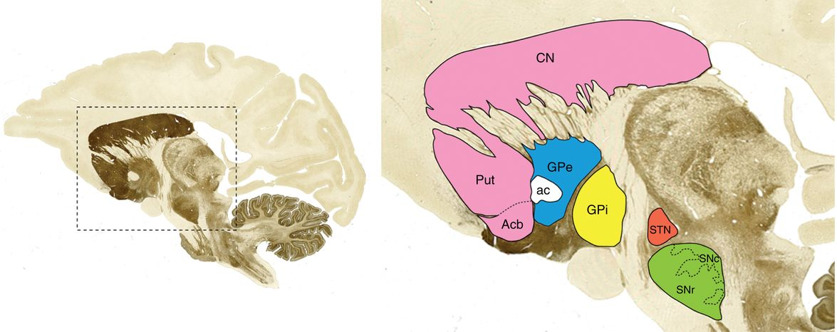
STR consists mainly of spiny projection neurons. It receives input from the cerebral cortex, sensorimotor and motivational regions of the brainstem. It also produces either inhibitory or excitatory effects on the GP and SN, depending on the receptor type(D1 and D2-type) in the postsynaptic cell.
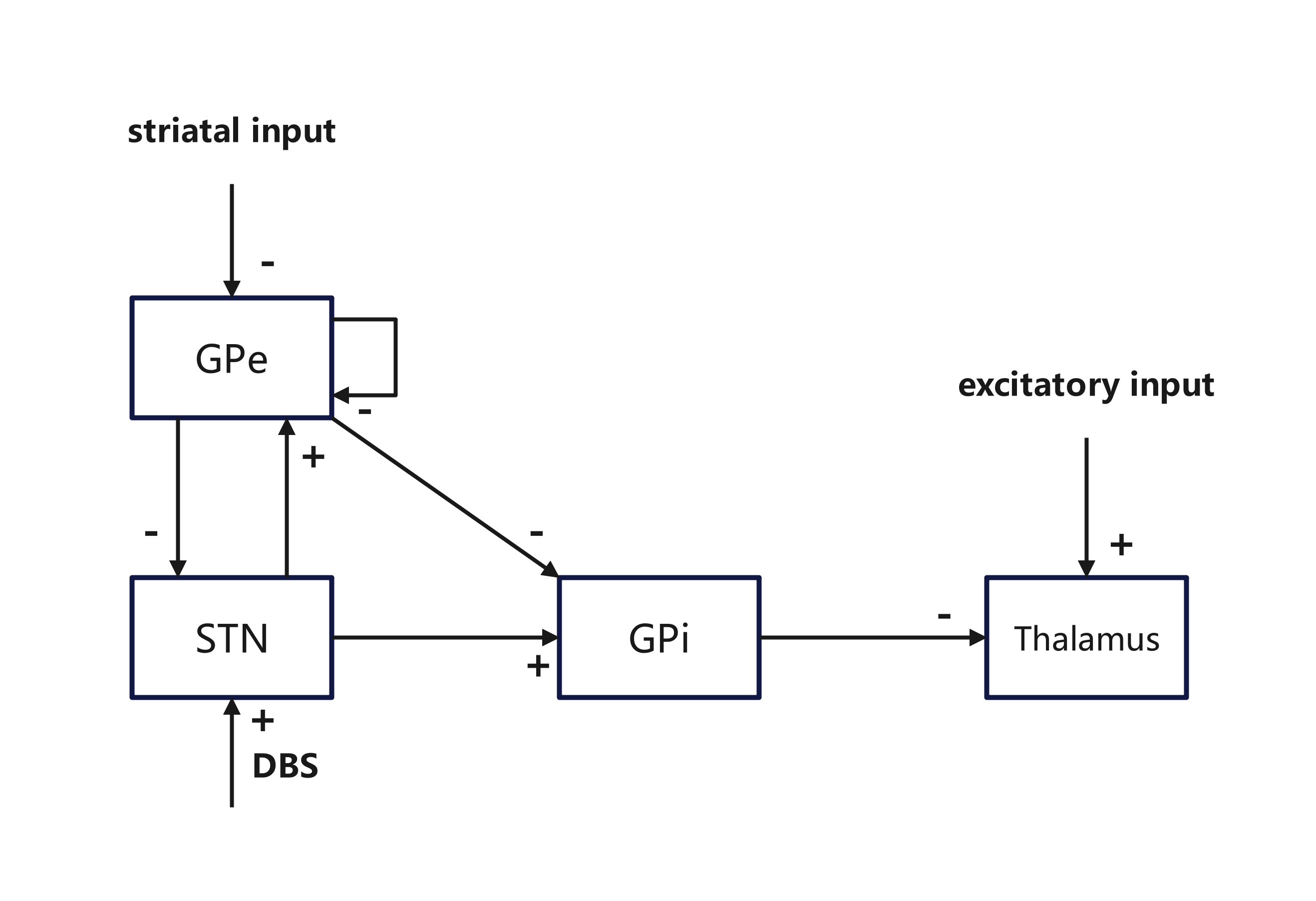
STN consists mainly of glutamatergic projection neurons. It receives input from cortical, thalamic, and brainstem structures and sends excitatory projections to the BG output nucleus and GP (Nambu et al. 2002).
GPi consists mainly of GABA neurons and exerts strong inhibitory effects on neurons in the thalamus and brainstem (Parent et al. 1999). It receives STR input, inhibitory GABA input from GPe, and excitatory glutamatergic input from STN. The function of SNr is similar to that of GPi, and GPi/SNr is usually regarded as a single output structure of BG.
GPe consists mostly of large projection neurons. It is considered an important relay nucleus of the BG. It receives inhibitory input from the striatum and excitatory input from the subthalamus. It provides GABA inhibitory efferent connections to the input and output nuclei of BG (Chan et al. 2005).
SNc consists of large dopamine cells. It provides important regulatory signals to other BG nuclei and external structures (frontal cortex, septal area, amygdala, Etc.) and is an important component of the dopaminergic regulatory system in the BG (Sulzer 2005).
The BG comprises two principal input nuclei, STR and STN(also an important relay), and two principal output nuclei, SNr and GPi. GPe establishes connections between the output nuclei and other nuclei, acting as a relay. Dopaminergic neurons in SNc and VTA provide important regulatory signals to other BG nuclei.
2.2 Synaptic connections and neural circuits in the BG
Albin (Albin et al. 1989) and DeLong (DeLong 1990)proposed two correlational theories in 1989 and 1990: The activity of subpopulations of STR projection neurons is differentially regulated by its afferent nerves. Different subpopulations of STR projection neurons may direct different aspects of motor control. Two important pathways of BG have been proposed: the Direct pathway and the Indirect pathway, known as the classical model of BG function. The classical model assumes that the output of BG is mediated by a direct pathway that promotes cortex activity (cortex-STR-GPi/SNr-thalamus-cortex) and an indirect pathway that inhibits cortex activity (cortex-STR-GPe-STN-GPi/SNr-thalamus-cortex). After the classical model was proposed, it was successively refined. The hyper-direct pathway was added (Nambu et al. 2002).DeLong extended the concept of parallel loops proposed by Alexander et al. (DeLong and Wichmann 2010) (Alexander et al. 1986). Most researchers established the understanding that the indirect pathway inhibits movement while the direct pathway facilitates it. The selection mechanism for decision-making was further developed as the direct pathway is used for selection while the indirect pathway is used for control(Gurney et al. 2001).
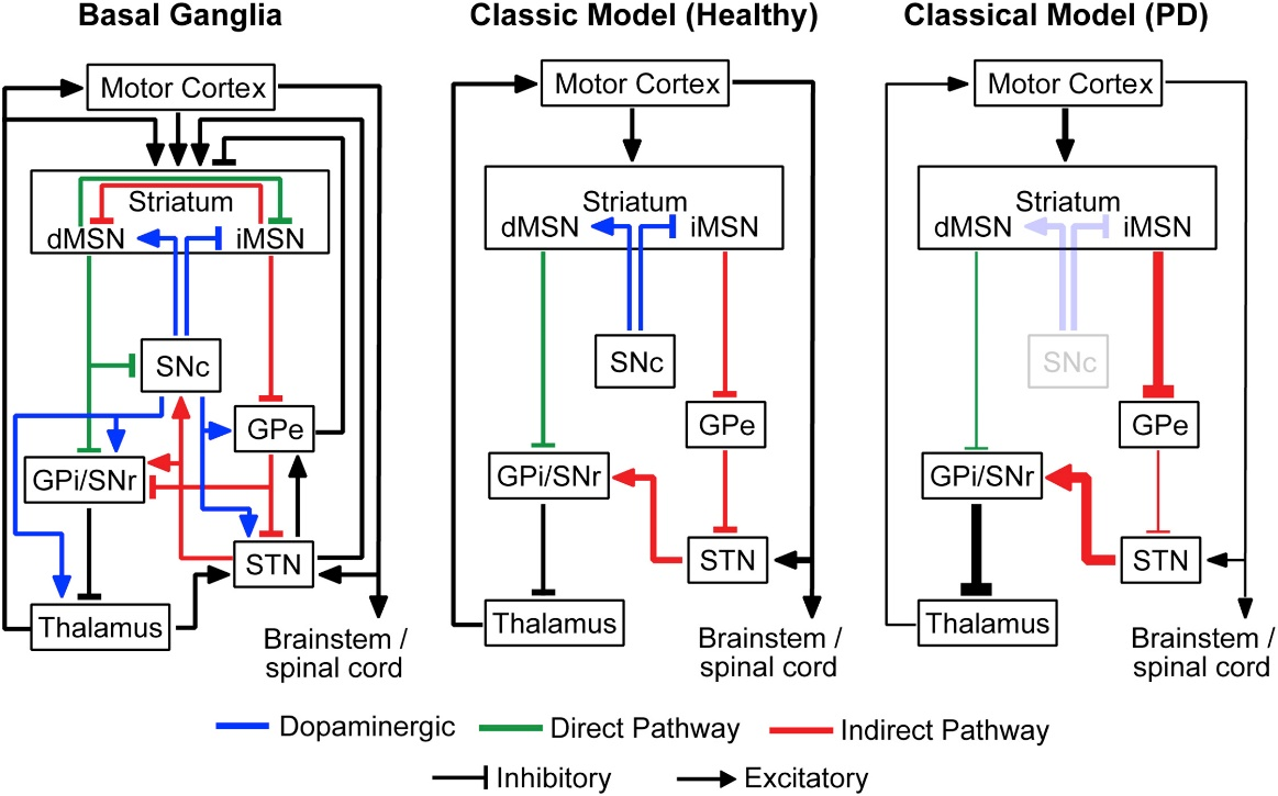
2.3 Pathology of BG in PD
The degeneration of dopaminergic neurons of the SNc in BG of PD patients will decrease the dopamine levels in BG (Jankovic 2008). It may change the BG function and cause an imbalance between direct pathway and indirect pathway. Studies have shown that the loss of BG dopamine in PD patients/primates is associated with sustained changes in firing rates and oscillatory synchrony between BG nuclei (Boraud et al. 2005) (Brown 2007) (Raz et al. 2000). The direct pathway’s effectiveness decreases while the indirect pathway’s effectiveness increases while the firing rates of GPi and STN increase but the firing rate of GPe decreases. The imbalance in the firing activity of these neurons may cause the suppression of the motor system, resulting in various movement disorders (Parker et al. 2018). All of which have been clinically demonstrated (Plenz and Kital 1999).
When the firing rate of BG neurons changes, the firing pattern also changes. Abnormal local field potential (LFP) oscillatory activity occurs in the BG. The activities usually manifest as 13-30 Hz oscillations ( oscillations), particularly pronounced in GPi, GPe, STN, and SNr (Weinberger et al. 2006). Symptoms of motor bradykinesia and rigidity in PD are thought to be closely related to oscillations in the BG (Boraud et al. 2005). Unlike the firing rate changes in the classical model of PD, the mechanism by which oscillations are generated remains unclear. Theoretically, any neuronal network containing negative feedback loops with delays can generate oscillatory activity patterns (Ermentrout et al. 2001). It has been suggested that oscillations may originate from a network of STN and GPe (Gillies et al. 2002). Some computational models of PD-related neural oscillations have been proposed in the academic community (Gillies and Willshaw 2007) (van Albada and Robinson 2009) (Sharott et al. 2005) (Leblois et al. 2006) (Holgado et al. 2010).
3 Mathematical theoretical model
Many computational models have been developed to explain the state of BG-cortical loops in health and disease. It may also contribute to the understanding of the mechanism of DBS. The starting point for modeling is often based on explaining the neuronal mechanisms of BG function and PD symptoms. Since consensus on BG function is still pending, different scholars have chosen different theoretical approaches, resulting in computational models that differ in many aspects.
Making assumptions and simplifying relevant parameters in computational models is always important. Simplifying the necessary components to a minimum can improve the efficiency of computational models. To simulate the changes observed in the firing patterns of pathological BG, we can study neuronal activity at the neuronal level based on neurophysiological and neuroanatomical data. Thanks to the pioneering work of Alan Hodgkin and Andrew Huxley in explaining the generation and propagation of action potentials (Hodgkin and Huxley 1952), scholars have developed a well-established system of neurodynamic theory (Gerstner et al. 2014) (Devaney 2010) (Gerstner et al. 2002). Most mathematical theoretical models have been developed on it. In other words, these models are based on the framework of biological theories rather than on specific patient conditions.
3.1 Explore mechanisms
3.1.1 DBS restores the TC relay function
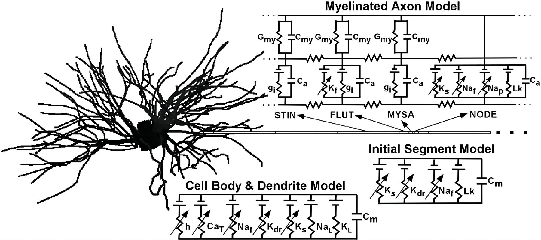
To explore the effect of high-frequency stimulation of the STN on the STN-GPe loop and how their combined output changes the GPi output to the thalamus, Rubin and Terman developed a computational model in 2004 (Rubin and Terman 2004). The model expanded from the previous STN-GPe model they developed in 2002 (Terman et al. 2002). GPi neurons and thalamocortical (TC) neurons were added to the model. Each neuron is constructed in a Hodgkin-Huxley type conductance-based model, represented as a set of nonlinear differential equations. Model neurons can replicate the firing phenomena of real neurons. TC cells of the model received a excitatory input train of spikes and inhibitory inputs from GPi. Figure 4 briefly represents the structure of the model neurons.
The authors quantify the curative effect of DBS by using the error index, which represents the fidelity of the relay of TC cells to their excitatory inputs. The model has three states: normal, PD, and DBS. The output of GPi was irregular and uncorrelated in the normal state. Such output had a low inhibitory effect on thalamic cells, which can transmit excitatory sensorimotor input to cortical areas. The STR input and the GPi inhibition level were adjusted in PD state. The setting produced regular synchronized oscillatory activity in the STN-GPe network. The increased rhythmicity of STN and GPe firing resulted in rhythmic GPi firing. Experimental results suggested that the GPi neurons in the PD state induced -frequency bursting discharges. The error index showed that the switch from irregular to bursting activity in BG impaired the fidelity of TC relay. TC cells were phase-inhibited and no longer completely transmitted motor sensory input of the cortex. In DBS state, the authors used high-frequency DBS at 167 Hz to stimulate STN neurons. The model implies that high-frequency stimulation induced a high-frequency firing rate of inhibitory GPi cells, which resulted in a strong but tonic inhibition. The tonic inhibition may have a much weaker effect on the TC’s ability to transmit motor sensory input than frequency inhibition. In other words, these changes improve TC cells’ responsiveness to excitatory inputs. Thus, the authors concluded that DBS might effectively reduce motor symptoms in PD by eliminating the oscillatory nature of inhibitory inputs (e.g., GPi) to TC cells.
Rubin and Termen provide a better explanation of how STN DBS produces efficacy. Experiments have shown that high-frequency stimulation can promote the discharge of its stimulating target, or at least elicit effects consistent with its output enhancement(Hashimoto et al. 2003)(Hershey et al. 2003)(Windels et al. 2000)(Garcia et al. 2005)(Alavi et al. 2022). If STN DBS enhances the GPi discharge so that it produces sustained, regular oscillations of GPi firing. Then according to the error index, the function of the thalamic relay is significantly improved. The idea provides the first mechanistic theory of DBS that regular spike trains in the BG affect the normal transmission of information in the brain. This mechanism hypothesis suggests that the regularization of GPi inhibitory input is more important than the level of inhibition itself. Thus, disrupting or eliminating this firing pattern may have a significant impact on the brain.
The results of the Rubin-Termen model are very valuable, but it should be noted that the model still has many shortcomings due to the limited hardware and theoretical development at that time. The model can simulate the burst-like firing pattern, but the frequency dependent effects of STN-DBS on PD cannot be reproduced. It does not think about 3D orientation of different neural nuclei and location of stimulation electrodes. So et al. 2012 revised this model(So et al. 2012), but when the DBS frequency was lower as 20 Hz, the stimulation became effective. Thibeault et al. 2013 replaced the neuron models with Izhikevich hybrid neurons for less computational cost in large-scale biophysical models(Thibeault and Srinivasa 2013). Karthik et al. 2016 again revised the model, to better simulate experiment results of 6-OHDA lesioned model(Kumaravelu et al. 2016).
3.1.2 DBS activates soma and axon separately
Close to the time when the model was proposed by Rubin et al., McIntyre and M. Grill used a finite-element model and a multicompartment cable model to study DBS (McIntyre et al. 2004). They focused on individual neuronal responses to DBS. They studied electrophysiological properties of the thalamic cortex, resting neurons and firing neurons with DBS administration. They concluded that the neuronal response to DBS depends mainly on the position and orientation of the axon relative to the electrode and the stimulation parameters.
In McIntyre’s model, direct activation of TC relay neurons by subthreshold stimulation is inhibited by intrinsic firing rate (spiking firing or bursting firing) activity in the stimulus train mediated by activation of presynaptic terminals. Suprathreshold stimulation causes inhibition of intrinsic firing rate in the soma but produces efferent output in axons at the stimulation frequency. In a nutshell, DBS could inhibit activity in the soma while stimulating axons. They considered the independent activation of efferent axons. In their results, axons and cell bodies are independent. The model showed that DBS overrides oscillatory pathological activity and replaces it with more regularized neuronal firing patterns. The concept was called ”Informational Lesion” by Grill et al.(Grill et al. 2004). The model provided a new basis for the hypothesis of a stimulus-induced regulation of cellular activity as a therapeutic mechanism for DBS.
3.1.3 Gpi burst activity in DBS
In 2010, Hahn and McIntyre built a point-neuron model of network dynamics to study the rate and pattern of subthalamopallidal network activity in DBS(Hahn and McIntyre 2010). They built the computational model of the hypothalamus-pallidum base on microelectrode recordings from the basal ganglia (BG) of non-human primates in order to better compare DBS network model predictions and the underlying neurophysiology. Each neuron is a current containing Hodgkin-Huxley type neuron. After training the model to fit in vivo recordings from PD monkeys, the model was used to assess the effects of STN-DBS. The model reproduced the result that reduced GPi activity during DBS in PD monkeys.
In their analysis of GPi burst activity, they proposed three main predictions: a) The volume of the DBS-activated STN is positively related to GPi burst activity. b) DBS stimulation frequency also reduces GPi burst activity and builds its best effect at clinical frequencies. c) Destruction of STN neurons could also produce the same effect as DBS treatment.
3.1.4 Resonant frequency determines the optimal DBS frequency
Although the models mentioned above are very representative, it contains a relatively small number of neurons. It assumes that the STN is uniformly activated for analysis. These settings may result in the model losing some critical information in the simulation of real situations. Therefore, some scholars have scaled the neuronal model to study the heterogeneous and macroscopic network effects of STN high-frequency stimuli when extended to the whole network (Modolo et al. 2007) (Humphries and Gurney 2012) (McIntyre and Hahn 2010). These models are usually large, containing hundreds of neurons.
Humphries and Gurney modified the rat basal ganglia network model they developed in 2006(Humphries et al. 2006) to study DBS(Humphries and Gurney 2012). They set the intensity of stimulation to neurons as related to the distance between that neuron and the stimulating electrode in their model. Humphries used the model to simulate the heterogeneous responses in STN high-frequency stimulation and the heterogeneity of the changes in spiking and bursting firing of GPi neurons. They replicated the changes recorded from primate GPi during STN high-frequency stimulation. These recorded changes suggested that the model predicts a mixed response in the neural network that is consistent with STN-DBS treatment. Finally, Humphries et al. concluded that STN-DBS resets a natural output balance to the GPi. These large network effect models also provided a possible explanation for why stimulation frequencies above 100 Hz are clinically effective stimulation frequencies: the optimal DBS frequency is the resonant frequency of the neuronal network.
Gradinaru et al. innovatively used optogenetic techniques (Gradinaru et al. 2009) to develop an optogenetic DBS model. They proposed a perspective on the effect of high-frequency pulses on DBS in the STN by driving the spiking activity of STN neurons expressing retinoid channels with 130-Hz light pulses. In their model, 130-Hz STN stimulation did not provide relief from behavioral deficits in PD rats. Their result challenged the conclusion drawn from the model above that regulated high-frequency STN spiking activity is critical for the effectiveness of DBS treatment. Humphries compared the optogenetic DBS model with his construction of an electrical DBS model (Humphries and Gurney 2012) (Humphries et al. 2018). They found that the DBS in the optogenetic DBS model and the electrical DBS model differ in their effects on the rate, regularity, and correlation of BG output.
From these models, we can learn that DBS can alleviate motor symptoms in PD, but not necessarily by restoring the diseased neurons to their normal state to achieve the effect.
3.2 Testing different stimulation targets and stimulation patterns
Therapeutic DBS provides continuous high-frequency cyclic stimulation of brain tissue in the target site of stimulation. The almost ”brutal” treatment provides good therapeutic results for patients while causing many undesirable side effects (Zarzycki and Domitrz 2020). Changing the stimulation target and designing new stimulation patterns may improve these side effects and increase efficacy. These improvements can improve the quality of life of patients. For example, new low-amplitude stimulation patterns may extend the life of the stimulator battery and reduce the DBS current spillover beyond the target site. However, the exploration of new modalities and targets can be met with greater resistance in the clinic due to ethical constraints. Computational models offer the possibility of many new stimulation targets and stimulation patterns. Many computational models have been proposed for testing new stimulation targets and modalities.
3.2.1 Different targets of DBS
Pirini et al. used a modified version of the Rubin-Terman model to investigate the performance of BG networks in different stimulus targets (Pirini et al. 2009). SMC sensorimotor cortex was added to the model, and the simple constant currents represent GPe, GPi, and STR inputs. DBS was modeled as a train of rectangular positive current pulses injected directly into cells belonging to the target site. Pirini applied DBS at 30 Hz, 120 Hz, and 180 Hz to three stimulation targets, STN, GPe, and GPi.
Experimental results showed that low-frequency GPe-DBS and GPi-DBS (30 and 60 Hz) were insufficient to drive STR to cause benign effects on TC relay function. STN low-frequency stimulation only partially restored TC relay function. It could partially explain the theory that lower than low-frequency DBS worsens PD symptoms (Perlmutter and Mink 2006). High-frequency STN-DBS restores TC relay function, whereas DBS in GPe and GPi over-activate and inhibit it. These simulation results are consistent with experimental results on DBS network effects achieved in human subjects and monkeys by microdialysis and extracellular recording procedures (Stefani et al. 2005) (Stefani et al. 2006).
3.2.2 Large network model
To investigate the effect of new stimulation patterns on the BG, Kumar and colleagues developed a large network computational model consisting of 3000 neurons (Kumar et al. 2011). They stimulated the STN with two different stimulation patterns. Then they observed and analyzed the role played by these two patterns on the network model. In the periodic stimulation mode, Kumar found that neither excitatory nor inhibitory STN stimulation inputs were critical to making a -oscillation reduction. In contrast, the excitatory and inhibitory inputs of periodic high-frequency stimulation above 100 Hz reduce -oscillation generation. Kumar then designed a non-periodic stimulation pattern such that the pulse period of DBS was . ( is the minimum interval between pulses, fluctuating between 0 and 15 ms. is a randomly chosen integer value between 0 and 3) They found that the stimulation could suppress pathological oscillations more effectively than periodic stimulation.
3.2.3 Test different stimulation patterns
Brocker and M. Grill et al. used the Rubin-Terman model to test new stimulation modalities. The three off-cycle stimulation modalities they designed showed promising properties (Brocker et al. 2013), including but not limited to having lower battery consumption, lower stimulation frequency, and better reduction of dyskinesia. The thalamic DBS mode with brief pauses was subsequently tested. It was found that stimulation pauses reduced the efficacy of open-loop DBS to suppress tremors, suggesting that thalamic DBS may reduce tremors by masking pathological burst discharges of PD in the BG (Swan et al. 2016).
Brocker and M. Grill used a genetic algorithm to apply these different stimulation modes to the model through multiple iterations. They assessed the effect of each stimulus pattern on the network signal transmission to select the best stimulus pattern for the lower frequency (Brocker et al. 2017). After that, Cassar and M. Grill proposed and evaluated several new modifications to the genetic algorithm. The proposed genetic algorithm modifications significantly reduced the number of iterations required for convergence and identified good stimulus patterns (Cassar et al. 2017).
3.2.4 Coordinated Reset
As mentioned above, abnormal neuronal synchronization is one of the hallmarks of PD, in addition to oscillations. Normal DBS stimulation is quickly followed by the reappearance of PD symptoms after cessation. To address these two points, Peter A Tass developed a new simulation method using a mathematical-theoretical model of a coupled phase oscillator that simulates synchronized neurons. The stimulation pattern leads to pathologically sustained desynchronization and ”forgetting” of synaptic connections, resulting in a good therapeutic effect for the patient. Tass called it ”coordinated reset” (CR) (Tass 2003). During CR stimulation, phase-shifted stimuli are delivered to multiple stimulation sites to counteract the synchronization of neurons. Compared to other stimulation modalities, CR does not require precisely timed stimulus delivery, has a better therapeutic effect on the patient, and can reduce side effects. Tass and colleagues then conducted an in-depth study of spike-time-dependent plasticity (STDP) in the nervous system. They found that an uncorrelated firing state and pathological synchrony state can coexist due to the STDP, making it possible for DBS’s therapeutic effects to persist even after stimulation is stopped (Tass and Majtanik 2006). After developing and testing the theory in a computational model, the method was tested in a non-human primate model of PD (Tass et al. 2012b) and PD patients (Adamchic et al. 2014). The results showed that DBS efficacy using the theory persisted for many days after stimulation was terminated. Later Tass et al. extended CR stimulation to the more general field of neuromodulation and found that sensory stimulation and auditory stimulation also improved pathological neuronal synchronization phenomena in PD patients (Popovych and Tass 2012) (Tass et al. 2012a).
3.3 Closed-loop DBS model
In clinical practice, most current DBS systems are open-loop, and the physician’s experience in the clinic mostly determines parameter adjustments. Therefore, DBS systems cannot automatically optimize the stimulation settings to provide different stimulation patterns to different patients (Ramirez-Zamora et al. 2018). Compared to open-loop DBS, closed-loop DBS is a more intelligent stimulation modality. Its stimulation parameters vary with the physiological variables monitored by the electrodes and have a more flexible stimulation pattern. Closed-loop algorithms that adjust stimulation parameters based on patient physiology can help to identify the optimal settings more systematically and efficiently. Computational models can act as virtual patients and are ideal platforms for the first testing and development of adaptive closed-loop algorithms before applying them to patients or even animals. Closed-loop control of DBS is more effective in treating PD than open-loop configurations (Rosin et al. 2011). However, the device and patient situation limit closed-loop algorithms and generally need to satisfy the following conditions (expanded by the guidelines proposed by Feng et al. (Feng et al. 2007)): (1) The algorithm must directly utilize clinical observations that can be measured quickly. (2) The algorithm must validate or falsify the hypothesis in a computational model, in vitro or in vivo. (3) The algorithm needs global search capability to fully utilize the DBS parameter space. (4) The algorithm should be able to operate with different types of DBS stimulation patterns (e.g., periodic and non-periodic stimulation) (5) The algorithm should be able to have good advantages over open-loop mechanical stimulation. (6) The algorithm should have good robustness and be less susceptible to dramatic fluctuations from changes in clinical observables.
A key step in developing such a system is to identify signal characteristics or ”biomarkers” that may quantify the clinical state. In the closed-loop control of DBS in PD, one of the most promising signal features is the level of -band (13-30 Hz) oscillatory activity in the STN and cortex-BG networks (Chen et al. 2006) (Kühn et al. 2008) (Kühn et al. 2009). In addition to the local field potential (LFP), signal features such as microelectrode recordings (MER), imaging data, and external sensors are worth exploring. Moreover, there are various methods to adjust stimulation parameters, such as recursive extended least square method (Zhu et al. 2021), machine learning (Merk et al. 2022), proportional-integral controllers (Fleming et al. 2020), fractional-order controllers (Coronel-Escamilla et al. 2020), and genetic algorithms (Cassar et al. 2017).
3.3.1 Phase response curves in closed-loop DBS
Holt and Netoff used the model of DBS developed by Hahn and McIntyre to investigate closed-loop DBS(Holt and Netoff 2014). They created a mean-field model of the closed-loop system, consisting of discrete time models and their Z-transforms. The simplified mean-field model faithfully reproduces the features of Hahn and McIntyre’s model. The authors proposed that the phase response curve of the oscillation’s response to subthreshold DBS-like pulses is a physiological measure that can be used to predict optimal stimulus frequencies and developed a new method for determining phase response curves from population data. The Fourier coefficients of spike times at 34Hz around low-frequency stimulation were used to estimate the phase of the population and instantaneous amplitude. Curve fitting was then performed on the phase change as a function of the stimulus phase to estimate the phase response curve. The authors showed that the phase response curve can be used to predict the effect of stimulation frequency on the oscillation amplitude. In 2016, they further studied(Holt et al. 2016), showing that the PRC can be used to predict the effects of phasic stimulation on the amplitude of the 34 Hz oscillation, and proposed a phasic burst stimulation protocol optimized using the PRC. The power of pathological 50 Hz oscillations was reduced by nearly 34 with phasic burst stimulation.
3.3.2 Nonlinear predictive control and radial basis function guide
Wang Jiang et al. used the basal ganglion network model developed by Rosa et al. to test the closed-loop stimulation method(Su et al. 2018). They used a nonlinear predictive control algorithm to achieve online tuning of stimulus parameters and then tried to understand the difference between conventional DBS and closed-loop stimulation from the distribution of interspike intervals of BG neurons. The LFP signal of the GPi was used as a feedback signal. The authors assumed that the relationship between the input stimulus signal and the output LFP signal was nonlinear, so a second-order autoregressive Volterra model was used to identify the input-output relationship. By comparing the inter-spike-intervals distributions of various stimulation frameworks, the authors found that their proposed closed-loop system was more therapeutically effective than other stimulation modalities. In 2012, Wang Jiang et al. collaborated with M. Grill to test the new closed-loop stimulation algorithm in a biophysically-based model of the cortex-basal ganglia-thalamus network developed by Kumaravelu et al(Zhu et al. 2021)(Kumaravelu et al. 2016). Guided by the radial basis function network, both the p and pi controllers effectively suppressed the generation of oscillations in the GPi nucleus cluster. These two attempts partially demonstrated the feasibility of power as a feedback signal in DBS.
4 Clinical Predictive Models
In contrast to theoretical mathematical models, patient-based computational models are mostly used in the clinical field. These clinical prediction models help clinicians to predict patient-specific outcomes of DBS and to understand the variability of outcomes across patients (McIntyre et al. 2007). They allow a non-invasive quantitative description of 130the theoretical responses of various neural elements to different stimulus settings. Since the success of DBS surgery depends largely on the accurate placement of electrodes in the brain, 3D brain models generated from imaging data and patient signs can effectively assist the surgeon in the procedure and thus reduce patient risk. Also, because these models simulate the anatomy of the electrodes and surrounding tissue, fitting the models to clinical data allows exploration of the volume of tissue activation (VTA) produced by stimulation and helps to adjust stimulation parameters. Finally, new electrode lead designs can be tested in computational models first.
4.1 Optimize DBS surgery
Presently, the electrode placement and stimulation parameter adjustment of DBS are mostly based on the experience of doctors. The calculation model based on patient imaging data helps doctors understand the situation of patients and help doctors accumulate experience. MiocinovicS and colleagues developed a modeling tool, Cicerone. It is based on Microsoft Windows for clinicians or researchers to establish DBS-related models more conveniently and effectively. It can predict VTA to help doctors visualize the impact of various stimulus parameters on surrounding anatomical structures (Butson et al. 2007) (Miocinovic et al. 2007). Cicerone can also visualize MRI, CT scan, 3D brain atlas, MER data, and the interaction between DBS electrode and VTA in three-dimensional, showing patients’ specific situations more clearly in the eyes of doctors and researchers. The software or other similar systems (Finnis et al. 2003) (D’Haese et al. 2005) is an example of how to use the DBS model to enhance DBS surgery in the future. Preoperative planning allows the definition of stereotactic anatomical targets and trajectories. During operation, stereotactic micro drive coordinates and MER data can be input to realize 3D real-time interactive visualization of electrode position relative to peripheral neuroanatomy and neurophysiology. In addition, neurosurgeons can combine anatomical (such as MRI/CT/3D brain atlas), neurophysiological (such as MER), and electrical (such as DBS VTA) data to optimize the preoperative planning of DBS electrodes before 130permanent implantation.
There are many short-term adverse effects after DBS (Hu et al. 2017a), and more accurate postoperative modeling may help physicians understand the causes of these adverse outcomes and help patients alleviate their symptoms. LEAD-DBS, a MATLAB toolbox, is a similar and relatively novel system with similar functionality to Cicerone (Horn and Kühn 2015). Horn developed the automated DBS electrode reconstruction algorithm in 2015 after analyzing 50 patients who underwent DBS procedures. The toolbox focuses on semi-automatic DBS electrode reconstruction based on postoperative MR or CT data and can visualize DBS lead trajectories and mapping data. The toolbox was used in various clinical cases (Kokkinos et al. 2020) (Al Awadhi et al. 2022) (Yu et al. 2022) or biological models such as rats (Andree et al. 2022), all with good results.
4.2 The model customized for patients.
A limitation of DBS optimization is the need to find clinically effective stimulus parameter settings within the stimuli window that do not cause patient damage, such as efficacious windows that cause side effects. The limitation is a tedious and time-consuming process. Patient-specific DBS computational models based on imaging data and volume conduction models can assist in the process of DBS program control. Quantitative theoretical predictions can be used to define effective stimulation parameter settings. Such models are tailored to the patient and maximize stimulation to defined target areas while minimizing stimulus propagation to non-target areas.
Patient-tailored model contains two important components: imaging data and volume of tissue activation (VTA); imaging data have been described above. Since the effects of electric currents on brain tissue are quite complex and there is still much uncertainty in the academic community about such effects, acquiring VTA requires a combination of means. Finite element models can be used to predict how electric fields penetrate brain tissue (McIntyre and Grill 2001), so generating finite element models from imaging data and pairing them with physiologically realistic neuronal models can accurately predict VTA (Butson et al. 2005).
Although DBS has been increasingly used in the clinic, researchers and physicians still know little about the neural response to applied voltage distribution. They have difficulty relating the modulation of different brain pathways to clinical prognosis. In other words, we want to understand how the specific axonal pathways activated in DBS affect the treatment outcome. To address these issues, Gunalan and McIntyre et al. proposed the pathway activation model (PAM) (Gunalan et al. 2017) based on previous work (Lujan et al. 2012) (Lujan et al. 2013). The PAM considers many factors representing the axonal response in DBS, which are more comprehensive than some diffusion-weighted (DWI)-based fibre bundle, imaging models. These factors include
-
1.
the electrode configuration,
-
2.
the shape, duration, and frequency of the applied stimuli,
-
3.
the electrical conduction properties of the brain tissue medium,
-
4.
the geometry and trajectory of the axons,
-
5.
the membrane biophysics of the axons.
Gunalan and McIntyre et al. attempted to simulate changes in the hyper-direct pathway in DBS with the model. They validated the model in a 67-year-old male patient who had PD for about 11 years and found that their proposed model predictions were in good agreement with the clinical hypothesis. That is, the hyper-direct pathway is directly activated during therapeutic STN-DBS, in agreement with the theory of Li et al.
These patient-specific field-cable pathway activation models are very accurate for quantifying cellular responses to applied electric fields, but like other models, they have several limitations and drawbacks. For example, PAM cannot quantify the network-level modulation of DBS, and its Multi-chamber cable model of axons ignores some of the complex branching patterns of real axons. In addition, PAM is so technically demanding to implement that its use in clinical studies is limited. Gunalan and McIntyre later developed the DF-Howell algorithm to improve the deficiency (Howell et al. 2019).
4.3 Driving new DBS electrode designs
The therapeutic effect of DBS and the amount of brain tissue activation are influenced by the physical properties of the electrodes and the stimulation parameters (McIntyre and Grill 2002). Most of the DBS electrodes currently used surgically are cylindrical with 4-8 contacts around the lead, equally spaced along the length of the lead. Computational models can be used to investigate whether different lead designs lead to differences in electric field distribution (Alonso et al. 2018) and help analyze the effects caused by them. Partitioning the stacked cylindrical electrodes into three-dimensional electrode arrays along and around the DBS electrodes may facilitate more spatially targeted stimulation and recording from deep brain structures. Computational modeling studies have shown that subdividing the circumference of each cylindrical electrode into four rectangular electrodes (Brinda et al. 2021) or four oval electrodes (Vitek and Johnson 2019) (Peña et al. 2017) (Lehto et al. 2017) can improve the ability of the lead to control, move, and affect areas of stimulated tissue within it. Connolly et al. applied the design to the clinical setting by designing new electrodes that segment the ring around the electrodes and found experimentally that such electrodes can control current more effectively (Connolly et al. 2015). Zitella and colleagues tested two computational models and found that current control using segmented electrodes may help focus stimulation when the electrodes are slightly off-target or targeted to brain regions with complex geometry (Zitella et al. 2013). The DBS electrodes with radially distributed electrodes have the potential to improve clinical outcomes by more selectively targeting pathways and networks within the brain (Teplitzky et al. 2016).
Directional DBS electrode designs are now commonly used in clinical practice, with newer directional electrodes segmenting the cylindrical band into three separate electrode contacts. These segmented electrodes have shown promise in redirecting stimulation to the treatment area and away from areas known to produce side effects. Thus, directional DBS electrodes can increase the therapeutic window for clinical stimulation by decreasing the efficacy and side effect threshold. Recently Gilbert et al. compared different approaches for representing targeted DBS electrodes in a finite element volume conductor (VC) model (Frankemolle-Gilbert et al. 2021). They evaluated 15 different variants of the DBSVC model and quantified how their differences affected estimates of the spatial extent of DBS axon activation. The result supposes that different DBS-VC models yielded significant differences in their voltage distributions and axon thresholds. For example, more complex VC models take 2-3 times longer to mesh, construct, and solve for the DBS voltage distribution compared to simpler VC models.
5 Discussion
After more than three decades of development, DBS has become an established intervention for PD and is a well-established treatment recognized by the medical community. However, the exact mechanism of DBS and the optimal site and stimulation parameters remain unresolved, so most treatments are still largely based on trial and error. During the first two decades of DBS development, the developing of stimulation sites and devices for DBS was slow. In any case, in the last decade, computational models have contributed to some extent to the emergence of multiple new stimulation methods and devices that have effectively advanced DBS technology. DBS models have extensively explored major phenomena in PD pathologies, such as beta oscillations, dopamine depletion, and dysfunction of direct and indirect pathways. This review reviews several representative computational models of DBS. Their purposes include, but are not limited to, exploring the mechanisms of DBS, investigating better stimulation targets, optimizing treatment options for clinical patients, and designing new DBS stimulation devices. Although the work to date does not provide an authoritative explanation of the exact mechanisms of DBS concerning the aetiology of PD, the variety of computational models proposed by scholars targeting specific PD phenomena and DBS effects provides a wealth of tools for researchers and physicians.
Each model has its limitations. For example, the basic connectivity within the BG network is much more complex than the model, the model does not take into account the differences between neurons, and the effects of DBS electrical stimulation are oversimplified in the model. Despite these shortcomings, the DBS computational model still provides great help when studying the pathophysiological mechanisms of PD and the potential mechanisms of DBS action. After comparing the models in the review, it is easy to see that mathematical theoretical models attempt to simplify the various influences in DBS in order to analyze them mathematically and thus understand the nature of DBS. In contrast, clinical prediction models tend to consider multiple influences to provide a better analysis of the patient’s condition and to obtain a better treatment outcome, making their models very complex and difficult to parse. Nevertheless, the connection between the two models is much closer than we might expect. Purely mathematical theoretical models must still contain useful clinical parameters, while purely clinical models aimed at improving clinical parameters still need mathematical and neurodynamic theory support. The relation between them also raises the question of how to link mathematical theoretical models more closely to clinical predictive models. It can be seen as a ”from bench to bedside” problem, i.e., how to bridge the gap between theoretical research and clinical applications so that scientific results can better help human beings.
Moreover, since DBS was originally applied in PD, studying DBS is usually closely linked to studying PD. However, DBS researchers can understand the physiological model of BG in addition to focusing on the pathological BG structure. Translating existing physiological models of BG into DBS models seems to be very promising for research. Due to ethical constraints, researchers have difficulty obtaining DBS data from healthy populations when physiological models of BG may be helpful for the research direction. The assumptions of previous models are constantly disproved because of the continuous development of physiological theories. For example, scholars have discovered the importance of GPe in BG (Gittis et al. 2014). They divided the GPe into two major subpopulations of neurons: prototypic and arkypallidal cells. Recent research results used optogenetic stimulation to study the structure of the GPe (Ketzef and Silberberg 2021).Rubchinsky found that the chaotic signals can also disrupt the periodic synchronization of neuron (Rubchinsky et al. 2012). McCarthy et al. focused on the rich kinetic phenomena in the striatum(McCarthy et al. 2011). In their latest model DBS can effectively recover the dynamics of striatal networks(Adam et al. 2022). These novel research results can be well used in computational models.
Furthermore, because of the good results of DBS for PD, physicians and scholars are now applying DBS to more than 30 other diseases (Hariz and Hariz 2013), especially in the group of patients who are poorly controlled despite trying multiple drugs and non-invasive treatments, such as epilepsy, urinary incontinence and depression. In addition, there is increasing academic interest in the mechanisms and optimization of non-invasive stimulation therapies, such as transcranial direct current stimulation, transcutaneous electrical nerve stimulation and transcranial magnetic stimulation. While the benefits of stimulation are clear and robust for some diseases (e.g., PD), there have been some failures in clinical trials for other applications. Furthermore, since the relatively short history of developing DBS treatments for other diseases prevents empiricism from playing a large role, computational models of DBS based on different diseases can be effective in helping to optimize these therapies.
DBS computational models will evolve, perhaps resulting in more concise mathematical formulas or more influential conditions to better match physiological realities. However, in any case, these models can help us uncover more of the brain’s secrets. In the future, computational models could act as virtual patients. Doctors and researchers can test new treatment options on the platform and get good results before implementing them on patients.
Ethics approval
Written informed consent for publication of this paper was obtained from the South China University of Technology, Ruijin hospital and all authors.
Disclosure statement
The authors declare there is no conflicts of interest.
Funding
This study was supported by Medical and Engineering Cross Research Fund from Shanghai Jiao Tong University (KH, YG2019QNA31), Shanghai Municipal Health Commission Clinical Study Special Fund (KH, 20194Y0067), Ruijin Youth NSFC Cultivation Fund (KH, 20212019), Ruijin Hospital Guangci Excellence Youth Training Program (KH, GCQN-2019-B10), Shanghai Pujiang Program (KH, 19PJ1407500), National Natural Science Foundation of China (Grant Nos.: 11572127 and 11872183).
Notes on contributor(s)
First Author and Second Author contribute equally to this work.
References
- Adam et al. (2022) Adam EM, Brown EN, Kopell N, McCarthy MM. 2022. Deep brain stimulation in the subthalamic nucleus for parkinson’s disease can restore dynamics of striatal networks. Proceedings of the National Academy of Sciences. 119(19):e2120808119. https://doi.org/10.1073/pnas.2120808119.
- Adamchic et al. (2014) Adamchic I, Hauptmann C, Barnikol UB, Pawelczyk N, Popovych O, Barnikol TT, Silchenko A, Volkmann J, Deuschl G, Meissner WG, et al. 2014. Coordinated reset neuromodulation for parkinson’s disease: proof-of-concept study. Movement disorders. 29(13):1679–1684. https://doi.org/10.1002/mds.25923.
- Aird (2000) Aird T. 2000. Functional anatomy of the basal ganglia. Journal of Neuroscience Nursing. 32(5):250. https://doi.org/10.1097/01376517-200010000-00002.
- Al Awadhi et al. (2022) Al Awadhi A, Tyrand R, Horn A, Kibleur A, Vincentini J, Zacharia A, Burkhard PR, Momjian S, Boëx C. 2022. Electrophysiological confrontation of lead-dbs-based electrode localizations in patients with parkinson’s disease undergoing deep brain stimulation. NeuroImage: Clinical. 34:102971. https://doi.org/10.1016/j.nicl.2022.102971.
- Alavi et al. (2022) Alavi SM, Mirzaei A, Valizadeh A, Ebrahimpour R. 2022. Excitatory deep brain stimulation quenches beta oscillations arising in a computational model of the subthalamo-pallidal loop. Scientific reports. 12(1):1–20. https://doi.org/10.1038/s41598-022-10084-4.
- Albin et al. (1989) Albin RL, Young AB, Penney JB. 1989. The functional anatomy of basal ganglia disorders. Trends in neurosciences. 12(10):366–375. https://doi.org/10.1016/0166-2236(89)90074-X.
- Alexander et al. (1986) Alexander GE, DeLong MR, Strick PL. 1986. Parallel organization of functionally segregated circuits linking basal ganglia and cortex. Annual review of neuroscience. 9(1):357–381. https://doi.org/10.1146/annurev.neuro.9.1.357.
- Alonso et al. (2018) Alonso F, Vogel D, Johansson J, Wårdell K, Hemm S. 2018. Electric field comparison between microelectrode recording and deep brain stimulation systems—a simulation study. Brain sciences. 8(2):28. https://doi.org/10.3390/brainsci8020028.
- Andree et al. (2022) Andree A, Li N, Butenko K, Kober M, Chen JZ, Higuchi T, Fauser M, Storch A, Ip CW, Kühn AA, et al. 2022. Deep brain stimulation electrode modeling in rats. Experimental Neurology. 350:113978. https://doi.org/10.1016/j.expneurol.2022.113978.
- Ashkan et al. (2017) Ashkan K, Rogers P, Bergman H, Ughratdar I. 2017. Insights into the mechanisms of deep brain stimulation. Nature Reviews Neurology. 13(9):548–554. https://doi.org/10.1038/nrneurol.2017.105.
- Aum and Tierney (2018) Aum DJ, Tierney TS. 2018. Deep brain stimulation: foundations and future trends. Frontiers in Bioscience-Landmark. 23(1):162–182. https://doi.org/10.2741/4586.
- Benabid et al. (1987) Benabid AL, Pollak P, Louveau A, Henry S, De Rougemont J. 1987. Combined (thalamotomy and stimulation) stereotactic surgery of the vim thalamic nucleus for bilateral parkinson disease. Stereotactic and functional neurosurgery. 50(1-6):344–346. https://doi.org/10.1159/000100803.
- Bogacz and Gurney (2007) Bogacz R, Gurney K. 2007. The basal ganglia and cortex implement optimal decision making between alternative actions. Neural computation. 19(2):442–477. https://doi.org/10.1162/neco.2007.19.2.442.
- Boraud et al. (2005) Boraud T, Brown P, Goldberg JA, Graybiel AM, Magill PJ. 2005. Oscillations in the basal ganglia: the good, the bad, and the unexpected. In: The basal ganglia viii. Springer; p. 1–24. https://doi.org/10.1007/0-387-28066-9_1.
- Brinda et al. (2021) Brinda AK, Doyle AM, Blumenfeld M, Krieg J, Alisch JS, Spencer C, Lecy E, Wilmerding LK, DeNicola A, Johnson LA, et al. 2021. Longitudinal analysis of local field potentials recorded from directional deep brain stimulation lead implants in the subthalamic nucleus. Journal of neural engineering. 18(4):046050. https://doi.org/10.1088/1741-2552/abfc1c.
- Brocker et al. (2017) Brocker DT, Swan BD, So RQ, Turner DA, Gross RE, Grill WM. 2017. Optimized temporal pattern of brain stimulation designed by computational evolution. Science translational medicine. 9(371):eaah3532. https://doi.org/10.1126/scitranslmed.aah3532.
- Brocker et al. (2013) Brocker DT, Swan BD, Turner DA, Gross RE, Tatter SB, Koop MM, Bronte-Stewart H, Grill WM. 2013. Improved efficacy of temporally non-regular deep brain stimulation in parkinson’s disease. Experimental neurology. 239:60–67. https://doi.org/10.1016/j.expneurol.2012.09.008.
- Brown (2007) Brown P. 2007. Abnormal oscillatory synchronisation in the motor system leads to impaired movement. Current opinion in neurobiology. 17(6):656–664. https://doi.org/10.1016/j.conb.2007.12.001.
- Brown and Marsden (1998) Brown P, Marsden C. 1998. What do the basal ganglia do? The Lancet. 351(9118):1801–1804. https://doi.org/10.1016/S0140-6736(97)11225-9.
- Brundin and Bloem (2018) Brundin P, Bloem BR. 2018. The times they are a-changin’: Parkinson’s disease 20 years from now. Journal of Parkinson’s disease. 8(Suppl 1):S1. https://doi.org/10.3233/JPD-189002.
- Butson et al. (2005) Butson CR, Cooper S, McIntyre C. 2005. Deep brain stimulation of the subthalamic nucleus: patient-specific analysis of the volume of tissue activated. In: Proceedings of the 10th Annual Conference of the International FES Society.
- Butson et al. (2007) Butson CR, Noecker AM, Maks C, McIntyre CC. 2007. Stimexplorer: deep brain stimulation parameter selection software system. Operative neuromodulation:569–574. https://doi.org/10.1007/978-3-211-33081-4_66.
- Cassar et al. (2017) Cassar IR, Titus ND, Grill WM. 2017. An improved genetic algorithm for designing optimal temporal patterns of neural stimulation. Journal of Neural Engineering. 14(6):066013. https://doi.org/10.1088/1741-2552/aa8270.
- Chan et al. (2005) Chan CS, Surmeier DJ, Yung WH. 2005. Striatal information signaling and integration in globus pallidus: timing matters. Neurosignals. 14(6):281–289. https://doi.org/10.1159/000093043.
- Chen et al. (2006) Chen C, Kühn A, Hoffmann KT, Kupsch A, Schneider GH, Trottenberg T, Krauss J, Wöhrle J, Bardinet E, Yelnik J, et al. 2006. Oscillatory pallidal local field potential activity correlates with involuntary emg in dystonia. Neurology. 66(3):418–420. https://doi.org/10.1212/01.wnl.0000196470.00165.7d.
- Connolly et al. (2015) Connolly AT, Vetter RJ, Hetke JF, Teplitzky BA, Kipke DR, Pellinen DS, Anderson DJ, Baker KB, Vitek JL, Johnson MD. 2015. A novel lead design for modulation and sensing of deep brain structures. IEEE Transactions on Biomedical Engineering. 63(1):148–157. https://doi.org/10.1109/TBME.2015.2492921.
- Coronel-Escamilla et al. (2020) Coronel-Escamilla A, Gomez-Aguilar JF, Stamova I, Santamaria F. 2020. Fractional order controllers increase the robustness of closed-loop deep brain stimulation systems. Chaos, Solitons & Fractals. 140:110149. https://doi.org/10.1016/j.chaos.2020.110149.
- DeLong and Wichmann (2010) DeLong M, Wichmann T. 2010. Changing views of basal ganglia circuits and circuit disorders. Clinical EEG and neuroscience. 41(2):61–67. https://doi.org/10.1177/155005941004100204.
- DeLong (1990) DeLong MR. 1990. Primate models of movement disorders of basal ganglia origin. Trends in neurosciences. 13(7):281–285. https://doi.org/10.1016/0166-2236(90)90110-V.
- Devaney (2010) Devaney AJ. 2010. Mathematical foundations of neuroscience. Mathematical Foundations of Neuroscience.
- D’Haese et al. (2005) D’Haese PF, Cetinkaya E, Konrad PE, Kao C, Dawant BM. 2005. Computer-aided placement of deep brain stimulators: from planningto intraoperative guidance. IEEE transactions on medical imaging. 24(11):1469–1478. https://doi.org/10.1016/10.1109/TMI.2005.856752.
- Dorsey et al. (2018) Dorsey ER, Elbaz A, Nichols E, Abbasi N, Abd-Allah F, Abdelalim A, Adsuar JC, Ansha MG, Brayne C, Choi JYJ, et al. 2018. Global, regional, and national burden of parkinson’s disease, 1990–2016: a systematic analysis for the global burden of disease study 2016. The Lancet Neurology. 17(11):939–953. https://doi.org/10.1016/10.1016/S1474-4422(18)30295-3.
- Ermentrout et al. (2001) Ermentrout B, Pascal M, Gutkin B. 2001. The effects of spike frequency adaptation and negative feedback on the synchronization of neural oscillators. Neural computation. 13(6):1285–1310. https://doi.org/10.1162/08997660152002861.
- Feng et al. (2007) Feng Xj, Greenwald B, Rabitz H, Shea-Brown E, Kosut R. 2007. Toward closed-loop optimization of deep brain stimulation for parkinson’s disease: concepts and lessons from a computational model. Journal of neural engineering. 4(2):L14. https://doi.org/10.1088/1741-2560/4/2/L03.
- Finnis et al. (2003) Finnis KW, Starreveld YP, Parrent AG, Sadikot AF, Peters TM. 2003. Three-dimensional database of subcortical electrophysiology for image-guided stereotactic functional neurosurgery. IEEE transactions on medical imaging. 22(1):93–104. https://doi.org/10.1109/TMI.2002.806567.
- Fleming et al. (2020) Fleming JE, Dunn E, Lowery MM. 2020. Simulation of closed-loop deep brain stimulation control schemes for suppression of pathological beta oscillations in parkinson’s disease. Frontiers in neuroscience. 14:166. https://doi.org/10.3389/fnins.2020.00166.
- Frank et al. (2004) Frank MJ, Seeberger LC, O’reilly RC. 2004. By carrot or by stick: cognitive reinforcement learning in parkinsonism. Science. 306(5703):1940–1943. https://doi.org/10.1126/science.1102941.
- Frankemolle-Gilbert et al. (2021) Frankemolle-Gilbert AM, Howell B, Bower KL, Veltink PH, Heida T, McIntyre CC. 2021. Comparison of methodologies for modeling directional deep brain stimulation electrodes. Plos one. 16(12):e0260162. https://doi.org/10.1371/journal.pone.0260162.
- Garcia et al. (2005) Garcia L, d’Alessandro G, Bioulac B, Hammond C. 2005. High-frequency stimulation in parkinson’s disease: more or less? Trends in neurosciences. 28(4):209–216. https://doi.org/10.1016/j.tins.2005.02.005.
- Gerstner et al. (2002) Gerstner, Wulfram, Kistler, Werner M. 2002. Spiking neuron models: Single neurons, populations, plasticity. Spiking Neuron Models. https://doi.org/10.1108/k.2003.06732gae.003.
- Gerstner et al. (2014) Gerstner W, Kistler WM, Naud R, Paninski L. 2014. Neuronal dynamics: From single neurons to networks and models of cognition. Cambridge University Press. https://doi.org/10.1017/CBO9781107447615.001.
- Gillies et al. (2002) Gillies, Willshaw D, Li Z. 2002. Subthalamic–pallidal interactions are critical in determining normal and abnormal functioning of the basal ganglia. Proceedings of the Royal Society of London Series B: Biological Sciences. 269(1491):545–551. https://doi.org/10.1098/rspb.2001.1817.
- Gillies and Willshaw (2007) Gillies A, Willshaw D. 2007. Neuroinformatics and modeling of the basal ganglia: bridging pharmacology and physiology. Expert review of medical devices. 4(5):663–672. https://doi.org/10.1586/17434440.4.5.663.
- Gittis et al. (2014) Gittis AH, Berke JD, Bevan MD, Chan CS, Mallet N, Morrow MM, Schmidt R. 2014. New roles for the external globus pallidus in basal ganglia circuits and behavior. Journal of Neuroscience. 34(46):15178–15183. https://doi.org/10.1523/JNEUROSCI.3252-14.2014.
- Goetz et al. (2005) Goetz CG, Poewe W, Rascol O, Sampaio C. 2005. Evidence-based medical review update: pharmacological and surgical treatments of parkinson’s disease: 2001 to 2004. Movement disorders: official journal of the Movement Disorder Society. 20(5):523–539. https://doi.org/10.1002/mds.20464.
- Gradinaru et al. (2009) Gradinaru V, Mogri M, Thompson KR, Henderson JM, Deisseroth K. 2009. Optical deconstruction of parkinsonian neural circuitry. science. 324(5925):354–359. https://doi.org/10.1126/science.1167093.
- Grill et al. (2004) Grill WM, Snyder AN, Miocinovic S. 2004. Deep brain stimulation creates an informational lesion of the stimulated nucleus. Neuroreport. 15(7):1137–1140. https://doi.org/10.1097/00001756-200405190-00011.
- Gunalan et al. (2017) Gunalan K, Chaturvedi A, Howell B, Duchin Y, Lempka SF, Patriat R, Sapiro G, Harel N, McIntyre CC. 2017. Creating and parameterizing patient-specific deep brain stimulation pathway-activation models using the hyperdirect pathway as an example. PloS one. 12(4):e0176132. https://doi.org/10.1371/journal.pone.0176132.
- Gurney et al. (2001) Gurney K, Prescott TJ, Redgrave P. 2001. A computational model of action selection in the basal ganglia. i. a new functional anatomy. Biological Cybernetics. 84(6):401–410. https://doi.org/10.1007/PL00007984.
- Hahn and McIntyre (2010) Hahn PJ, McIntyre CC. 2010. Modeling shifts in the rate and pattern of subthalamopallidal network activity during deep brain stimulation. Journal of computational neuroscience. 28(3):425–441. https://doi.org/10.1007/s10827-010-0225-8.
- Hariz and Hariz (2013) Hariz MI, Hariz GM. 2013. Therapeutic stimulation versus ablation. Handbook of clinical neurology. 116:63–71. https://doi.org/10.1016/B978-0-444-53497-2.00006-1.
- Hashimoto et al. (2003) Hashimoto T, Elder CM, Okun MS, Patrick SK, Vitek JL. 2003. Stimulation of the subthalamic nucleus changes the firing pattern of pallidal neurons. Journal of neuroscience. 23(5):1916–1923. https://doi.org/10.1523/JNEUROSCI.23-05-01916.2003.
- Hely et al. (2008) Hely MA, Reid WG, Adena MA, Halliday GM, Morris JG. 2008. The sydney multicenter study of parkinson’s disease: the inevitability of dementia at 20 years. Movement disorders. 23(6):837–844. https://doi.org/10.1002/mds.21956.
- Hershey et al. (2003) Hershey T, Revilla F, Wernle A, McGee-Minnich L, Antenor J, Videen T, Dowling J, Mink J, Perlmutter J. 2003. Cortical and subcortical blood flow effects of subthalamic nucleus stimulation in pd. Neurology. 61(6):816–821. https://doi.org/10.1212/01.WNL.0000083991.81859.73.
- Hodgkin and Huxley (1952) Hodgkin AL, Huxley AF. 1952. A quantitative description of membrane current and its application to conduction and excitation in nerve. The Journal of physiology. 117(4):500. https://doi.org/10.1113/jphysiol.1952.sp004764.
- Holgado et al. (2010) Holgado AJN, Terry JR, Bogacz R. 2010. Conditions for the generation of beta oscillations in the subthalamic nucleus–globus pallidus network. Journal of Neuroscience. 30(37):12340–12352. https://doi.org/10.1523/JNEUROSCI.0817-10.2010.
- Holt and Netoff (2014) Holt AB, Netoff TI. 2014. Origins and suppression of oscillations in a computational model of parkinson’s disease. Journal of computational neuroscience. 37:505–521. https://doi.org/10.1007/s10827-014-0523-7.
- Holt et al. (2016) Holt AB, Wilson D, Shinn M, Moehlis J, Netoff TI. 2016. Phasic burst stimulation: a closed-loop approach to tuning deep brain stimulation parameters for parkinson’s disease. PLoS computational biology. 12(7):e1005011. https://doi.org/10.1371/journal.pcbi.1005011.
- Horn and Kühn (2015) Horn A, Kühn AA. 2015. Lead-dbs: a toolbox for deep brain stimulation electrode localizations and visualizations. Neuroimage. 107:127–135. https://doi.org/10.1016/j.neuroimage.2014.12.002.
- Howell et al. (2019) Howell B, Gunalan K, McIntyre CC. 2019. A driving-force predictor for estimating pathway activation in patient-specific models of deep brain stimulation. Neuromodulation: Technology at the Neural Interface. 22(4):403–415. https://doi.org/10.1111/ner.12929.
- Hu et al. (2017a) Hu K, Moses ZB, Hutter MM, Williams Z. 2017a. Short-term adverse outcomes after deep brain stimulation treatment in patients with parkinson disease. World Neurosurgery. 98:365–374. https://doi.org/10.1016/j.wneu.2016.10.138.
- Hu et al. (2017b) Hu K, Moses ZB, Xu W, Williams Z. 2017b. Bibliometric profile of deep brain stimulation. British Journal of Neurosurgery. 31(5):587–592. https://doi.org/10.1080/02688697.2017.1324109.
- Humphries and Gurney (2012) Humphries MD, Gurney K. 2012. Network effects of subthalamic deep brain stimulation drive a unique mixture of responses in basal ganglia output. European journal of neuroscience. 36(2):2240–2251. https://doi.org/10.1111/j.1460-9568.2012.08085.x.
- Humphries et al. (2018) Humphries MD, Obeso JA, Dreyer JK. 2018. Insights into parkinson’s disease from computational models of the basal ganglia. Journal of Neurology, Neurosurgery & Psychiatry. 89(11):1181–1188. https://doi.org/10.1136/jnnp-2017-315922.
- Humphries et al. (2006) Humphries MD, Stewart RD, Gurney KN. 2006. A physiologically plausible model of action selection and oscillatory activity in the basal ganglia. Journal of Neuroscience. 26(50):12921–12942. https://doi.org/10.1523/JNEUROSCI.3486-06.2006.
- Jaeger et al. (1993) Jaeger D, Gilman S, Aldridge JW. 1993. Primate basal ganglia activity in a precued reaching task: preparation for movement. Experimental Brain Research. 95(1):51–64. https://doi.org/10.1007/BF00229653.
- Jakobs et al. (2019) Jakobs M, Fomenko A, Lozano AM, Kiening KL. 2019. Cellular, molecular, and clinical mechanisms of action of deep brain stimulation—a systematic review on established indications and outlook on future developments. EMBO molecular medicine. 11(4):e9575. https://doi.org/10.15252/emmm.201809575.
- Jankovic (2008) Jankovic J. 2008. Parkinson’s disease: clinical features and diagnosis. Journal of neurology, neurosurgery & psychiatry. 79(4):368–376. https://doi.org/10.1136/jnnp.2007.131045.
- Kawagoe et al. (1998) Kawagoe R, Takikawa Y, Hikosaka O. 1998. Expectation of reward modulates cognitive signals in the basal ganglia. Nature neuroscience. 1(5):411–416. https://doi.org/10.1038/1625.
- Ketzef and Silberberg (2021) Ketzef M, Silberberg G. 2021. Differential synaptic input to external globus pallidus neuronal subpopulations in vivo. Neuron. 109(3):516–529. https://doi.org/10.1016/j.neuron.2020.11.006.
- Kokkinos et al. (2020) Kokkinos V, Urban A, Sisterson ND, Li N, Corson D, Richardson RM. 2020. Responsive neurostimulation of the thalamus improves seizure control in idiopathic generalized epilepsy: a case report. Neurosurgery. 87(5):E578–E583. https://doi.org/10.1093/neuros/nyaa001.
- Kühn et al. (2008) Kühn AA, Kempf F, Brücke C, Doyle LG, Martinez-Torres I, Pogosyan A, Trottenberg T, Kupsch A, Schneider GH, Hariz MI, et al. 2008. High-frequency stimulation of the subthalamic nucleus suppresses oscillatory activity in patients with parkinson’s disease in parallel with improvement in motor performance. Journal of Neuroscience. 28(24):6165–6173. https://doi.org/10.1523/JNEUROSCI.0282-08.2008.
- Kühn et al. (2009) Kühn AA, Tsui A, Aziz T, Ray N, Brücke C, Kupsch A, Schneider GH, Brown P. 2009. Pathological synchronisation in the subthalamic nucleus of patients with parkinson’s disease relates to both bradykinesia and rigidity. Experimental neurology. 215(2):380–387. https://doi.org/10.1016/j.expneurol.2008.11.008.
- Kumar et al. (2011) Kumar A, Cardanobile S, Rotter S, Aertsen A. 2011. The role of inhibition in generating and controlling parkinson’s disease oscillations in the basal ganglia. Frontiers in systems neuroscience. 5:86. https://doi.org/10.3389/fnsys.2011.00086.
- Kumaravelu et al. (2016) Kumaravelu K, Brocker DT, Grill WM. 2016. A biophysical model of the cortex-basal ganglia-thalamus network in the 6-ohda lesioned rat model of parkinson’s disease. Journal of computational neuroscience. 40(2):207–229. https://doi.org/10.1007/s10827-016-0593-9.
- Lanciego et al. (2012) Lanciego JL, Luquin N, Obeso JA. 2012. Functional neuroanatomy of the basal ganglia. Cold Spring Harbor perspectives in medicine. 2(12):a009621. https://doi.org/10.1101/cshperspect.a009621.
- Leblois et al. (2006) Leblois A, Boraud T, Meissner W, Bergman H, Hansel D. 2006. Competition between feedback loops underlies normal and pathological dynamics in the basal ganglia. Journal of Neuroscience. 26(13):3567–3583. https://doi.org/10.1523/JNEUROSCI.5050-05.2006.
- Lehéricy et al. (2005) Lehéricy S, Benali H, Van de Moortele PF, Pélégrini-Issac M, Waechter T, Ugurbil K, Doyon J. 2005. Distinct basal ganglia territories are engaged in early and advanced motor sequence learning. Proceedings of the National Academy of Sciences. 102(35):12566–12571. https://doi.org/10.1073/pnas.0502762102.
- Lehto et al. (2017) Lehto LJ, Slopsema JP, Johnson MD, Shatillo A, Teplitzky BA, Utecht L, Adriany G, Mangia S, Sierra A, Low WC, et al. 2017. Orientation selective deep brain stimulation. Journal of neural engineering. 14(1):016016. https://doi.org/10.1088/1741-2552/aa5238.
- Lozano et al. (2019) Lozano AM, Lipsman N, Bergman H, Brown P, Chabardes S, Chang JW, Matthews K, McIntyre CC, Schlaepfer TE, Schulder M, et al. 2019. Deep brain stimulation: current challenges and future directions. Nature Reviews Neurology. 15(3):148–160. https://doi.org/10.1038/s41582-018-0128-2.
- Lujan et al. (2013) Lujan JL, Chaturvedi A, Choi KS, Holtzheimer PE, Gross RE, Mayberg HS, McIntyre CC. 2013. Tractography-activation models applied to subcallosal cingulate deep brain stimulation. Brain stimulation. 6(5):737–739. https://doi.org/10.1016/j.brs.2013.03.008.
- Lujan et al. (2012) Lujan JL, Chaturvedi A, Malone DA, Rezai AR, Machado AG, McIntyre CC. 2012. Axonal pathways linked to therapeutic and nontherapeutic outcomes during psychiatric deep brain stimulation. Human brain mapping. 33(4):958–968. https://doi.org/10.1002/hbm.21262.
- McCarthy et al. (2011) McCarthy M, Moore-Kochlacs C, Gu X, Boyden E, Han X, Kopell N. 2011. Striatal origin of the pathologic beta oscillations in parkinson’s disease. Proceedings of the National Academy of Sciences. 108(28):11620–11625. https://doi.org/10.1073/pnas.1107748108.
- McGregor and Nelson (2019) McGregor MM, Nelson AB. 2019. Circuit mechanisms of parkinson’s disease. Neuron. 101(6):1042–1056. https://doi.org/10.1016/j.neuron.2019.03.004.
- McIntyre and Grill (2001) McIntyre CC, Grill WM. 2001. Finite element analysis of the current-density and electric field generated by metal microelectrodes. Annals of biomedical engineering. 29(3):227–235. https://doi.org/10.1114/1.1352640.
- McIntyre and Grill (2002) McIntyre CC, Grill WM. 2002. Extracellular stimulation of central neurons: influence of stimulus waveform and frequency on neuronal output. Journal of neurophysiology. https://doi.org/10.1152/jn.2002.88.4.1592.
- McIntyre et al. (2004) McIntyre CC, Grill WM, Sherman DL, Thakor NV. 2004. Cellular effects of deep brain stimulation: model-based analysis of activation and inhibition. Journal of neurophysiology. 91(4):1457–1469. https://doi.org/10.1152/jn.00989.2003.
- McIntyre and Hahn (2010) McIntyre CC, Hahn PJ. 2010. Network perspectives on the mechanisms of deep brain stimulation. Neurobiology of disease. 38(3):329–337. https://doi.org/10.1016/j.nbd.2009.09.022.
- McIntyre et al. (2007) McIntyre CC, Miocinovic S, Butson CR. 2007. Computational analysis of deep brain stimulation. Expert review of medical devices. 4(5):615–622. https://doi.org/10.1586/17434440.4.5.615.
- McNab and Klingberg (2008) McNab F, Klingberg T. 2008. Prefrontal cortex and basal ganglia control access to working memory. Nature neuroscience. 11(1):103–107. https://doi.org/10.1038/nn2024.
- Merk et al. (2022) Merk T, Peterson V, Köhler R, Haufe S, Richardson RM, Neumann WJ. 2022. Machine learning based brain signal decoding for intelligent adaptive deep brain stimulation. Experimental Neurology:113993. https://doi.org/10.1016/j.expneurol.2022.113993.
- Miocinovic et al. (2007) Miocinovic S, Noecker A, Maks C, Butson C, McIntyre CC. 2007. Cicerone: stereotactic neurophysiological recording and deep brain stimulation electrode placement software system. Operative neuromodulation:561–567. https://doi.org/10.1007/978-3-211-33081-4_65.
- Miyasaki et al. (2002) Miyasaki J, Martin W, Suchowersky O, Weiner W, Lang A. 2002. Practice parameter: initiation of treatment for parkinson’s disease: an evidence-based review: report of the quality standards subcommittee of the american academy of neurology. Neurology. 58(1):11–17. https://doi.org/10.1212/WNL.58.1.11.
- Modolo et al. (2007) Modolo J, Mosekilde E, Beuter A. 2007. New insights offered by a computational model of deep brain stimulation. Journal of Physiology-Paris. 101(1-3):56–63. https://doi.org/10.1016/j.jphysparis.2007.10.007.
- Nambu et al. (2002) Nambu A, Tokuno H, Takada M. 2002. Functional significance of the cortico–subthalamo–pallidal ‘hyperdirect’pathway. Neuroscience research. 43(2):111–117. https://doi.org/10.1016/S0168-0102(02)00027-5.
- Nelson and Kreitzer (2014) Nelson AB, Kreitzer AC. 2014. Reassessing models of basal ganglia function and dysfunction. Annual review of neuroscience. 37:117. https://doi.org/10.1146/annurev-neuro-071013-013916.
- Pahwa et al. (2006) Pahwa R, Factor S, Lyons K, Ondo W, Gronseth G, Bronte-Stewart H, Hallett M, Miyasaki J, Stevens J, Weiner W. 2006. Practice parameter: Treatment of parkinson disease with motor fluctuations and dyskinesia (an evidence-based review):[retired]: Report of the quality standards subcommittee of the american academy of neurology. Neurology. 66(7):983–995. https://doi.org/10.1212/01.wnl.0000215250.82576.87 .
- Parent et al. (1999) Parent M, Lévesque M, Parent A. 1999. The pallidofugal projection system in primates: evidence for neurons branching ipsilaterally and contralaterally to the thalamus and brainstem. Journal of chemical neuroanatomy. 16(3):153–165. https://doi.org/10.1016/S0891-0618(99)00008-3.
- Parker et al. (2018) Parker JG, Marshall JD, Ahanonu B, Wu YW, Kim TH, Grewe BF, Zhang Y, Li JZ, Ding JB, Ehlers MD, et al. 2018. Diametric neural ensemble dynamics in parkinsonian and dyskinetic states. Nature. 557(7704):177–182. https://doi.org/10.1038/s41586-018-0090-6.
- Peña et al. (2017) Peña E, Zhang S, Deyo S, Xiao Y, Johnson MD. 2017. Particle swarm optimization for programming deep brain stimulation arrays. Journal of neural engineering. 14(1):016014. https://doi.org/10.1088/1741-2552/aa52d1.
- Perlmutter and Mink (2006) Perlmutter JS, Mink JW. 2006. Deep brain stimulation. Annual review of neuroscience. 29:229. https://doi.org/10.1146/annurev.neuro.29.051605.112824.
- Pirini et al. (2009) Pirini M, Rocchi L, Sensi M, Chiari L. 2009. A computational modelling approach to investigate different targets in deep brain stimulation for parkinson’s disease. Journal of computational neuroscience. 26(1):91–107. https://doi.org/10.1007/s10827-008-0100-z.
- Plenz and Kital (1999) Plenz D, Kital ST. 1999. A basal ganglia pacemaker formed by the subthalamic nucleus and external globus pallidus. Nature. 400(6745):677–682. https://doi.org/10.1038/23281.
- Popovych and Tass (2012) Popovych OV, Tass PA. 2012. Desynchronizing electrical and sensory coordinated reset neuromodulation. Frontiers in human neuroscience. 6:58. https://doi.org/10.3389/fnhum.2012.00058.
- Postuma et al. (2015) Postuma RB, Berg D, Stern M, Poewe W, Olanow CW, Oertel W, Obeso J, Marek K, Litvan I, Lang AE, et al. 2015. Mds clinical diagnostic criteria for parkinson’s disease. Movement disorders. 30(12):1591–1601. https://doi.org/10.1002/mds.26424.
- Ramirez-Zamora et al. (2018) Ramirez-Zamora A, Giordano JJ, Gunduz A, Brown P, Sanchez JC, Foote KD, Almeida L, Starr PA, Bronte-Stewart HM, Hu W, et al. 2018. Evolving applications, technological challenges and future opportunities in neuromodulation: proceedings of the fifth annual deep brain stimulation think tank. Frontiers in Neuroscience. 11:734. https://doi.org/10.3389/fnins.2017.00734.
- Raz et al. (2000) Raz A, Vaadia E, Bergman H. 2000. Firing patterns and correlations of spontaneous discharge of pallidal neurons in the normal and the tremulous 1-methyl-4-phenyl-1, 2, 3, 6-tetrahydropyridine vervet model of parkinsonism. Journal of Neuroscience. 20(22):8559–8571. https://doi.org/10.1523/JNEUROSCI.20-22-08559.2000.
- Rosin et al. (2011) Rosin B, Slovik M, Mitelman R, Rivlin-Etzion M, Haber SN, Israel Z, Vaadia E, Bergman H. 2011. Closed-loop deep brain stimulation is superior in ameliorating parkinsonism. Neuron. 72(2):370–384. https://doi.org/10.1016/j.neuron.2011.08.023.
- Rubchinsky et al. (2012) Rubchinsky LL, Park C, Worth RM. 2012. Intermittent neural synchronization in parkinson’s disease. Nonlinear dynamics. 68(3):329–346. https://doi.org/10.1007/s11071-011-0223-z.
- Rubin and Terman (2004) Rubin JE, Terman D. 2004. High frequency stimulation of the subthalamic nucleus eliminates pathological thalamic rhythmicity in a computational model. Journal of computational neuroscience. 16(3):211–235. https://doi.org/10.1023/B:JCNS.0000025686.47117.67.
- Seger (2008) Seger CA. 2008. How do the basal ganglia contribute to categorization? their roles in generalization, response selection, and learning via feedback. Neuroscience & Biobehavioral Reviews. 32(2):265–278. https://doi.org/10.1016/j.neubiorev.2007.07.010.
- Sharott et al. (2005) Sharott A, Magill PJ, Harnack D, Kupsch A, Meissner W, Brown P. 2005. Dopamine depletion increases the power and coherence of -oscillations in the cerebral cortex and subthalamic nucleus of the awake rat. European Journal of Neuroscience. 21(5):1413–1422. https://doi.org/10.1111/j.1460-9568.2005.03973.x.
- Shohamy et al. (2008) Shohamy D, Myers C, Kalanithi J, Gluck M. 2008. Basal ganglia and dopamine contributions to probabilistic category learning. Neuroscience & Biobehavioral Reviews. 32(2):219–236. https://doi.org/10.1016/j.neubiorev.2007.07.008.
- So et al. (2012) So RQ, Kent AR, Grill WM. 2012. Relative contributions of local cell and passing fiber activation and silencing to changes in thalamic fidelity during deep brain stimulation and lesioning: a computational modeling study. Journal of computational neuroscience. 32(3):499–519. https://doi.org/10.1007/s10827-011-0366-4.
- Stefani et al. (2005) Stefani A, Fedele E, Galati S, Pepicelli O, Frasca S, Pierantozzi M, Peppe A, Brusa L, Orlacchio A, Hainsworth AH, et al. 2005. Subthalamic stimulation activates internal pallidus: evidence from cgmp microdialysis in pd patients. Annals of neurology. 57(3):448–452. https://doi.org/10.1002/ana.20402.
- Stefani et al. (2006) Stefani A, Fedele E, Galati S, Raiteri M, Pepicelli O, Brusa L, Pierantozzi M, Peppe A, Pisani A, Gattoni G, et al. 2006. Deep brain stimulation in parkinson’s disease patients: biochemical evidence. In: Parkinson’s disease and related disorders. Springer; p. 401–408. https://doi.org/10.1007/978-3-211-45295-0_60.
- Su et al. (2018) Su F, Wang J, Niu S, Li H, Deng B, Liu C, Wei X. 2018. Nonlinear predictive control for adaptive adjustments of deep brain stimulation parameters in basal ganglia–thalamic network. Neural Networks. 98:283–295. https://doi.org/10.1016/j.neunet.2017.12.001.
- Sulzer (2005) Sulzer D. 2005. The complex regulation of dopamine output: A review of current themes. Clinical Neuroscience Research. 5(2-4):117–121. https://doi.org/10.1016/j.cnr.2005.08.007.
- Swan et al. (2016) Swan BD, Brocker DT, Hilliard JD, Tatter SB, Gross RE, Turner DA, Grill WM. 2016. Short pauses in thalamic deep brain stimulation promote tremor and neuronal bursting. Clinical Neurophysiology. 127(2):1551–1559. https://doi.org/10.1016/j.clinph.2015.07.034.
- Tass (2003) Tass PA. 2003. A model of desynchronizing deep brain stimulation with a demand-controlled coordinated reset of neural subpopulations. Biological cybernetics. 89(2):81–88. https://doi.org/10.1007/s00422-003-0425-7.
- Tass et al. (2012a) Tass PA, Adamchic I, Freund HJ, von Stackelberg T, Hauptmann C. 2012a. Counteracting tinnitus by acoustic coordinated reset neuromodulation. Restorative neurology and neuroscience. 30(2):137–159. https://doi.org/10.3233/RNN-2012-110218.
- Tass and Majtanik (2006) Tass PA, Majtanik M. 2006. Long-term anti-kindling effects of desynchronizing brain stimulation: a theoretical study. Biological cybernetics. 94(1):58–66. https://doi.org/10.1007/s00422-005-0028-6.
- Tass et al. (2012b) Tass PA, Qin L, Hauptmann C, Dovero S, Bezard E, Boraud T, Meissner WG. 2012b. Coordinated reset has sustained aftereffects in parkinsonian monkeys. Annals of neurology. 72(5):816–820. https://doi.org/10.1002/ana.23663.
- Teplitzky et al. (2016) Teplitzky BA, Zitella LM, Xiao Y, Johnson MD. 2016. Model-based comparison of deep brain stimulation array functionality with varying number of radial electrodes and machine learning feature sets. Frontiers in Computational Neuroscience. 10:58. https://doi.org/10.3389/fncom.2016.00058.
- Terman et al. (2002) Terman D, Rubin JE, Yew A, Wilson C. 2002. Activity patterns in a model for the subthalamopallidal network of the basal ganglia. Journal of Neuroscience. 22(7):2963–2976. https://doi.org/10.1523/JNEUROSCI.22-07-02963.2002.
- Thibeault and Srinivasa (2013) Thibeault CM, Srinivasa N. 2013. Using a hybrid neuron in physiologically inspired models of the basal ganglia. Frontiers in computational neuroscience. 7:88. https://doi.org/10.3389/fncom.2013.00088.
- Turner and Desmurget (2010) Turner RS, Desmurget M. 2010. Basal ganglia contributions to motor control: a vigorous tutor. Current opinion in neurobiology. 20(6):704–716. https://doi.org/10.1016/j.conb.2010.08.022.
- van Albada and Robinson (2009) van Albada SJ, Robinson PA. 2009. Mean-field modeling of the basal ganglia-thalamocortical system. i: Firing rates in healthy and parkinsonian states. Journal of theoretical biology. 257(4):642–663. https://doi.org/10.1016/j.jtbi.2008.12.018.
- Vitek and Johnson (2019) Vitek JL, Johnson LA. 2019. Understanding parkinson’s disease and deep brain stimulation: Role of monkey models. Proceedings of the National Academy of Sciences. 116(52):26259–26265. https://doi.org/10.1073/pnas.1902300116.
- Weinberger et al. (2006) Weinberger M, Mahant N, Hutchison WD, Lozano AM, Moro E, Hodaie M, Lang AE, Dostrovsky JO. 2006. Beta oscillatory activity in the subthalamic nucleus and its relation to dopaminergic response in parkinson’s disease. Journal of neurophysiology. 96(6):3248–3256. https://doi.org/10.1152/jn.00697.2006.
- Windels et al. (2000) Windels F, Bruet N, Poupard A, Urbain N, Chouvet G, Feuerstein C, Savasta M. 2000. Effects of high frequency stimulation of subthalamic nucleus on extracellular glutamate and gaba in substantia nigra and globus pallidus in the normal rat. European Journal of Neuroscience. 12(11):4141–4146. https://doi.org/10.1046/j.1460-9568.2000.00296.x.
- Yu et al. (2022) Yu K, Ren Z, Hu Y, Guo S, Ye X, Li J, Li Y. 2022. Efficacy of caudal pedunculopontine nucleus stimulation on postural instability and gait disorders in parkinson’s disease. Acta Neurochirurgica. 164(2):575–585. https://doi.org/10.1007/s00701-022-05117-w.
- Zarzycki and Domitrz (2020) Zarzycki MZ, Domitrz I. 2020. Stimulation-induced side effects after deep brain stimulation–a systematic review. Acta Neuropsychiatrica. 32(2):57–64. https://doi.org/10.1017/neu.2019.35.
- Zhu et al. (2021) Zhu Y, Wang J, Chang S, Li H, Deng B, Liu C. 2021. Adaptive parameter modulation of deep brain stimulation in a computational model of basal ganglia–thalamic network. Nonlinear Dynamics. 106(1):945–958. https://doi.org/10.1007/s11071-021-06833-1.
- Zitella et al. (2013) Zitella LM, Mohsenian K, Pahwa M, Gloeckner C, Johnson MD. 2013. Computational modeling of pedunculopontine nucleus deep brain stimulation. Journal of neural engineering. 10(4):045005. https://doi.org/10.1088/1741-2560/10/4/045005.