Role of substrate clamping on anisotropy and domain structure in the canted antiferromagnet -Fe2O3
Abstract
Antiferromagnets have recently been propelled to the forefront of spintronics by their high potential for revolutionizing memory technologies. For this, understanding the formation and driving mechanisms of the domain structure is paramount. In this work, we investigate the domain structure in a thin-film canted antiferromagnet -Fe2O3. We find that the internal destressing fields driving the formation of domains do not follow the crystal symmetry of -Fe2O3, but fluctuate due to substrate clamping. This leads to an overall isotropic distribution of the Néel order with locally varying effective anisotropy in antiferromagnetic thin films. Furthermore, we show that the weak ferromagnetic nature of -Fe2O3 leads to a qualitatively different dependence on magnetic field compared to collinear antiferromagnets such as NiO. The insights gained from our work serve as a foundation for further studies of electrical and optical manipulation of the domain structure of antiferromagnetic thin films.
I Introduction
Antiferromagnets (AFMs) exhibit highly favorable properties such as robustness to external magnetic field, no stray field, and ultra-fast switching in the terahertz regime making them promising candidates for novel memory technologies [1, 2, 3, 4, 5]. Recent interest in this field, and the canted antiferromagnet -Fe2O3 in particular, was sparked by the experimental demonstration of electrical control and readout of the Néel order in metallic AFMs [6, 7, 8], insulating AFM/heavy-metal bilayers [9, 10, 11, 12, 13, 14, 15], and the observation of topological spin textures [16, 17, 18] paving the path towards using AFMs as active materials for spintronic devices.
Analogously to demagnetizing fields in ferromagnets, destressing fields have been identified as the dominant driving force behind domain formation in AFMs [19, 20]. So far, it was assumed that the domains are oriented along the magnetocrystalline easy-axes. However, the underlying model is strictly true only for free layers. Our experimental findings on -Fe2O3 thin films imply that substrate clamping results in a far more complex and disordered domain structure. We show that this leads to long-range interactions that impose a locally-varying effective anisotropy. The local fluctuations of the easy-axis orientations qualitatively impact the domain structure and magnetization process. These previously unidentified effects should generally be present and in competition with the magnetocrystalline anisotropy in antiferromagnetic oxide thin films based on their similarities in magnetic, chemical and structural properties [21, 22]. Therefore, the insights and the refined model developed in this work will allow for a more informed understanding of magnetic switching and complex spin textures in easy-plane antiferromagnets.
II Thin film characterization
Our epitaxial -Fe2O3 films (10 nm thick unless mentioned otherwise) were grown at 640 ∘C on -axis (001) oriented Al2O3 with off-axis magnetron sputtering. In order to validate the quality of the thin films, we have performed structure and composition characterization.
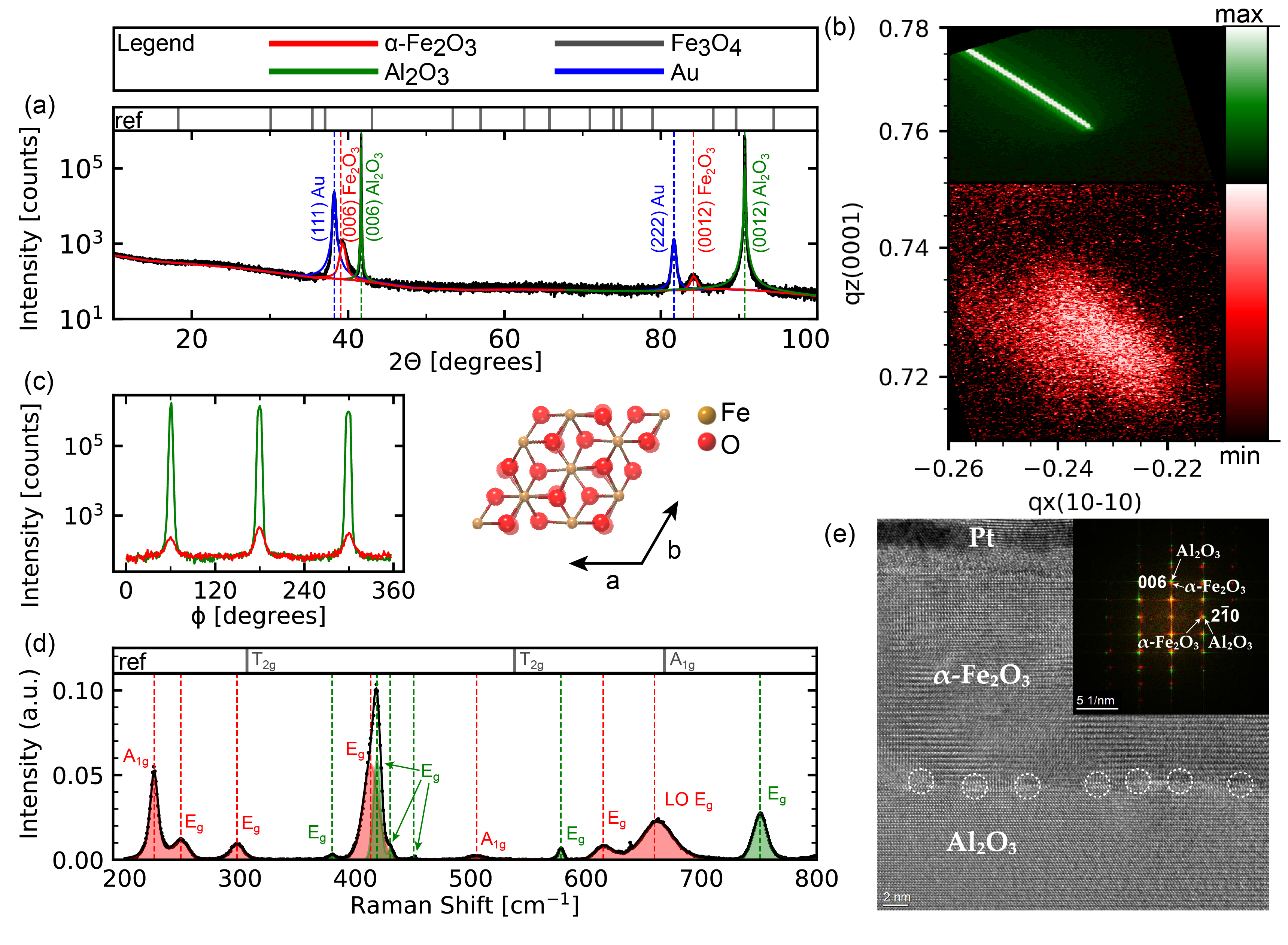
Figure 1a-c show the structure characterization of the -Fe2O3 thin films using x-ray diffraction (XRD) confirming the high epitaxial quality of the thin films (see supplemental material for details [23]). The (006)/(0012) symmetric 2 scan shown in Fig. 1a was used to determine the c lattice parameters of the magnetic thin film and the substrate (c = 13.79 Å and c = 12.99 Å). The film thickness was confirmed to be 11.88 nm (nominally 10 nm) using the Scherrer method which relates peak broadening to the crystallite size. The peaks from gold appear due to the gold contacts on the sample. The strain state and degree of relaxation was determined from a reflection with in-plane and out-of-plane components. In the (1 0 10)- reciprocal space map (Fig. 1b), shifting of the -Fe2O3 film peak around q (red) towards the origin is indicative of film relaxation with respect to the substrate at q (green). At the substrate-film interface, the film experiences strain due to clamping on the substrate, as shown by the shadow peak located along q. Through the thickness of the -Fe2O3 film, the strain relaxes by 93.7 % to a nearly fully relaxed film. The phi-scan (Fig. 1c) confirms the six-fold symmetry in the -Fe2O3 and Al2O3 lattices.
To characterize the film-substrate interface in more detail, we acquired cross-sectional high-resolution transmission electron microscopy (HRTEM) images on a 20 nm -Fe2O3 film (see supplemental material for details [23]). Fig. 1e shows an HRTEM image exhibiting clear atomic lattice plane contrast. The Fourier transform shown as inset in Fig. 1e confirms the high crystallinity of the -Fe2O3 film. To quantify the strain distribution along the thickness of the -Fe2O3 film, we conducted a geometric phase analysis [24]. We find a sharp relaxation after 1 nm of the -Fe2O3 lattice clamped at the Al2O3 interface. This is facilitated by the presence of misfit dislocations and other crystal defects indicated with circles in the HRTEM image in Fig. 1e. Moreover, we observe an almost constant in- and out-of-plane dilation in the -Fe2O3 lattice with respect to the smaller Al2O3 substrate lattice. The relative dilation by 6 % agrees well with the ratio of the lattice plane distances for in-plane and out-of-plane directions. The sharp relaxation and dilation of the -Fe2O3 film support the findings from the XRD measurements described above.
Figure 1d shows the Raman measurement data (black dots) taken using an excitation laser wavelength of 532 nm with fits using Voigt functions (solid line). The center of the peaks are labelled by vertical dashed lines corresponding to the identified Raman modes based on previous reports in literature [25, 26, 27, 28, 29] (see supplemental material for detailed peak information [23]).
In particular, we note the presence of a wide peak at 660 cm-1 corresponding to the Raman-forbidden longitudinal optical (LO) mode in -Fe2O3 due to defect-induced scattering [27, 30, 31].
We find that the experimental data is accurately fitted with Raman peaks corresponding to -Fe2O3 and Al2O3, with a small shift of up to 6 cm-1 for -Fe2O3 compared to the bulk values. This shift agrees well with previous reports that compressive strain as observed in our in -Fe2O3 films in the XRD measurements leads to higher peak center values [25, 32].
We note that the structure and composition characterization based on XRD, HRTEM, and Raman measurements presented here confirm the high epitaxial quality and phase-purity of the -Fe2O3 thin films grown on Al2O3 substrates investigated in this work. Importantly, we do not observe any evidence of impurity phases of Fe3O4 (see reference marks in Fig. 1a,d).
III Experiment
At room temperature, there is a strong easy-plane anisotropy in -Fe2O3 forcing the magnetic moments to lie in the basal plane. Additionally, there is a weak six-fold magnetocrystalline anisotropy within this easy-plane with an anisotropy field of T [33, 34, 35, 36, 37]. The easy-axes are labeled E1, E2, and E3 in the inset of Fig. 3b.
The two magnetic sublattices and of -Fe2O3 are slightly canted due to the Dzyaloshinskii-Moriya interaction (DMI) [38, 39] with a canting angle [40], giving rise to a small FM moment in the basal plane of the hexagonal cell (see inset of Fig. 2b).
Moreover, -Fe2O3 exhibits pronounced magnetoelastic coupling with a magnetostriction constant originating from spontaneous strain [41, 42].
The AFM state is commonly read electrically by measuring the angle-dependent spin Hall magnetoresistance (SMR) in -Fe2O3/Pt bilayers where the perpendicular alignment of the Néel vector with respect to the external magnetic field results in a negative sign of the SMR signal [43, 44, 45, 46]. The electrical measurements presented in this work were performed by applying an AC probing current A and measuring the transverse resistance of the Pt (5 nm thick and 20 m wide) crossbar grown on -Fe2O3 by magnetron sputtering.
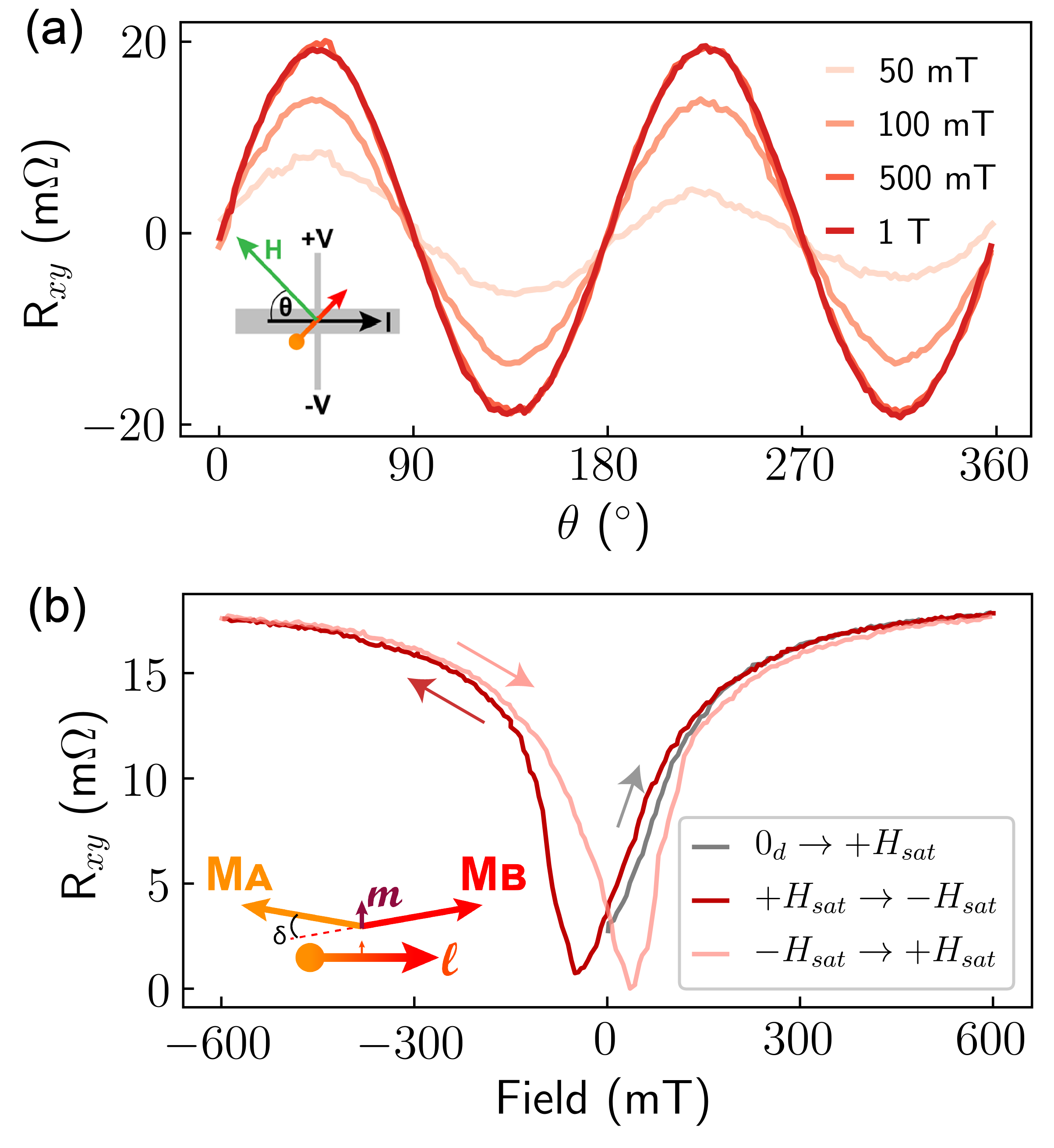
As shown in Fig. 2a, the SMR shows the characteristic dependence, where denotes the angle between the current direction (along easy-axis E2) and the external in-plane magnetic field. A magnetic field smaller than 500 mT is not sufficient to fully reorient the Néel order perpendicular to the field, resulting in a smaller amplitude of the angle-dependent SMR.
Fig. 2b shows a measurement of the field dependence of the SMR at a fixed angle .
Starting from a demagnetized state at zero field , initially increases linearly with magnetic field and saturates at mT. Above , the Néel order is oriented fully perpendicular to the magnetic field. When the field is reduced back to zero, the SMR signal decreases to nearly the initial value at , signaling that the sample has demagnetized into a multi-domain state with nearly zero net Néel vector. This implies the existence of strong internal fields driving the formation of domains.
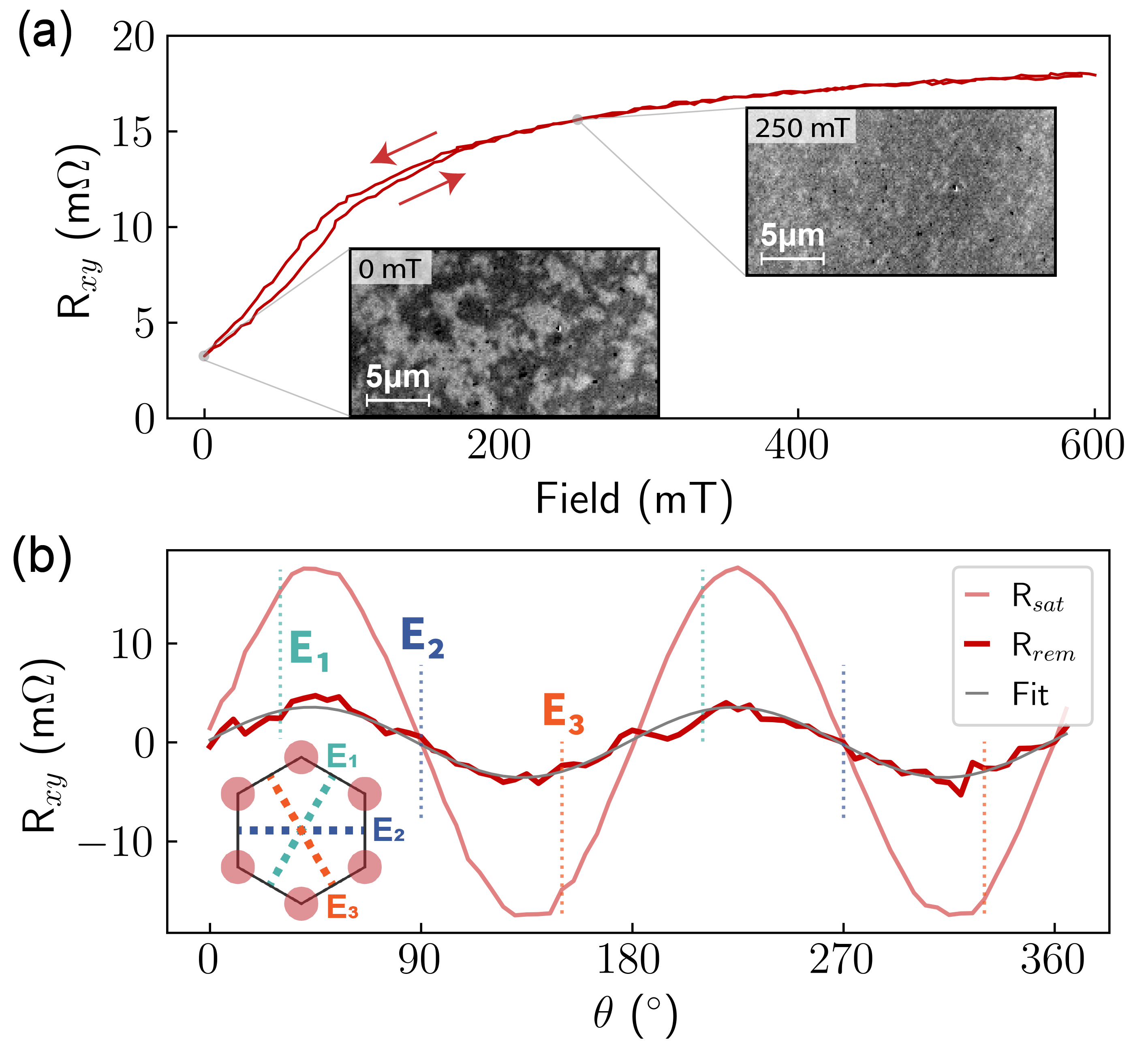
We have confirmed the strong dependence of the AFM order on the magnetic field by measuring x-ray magnetic linear dichroism (XMLD) contrast images of the domain structure of -Fe2O3 by total electron yield using a scanning x-ray microscope (Maxymus) (see Fig. 3a).
In contrast to collinear AFMs such as NiO, we observe clear hysteresis in the SMR signal in -Fe2O3/Pt. We define the location of the minima of to be the coercive field mT. The hysteresis is a signature of the weak ferromagnetic nature of -Fe2O3. In collinear AFMs with an applied field greater than the spin-flop field, the Néel vector can adopt either of the two directions orthogonal to the field. In canted AFMs, on the contrary, the small FM moment prefers to align with the external magnetic field, and hence the degeneracy of the orientation of the Néel order is lifted due to the coupling of the AFM and FM moments by DMI. As a consequence, the interpretation of angle-dependent SMR at low fields, where hysteresis plays a significant role, is not straightforward in -Fe2O3.
Therefore, we focus on a field regime with a single polarity, where all Néel vectors are oriented within a semicircle defined by the direction of the preceding saturating magnetic field. Fig. 3a shows the SMR signal for an in-plane field sweep from to zero and back to oriented at . initially increases linearly with magnetic field. This linear dependence is in stark contrast to the experimentally observed and theoretically modeled quadratic dependence on the magnetic field in the low-field regime previously reported in collinear AFMs such as NiO [44]. We note that there is a small hysteresis below the irreversibility field mT. At low fields , the reorientation of the Néel order is dominated by domain wall movement. Pinning of the AFM domain walls induces irreversibility which manifests in the slight hysteresis of the SMR signal. We can extract an effective pinning field of the AFM domain walls of mT. For , is independent of the preceding magnetic field. In this regime, the reorientation of the Néel order is dominated by coherent rotation of the Néel vectors within the domains and is, hence, fully reversible.
To elucidate further details of the equilibrium AFM domain structure, we have investigated the angular dependence of the remanent state. We applied an external in-plane magnetic field T (light red curve in Fig. 3b) and measured the remanent SMR signal (dark red curve) as a function of the angle of the preceding field. If magneto-crystalline anisotropy were the driving mechanism for relaxation in this system, one would expect the Néel vector to relax towards the closest anisotropy axis depending on the direction of the preceding magnetic field. This would result in maxima and minima in R along E1 and E3 respectively. However, does not show any signature of the crystal symmetry. Fitting to (gray) shows good agreement, implying a simple scaling with between the two curves. This suggests that the remanent signal arises from a small fraction of domains that are oriented perpendicular to the preceding saturating magnetic field direction.
IV Theoretical discussion
In order to identify the different contributions to the formation of the domains, we have investigated the underlying driving mechanisms in more detail. The formation of a multi-domain state when the magnetic field is reduced has been investigated recently in several AFMs [47, 43, 48, 44, 49, 13, 50]. So far, it has been presumed that the domains are oriented along the magneto-crystalline anisotropy axes. The hysteresis and formation of well-defined small domains in zero external magnetic field are evidence for the existence of anisotropy in our system. The lack of signatures of the six-fold symmetry of the magnetocrystalline anisotropy despite the high-quality crystallinity of the epitaxially grown films, however, implies that different contributions are dominant for the effective anisotropy. In the following section, we show that the effective anisotropy is composed of a competition of the short-range magnetocrystalline anisotropy and a long-range contribution of magnetoelastic nature.
First, we note that the spontaneous magnetoelastic strains in the AFM layer are incompatible with the non-strained nonmagnetic substrate and create a long-range so-called destressing field which is similar to the stray field in ferromagnets [19, 20] (see supplemental material for details [23]). The local orientation of the easy magnetic axes is parameterized by the angle that is calculated from the easy-axis E2 within the film plane. By minimizing the energy of the antiferromagnetic layer, we get the following equation for :
| (1) | |||
where denotes the in-plane magnetic anisotropy and is the destressing field which is proportional to the magnetoelastic constant , and is the derivative of the Dirac delta function. Here, we neglect the contributions from the AFM domain walls within the plane.
In the second step, we analyse the possible distribution of the effective anisotropy by solving Eq. (IV). The first term in Eq. (IV) stems from the six-fold crystallographic anisotropy and turns to zero for (direction along E2), (E1), or (E3). The second term originates from the magnetoelastic coupling and depends on the distribution of the magnetic texture in the whole sample. To estimate this term, we note that the destressing fields , satisfy the Poisson equations
| (2) | |||||
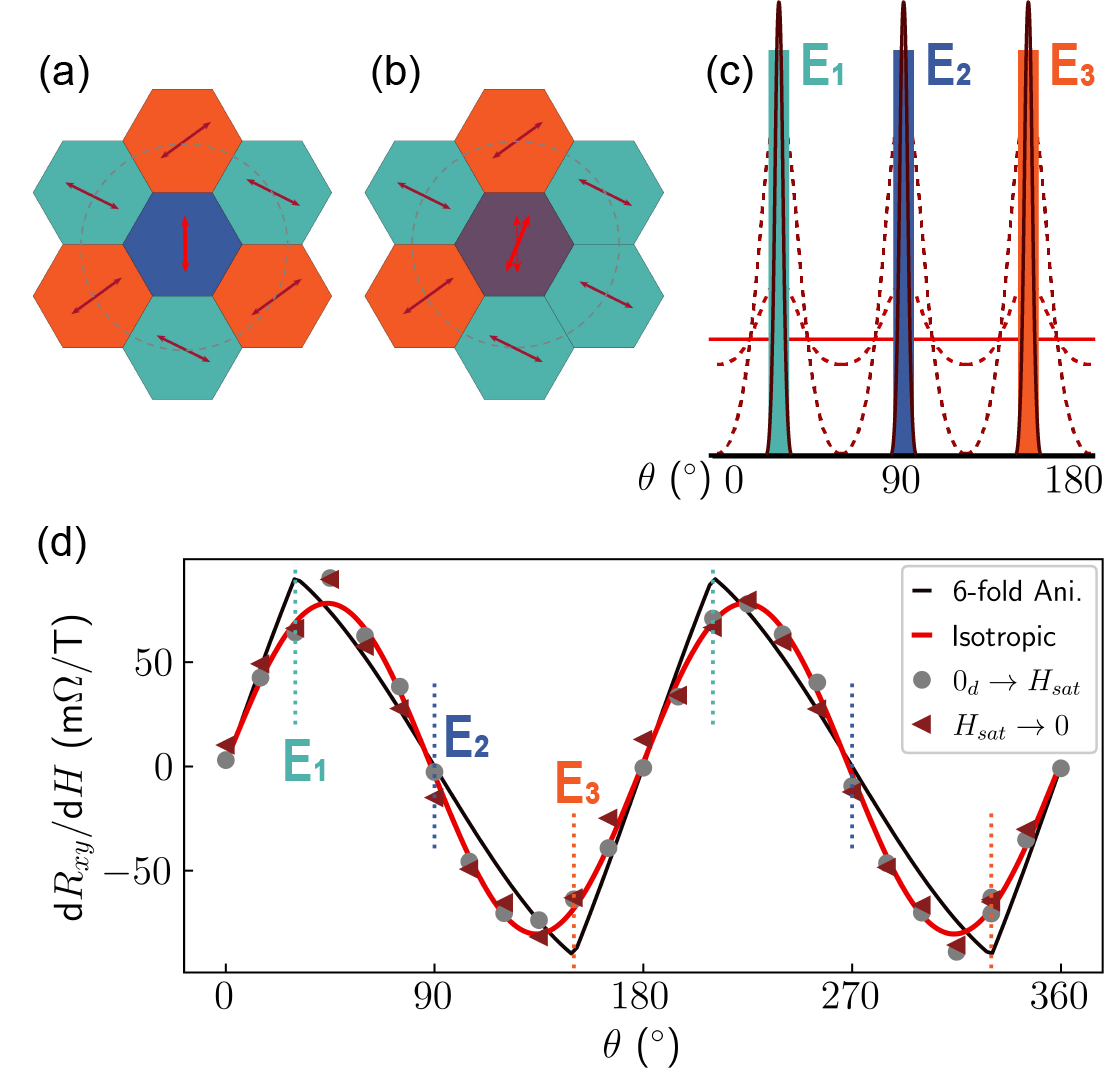
in which the functions , play the role of magnetoelastic charges. Based on the analogy with electrostatics, we conclude that the crystalline anisotropy defined by the first term in Eq. (IV) can only be restored by either a vanishingly small destressing field or an equiprobable distribution of domains in which case the average charges in Eq. (2) vanish. However, in AFM thin-films with finite-sized domains, such averaging can only take place by considering a sufficiently large area including enough different domains. On a smaller scale, the local anisotropy is very sensitive to the direct surroundings. A local imbalance between the domains E1, E2, and E3 creates a local nonzero density of charges leading to a rotation of the local anisotropy axis away from the magnetocrystalline anisotropy axis, as illustrated schematically in Fig. 4a,b. In this example, the Néel vector in the central domain in Fig. 4a is oriented along E2 as long as the areas and of the surrounding domains along E1 and E3 respectively are equal. In this configuration, the magnetoelastic charge is fully compensated.111 The noncompensated charge in this case creates a field that affects the value of the anisotropy but not the direction. However, an imbalance between the areas along E1 and E3 creates a nonzero charge and a related field . As a result, the equilibrium orientation of the Néel vector in the central domain rotates away from the crystalline axis E2 by (Fig. 4b). Hence, spatially fluctuating magnetoelastic charges result in local anisotropy axes which do not have to coincide with the crystalline anisotropy axes [35]. Depending on the relative strength of the crystalline anisotropy and the fluctuating destressing fields, the domains in an AFM thin-film can range from distributed sharply around the crystalline anisotropy axes up to an isotropic distribution as illustrated in Fig. 4c.
Based on our model, we can introduce a characteristic ”Debye radius” at which the effect of fluctuations of the magnetoelastic charges is fully screened. To estimate , we assume that the orientations of different domains are statistically independent. We can model the probability to find a domain with a given orientation using the Boltzmann distribution with effective temperature . In analogy to the Debye model, we estimate the Debye radius , where is the thickness of the AFM layer, and is the sublattice magnetization.
We associate the statistical independence of the domain distribution with the process of the magnetic ordering when cooling through the Néel temperature. However, on a short length scale (below the Debye radius ) temperature-induced redistribution of the domains can be blocked e.g. by domain wall pinning. In this case correlations between the different domain types result in local fluctuations around the equilibrium distribution and rotation of the Néel vector away from the crystallographic directions, as explained above.
In order to verify this model, we investigate the response of the domain structure to an external in-plane magnetic field. For this, we have refined the existing theoretical framework [52, 44, 53], which was developed for a compensated three-domain collinear AFM. Here, we consider the competition between the destressing energy and Zeeman energy. Starting from an equiprobable domain distribution at zero field, the existing theory predicts that the dominant domain fraction increases as (see Eq. S27 in SI) [45]. The DMI in -Fe2O3 and the corresponding nonzero FM moment give rise to an additional Zeeman term proportional to . Our estimations show that the linear Zeeman term dominates over the quadratic term for magnetic fields T. This agrees with our experimental findings that the SMR signal depends linearly on magnetic field in the low field regime (see Figs. 2b and 3a).
To calculate the angular dependence of the SMR signal, we add the Zeeman contribution to Eq. (IV) (see SI). We consider two limiting cases of the equilibrium domain distribution schematically shown in Fig. 4d: i) isotropic distribution of all values due to destressing effects (red line); ii) sharp peaks corresponding to six-fold crystalline anisotropy (dark grey line). Fig. 4d shows the results of the theoretical models and the experimental data of the slope at low field. The experimentally extracted slope dd of the field-dependent SMR signal in the linear regime (triangles and dots) shows the qualitative angle-dependence irrespective of the preceding magnetic field history. This dependence is in agreement with the theoretical prediction
| (3) |
for an isotropic distribution with the maxima located at and . In contrast, the six-fold anisotropy leads to peaks at and 150∘. This further confirms that the orientation of the domains in AFM thin films does not follow the crystal symmetry but lies along isotropically distributed local anisotropy axes due to substrate clamping.
V Experimental verification
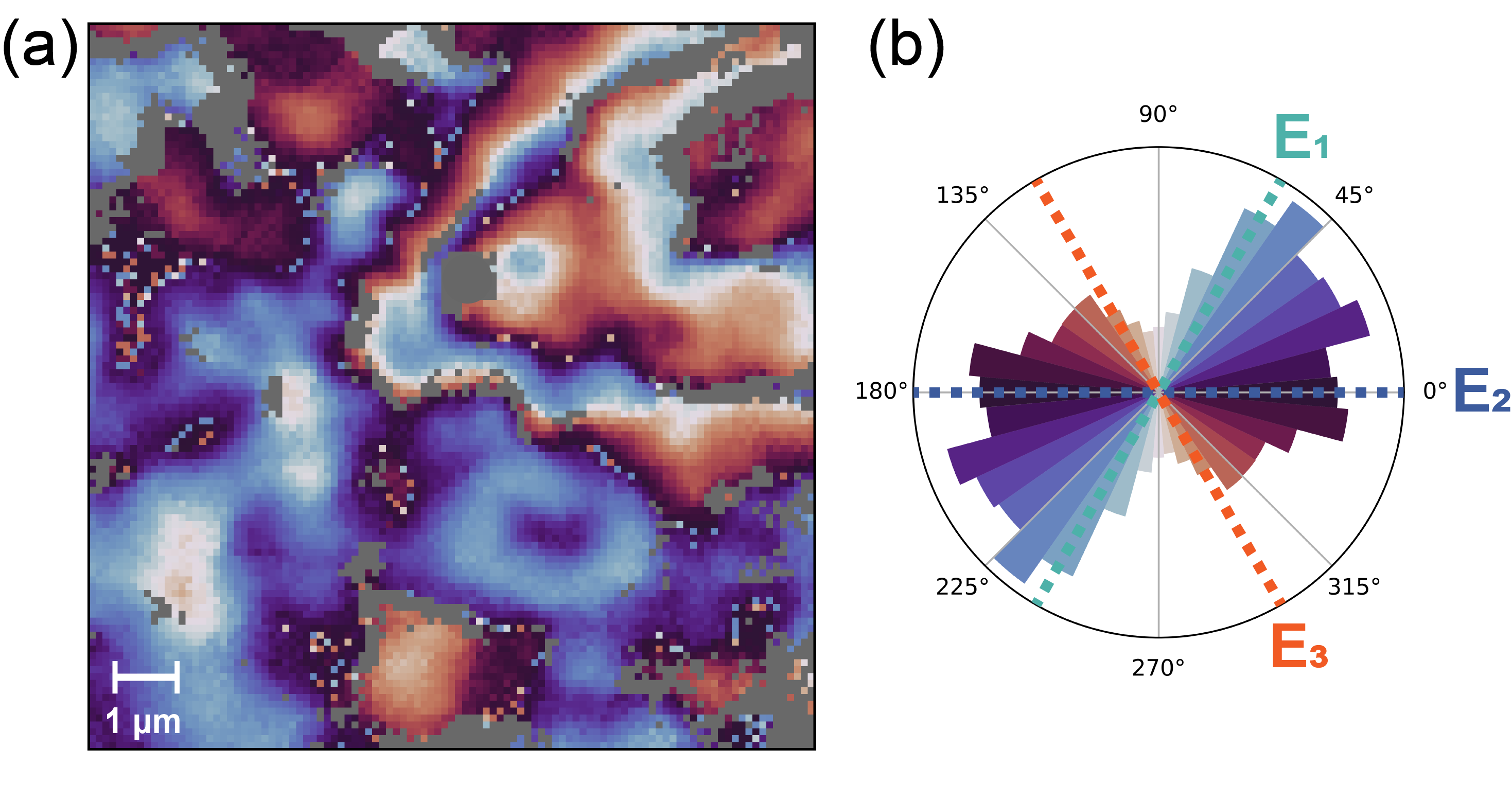
In order to investigate the orientation of the Néel vectors in the multi-domain state, we have performed angle-dependent XMLD x-ray photoemission electron microscopy (XPEEM) on -Fe2O3 (20 nm with 2 nm Carbon capping). Varying the angle between the linear polarization of the incident x-rays and the sample allows reconstruction of the orientation of the Néel orientation (see supplemental material for details of analysis [23]).
Fig. 5a shows a vector map of the antiferromagnetic domain structure. The orientation of the Néel vectors encoded by different colors is shown in the histogram in Fig. 5b. While the histogram does not show any signatures of the hexagonal crystal structure of -Fe2O3, the distribution of domain orientation is not fully isotropic either in the limited field of view of the figure. This agrees well with the model of locally varying effective anisotropies we introduce above.
VI Conclusions
This work highlights the impact of strain effects on the equilibrium domain structure in thin-film AFMs. Combining imaging of the AFM domain structure with electrical measurements, we find that the formation of domains is predominantly driven by destressing fields which do not follow the hexagonal crystal symmetry of -Fe2O3 but rather result in an overall isotropic distribution of the Néel vector orientations with fluctuating easy-axes depending on the local surrounding domains. Our work demonstrates that understanding the formation of the domain structure and effective anisotropy in thin-films is paramount for realizing AFM-based memory technologies.
Acknowledgements.
The authors thank M. Birch, S. Meyer, D. Bono, and B. Neltner for their valuable contributions to this work. We thank A. Lee for the use of his Raman spectrometer and P. Askeland for technical assistance. The x-ray imaging measurements were carried out at the UE-49-PGM-SPEEM (XPEEM) and UE46 MAXYMUS (STXM) beamlines at Helmholtz-Zentrum Berlin. We thank HZB for the allocation of synchrotron radiation beamtime. This work is supported in part by SMART, one of seven centers of nCORE, a Semiconductor Research Corporation program, sponsored by the National Institute of Standards and Technology (NIST), and by the DARPA TEE program. A. W. acknowledges financial support from the Swiss National Science Foundation. O.G. acknowledges support of the ERC Synergy Grant SC2 (No. 610115), the Deutsche Forschungsgemeinschaft (DFG, German Research Foundation) - TRR 173 – 268565370 (project B12) and TRR 288 – 422213477 (project A09). A.L. and D.W. acknowledge funding from the Würzburg-Dresden Cluster of Excellence on Complexity and Topology in Quantum Matter—ct.qmat (EXC 2147, project no. 390858490). F.B. acknowledges financial support from the Helmholtz Young Investigator Group Program. N. B. thanks G.S.D. Beach and his group for their hospitality during his sabbatical visit to MIT.References
- Olejník et al. [2018] K. Olejník, T. Seifert, Z. Kašpar, V. Novák, P. Wadley, R. P. Campion, M. Baumgartner, P. Gambardella, P. Němec, J. Wunderlich, J. Sinova, P. Kužel, M. Müller, T. Kampfrath, and T. Jungwirth, Science Advances 4, eaar3566 (2018).
- Jungwirth et al. [2016] T. Jungwirth, X. Marti, P. Wadley, and J. Wunderlich, Nature Nanotechnology 11, 231 (2016).
- Baltz et al. [2018] V. Baltz, A. Manchon, M. Tsoi, T. Moriyama, T. Ono, and Y. Tserkovnyak, Reviews of Modern Physics 90, 015005 (2018).
- Jungwirth et al. [2018] T. Jungwirth, J. Sinova, A. Manchon, X. Marti, J. Wunderlich, and C. Felser, Nature Physics 14, 200 (2018).
- Železný et al. [2018] J. Železný, P. Wadley, K. Olejník, A. Hoffmann, and H. Ohno, Nature Physics 14, 220 (2018).
- Wadley et al. [2016] P. Wadley, B. Howells, J. Elezny, C. Andrews, V. Hills, R. P. Campion, V. Novak, K. Olejnik, F. Maccherozzi, S. S. Dhesi, S. Y. Martin, T. Wagner, J. Wunderlich, F. Freimuth, Y. Mokrousov, J. Kune, J. S. Chauhan, M. J. Grzybowski, A. W. Rushforth, K. W. Edmonds, B. L. Gallagher, and T. Jungwirth, Science 351, 587 (2016).
- Bodnar et al. [2018] S. Y. Bodnar, L. Šmejkal, I. Turek, T. Jungwirth, O. Gomonay, J. Sinova, A. A. Sapozhnik, H.-J. Elmers, M. Kläui, and M. Jourdan, Nature Communications 9, 348 (2018).
- Godinho et al. [2018] J. Godinho, H. Reichlová, D. Kriegner, V. Novák, K. Olejník, Z. Kašpar, Z. Šobáň, P. Wadley, R. P. Campion, R. M. Otxoa, P. E. Roy, J. Železný, T. Jungwirth, and J. Wunderlich, Nature Communications 9, 4686 (2018).
- Chen et al. [2018] X. Z. Chen, R. Zarzuela, J. Zhang, C. Song, X. F. Zhou, G. Y. Shi, F. Li, H. A. Zhou, W. J. Jiang, F. Pan, and Y. Tserkovnyak, Physical Review Letters 120, 207204 (2018).
- Gray et al. [2019] I. Gray, T. Moriyama, N. Sivadas, G. M. Stiehl, J. T. Heron, R. Need, B. J. Kirby, D. H. Low, K. C. Nowack, D. G. Schlom, D. C. Ralph, T. Ono, and G. D. Fuchs, Physical Review X 9, 041016 (2019).
- Baldrati et al. [2019] L. Baldrati, O. Gomonay, A. Ross, M. Filianina, R. Lebrun, R. Ramos, C. Leveille, F. Fuhrmann, T. R. Forrest, F. Maccherozzi, S. Valencia, F. Kronast, E. Saitoh, J. Sinova, and M. Kläui, Physical Review Letters 123, 177201 (2019).
- Cheng et al. [2020] Y. Cheng, S. Yu, M. Zhu, J. Hwang, and F. Yang, Physical Review Letters 124, 027202 (2020).
- Zhang et al. [2019] P. Zhang, J. Finley, T. Safi, and L. Liu, Physical Review Letters 123, 247206 (2019).
- Chiang et al. [2019] C. C. Chiang, S. Y. Huang, D. Qu, P. H. Wu, and C. L. Chien, Physical Review Letters 123, 227203 (2019).
- Churikova et al. [2020] A. Churikova, D. Bono, B. Neltner, A. Wittmann, L. Scipioni, A. Shepard, T. Newhouse-Illige, J. Greer, and G. S. D. Beach, Applied Physics Letters 116, 022410 (2020).
- Chmiel et al. [2018] F. P. Chmiel, N. Waterfield Price, R. D. Johnson, A. D. Lamirand, J. Schad, G. van der Laan, D. T. Harris, J. Irwin, M. S. Rzchowski, C.-b. Eom, and P. G. Radaelli, Nature Materials 17, 581 (2018).
- Jani et al. [2021] H. Jani, J.-C. Lin, J. Chen, J. Harrison, F. Maccherozzi, J. Schad, S. Prakash, C.-B. Eom, A. Ariando, T. Venkatesan, and P. G. Radaelli, Nature 590, 74 (2021).
- Ross et al. [2020] A. Ross, R. Lebrun, C. Ulloa, D. A. Grave, A. Kay, L. Baldrati, F. Kronast, S. Valencia, A. Rothschild, and M. Kläui, Physical Review B 102, 094415 (2020).
- Gomonay and Loktev [2007] H. V. Gomonay and V. M. Loktev, Physical Review B 75, 174439 (2007).
- Gomonay et al. [2014] O. Gomonay, S. Kondovych, and V. Loktev, Journal of Magnetism and Magnetic Materials 354, 125 (2014).
- Rao [1989] C. N. R. Rao, Annual Review of Physical Chemistry 40, 291 (1989).
- Duò et al. [2010] L. Duò, M. Finazzi, and F. Ciccacci, Magnetic Properties of Antiferromagnetic Oxide Materials: Surfaces, Interfaces, and Thin Films, edited by L. Duò, M. Finazzi, and F. Ciccacci (Wiley, 2010).
- [23] See supplemental Material at xxx for more detailed information .
- Hÿtch et al. [2011] M. Hÿtch, F. Houdellier, F. Hüe, and E. Snoeck, Ultramicroscopy 111, 1328 (2011).
- Lübbe et al. [2010] M. Lübbe, A. M. Gigler, R. W. Stark, and W. Moritz, Surface Science 604, 679 (2010).
- Cvejic et al. [2009] Z. Cvejic, B. Antic, A. Kremenovic, S. Rakic, G. Goya, H. Rechenberg, C. Jovalekic, and V. Spasojevic, Journal of Alloys and Compounds 472, 571 (2009).
- Jubb and Allen [2010] A. M. Jubb and H. C. Allen, ACS Applied Materials and Interfaces 2, 2804 (2010).
- Massey et al. [1990] M. J. Massey, U. Baier, R. Merlin, and W. H. Weber, Physical Review B 41, 7822 (1990).
- Porto and Krishnan [1967] S. P. S. Porto and R. S. Krishnan, The Journal of Chemical Physics 47, 1009 (1967).
- Chernyshova et al. [2007] I. V. Chernyshova, M. F. Hochella Jr, and A. S. Madden, Physical Chemistry Chemical Physics 9, 1736 (2007).
- Xu et al. [2009] Y. Xu, D. Zhao, X. Zhang, W. Jin, P. Kashkarov, and H. Zhang, Physica E: Low-dimensional Systems and Nanostructures 41, 806 (2009).
- Angel et al. [2019] R. J. Angel, M. Murri, B. Mihailova, and M. Alvaro, Zeitschrift fur Kristallographie - Crystalline Materials 234, 129 (2019).
- Tasaki and Iida [1963] A. Tasaki and S. Iida, Journal of the Physical Society of Japan 18, 1148 (1963).
- Flanders and Remeika [1965] P. J. Flanders and J. P. Remeika, The Philosophical Magazine: A Journal of Theoretical Experimental and Applied Physics 11, 1271 (1965).
- Besser et al. [1967] P. J. Besser, A. H. Morrish, and C. W. Searle, Physical Review 153, 632 (1967).
- Chen et al. [2012] P. Chen, N. Lee, S. McGill, S. W. Cheong, and J. L. Musfeldt, Physical Review B - Condensed Matter and Materials Physics 85, 1 (2012).
- Cheng et al. [2019] Y. Cheng, S. Yu, A. S. Ahmed, M. Zhu, Y. Rao, M. Ghazisaeidi, J. Hwang, and F. Yang, Physical Review B 100, 220408 (2019).
- Dzyaloshinsky [1958] I. Dzyaloshinsky, Journal of Physics and Chemistry of Solids 4, 241 (1958).
- Moriya [1960] T. Moriya, Physical Review 120, 91 (1960).
- Morrish [1995] A. H. Morrish, Canted Antiferromagnetism: Hematite (WORLD SCIENTIFIC, 1995).
- Urquhart and Goldman [1956] H. M. A. Urquhart and J. E. Goldman, Physical Review 101, 1443 (1956).
- Voskanyan et al. [1968] R. Voskanyan, R. Levitin, and V. Shchurov, Soviet Journal of Experimental and Theoretical Physics 27, 790 (1968).
- Hoogeboom et al. [2017] G. R. Hoogeboom, A. Aqeel, T. Kuschel, T. T. M. Palstra, and B. J. van Wees, Applied Physics Letters 111, 052409 (2017).
- Fischer et al. [2018] J. Fischer, O. Gomonay, R. Schlitz, K. Ganzhorn, N. Vlietstra, M. Althammer, H. Huebl, M. Opel, R. Gross, S. T. B. Goennenwein, and S. Geprägs, Physical Review B 97, 014417 (2018).
- Lebrun et al. [2019] R. Lebrun, A. Ross, O. Gomonay, S. A. Bender, L. Baldrati, F. Kronast, A. Qaiumzadeh, J. Sinova, A. Brataas, R. A. Duine, and M. Kläui, Communications Physics 2, 50 (2019).
- Geprägs et al. [2020a] S. Geprägs, M. Opel, J. Fischer, O. Gomonay, P. Schwenke, M. Althammer, H. Huebl, and R. Gross, Journal of Applied Physics 127, 243902 (2020a).
- Han et al. [2014] J. H. Han, C. Song, F. Li, Y. Y. Wang, G. Y. Wang, Q. H. Yang, and F. Pan, Physical Review B 90, 144431 (2014).
- Baldrati et al. [2018] L. Baldrati, A. Ross, T. Niizeki, C. Schneider, R. Ramos, J. Cramer, O. Gomonay, M. Filianina, T. Savchenko, D. Heinze, A. Kleibert, E. Saitoh, J. Sinova, and M. Kläui, Physical Review B 98, 024422 (2018).
- Hajiri et al. [2019] T. Hajiri, L. Baldrati, R. Lebrun, M. Filianina, A. Ross, N. Tanahashi, M. Kuroda, W. L. Gan, T. O. Menteş, F. Genuzio, A. Locatelli, H. Asano, and M. Kläui, Journal of Physics: Condensed Matter 31, 445804 (2019).
- Fischer et al. [2020] J. Fischer, M. Althammer, N. Vlietstra, H. Huebl, S. T. Goennenwein, R. Gross, S. Geprägs, and M. Opel, Physical Review Applied 13, 014019 (2020).
- Note [1] The noncompensated charge in this case creates a field that affects the value of the anisotropy but not the direction.
- Gomonay and Loktev [2002] H. Gomonay and V. M. Loktev, Journal of Physics: Condensed Matter 14, 3959 (2002).
- Geprägs et al. [2020b] S. Geprägs, M. Opel, J. Fischer, O. Gomonay, P. Schwenke, M. Althammer, H. Huebl, and R. Gross, Journal of Applied Physics 127, 243902 (2020b).