Opening Amyloid-Windows to the Secondary Structure of Proteins: The Amyloidogenecity Increases Tenfold Inside Beta-Sheets
Abstract
Methods from artificial intelligence (AI), in general, and machine learning, in particular, have kept conquering new territories in numerous areas of science. Most of the applications of these techniques are restricted to the classification of large data sets, but new scientific knowledge can seldom be inferred from these tools. Here we show that an AI-based amyloidogenecity predictor can strongly differentiate the border- and the internal hexamers of -pleated sheets when screening all the Protein Data Bank-deposited homology-filtered protein structures. Our main result shows that more than 30% of internal hexamers of sheets are predicted to be amyloidogenic, while just outside the border regions, only 3% are predicted as such. This result may elucidate a general protection mechanism of proteins against turning into amyloids: if the borders of -sheets were amyloidogenic, then the whole sheet could turn more easily into an insoluble amyloid-structure, characterized by periodically repeated parallel -sheets. We also present that no analogous phenomenon exists on the borders of -helices or randomly chosen subsequences of the studied protein structures.
Introduction
The amyloid status of proteins are studied for a long time because of its relevance in human diseases, including neurodegenerative ailments [1, 2, 3, 4], amyloidoses [5] and other diseases, listed in Table 1 of [2]. Recently, the amyloids gained increased attention as possible antiviral agents [6], or in one of the first successes in the human in vivo CRISPR-Cas9-based gene editing therapy of the transthyretin amyloidosis [7].
The insoluble amyloid state differs from the unstructured protein aggregates; it has a well-defined structure characterized by repetitive parallel -sheets [8, 9].
Numerous globular proteins may turn into insoluble amyloid structures in certain pH and temperature combinations. It is observed that the transition to the amyloid state is similar to the contagious prion-formation: it is related to a core-forming structure [2] for starting the amyloid-transition, and the subsequent propagation of misfolded, insoluble, parallel -sheets [10, 11]. The amyloid propagation is related to the spatially accessible, neighboring -pleated sheet secondary structural elements of proteins [12, 3, 4].
Naturally occurring, globular, soluble proteins need to possess certain structural properties which prevent them from turning into insoluble amyloid aggregates. Experimental studies, together with in silico methods able to predict aggregation-prone regions (APRs) in protein sequences. In the literature, certain residues acting as “gatekeepers” are described as preventing those conformational changes to amyloid structures: aspartic acid and glycine in bacterial curlin subunits CsgA and CsgB [13], lysine in transthyretin [14] or in peptides Trpzip1 and Trpzip2 [15].
Our hypothesis is that the borders of -sheets in globular, soluble proteins in general (and not only in the specific examples listed above) need to be protected against turning to amyloid structures; otherwise the whole polypeptide chain bordering the -sheet subsequences would be transformed to amyloids. We examine this hypothesis in the present work by screening the starting and ending subsequences, or in other terminology, the prefixes, and suffixes of the -sheets of the homology-filtered Protein Data Bank (PDB) entries.
Previous work
In [8], a clean geometrical formulation was given for the characterization of the amyloid-like structures in length, distance, and parallelism of their -sheets, and by the search of these geometric properties, an automatically updated list of the PDB-entries was published and maintained at the address https://pitgroup.org/amyloid/.
In [9], the amyloid-precursor molecules of https://pitgroup.org/amyloid/ were classified by the prefixes of their parallel -sheet subsequences, and these prefixes were also searched for in the whole Protein Data Bank [16]. The remarkable findings include the major histocompatibility complexes MHC-1 and MHC-2, the p53 tumor suppressor protein, and an anti-coagulant peptide molecule.
With a prediction tool, based on artificial intelligence, in [17], we partitioned all the possible hexapeptides into two classes: “amyloidogenic” and “non-amyloidogenic” with more than 84% correctness. The prediction tool, which applies a linear Support Vector Machine (SVM) [18], was trained by the Waltz dataset of 514 amyloidogenic and 901 non-amyloidogenic hexapeptides [19, 20]. The corresponding web-based tool is available as the Budapest Amyloid Predictor at https://pitgroup.org/bap.
As a surprising application of the Budapest Amyloid Predictor, numerous amyloid- and non-amyloid hexapeptide patterns were identified in [21]. For example, it was shown that for all independent amino acid substitutions for the positions marked by “x”, the CxFLWx or FxFLFx patterns describe amyloidogenic hexamers, while the patterns PxDxxx or xxKxEx describe non-amyloidogenic hexamers. Note that each pattern with two x’s describes , while those with four x’s describe different hexapeptides succinctly if for the positions x, we can substitute the 20 amino acid residues.
In the present work, we study the prefixes and suffixes of the -sheet subsequences of the PDB-deposited structures and apply the Budapest Amyloid Predictor to the hexamers of the border regions on these -sheets. As we show below, the number of the amyloidogenic hexapeptides of the border regions of the -sheets is one-tenth of the same number in the inner parts of those sequences by screening the homology-filtered Protein Data Bank entries. We believe that this finding is a very strong evidence for our hypothesis.
We remark that applying the artificial intelligence-based Budapest Amyloid Predictor for the 64 million () possible hexapeptides is a crucial step here: the largest experimental dataset that labels the hexapeptides by their amyloidogenic propensity, the Waltz dataset, contains only 1415 hexapeptides [19, 20], while we analyzed the prefixes and suffixes of more than 110,000 -sheets from the homology-filtered PDB. Gaining experimental data for that many hexapeptides is intractable today.
Methods
For the statistical analysis involving the entries of the Protein Data Bank (PDB) [16], usually some “non-redundant” homology-filtered PDB-subsets are applied because otherwise, the multiple depositions of more important or more researched protein structures (e.g., several thousand copies of the HIV-1 protease) would impact the results. In this study, we have made use of the representative polypeptide chains of the 30% homology-filtered set, which was available at the URL https://www.rcsb.org/pdb/rest/representatives?cluster=30 (downloaded on May 21, 2020).
Technical note for reproducibility of the present work: At the time of writing this section, we were informed that the administrators of the RCSB PDB have removed the link target indicated above, but one can still generate the similar non-redundant set with an advanced search feature of the RCSB PDB, namely https://search.rcsb.org/#group-by-context, by choosing 30% sequence identity.
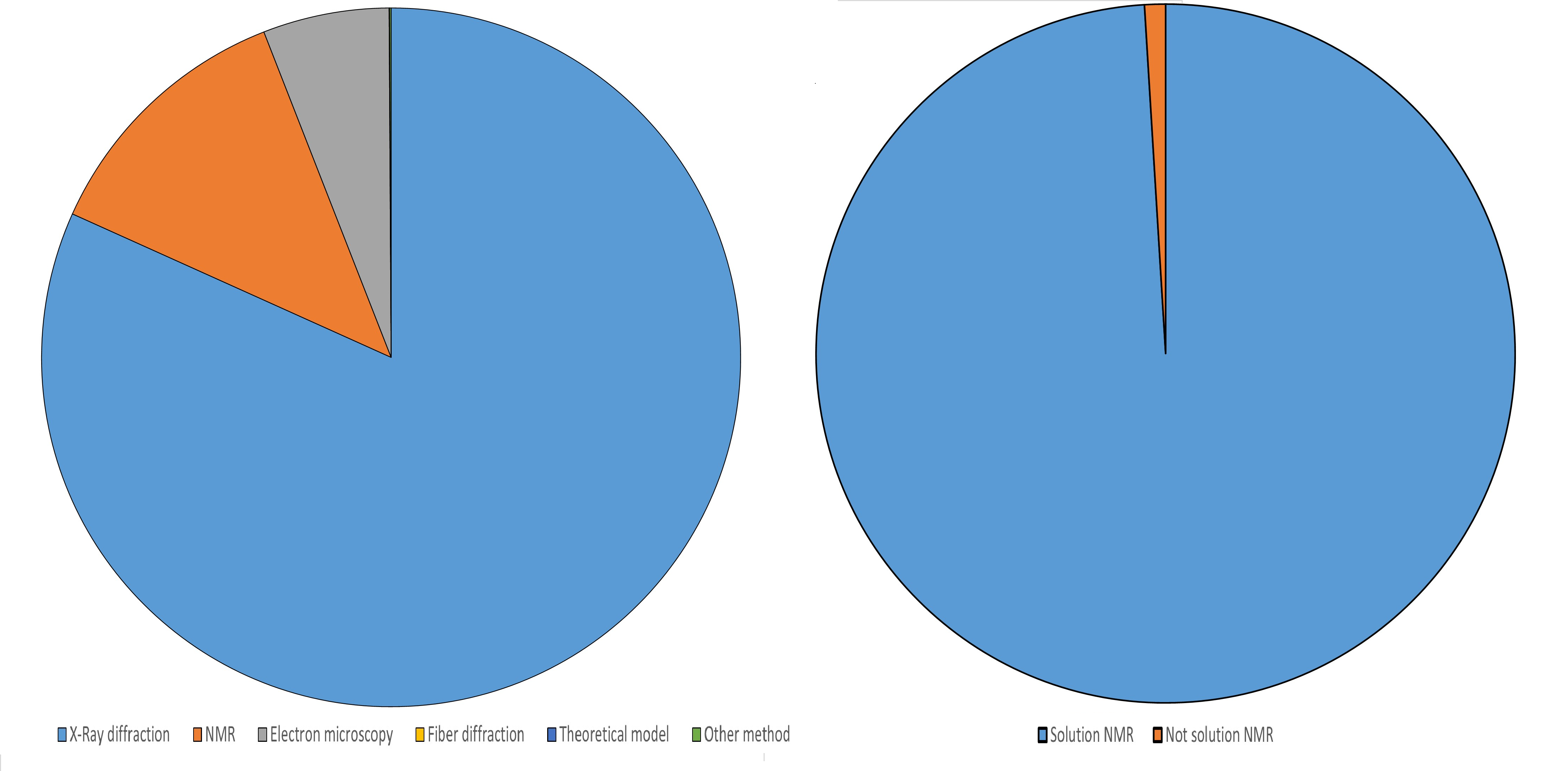
The structure of almost all representative members of this non-redundant set was identified in a soluble state, so almost all proteins in the set are water-soluble (see Figure S3 in the supporting material).
From the representative sequences of the non-redundant set, we have identified the maximal contiguous subsequences corresponding to -sheets by applying the “SHEET” records in the PDB file. In other words, all the non-expandable contiguous -sheet subsequences were identified. Those subsequences whose lengths are less than 6 were discarded. This way, we have identified as many as 117,467 subsequences.
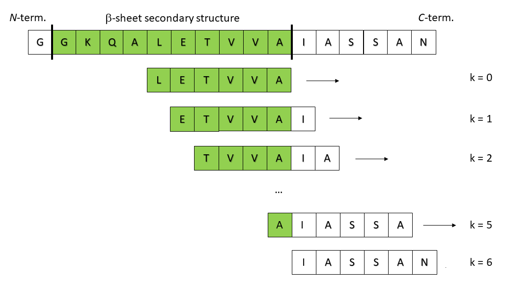
In the next step, we listed the six-residue long sliding windows through the borders of these 117,467 -sheet subsequences, as it is visualized in Figure 1.
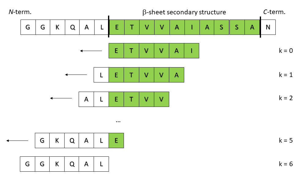
The six residue-long windows map the border regions of the -sheets, identified from the “SHEET” record of the PDB file. For each contiguous -sheet section, we consider the sliding windows of its left border (Figure 1), called prefixes, and the seven sliding windows at the right border, called suffixes. That is, for each of the 117,467 unique subsequences, we have listed the seven prefix and the seven suffix sequences (for ), and applied the Budapest Amyloid Predictor [17] at https://pitgroup.org/bap for assigning one of the “amyloidogenic” or “non-amyloidogenic” labels for each of the seven length-6 windows at both ends of the 117,467 subsequences.
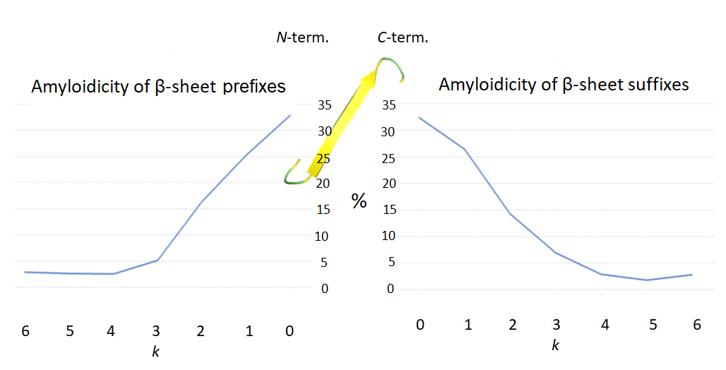
Next, we have drawn a diagram, Figure 2, which contains at the axis the value (i.e., the length of the window over (or outside) the -sheet sequence (colored by green in Figure 1). On the axis, we give the percentage of the predicted amyloidogenic length-6 windows, computed by the Budapest Amyloid Predictor. In other words, the value gives the fraction of the hexapeptides with exactly residues outside the -sheet region, predicted to be amyloidogenic.
Discussion and results
The rapid development of artificial intelligence tools may open up new insights into the structural properties of biological macromolecules. For example, the largest available hexapeptide database, the Waltz dataset [19, 20] today contains 1415 hexapeptides, labeled as “amyloidogenic” or “non-amyloidogenic”, based on different types of experimental data. The total number of all the hexapeptides from the biologically most relevant 20 amino acids is . Therefore, it is improbable that experimental evidence could be gained for the amyloid-forming properties of all of these hexapeptides.
By artificial intelligence tools, however, we were able to build the Budapest Amyloid Predictor, which predicts the amyloidocity of any hexapeptide from these 64 million with an 84% success rate [17], https://pitgroup.org/bap.
The BAP predictor makes it possible to assign amyloidogenecity labels to any hexapeptides, not only the experimentally identified ones. Consequently, we may screen all the given secondary structures deposited in the PDB and compute the prediction for any hexapeptides in them. These actions were not possible without highly developed artificial intelligence tools.
Our results are summarized in Figure 2.
Figure 2 shows a monotonic tenfold increase of the amyloidogenecity of the hexapeptides, described by the sliding windows, for positions entirely in the -sheet sequences (), relative to the other positions, when the largest part of the windows are outside of the -sheet region . This observation validates our hypothesis, described in the Introduction, namely, the prefixes and the suffixes of the -sheet subsequences need to be protected against turning into amyloid structures Otherwise, the whole polypeptide chain bordering the -sheet subsequences would be transformed to amyloids.
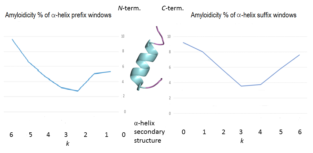
The -helix control
For a simple control of our hypothesis, we also prepared the sliding prefix- and suffix windows for the -helices, found by screening all the homology-filtered PDB entries. As the supporting Figure S2 shows in the Supporting Material, no similar observation can be done in the case of the -helices.
The random control
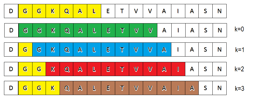
For another simple control, let us consider random (contiguous) subsequences of the PDB-deposited proteins instead of subsequences of -sheets or -helices and evaluate their prefixes or suffixes for amyloidogenecity. The following reasoning shows that in this case, the amylodocity percentage of prefixes or suffixes of different values are almost the same (the “almost” adjective is explained below). We consider prefixes here; the proof for suffixes is analogous.
We refer to Figure S4 in the Supporting Material for visualizing the reasoning.
First, assume that we consider only fixed-length subsequences (of length ). Let us consider a hexapeptide, which is not too close to the right end of the protein sequence, more exactly, whose first position is at least 6+ residues from the right end (denoted by yellow in the first row of Fig. S4).
It is easy to see that the very same (yellow) hexapeptide may be a prefix-window with for the green, with for the blue, with for the red, or with for the brown subsequence of length . That is, if a hexapeptide is not too close to the right end of a protein chain, then it appears with for different length sequences. Let us remark that the amyloidocity of a hexapeptide depends only on its sequence, so there would not be any difference of prefix-amyloidocities for different values, unlike in the case of -sheets in Fig. 2. This reasoning works also for random subsequences of different lengths, and also for suffixes. Small variability of the appearance percentage of a given hexapeptide may occur only if it is close to the right end of the protein-sequence and, consequently, cannot appear with larger or values.
Conclusions
Here we screened all the prefix- and suffix sequences of length-6 of the -sheets in the homology-filtered Protein Data Bank and have found that by crossing the boundaries of the -sheet sequences the amyloidogenic ratio of the hexapeptides, characterized by the length-6 sliding windows, moving from outside to inside of the -sheet sequence, increases more than tenfolds.
No similar phenomenon can be observed in the case of -helices or random subsequences. We strongly believe that this fact shows the validity of our hypothesis, namely, the prefixes and the suffixes of the -sheet subsequences need to be strongly protected against turning into amyloid structures, otherwise the whole polypeptide chain, bordering the -sheet subsequences would possibly be transformed into amyloids.
Data availability
The Budapest Amyloid Predictor webserver is available freely at https://pitgroup.org/bap.
Funding
KT, BV, and VG were partially supported by the NKFI-127909 grant of the National Research, Development and Innovation Office of Hungary. KT, BV, AP, VF, and VG were partially supported by the ELTE Thematic Excellence Programme (Szint+) subsidized by the Hungarian Ministry for Innovation and Technology.
Author Contribution
AP, VF, and VG initiated the study and evaluated results; KT collected & identified the sequences to be analyzed, generated the sliding windows and analyzed the results, created the figures, and provided the control studies; BV assigned the amyloidogenecity labels to hexapeptides, VG has overseen the work and wrote the first version of the paper. AP, VF, and VG secured funding & improved the manuscript.
Conflicting interest
The authors declare no conflicting interests.
References
- Nelson et al. [2005] Rebecca Nelson, Michael R Sawaya, Melinda Balbirnie, Anders Ø Madsen, Christian Riekel, Robert Grothe, and David Eisenberg. Structure of the cross- spine of amyloid-like fibrils. Nature, 435(7043):773, 2005.
- Eisenberg and Jucker [2012] David Eisenberg and Mathias Jucker. The amyloid state of proteins in human diseases. Cell, 148(6):1188–1203, 2012.
- Horvath et al. [2019] Daniel Horvath, Dora Menyhard, and Andras Perczel. Protein aggregation in a nutshell: The splendid molecular architecture of the dreaded amyloid fibrils. Current Protein and Peptide Science, 20(11):1077–1088, 2019.
- Taricska et al. [2020] Nora Taricska, Daniel Horvath, Dora K Menyhard, Hanna Akontz-Kiss, Masahiro Noji, Masatomo So, Yuji Goto, Toshimichi Fujiwara, and Andras Perczel. The route from the folded to the amyloid state: Exploring the potential energy surface of a drug-like miniprotein. Chemistry A European Journal, 26:1968–1978, February 2020. ISSN 1521-3765. doi: 10.1002/chem.201903826. URL https://doi.org/10.1002/chem.201903826.
- Hazenberg [2013] Bouke P. C. Hazenberg. Amyloidosis:: A clinical overview. RHEUMATIC DISEASE CLINICS OF NORTH AMERICA, 39(2):323–345, May 2013. ISSN 0889-857X. doi: 10.1016/j.rdc.2013.02.012.
- Michiels et al. [2020] Emiel Michiels, Kenny Roose, Rodrigo Gallardo, Ladan Khodaparast, Laleh Khodaparast, Rob van der Kant, Maxime Siemons, Bert Houben, Meine Ramakers, Hannah Wilkinson, Patricia Guerreiro, Nikolaos Louros, Suzanne J F Kaptein, Lorena Itati Ibanez, Anouk Smet, Pieter Baatsen, Shu Liu, Ina Vorberg, Guy Bormans, Johan Neyts, Xavier Saelens, Frederic Rousseau, and Joost Schymkowitz. Reverse engineering synthetic antiviral amyloids. Nature Communications, 11:2832, June 2020. ISSN 2041-1723. doi: 10.1038/s41467-020-16721-8.
- Gillmore et al. [2021] Julian D. Gillmore, Ed Gane, Jorg Taubel, Justin Kao, Marianna Fontana, Michael L. Maitland, Jessica Seitzer, Daniel O’Connell, Kathryn R. Walsh, Kristy Wood, Jonathan Phillips, Yuanxin Xu, Adam Amaral, Adam P. Boyd, Jeffrey E. Cehelsky, Mark D. McKee, Andrew Schiermeier, Olivier Harari, Andrew Murphy, Christos A. Kyratsous, Brian Zambrowicz, Randy Soltys, David E. Gutstein, John Leonard, Laura Sepp-Lorenzino, and David Lebwohl. Crispr-cas9 in vivo gene editing for transthyretin amyloidosis. New England Journal of Medicine, 385(0):493–502, 2021. doi: 10.1056/NEJMoa2107454. URL https://doi.org/10.1056/NEJMoa2107454.
- Takács et al. [2019] Kristóf Takács, Bálint Varga, and Vince Grolmusz. Pdb _amyloid: an extended live amyloid structure list from the PDB. FEBS Open Bio, 9(1):185–190, 2019.
- Takacs and Grolmusz [2021] Kristof Takacs and Vince Grolmusz. On the border of the amyloidogenic sequences: Prefix analysis of the parallel beta sheets in the PDB_Amyloid collection. Journal of Integrative Bioinformatics, 2021. URL https://doi.org/10.1515/jib-2020-0043.
- Ban et al. [2006] Tadato Ban, Keiichi Yamaguchi, and Yuji Goto. Direct observation of amyloid fibril growth, propagation, and adaptation. Accounts of chemical research, 39:663–670, September 2006. ISSN 0001-4842. doi: 10.1021/ar050074l.
- Knowles et al. [2011] Tuomas P. J. Knowles, Duncan A. White, Adam R. Abate, Jeremy J. Agresti, Samuel I. A. Cohen, Ralph A. Sperling, Erwin J. De Genst, Christopher M. Dobson, and David A. Weitz. Observation of spatial propagation of amyloid assembly from single nuclei. Proceedings of the National Academy of Sciences of the United States of America, 108:14746–14751, September 2011. ISSN 1091-6490. doi: 10.1073/pnas.1105555108.
- Chiti and Dobson [2017] Fabrizio Chiti and Christopher M. Dobson. Protein misfolding, amyloid formation, and human disease: A summary of progress over the last decade. Annual review of biochemistry, 86:27–68, June 2017. ISSN 1545-4509. doi: 10.1146/annurev-biochem-061516-045115.
- Wang et al. [2010] Xuan Wang, Yizhou Zhou, Juan-Jie Ren, Neal D Hammer, and Matthew R Chapman. Gatekeeper residues in the major curlin subunit modulate bacterial amyloid fiber biogenesis. Proceedings of the National Academy of Sciences, 107(1):163–168, 2010.
- Sant’Anna et al. [2014] Ricardo Sant’Anna, Carolina Braga, Nathalia Varejão, Karinne M Pimenta, Ricardo Graña-Montes, Aline Alves, Juliana Cortines, Yraima Cordeiro, Salvador Ventura, and Debora Foguel. The importance of a gatekeeper residue on the aggregation of transthyretin. Journal of Biological Chemistry, 289(41):28324–28337, 2014.
- Markiewicz et al. [2014] Beatrice N Markiewicz, Rolando Oyola, Deguo Du, and Feng Gai. Aggregation gatekeeper and controlled assembly of trpzip -hairpins. Biochemistry, 53(7):1146–1154, 2014.
- Berman et al. [2000] H.M. Berman, J. Westbrook, Z. Feng, G. Gilliland, T.N. Bhat, H. Weissig, I.N. Shindyalov, and P.E. Bourne. The Protein Data Bank. Nucleic Acids Research, 28:235–242, 2000.
- [17] Laszlo Keresztes, Evelin Szogi, Balint Varga, Viktor Farkas, Andras Perczel, and Vince Grolmusz. The Budapest Amyloid Predictor and its applications. Biomolecules, 11(4):500. URL https://doi.org/10.3390/biom11040500.
- Cortes and Vapnik [1995] Corinna Cortes and Vladimir Vapnik. Support-vector networks. Machine Learning, 20(3):273–297, 1995.
- Beerten et al. [2015] Jacinte Beerten, Joost Van Durme, Rodrigo Gallardo, Emidio Capriotti, Louise Serpell, Frederic Rousseau, and Joost Schymkowitz. WALTZ-DB: a benchmark database of amyloidogenic hexapeptides. Bioinformatics (Oxford, England), 31:1698–1700, May 2015. ISSN 1367-4811. doi: 10.1093/bioinformatics/btv027.
- Louros et al. [2020] Nikolaos Louros, Katerina Konstantoulea, Matthias De Vleeschouwer, Meine Ramakers, Joost Schymkowitz, and Frederic Rousseau. WALTZ-DB 2.0: an updated database containing structural information of experimentally determined amyloid-forming peptides. Nucleic Acids Research, 48:D389–D393, January 2020. ISSN 1362-4962. doi: 10.1093/nar/gkz758.
- Keresztes et al. [2022] Laszlo Keresztes, Evelin Szogi, Balint Varga, Viktor Farkas, Andras Perczel, and Vince Grolmusz. Succinct amyloid and non-amyloid patterns in hexapeptides. ACS Omega, 2022. URL https://doi.org/10.1021/acsomega.2c02513.
Appendix: Supplementary material
We present here Supplementary Figures for our work.
Figure S1 is the suffix-version of Figure 1 in the main text: it visualizes the sliding suffix windows of length 6.
Figure S2 is the -helix version of Figure 2 in the main text; it yields a control for our main result.
Figure S3 visualizes the percentage of the water-soluble proteins in the non-redundant PDB set we worked with.
Figure S4 explains the random control of our main result.