Orientation and dynamics of water molecules in beryl
Abstract
Behavior of individual molecules of normal and heavy water in beryl single crystals was studied by 1H and 2H nuclear magnetic resonance spectroscopy. From temperature dependences of the spectra we deduce that type-I water molecules embedded in the beryl voids are oriented quite differently from the view established in the literature. Namely, contrary to earlier assumptions, their H-H lines deviate by about \qty18 from the hexagonal axis. We suggest that this is due to the molecules attaching to the oxygen atoms forming the beryl structural voids by a hydrogen bond. Our analysis shows that the molecules perform two types of movement: (i) rapid librations around the axis of the hydrogen bond, and (ii) less frequent orientational jumps among the twelve possible binding sites in the beryl voids. The frequencies of the librational motions are evaluated from a simple thermodynamic model, providing a good quantitative agreement with the frequencies of librations from optical experiments reported earlier.
I Introduction
I.1 Water confined in beryl
Water confined in nanoscale volumes manifests many unusual properties and has recently drawn a considerable attention. Among the known corresponding structures, hydrated beryl Be3Al2Si6O18 is a system attracting a broad interest where tendencies to low-temperature ferroelectric ordering of the crystal water were clearly demonstrated 1, 2. In fact, the water molecules can occupy regularly spaced crystal sites which are enclosed by oxygen atoms (see Fig. 1). These sites define well the molecules’ positions; in contrast, their angular orientations are generally variable. Note also that, as a rule, only a partial beryl hydration is achieved. Thus, in this system, unlike in common condensed phases of water, hydrogen bonding among the water molecules is suppressed, and they interact predominantly via electric dipole–dipole interactions involving their dipole moments of \qty1.85D (\qty6.17e-30). Incipient ferroelectricity has been documented especially by observations of collective vibrations of the water molecules, producing a ferroelectric soft phonon mode. This soft mode was observed in the THz-range spectra of dielectric permittivity, obeying the usual Curie-Weiss and Cochran temperature dependences 2. In the reported case, a negative Curie temperature of \qty-20 was determined, so no ferroelectric state could be achieved. Additionally, at temperatures below about \qty20, the phonon no more softened; instead, its frequency was leveling off, which has been attributed to quantum tunneling 3, 4. For the above reasons, hydrated beryl has been one of the most studied crystal systems featuring confined water 5, 1, 2, 4, 6, 7, 3. Owing to its well defined geometry and interesting observed phenomena, hydrated beryl can serve as a model structure for more detailed spectroscopic and theoretical studies, with the aim of improving the current knowledge of the underlying phenomena which may favor or suppress the ordering of confined water molecules.
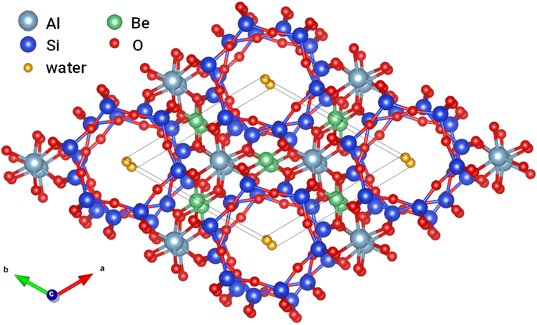
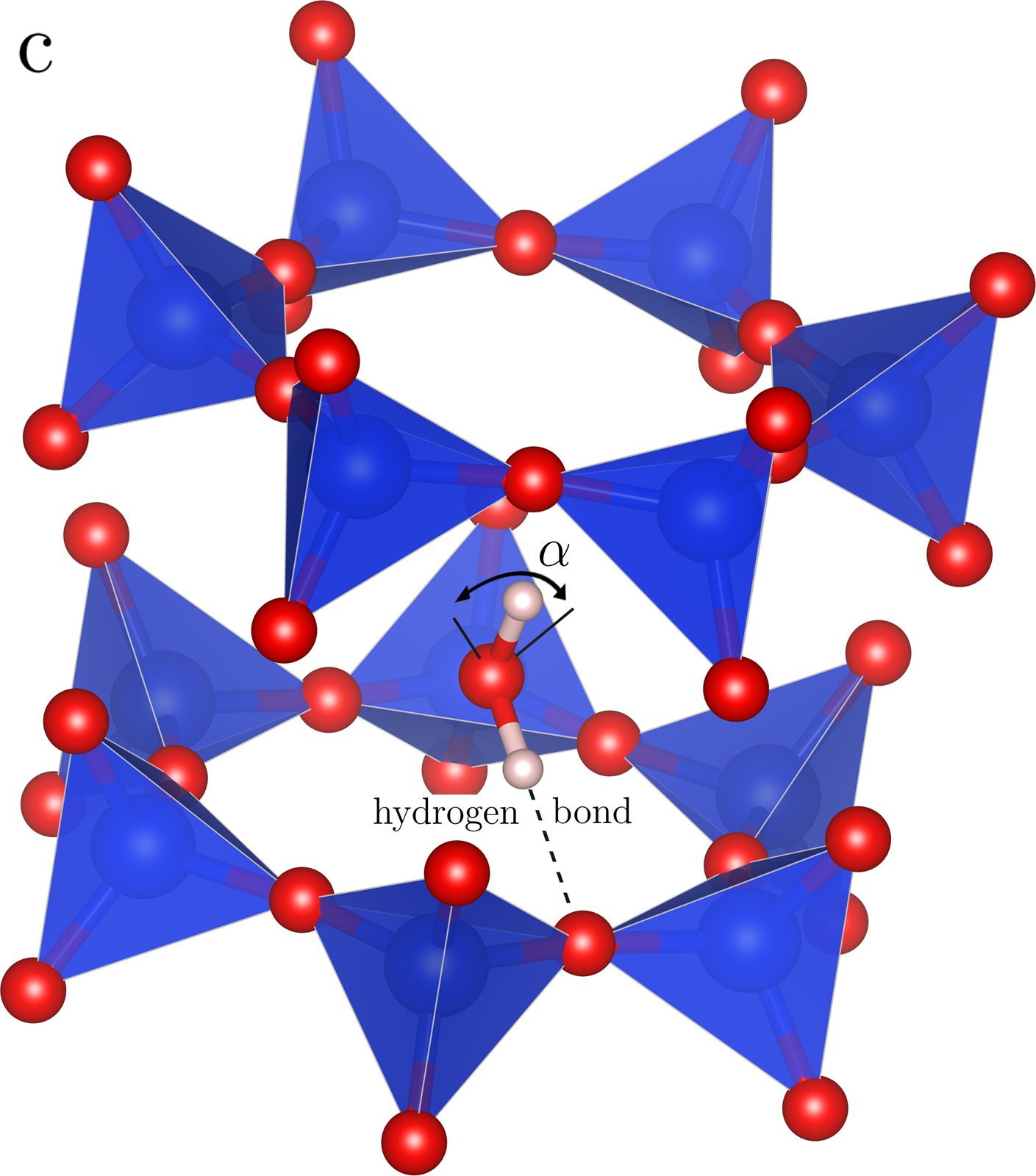
The crystal structure of beryl is hexagonal (space group ), and it contains channels of voids running along its hexagonal axis. Each of these voids may accommodate one water molecule. For more than fifty years, the water molecules have been supposed to take up two possible types of orientations within the voids, as hypothesized first by Wood and Nassau based on their infrared spectra analysis. 10 They concluded that the molecules will orient themselves with the H–H lines oriented either parallel to the hexagonal axis (“type-I water”), or perpendicular to it (“type-II water”); the latter type should be present especially in crystals with an additional doping, as the oxygen may bind to an impurity atom located within the channel. The earlier studies dealing with the interactions among the water molecules and their collective dynamics assumed the type-I molecules to rotate around the hexagonal axis of beryl, so the molecules’ planes remained parallel to the hexagonal axis. At the same time, it was supposed that the molecules are subjected, in their angular orientations, to a local potential exhibiting six equivalent minima separated by angles of \qty60. 5, 3, 11, 6, 4 Whereas the properties of the molecular ensemble were studied quite extensively—see the above references—, the orientations and dynamics of the individual molecules in the voids have been still less explored, despite being crucial for the bulk properties. Such local, molecular-level information can be provided namely by Nuclear Magnetic Resonance (NMR) spectroscopy.
I.2 NMR spectroscopy of water in beryl
Water molecules are expected to produce a single peak in the 1H NMR spectrum, because the spin of the 1H nucleus (proton) is ½ and both hydrogen atoms are equivalent due to symmetry. The peak may become split and/or shifted when anisotropic interactions are involved, e.g., dipolar interactions or anisotropy of chemical shielding. In cases when the water molecules are highly mobile, such as in liquid or gas phases, the effect of these anisotropic interactions on the NMR spectrum gets averaged by the fast molecular reorientations (motional narrowing). Then, the 1H nuclei in water molecules are exposed to the same, averaged local fields and the 1H NMR spectrum of water consists of a single, usually very narrow peak.12
Water molecules in solids have their dynamics significantly restricted. When the molecules are static or their reorientations are very slow (e.g., in ice or as a water of crystallization), the anisotropic interactions are not fully averaged and the NMR spectrum comprises contributions from all arrangements of the molecule present in the sample. For single crystals, the NMR spectra depend on the crystal orientation with respect to , while the NMR spectra of powders with randomly oriented grains display powder-pattern features.12 In terms of magnitude, the anisotropy of chemical shielding of 1H in water molecule ranges from 19 to \qty35ppm for various phases of water13, which for \qty10 corresponds to shifts or splittings of peaks in the 1H spectrum of about \qty10. The effect of dipolar interactions between 1H nuclei within the water molecule may be an order of magnitude larger, the dipolar interaction is thus often the dominant source of anisotropy in 1H NMR spectra of solids.12
The character of the 1H NMR spectrum of water molecules enclosed in the crystal voids is different from the two cases presented above. The confined molecules are not static as in ice, and the molecular reorientations are not isotropic as in liquid water. Thus, the anisotropic interactions are not fully averaged and the 1H NMR spectrum of water consists of more than one peak. This is the case of water molecules confined in the voids of beryl crystal, which was first studied using NMR by Paré and Ducros14 and by Sugitani et al.15 already in 1960s. In both these works, the 1H NMR spectra comprise doublet of peaks arising due to the nuclear dipolar interaction between the 1H nuclei within the water molecules. The signal was attributed to type-I water. Moreover, the observed dipolar splitting increased linearly with decreasing temperature and saturated below 100 K. The natural interpretation then was that the water molecules oscillate around their equilibrium positions when H–H line is parallel to the hexagonal axis, and that the amplitudes of oscillations increase with temperature14.
The idea that the type-I water molecules are simply oscillating around the hexagonal axis is, however, not entirely correct, since even at the lowest temperatures the observed dipolar splitting does not reach the expected value which is precisely given by the distance between the 1H nuclei within the water molecule. As we show in this paper, the movements of water molecules must be more complex. This deduction is based on measuring and analyzing the dipolar splitting in 1H NMR quantitatively, and especially by measuring and analyzing 2H NMR in beryl hydrated by heavy water.
The spin quantum number of the 2H nucleus equals 1 and so, in the presence of an external magnetic field, two transitions between the nuclear energy levels of the Zeeman multiplet are observable. Thus, the 2H NMR spectrum of heavy water in beryl is expected to comprise two doublets of peaks, one for each 2H nucleus. In contrast to the 1H case, the 2H dipolar interaction is small and a quadrupolar splitting occurs due to an interaction between the electric quadrupole moment of the 2H nucleus and the gradient of surrounding electric fields. The quadrupole splitting depends on the direction of the external magnetic field vector with respect to the axes of the electric field gradient (EFG) tensor. Whereas in the 1H case it is the orientation of the H–H line that determines the magnitude of dipolar splitting, in case of 2H the orientation of the O–H bond becomes important instead, since in the H2O molecule the principal axis of the EFG tensor at the hydrogen site points approximately along the O–H bond. This is an essential difference between the NMR interactions in H2O and D2O that allows for extracting more complete information about the orientation of water molecule in the beryl voids: the isotopes 1H and 2H serve as two completely different probes. Since the values of dipolar splitting in 1H and quadrupole splitting in the 2H NMR spectra depend significantly on the orientations of water molecules, we can evaluate the orientation and dynamics of the water molecules by analyzing the temperature dependences of the measured dipolar and quadrupolar splittings.
II Methods
Four high-quality synthetic beryl single crystals were studied: two samples containing normal water and two containing heavy water. The samples were grown from oxides on seed plates at \qty600 and \qty1.5kbar in a hermetically sealed gold vessel by the hydrothermal method of Thomas and Klyakhin16. All samples were cut into cylinders about \qty2 in diameter, with one sample for each hydrogen isotope having the hexagonal axis oriented parallel to the axis of cylinder and the other perpendicular to it. All beryl samples contained type-I water predominately; because of the artificial origin of the samples, the content of alkali atoms and other such impurities and hence also the content of type-II water was negligible.
NMR spectra of water-containing beryl single crystals were acquired in a magnetic field of \qty9.41 (Larmor frequency \qty400.185 for 1H and \qty61.431 for 2H) using a Bruker Avance II spectrometer. Solid echo pulse sequences (with lengths of the pulses 0.5–\qty1.2 for 1H and 3.5–\qty5.0\micro for 2H, and with a delay of –\qty100) were applied to excite the NMR signal in order to ensure proper refocusing of the spin magnetization in the presence of strong nuclear dipolar interaction (1H case) or electric quadrupole interaction (2H case). The trigger delays between the subsequent scans were adjusted at each temperature point in order to compensate the strong temperature dependence of the spin-lattice relaxation time. Its value amounted to about \qty1 and \qty20 for 1H and 2H, respectively, at the highest temperatures, and to more than \qty100 for both isotopes at the lowest temperatures. At each temperature point, 2–64 scans (1H spectra) or 8–1024 scans (2H spectra) were performed, and the acquired spin echo signals were coherently summed.
The temperature dependences were measured in a Janis helium continuous flow cryostat, and the temperature was monitored with Cernox CX-1030 temperature sensor in the proximity of the NMR coil. The orientation of the beryl crystal with respect to the external magnetic field was adjusted carefully, with an estimated error of less than \qty3. Using a goniometer, the angular dependence of 2H spectra was measured at room temperature.
Special care was taken to keep parasitic 1H signals at the minimum level. We used a custom-made NMR probe without any plastic or Teflon parts, and an NMR coil made from unvarnished silver wire. The probe and the sample were dehumidified prior to the experiments. The remaining small parasitic 1H signal near Larmor frequency most probably originated from the cap of the temperature sensor.
III Results
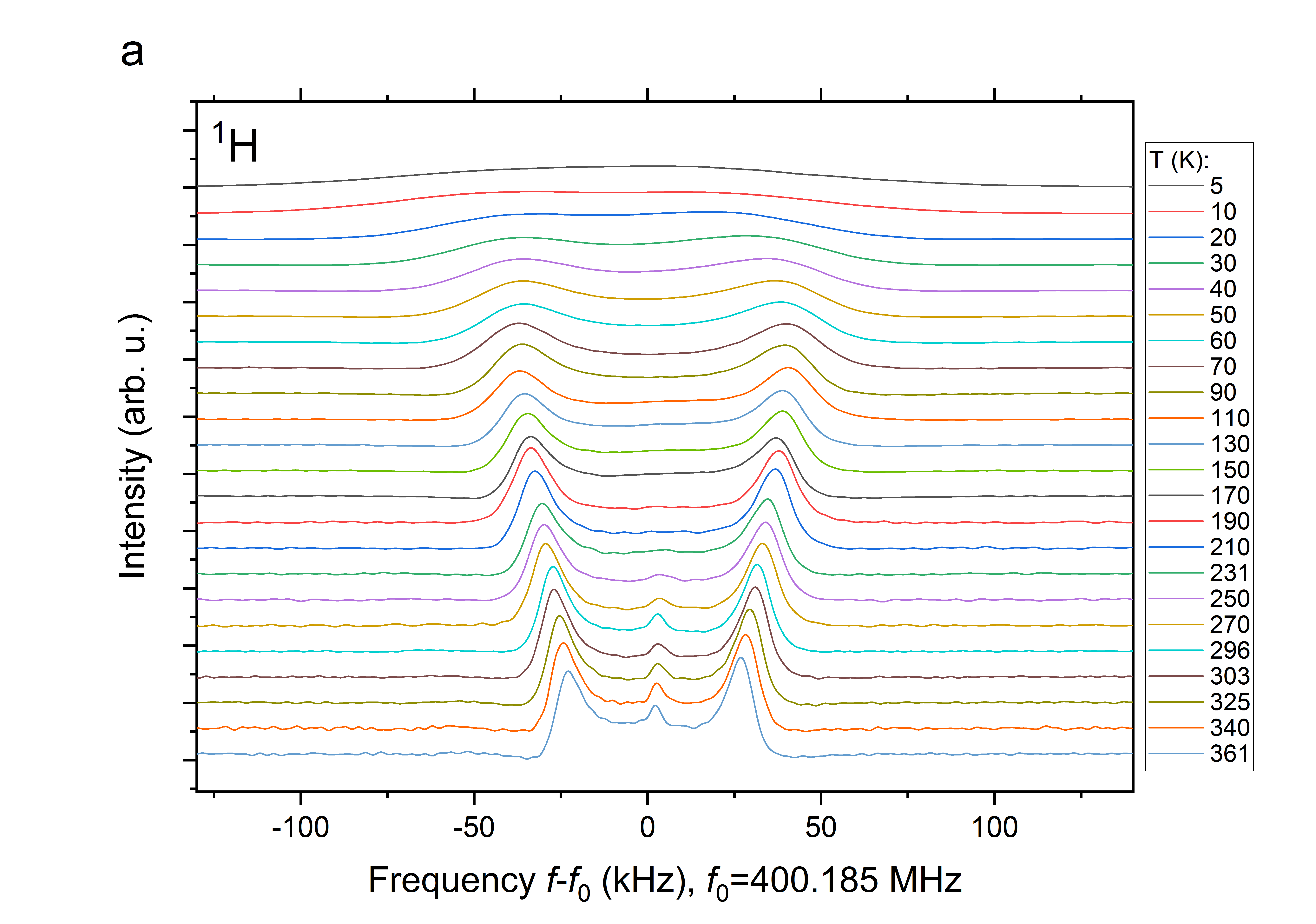
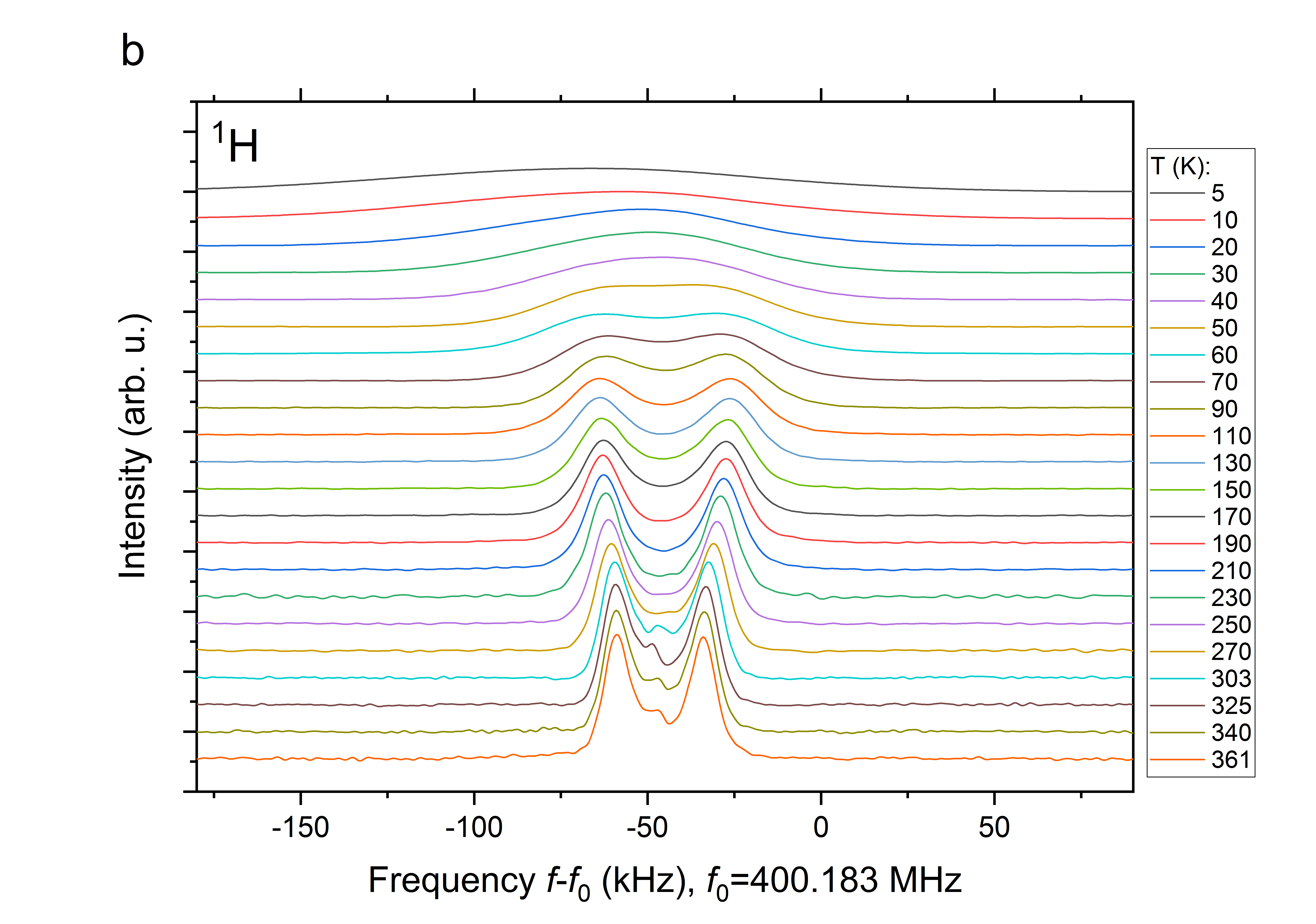
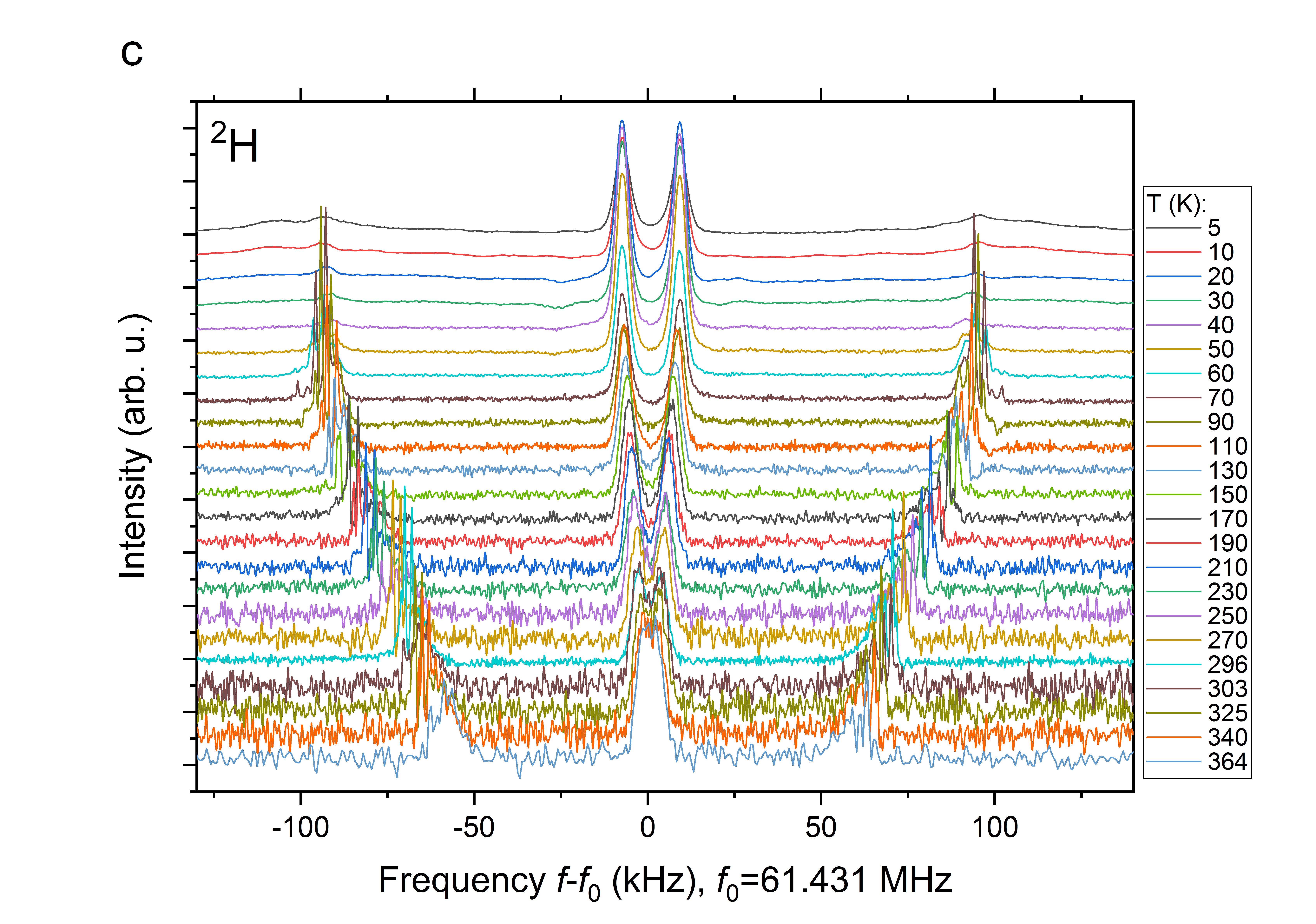
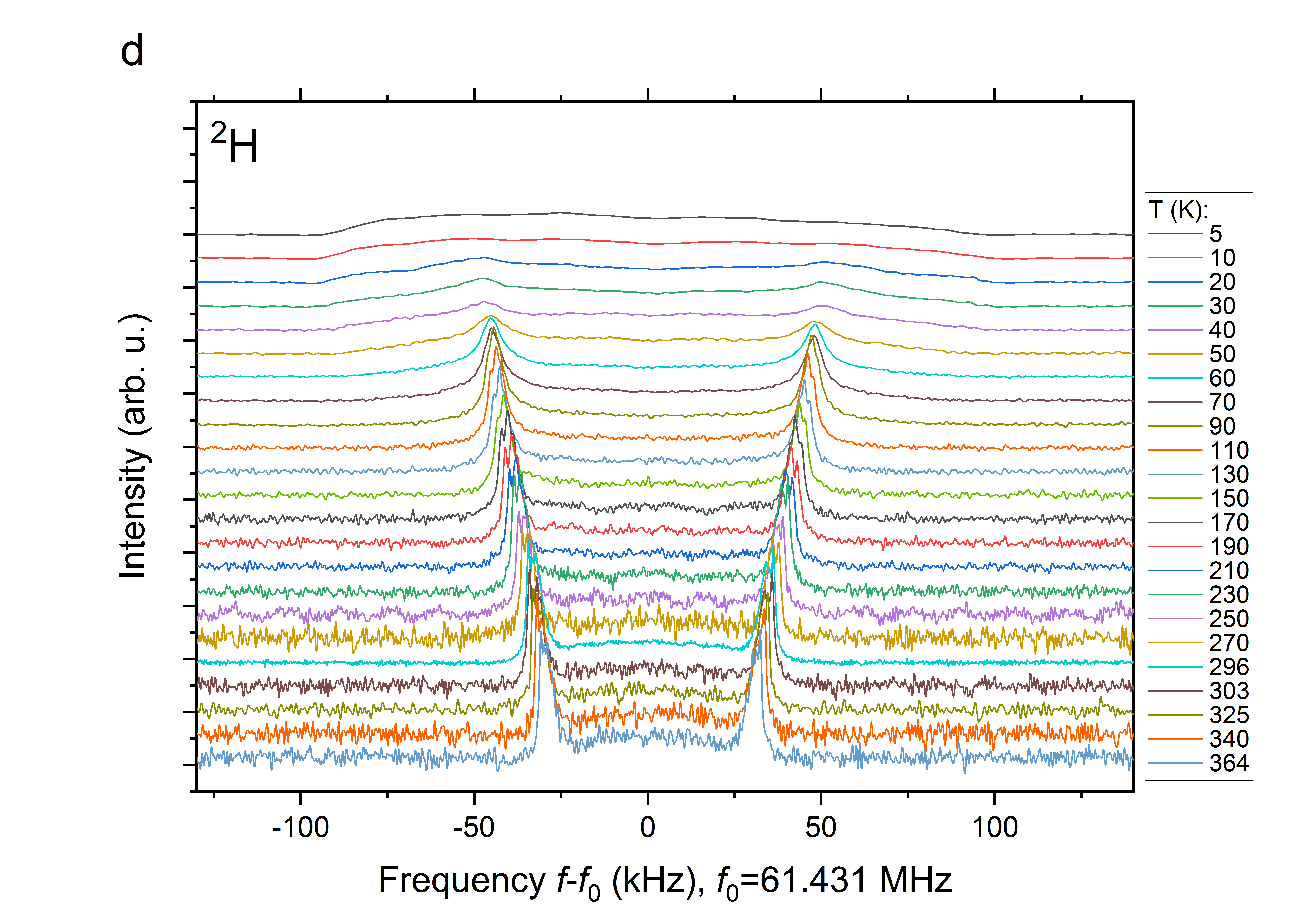
1H and 2H NMR spectra were measured in the temperature range 5–\qty360, and for each isotope two cylindrical samples were studied: in one, the hexagonal crystallographic axis was parallel to the cylinder axis and in the other perpendicular to it. We first describe and interpret the room temperature spectra of 1H and 2H, then we deal with their temperature dependences in the range of approximately 5–\qty360.
III.1 1H NMR spectra at room temperature
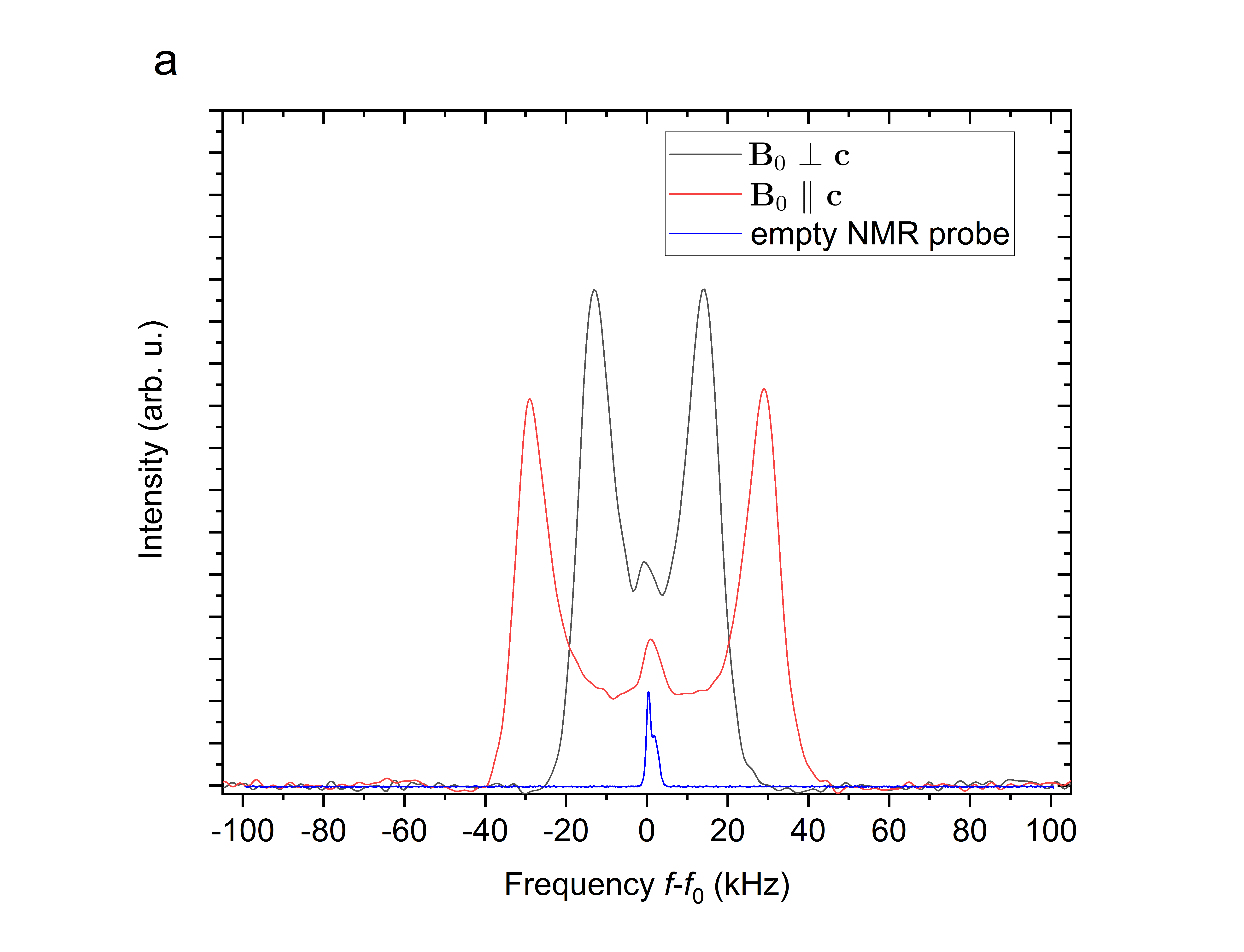
The room temperature 1H NMR spectrum of water molecules in beryl (Fig. 2a) consists of a doublet of peaks, originating in the magnetic dipolar interaction between the two 1H nuclei in the water molecule. The value of the dipolar splitting depends on the dipolar constant as well as on the orientation of the line connecting the interacting nuclei with respect to the direction of the external magnetic field , which is described by a polar angle 12:
| (1) |
The dipolar constant depends on the gyromagnetic ratio of the interacting nuclei, and it decays with the third power of their mutual distance . Clearly, the dipolar splitting in the measured spectra is different for the two displayed orientations of the crystal with respect to the external magnetic field, as the parallel orientation () corresponds to and the perpendicular one () to . The larger splitting observed for the parallel orientation implies that the lines connecting the hydrogen atoms (H–H) in the water molecules are preferentially oriented nearly along the hexagonal axis of beryl crystal, i.e., type-I water is observed in the 1H NMR spectra. No signals corresponding to type-II water sites were detected, which is in accord with the supposed negligible concentration of alkali metal atoms in the studied synthetic beryl crystals.
The room temperature values of the observed dipolar splitting \qty59 and \qty29 for the parallel and perpendicular orientations, respectively, are significantly smaller than expected for a dipolar interaction between two 1H nuclei in a type-I oriented water molecules. In fact, the dipolar constant between two 1H nuclei is given [see Eq. (1)] by their distance , which is relatively well known (\qty1.524). An orientation with the H–H line parallel () or perpendicular () to the external field would therefore yield a splitting of the doublet \qty117 and \qty58.7, respectively. Such discrepancy can be explained by (i) the actual value of the angle being appreciably different from 0 or 90∘, or (ii) by an averaging of the dipolar interaction strength due to rapid molecular motions. In the further text (section IV), we show that both mechanisms are responsible for the reduction of the observed dipolar splitting.
Additionally, the 1H nuclei in the water molecules are affected by dipolar interactions with other nuclear species in the surroundings, mainly the 1H nuclei of other water molecules in the neighboring voids and 27Al nuclei in the beryl crystal structure. However, these distant partners induce much weaker dipolar fields, and thus these interactions only lead to a broadening of the resonance peaks. Also, despite our efforts to avoid any hydrogen-containing materials in the vicinity of the radiofrequency coil, a small parasitic 1H line was detected close to the Larmor frequency. This originated probably from hydrogen atoms present in a small amount in the body of the NMR probe, or in the coating of the Cernox temperature sensor.
III.2 2H NMR spectra at room temperature
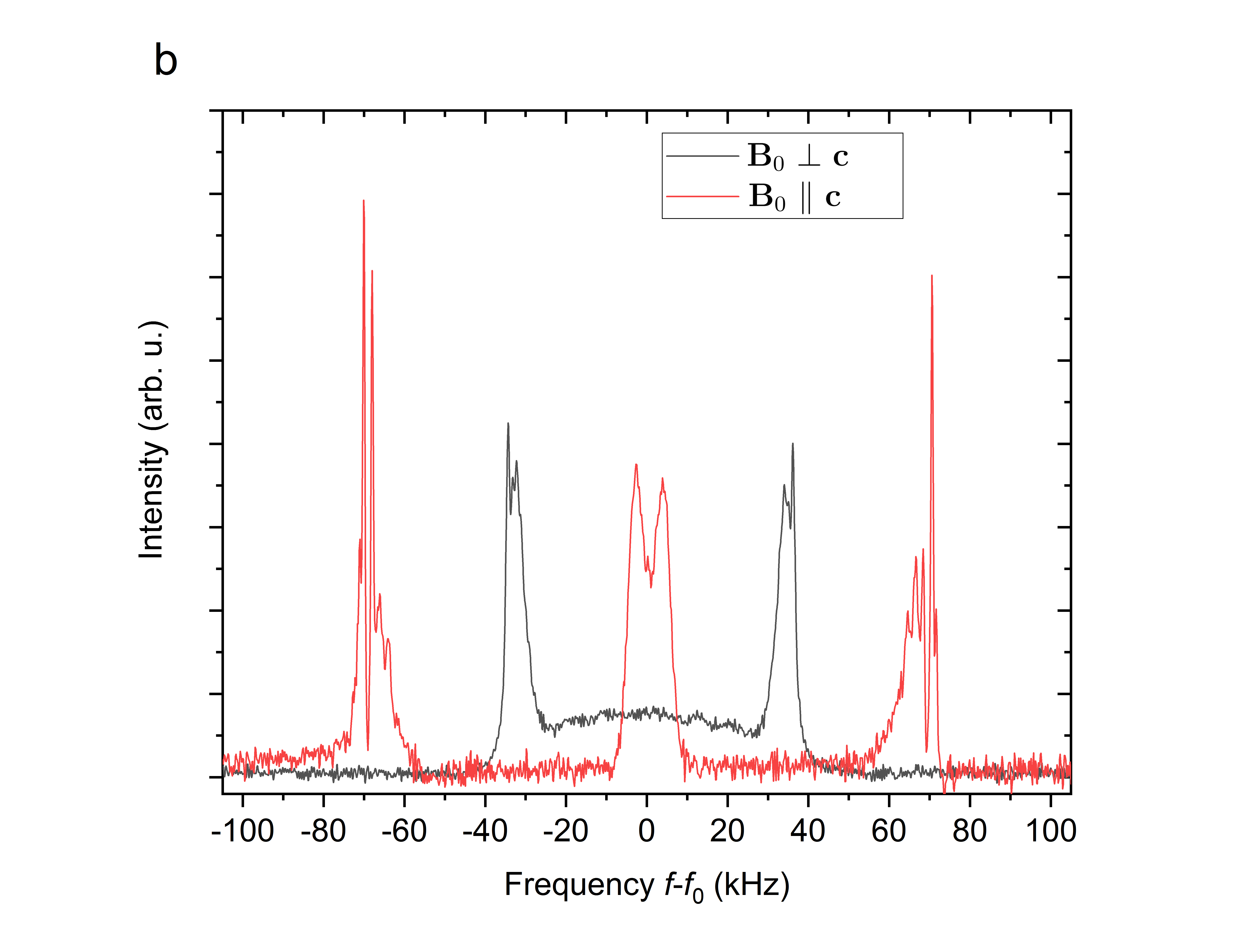
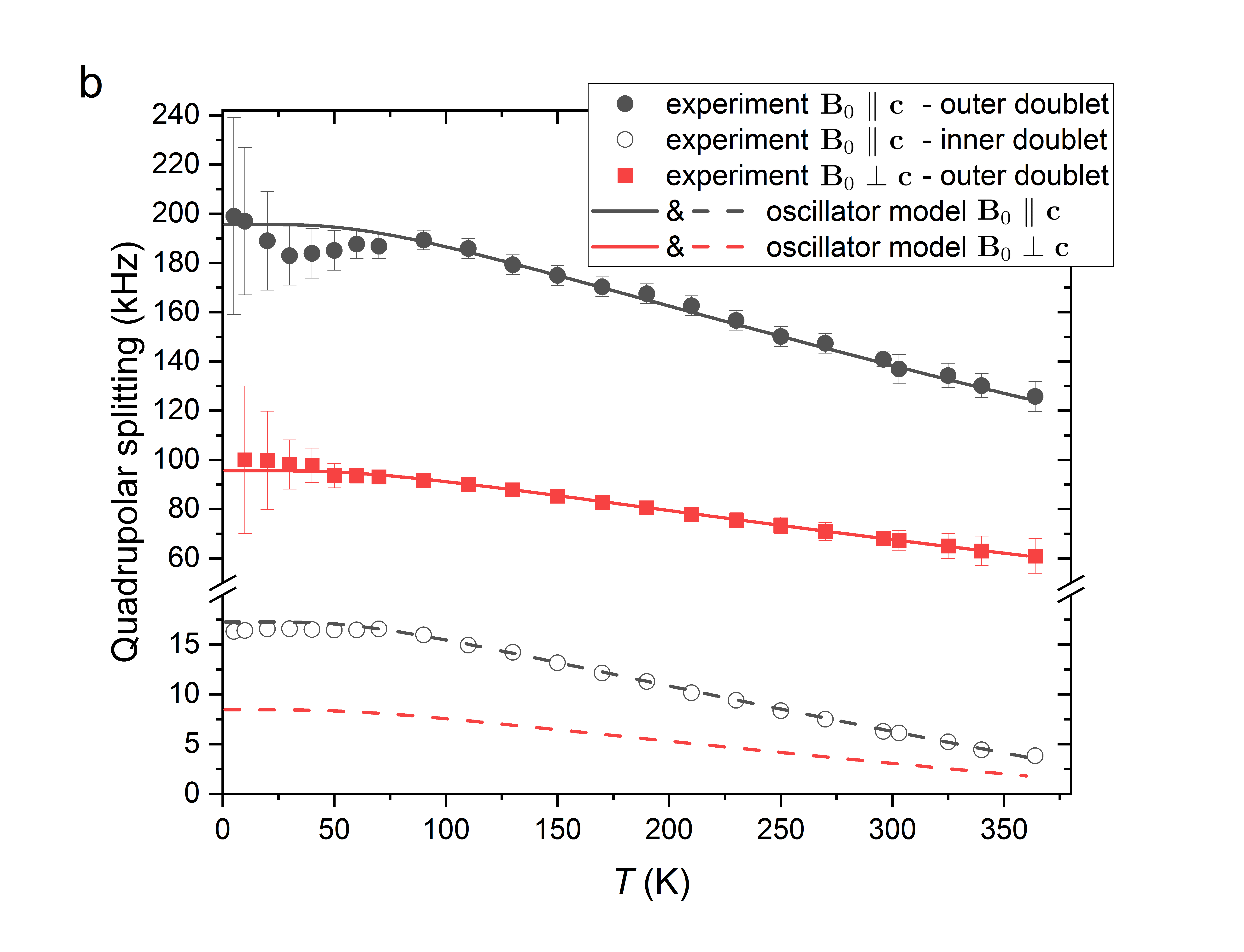
The 2H NMR spectrum of beryl containing deuterated water is more complex. When the hexagonal axis of beryl crystal is oriented parallel to the external magnetic field, a pair of well resolved doublets is observed in the 2H spectrum: an outer doublet with a larger splitting of \qty140 and an inner doublet with a splitting of \qty7. In contrast to the 1H case, these doublets are formed as a result of the electric quadrupole interaction at each 2H nucleus. The presence of two distinct quadrupole doublets in the spectrum implies that the two 2H nuclei in the water molecules are non-equivalent. This in turn means that the motion of water molecules cannot be described by simple oscillations about the hexagonal axis because, in such a case, the averaged local fields would be identical for both 2H nuclei. Similarly to the dipolar splitting in the 1H case, the electric quadrupole splitting also depends on the orientation of the water molecules with respect to the external magnetic field, yet in a more complex way:12, 17
| (2) | |||||
| (3) |
The polar and azimuthal angles characterize the direction of the external magnetic field within the principal axis system (PAS) of the EFG tensor , which is described by two parameters: the largest component , , and a dimensionless asymmetry factor , . For the hydrogen site in the water molecule, the principal axis belonging to of the tensor is pointing approximately along the O–H bond, and is perpendicular to the H2O molecular plane (Fig. 5a). The electric quadrupole interaction constant is given by the nuclear quadrupole moment of 2H, \qty2.85783(30)mb (milibarn, \qty1mb = \qtye-31\squared) 18, and by the value of at the 2H nucleus, which depends significantly on the phase of water. In the gas phase, \qty307.5(6) with 19, 20, in liquid water \qty250(7)21, 22 ( is not known in liquid, but its value is estimated to be similar to those in other phases), and \qty213.4(3) with in ice Ih23. The values of for 2H in liquid and ice are reduced compared to the gas phase due to numerous interactions with neighboring molecules via hydrogen bonds in the condensed phases. The quadrupole constant of 2H in heavy water confined within the beryl voids is unknown, but its value can be expected roughly around 200–\qty300. The magnitude of the observed quadrupole splitting at room temperature (Fig. 2b) is again diminished due to molecular motions, similarly as in the case of 1H dipolar splitting in H2O.
The dipolar interaction within the water molecule, which is the dominating interaction in the 1H spectra, is much weaker in case of 2H due to the magnetic moment of the 2H nucleus being smaller than the moment of 1H. The intramolecular 2H–2H dipolar interactions thus manifest only as a fine splitting (\qty2) of the spectral lines, which is well noticeable for the peaks of the outer quadrupole doublet [see Fig. 2(b)], and any further dipolar interactions may be neglected.
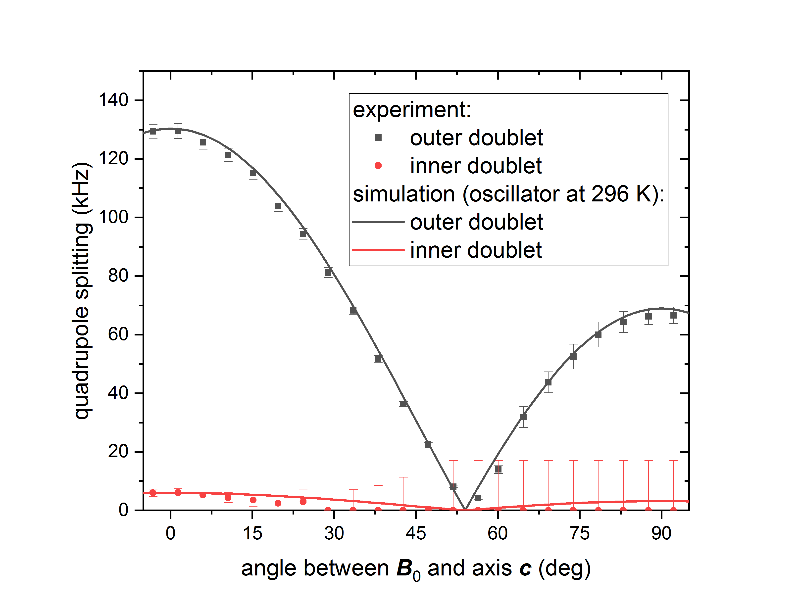
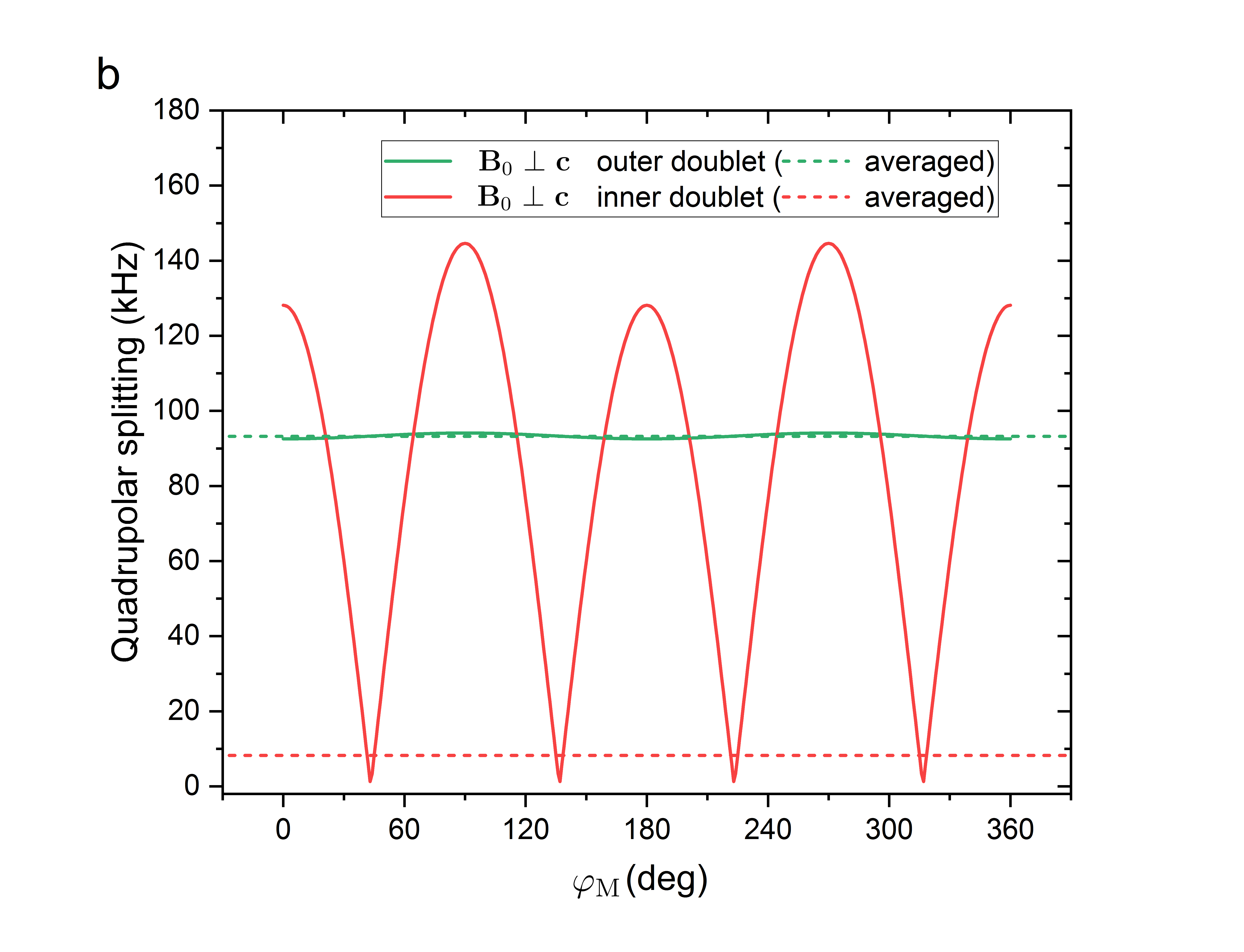
In the orientation (Fig. 2b), the 2H NMR spectrum also consists of two doublets, however, only the outer one is well resolved. The peaks of the inner doublet with a smaller splitting overlap and they are severely broadened, especially at lower temperatures. This behavior can be well observed in the angular dependence of the 2H quadrupole splitting measured for several angles between the two extreme cases and (Fig. 3 right). The peaks of the inner doublet are resolved only for angles between and below . The excessive broadening is caused by insufficient motional averaging with respect to the hexagonal axis, as we explained below in the section IV.
III.3 Temperature dependence of 1H and 2H NMR spectra
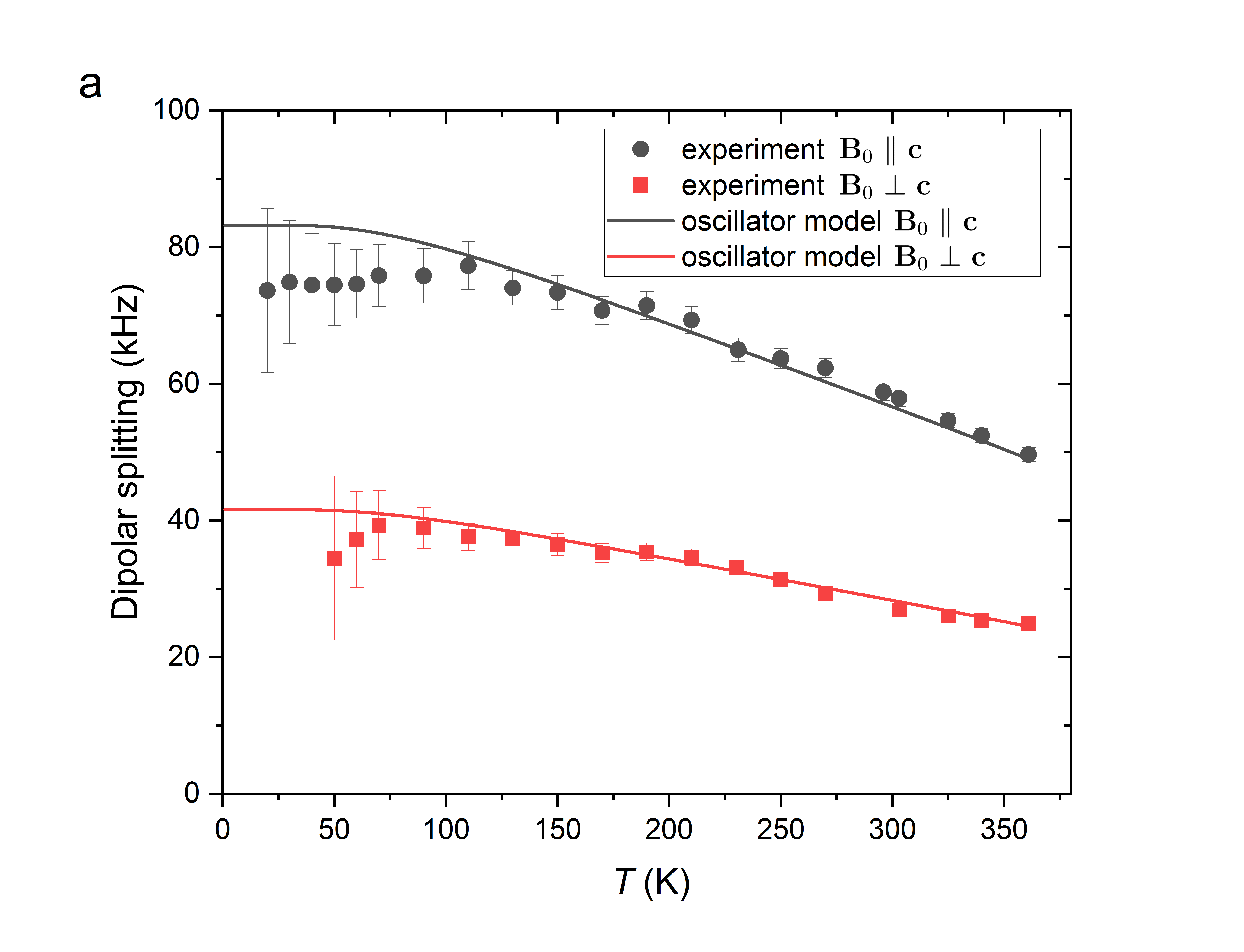
The temperature dependent 1H and 2H NMR spectra within 5–\qty364 are displayed in Fig. 4. The dipolar and quadrupole splittings , increase upon cooling, and they level off at about \qty70. The magnitude of the observed 1H dipolar splitting and its temperature behavior are in agreement with 1H NMR data available in the literature 14, 15, where this behavior was explained by oscillations of the water molecules around their equilibrium positions along the hexagonal axis. This interpretation, however, conflicts with the fact that the dipolar splittings do not reach the expected values even at the lowest temperatures. This leads us unambiguously to the conclusion that the motions of the water molecules have to be more complex than this. As for the 2H spectra, we are aware of no published measurements of the quadrupole splitting of 2H of D2O in beryl crystal. Our experiments reveal that the 2H spectra follow qualitatively the same pattern as the 1H ones—the splittings of the quadrupole doublets decrease upon cooling in a similar manner.
Below \qty70, the 1H and 2H spectral peaks become significantly broader, indicating a slowing down of the molecular motions so that the averaging of the anisotropic interactions (magnetic dipolar for 1H and electric quadrupole for 2H) becomes inefficient. Thus, at the lowest temperatures, the spectral shapes resemble features typical of static NMR spectra of randomly oriented powder samples. The temperature dependences of the measured NMR spectra and their spectral shapes are analyzed in more detail below.
IV Analysis and Discussion
In this section we show that the established notion of the behavior of water molecules in beryl, i.e., motions with equilibrium positions along the hexagonal axis, is not consistent with the measured NMR data. Furthermore, we propose a new type of movement when the water molecule is bonded via a hydrogen bond to an oxygen atom in the enclosing wall of the beryl void. A detailed analysis allows us to explain all the features observed in the NMR experiment.
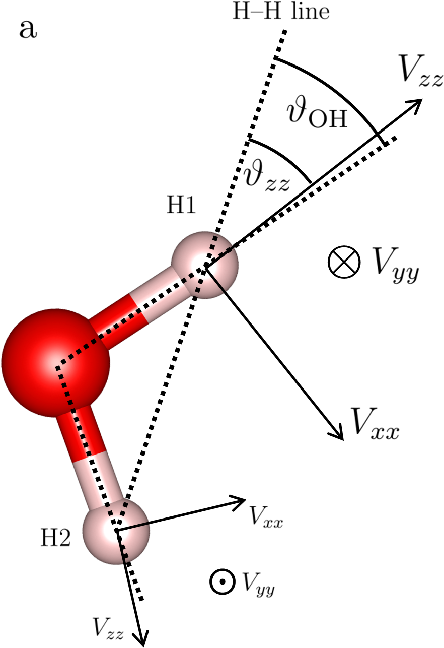
The key to determining the motions of the water molecule in beryl lies in a fundamental difference between the 1H and 2H NMR spectra. The dipolar splitting of 1H peaks depends on the orientation of the 1H–1H line with respect to the direction of external magnetic field (via the angle in Eq. 1). In contrast, the quadrupole splitting of 2H peaks depends on the direction of the external magnetic field within the PAS of the EFG tensor (via the angles and in Eq. 3). The hydrogen atoms in the water molecule are (chemically) equivalent, and therefore, the 2H EFG tensors are of the same magnitude. However, the orientation of the PAS differs for the two 2H positions in the water molecule since for both EFG tensors the principal axis points approximately along the respective O–H bond, and the water molecule is not linear (Fig. 5a). As a consequence, the angles and are in general different for the two 2H sites.
IV.1 Model for Motion of Water Molecules in Beryl Voids
For sake of spectra analysis, let us provisionally assume that the type-I water molecules perform the traditionally accepted type of motion within the void of the beryl crystal as follows. Let the equilibrium orientation of the molecules be such that the H–H line is aligned with the hexagonal axis of the beryl structure, and the molecules may rotate and oscillate around the H–H line within a limited spatial angle around axis —as dictated by the shape and dimensions of the void. Owing to such a motion, the angle is averaged to zero for the case when the external field , or to for , and a qualitative agreement is reached with the temperature dependences of 1H NMR (Fig. 4) and with 1H NMR data in previous works.14, 15 However, the observed dipolar splittings are somewhat lower than expected based on the distance of 1H nuclei in the water molecule, suggesting that the angle is on average different from 0 or . An even more convincing argument against the considered type of motion is revealed by 2H NMR where two distinct quadrupole doublets are observed in the spectra for (Fig. 2b). Because of the symmetry of the water molecules, any rotational or oscillatory motion about the H–H axis would average the angle to the same value for both 2H nuclei, and hence the quadrupole splitting of 2H would be the same. But it is clear from the measured 2H NMR spectra that the quadrupole splitting, and thus also the averaged angle , is significantly different for each of the two sites of deuterium in the water molecule.
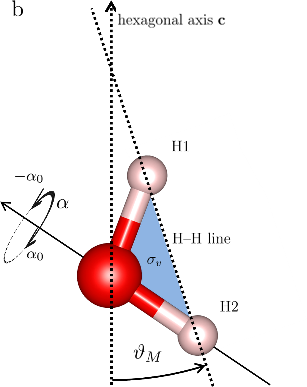
In principle, the large quadrupole splitting of the outer doublet in the 2H spectrum could be compatible with a situation where the H–H line lies approximately along the hexagonal axis, but the splitting of the second, inner doublet would not comply. The quadrupole splitting of the inner doublets is so small that it can only be reached if the magnetic field maintains a very specific orientation with respect to the PAS of EFG of such a 2H atom: the angle must be (on average) close to the value , so called ”magic angle” for which the expression in Eq. 3 becomes zero. This condition can be satisfied when the H–H line of the water molecule is deviated from the beryl hexagonal axis by an angle (Fig. 5b). Since the angle between the principal axis of the EFG tensor and the H–H line is , then , yielding the values of and , the first one being close to the magic angle.
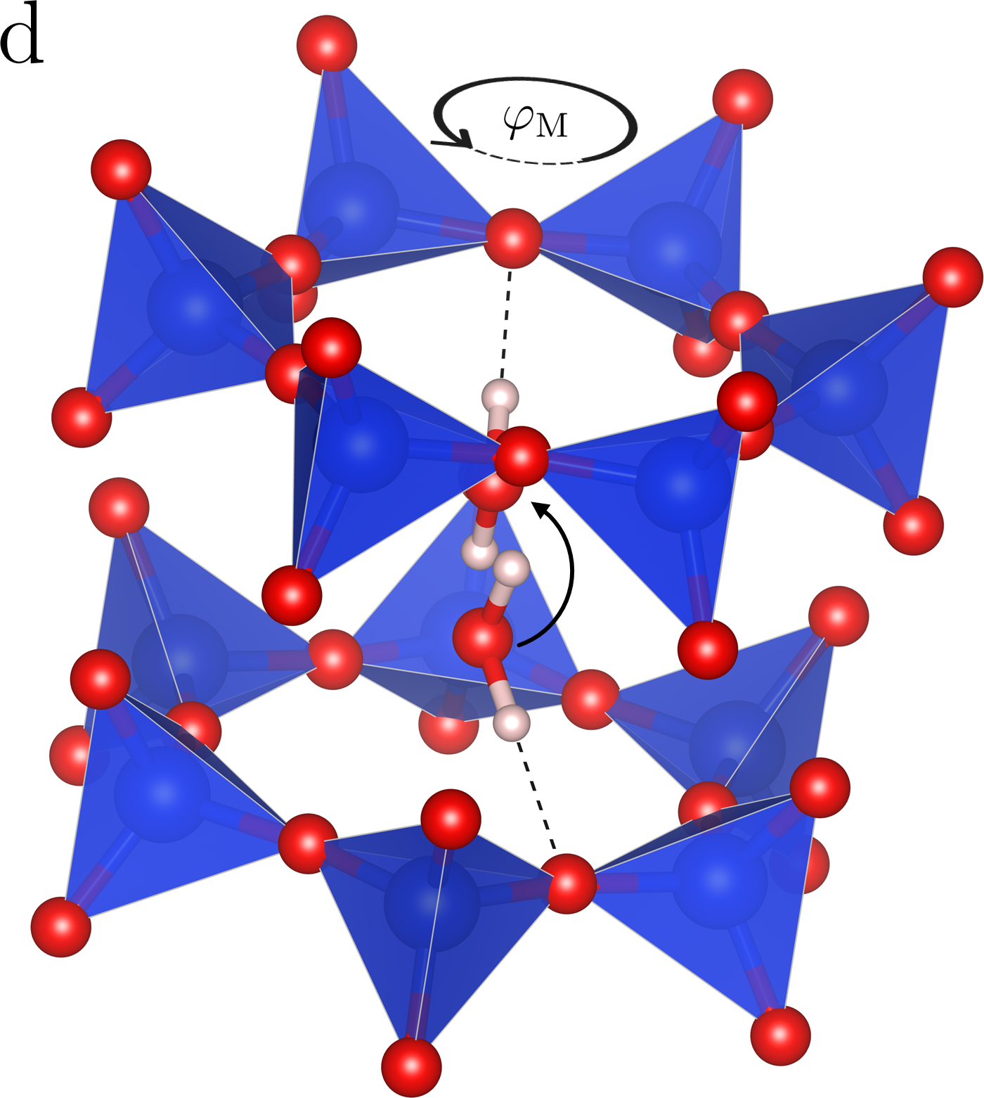
The reason for such peculiar arrangement of the water molecules is apparently the formation of a hydrogen bond between the deuterium atom in D2O and one of the oxygen atoms which form the walls of the beryl voids. Therefore, we propose a model in which the water molecules attach to an oxygen atom via a hydrogen bond, and the molecules perform two distinct motions: first, they librate rapidly about the formed hydrogen bond (Fig. 5c). Since hydrogen bonds tend to be highly directional,24 the axes of libration coincide with the O–H bonds of the molecules, and these axes deviate from the beryl hexagonal axis by an angle close to the magic angle . Second, since the water molecules may form the hydrogen bonds with any of the twelve oxygen atoms available in the walls of the voids, the molecules are also expected to jump among these available bonding sites (Fig. 5d), i.e., they effectively rotate about the beryl hexagonal axis. In the following analysis, we show that these two types of motion can explain the NMR experiment even quantitatively.
Naturally, the jumps accompanied by re-bonding of hydrogen bond have to be slower than the librations about the O–H bond, yet both these molecular motions are fast enough to cause averaging effects in NMR. The resonance frequencies of the nuclei are usually orders of magnitude slower than the changes of local fields induced by such molecular motions. As a consequence, the measured 1H and 2H NMR spectra of water in beryl are affected by averaged dipolar and quadrupole interactions, which is documented by the reduced values of splittings and by the presence of narrow spectral peaks at higher temperatures. Upon lowering the temperature, the frequencies and amplitudes of molecular motions decrease, therefore the splittings approach their nominal values, and the spectral peaks broaden.
IV.2 Analysis of Librations of Water Molecules
In order to analyze the first mode of molecular motion (i.e., the rapid librations of the molecule about the hydrogen bond), we denote by the angle describing the deviation of the H–H line from the hexagonal axis of the beryl crystal (Fig. 5b). The librations of the molecule about its axis O–H2 are described by the angle which is zero for the apex position (i.e., when the hexagonal axis is parallel to the mirror plane which contains all three atoms of the water molecule), and we suppose it librates around the mean position with an amplitude of , . For now we assume the angle to be distributed uniformly within its limits , which enables us to express the average dipolar and quadrupole splittings as:
| (4) |
The second type of motion is connected with re-bonding of the H2 atom to any of the remaining five oxygen sites or, instead, with H1 atom forming a hydrogen bond with one of the six oxygen sites in the other hemisphere (Fig. 5d). Such jumps cause averaging with respect to the hexagonal axis of beryl, which we can consider as rotational averaging over the angle in the full range. The dipolar and quadrupole splittings, averaged by both types of movements, can then be expressed as functions of the angles , , , and :
| (5) |
The averaged splittings and further depend also on the dipolar constant and the quadrupolar parameters and , respectively. The value of is given by the distance between the 1H nuclei (Eq. 1) and thus it is closely linked to the geometry of the water molecule. and are unknown for water confined in beryl but they should not be dramatically different from the values for D2O in various phases, i.e., –\qty300 and . Also the angles and are given by the geometry of water molecule (Fig. 5a). In further analysis, we assume the value of the H–O–H angle of and a H–O distance of \qty0.949, which was found by neutron diffraction experiments 25, 3. These parameters yield a value of for the angle between the H–H line and the O–H bond. The angle between the H–H line and the principal axis is then equal to , which corresponds to a small deviation () of the axis from the direction of the O–H bond, as determined from hyperfine structure measurements of heavy water20. The H–H distance is equal to \qty1.525 and thus \qty58.7, according to Eq. 1. Based on these values, the remaining parameters of interest, angles and , can be determined from our NMR experiments using a suitable model.
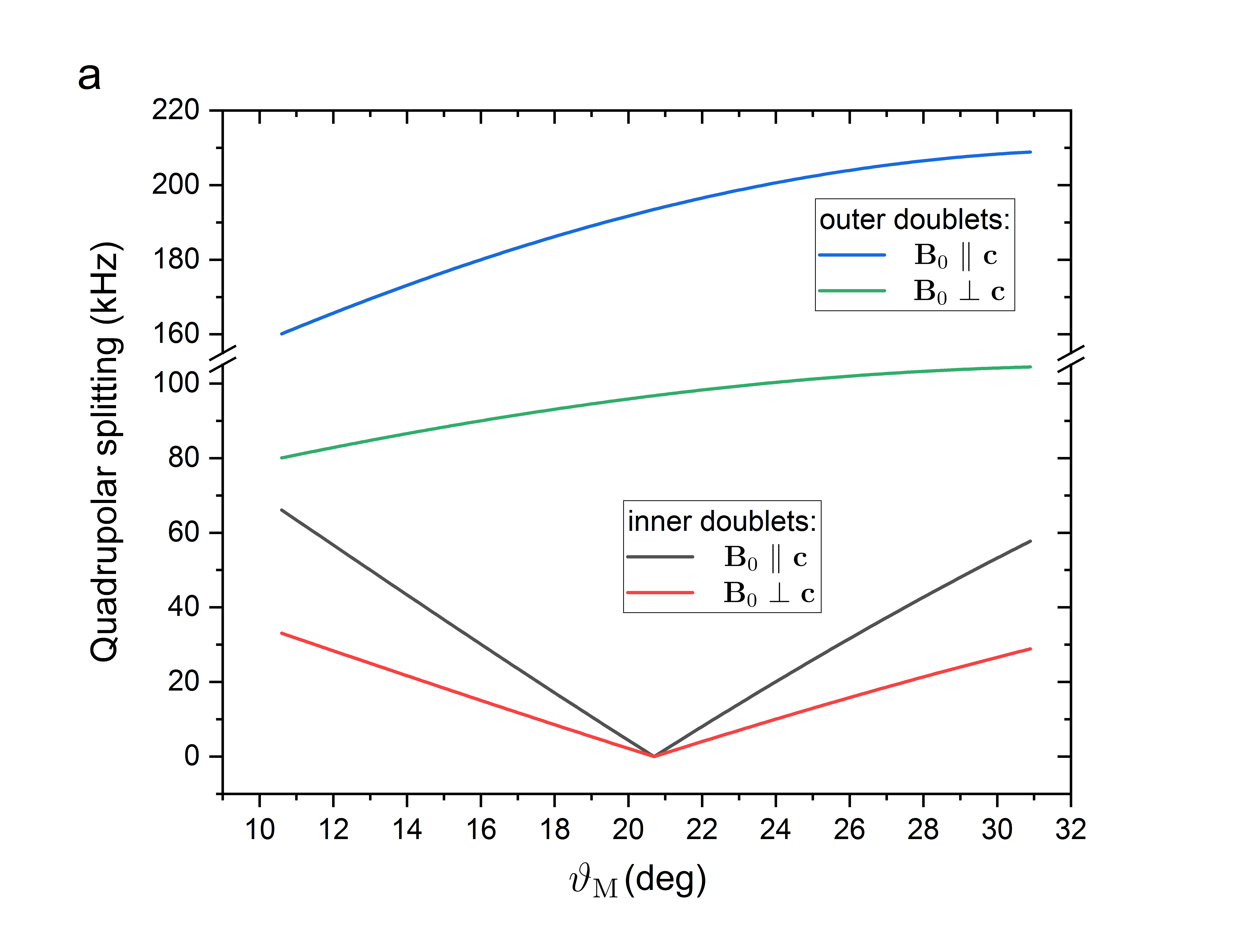
The angle determines the deviation of the libration axis O–H from the hexagonal axis and its value dictates, to a great extent, the specific character of the observed 2H spectra, i.e., the positions of the two doublets with distinctly different quadrupole splittings. Whereas the dipolar splitting in 1H spectra as well as the quadrupole splitting of outer doublets in 2H spectra do not vary much with deviation of the axis of libration (angle ), the small quadrupole splitting of the inner doublet in the 2H spectra is particularly sensitive to the changes in the angle (Fig. 6a). For both sample orientations and , the splitting of the inner doublet becomes zero when reaches the magic angle. One may notice that there are two possible values of that yield the small splitting of the inner doublets observed in experiment; this issue will be addressed in further analysis below.
The peaks of the inner doublet in the 2H spectrum become substantially broadened for (as well as for any orientations where the external magnetic field is deviated from the hexagonal axis by more than , see Fig. 3), and this broadening becomes even more pronounced with decreasing temperature. For , the splitting of the inner doublet depends strongly on the angle (Fig. 6b). Consequently, for the inner doublet, averaging over the angle becomes ineffective already at temperatures below room temperature, whereas the averaging is still effective enough for the outer doublet to be well resolved. The averaging of and in Eq. 5 is assumed to be continuous in angle , whereas in reality there are twelve discrete orientations in the single crystal, i.e., the angle should take values , where is an integer. However, neither this simplification nor the indeterminacy of the phase angle lead to any noticeable difference in the averaged values of the splittings.
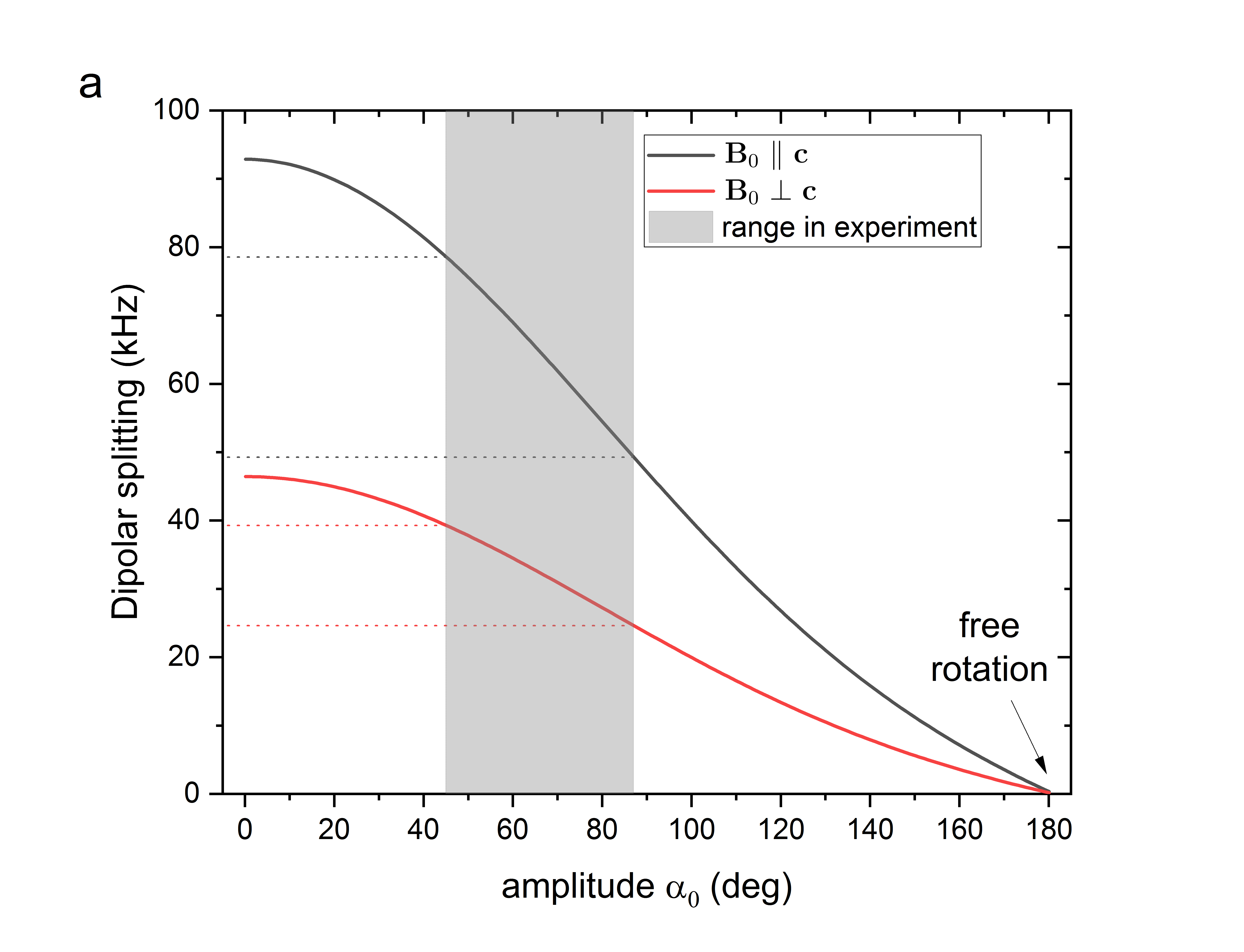
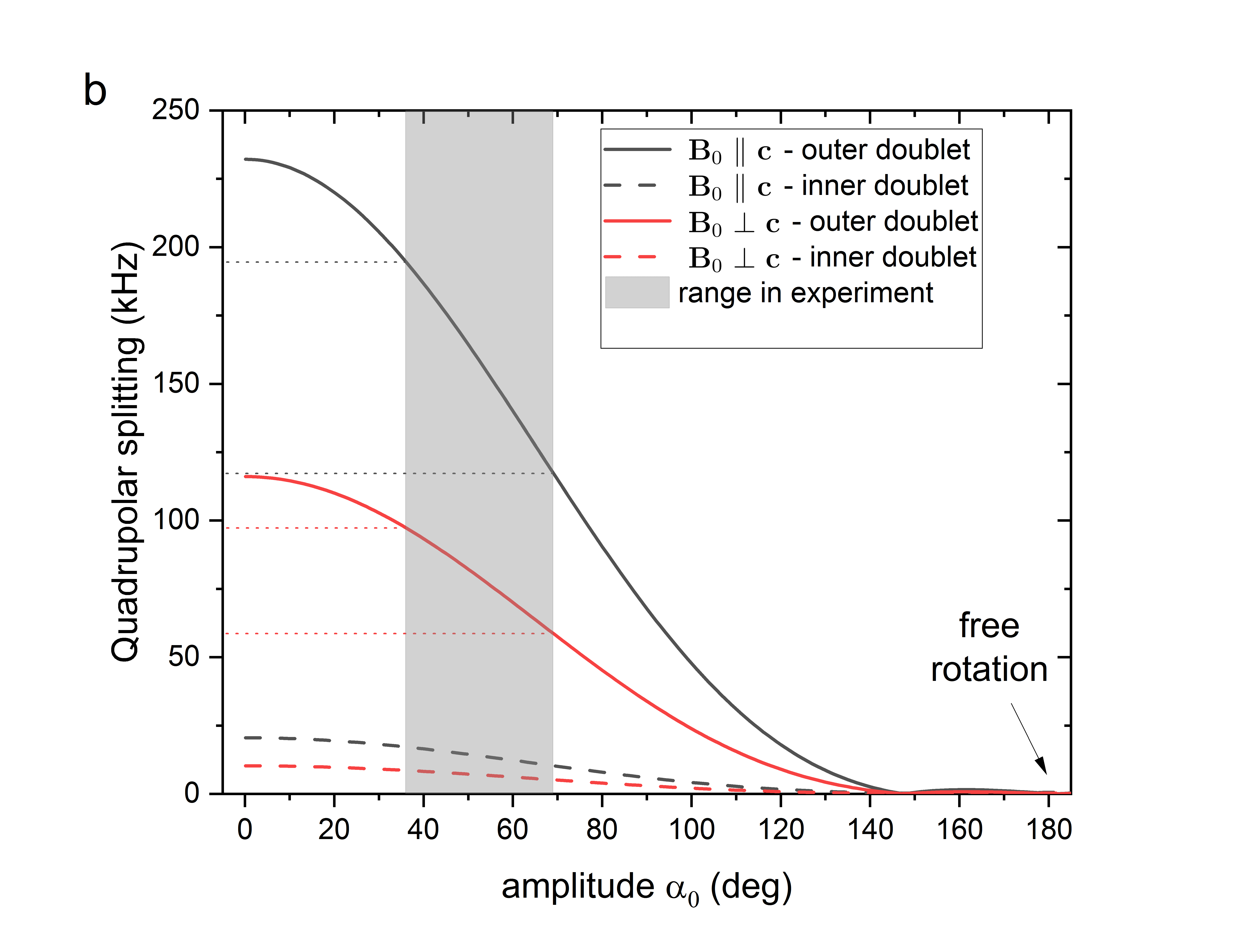
Although both and are expected to be temperature dependent, mainly the amplitude of librations is responsible for the decrease in dipolar and quadrupole splittings with temperature due to motional averaging. The averaged splittings and as functions of amplitude are displayed in Fig. 7; the grey areas denote the ranges of amplitudes corresponding to experimentally observed dipolar and quadrupole splittings. The splittings obtained by NMR experiments can thus be related to variations of the amplitude within the given range. But even if the calculated dependences roughly capture the range and basic character of experimental splittings (lower values of correspond to low temperatures and highest to \qty360), it is clear that there are systematic differences. First, the experimentally detected low-temperature plateaus are not present in the simulated dependences. Second, in the experiment the splitting of the inner quadrupole doublet decays faster with increasing temperature than the splitting of outer doublet—the ratio of splittings for these doublets gradually increases from 11.5 at low temperatures to more than 30 at high temperatures (Fig. 8).
Both these shortcomings can be removed by implementing a specific temperature dependence to angles and . Such dependence, however, may be realized in many different ways leading to similar levels of agreement with the experiment. We show that a good agreement (Fig. 8) is reached already with probably the simplest approach when the librational motion of the water molecule about one of its O–H axes is considered as that of a quantum harmonic oscillator.
A quantum harmonic oscillator provides quantized energies
| (6) |
where denotes the angular frequency of the librational motions of the water molecule about the O–H bond. The states with energies are populated with Boltzmann probabilities
| (7) |
the mean energy can be written as
| (8) |
and it is related to the amplitude as
| (9) |
where is the length of the O–H bond. The force constant of the oscillator is related to the frequency as , where is the mass of proton or deuteron for the H2O or D2O case, respectively. The value of is a free parameter in the harmonic oscillator model, and it accounts for the overall shape of the temperature dependences.
Eqs. 4 and 5, which assumed a uniformly distributed angle , now have to be modified because for a Boltzmann-averaged harmonic oscillator the angle is not distributed uniformly; instead, it has a Gaussian distribution26 with a dispersion . In our case only a few lowest energy levels of the oscillator are populated even at the highest temperature \qty365, therefore, the zero-point energy is considerable and a visible plateau up to about \qty50 appears in all simulated dependences (Fig. 8).
The fact that upon heating, the splitting of the inner quadrupole doublet decays faster than that of the outer doublet can be captured by making the angle temperature dependent. By increasing slightly with increasing temperature, the value of further approaches the magic angle, which reduces the splittings of the inner quadrupole doublets while leaving the splittings of the outer doublets relatively unchanged (see Fig. 6a). If we thus increase within the range of 18.1–19.2∘ linearly with temperature in range 5–\qty365, the ratio of simulated splittings follows very well the values observed in our experiment (Fig. 8).
In the dependence of quadrupole splittings on angle (Fig. 6a), there are apparently two values of and , bringing close to the magic angle and thus leading to the small splittings of inner doublets in the experiment. The larger value of can also lead to a good agreement of the model with experimental quadrupole splittings, using a different frequency , similar quadrupole parameters and , and a gradual decrease in with increasing temperature—again to approach the magic angle for , but this time from above. To discriminate between these two solutions we can use the dipolar splittings in 1H spectra, where this somewhat larger tilt angle does not yield a reasonable agreement. In order to match the experiment, we have to use \qty69.1 corresponding to a H–H distance of \qty1.444, which would lead to an unrealistic H–O–H angle of (for an O–H bond-length of \qty0.949). Moreover, a decrease in the tilt angle with increasing temperature would be required, which lacks a suitable justification. In contrast, for the solution, an increase in with temperature makes more physical sense: with increasing the hydrogen H1 points more towards the hexagonal axis (Fig. 5), which provides more space for the oscillating part of the molecule—increasing with temperature is thus a plausible consequence of larger oscillations at high temperatures.
The best match with the experiment is reached for linearly changing in the range 18.1–19.2∘ (from low to high temperatures), with \qty4.95e12 for H2O, which corresponds to a frequency of librations of \qty165cm^-1. As for the temperature dependence of the quadrupole splitting in D2O, the best match is reached using \qty4.34e12, corresponding to \qty145cm^-1. Both these wave numbers are in a reasonable agreement with values of libration modes of type-I water as observed in optical experiments 10, 5. Out of the remaining parameters, a part of them is determined by the water molecule geometry: \qty58.7, \qty0.949, and , and the last two parameters do not significantly differ from expectations: = \qty176 and .
We note that the ratio of harmonic frequencies does not reach the value , expected from the ratio of deuteron and proton masses. This is most probably caused by the limitation of our simple oscillator model, where the amplitudes of oscillations are relatively large at high temperatures. Only a few lowest energy levels of the oscillator ensemble are populated even at \qty365, as indicated by the sum of the first five Boltzmann probabilities , therefore the difference between quantum harmonic oscillator model and, e.g., particle in box model would not be large. However, the effect of walls of the beryl voids should be addressed, especially at high temperatures, by adding some particle-in-box features, which would lead to a more appropriate model such as oscillator confined in a box27.
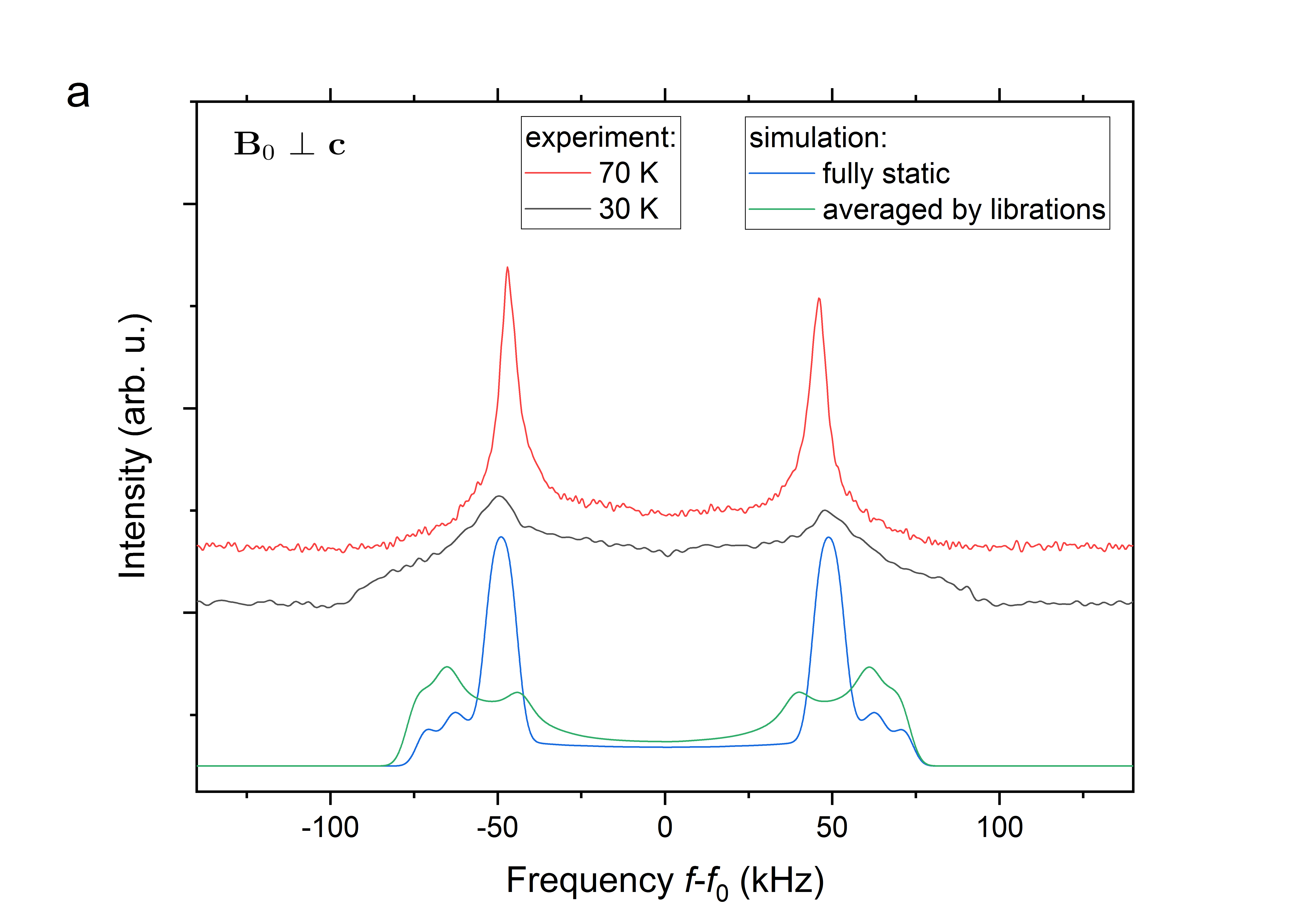
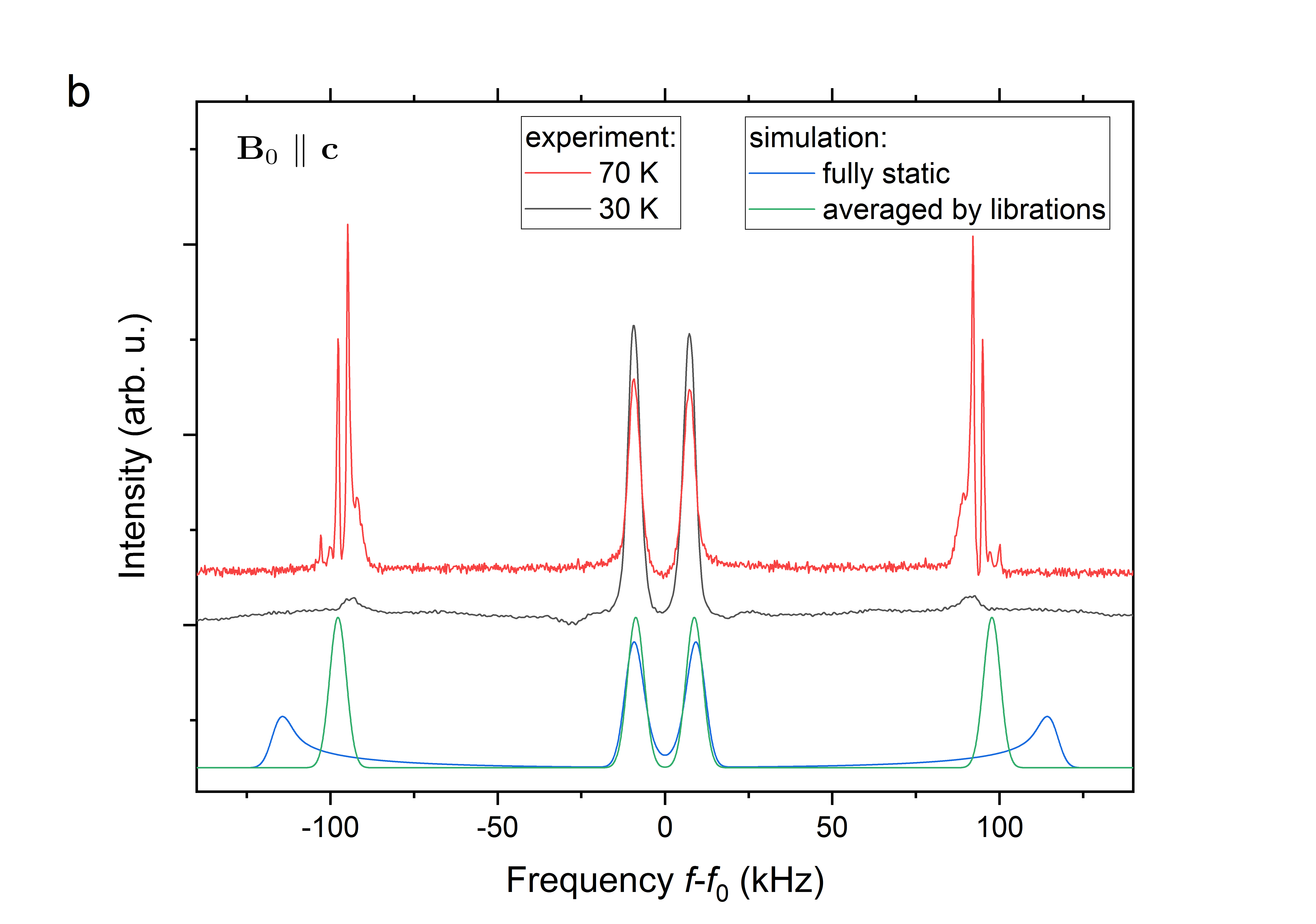
Another limitation of the applied model is that the frequency of linear harmonic oscillator is constant and only the amplitude of motion decreases with temperature. In reality however, the frequency of molecular motions and jumps also reduces on cooling, and at some point the motional averaging becomes inefficient. This is noticeable at temperatures below \qty70 where the character of NMR spectra changes and some of the well-resolved doublets become broad bands with features resembling spectra of a powder sample—instead of averaged values the spectra display the actual distributions of the local fields. The narrow peaks with resonance frequencies corresponding to the time-averaged fields are still apparent at \qty70, whereas the NMR spectra at \qty30 already contain broad bands (Fig. 9). In order to understand these spectral shapes we compare them with NMR spectra where the motional averaging was fully or partially removed. Using the spectroscopic and angular parameters obtained from the linear harmonic oscillator model, we constructed the 2H spectrum for (Fig. 9a) as a histogram where the angle has a Gaussian distribution (with dispersion ) and the angle is uniformly distributed within , i.e., a fully static spectrum. Additionally we simulated a spectrum where the jumps/re-bonding ceased, but the averaging due to librational motion is still effective. Apparently, the experimental spectrum at \qty30 contains features of both types of simulated spectra. Similar behavior can be seen for (Fig. 9b), where the outer doublet is broadened as in a fully static spectrum, yet it contains some residual peaks at about 100 and \qty-100 which stem from the librational averaging. On the other hand, the inner doublet for remains unchanged by these effects, which is expected, since it originates from the 2H nuclei of the H2 atoms which have a delta-function-like or very narrow distribution of motions during the librations—as imposed by the directionality of the hydrogen bond. The powder-like features in the low temperature spectra are thus in line with the proposed behavior of water molecules.
IV.3 Analysis of Jumps and Re-bonding of Water Molecules
The details of the second type of motion of water molecule in the voids—the re-bonding of hydrogen bond leading to effective rotations of the water molecules about the beryl hexagonal axis—are more difficult to obtain from our NMR experiments. The twelve bonding sites in the beryl void are equivalent by symmetry and thus cannot be distinguished directly in the NMR spectrum. From the dispersion of the quadrupole splitting with angle in case of (Fig. 6b) it is apparent that the re-bonding (considered as an instantaneous change in ) leads to variations of the splitting for the inner doublet that are about two orders of magnitude larger than those of the outer doublet. Therefore, the outer doublet is well resolved in the 2H NMR spectrum, i.e., the librations and re-bonding lead to sufficient averaging of local fields for the oscillating part of the water molecule (H1 site), whereas the inner doublet is severely broadened when , which means that the averaging by re-bonding is ineffective. From this we may estimate that the jumps within the beryl void are about two orders of magnitude slower than the molecular librations.
V Conclusions
The experimentally obtained 1H and 2H NMR spectra, measured at temperatures 5–\qty360, provided spatial and dynamic information on the behavior of water molecules in beryl crystal. We showed that although the water molecules in the undoped beryl, traditionally denoted as type-I molecules, are indeed oriented preferentially with their H–H lines along the hexagonal axis of beryl, the actual orientation of the H–H lines is significantly (by about 18∘) declined from the hexagonal axis. The reason consists apparently in the fact that the water molecules are oriented by their O–H bond towards one of twelve oxygen atoms forming the walls of the beryl structural voids. These positions are obviously energetically favorable, as hydrogen bonds between the H2O molecules and the void walls can be formed. In order to explain satisfactorily our experimental results, we proposed a model within which the molecules perform two types of movements: (i) librations around the axes of such hydrogen bonds, and (ii) less frequent orientational jumps among the twelve possible binding sites in the void. A simple thermodynamical model, with the water molecule considered as a quantum harmonic oscillator, correctly describes the observed experimental data at all temperatures, for both normal and heavy water. The frequencies of the oscillatory motions, evaluated from our thermodynamic model, agree well quantitatively with the frequencies of libration motions observed by optical experiments10, 5.
The implications of our work can be summarized as follows. First of all, the traditional view of type-I water molecules embedded in the beryl voids has to be corrected; a deviation of their H–H lines from the hexagonal axis with a mean value of about 18∘ must be accounted for. Consequently, in the search for ferroelectric order in the water subsystem, it appears important to bear in mind that, in principle, two components of spontaneous polarization may exist at low temperatures; a net ferroelectric moment may appear not only in the crystallographic planes but also along the hexagonal axis. Concerning the interactions between the water molecules and the atoms forming the structural voids in beryl, we have provided a very strong indirect indication that hydrogen bonds are formed between the molecules and any of the twelve closest oxygen atoms. This contradicts the earlier conjecture by Paré and Ducros 14 that no hydrogen bonds between the molecules and the beryl structure forms. Conversely, the presence of these hydrogen bonds supports the recent idea of a coupling mechanism between the water molecules and the beryl crystal lattice, suggested recently by Finkelstein et al.6, which agrees very well also with the observation of sum and difference frequencies in the infrared spectra 10. Consequently, our findings, as we believe, can serve as a firm basis for further investigations, including theoretical modeling, of the dynamics of isolated water molecules in beryl, and they can thus also deepen the understanding of mechanisms and interactions favoring the ferroelectric ordering of the water molecules.
Acknowledgements.
This work was supported by the Czech Science Foundation (project No. 20-1527S).References
References
- Gorshunov et al. [2014] B. P. Gorshunov, E. S. Zhukova, V. I. Torgashev, E. A. Motovilova, V. V. L. an d Anatoly S. Prokhorov, G. S. Shakurov, R. K. Kremer, V. V. Uskov, E. V. Pestrjakov, V. G. Thomas, D. A. Fursenko, C. Kadlec, F. Kadlec, and M. Dressel, “THz–IR spectroscopy of single molecules confined in nanocage of beryl crystal lattice,” Phase Trans. 87, 966–972 (2014).
- Gorshunov et al. [2016] B. P. Gorshunov, V. I. Torgashev, E. S. Zhukova, V. G. Thomas, M. A. Belyanchikov, C. Kadlec, F. Kadlec, M. Savinov, T. Ostapchuk, J. Petzelt, J. Prokleška, P. V. Tomas, D. A. Pestrjakov, E. V. an d Fursenko, G. S. Shakurov, A. S. Prokhorov, V. S. Gorelik, L. S. Kadyrov, V. V. Uskov, R. K. Kremer, and M. Dressel, “Incipient ferroelectricity of water molecules confined to nano-channels of beryl,” Nat. Commun. 7, 12842 (2016).
- Kolesnikov et al. [2016] A. I. Kolesnikov, G. F. Reiter, N. Choudhury, T. R. Prisk, E. Mamontov, A. Podlesnyak, G. Ehlers, A. G. Seel, D. J. Wesolowski, and L. M. Anovitz, “Quantum tunneling of water in beryl: A new state of the water molecule,” Phys. Rev. Lett. 116 (2016), 10.1103/physrevlett.116.167802.
- Zhukova et al. [2014] E. S. Zhukova, V. I. Torgashev, B. P. Gorshunov, V. V. Lebedev, G. S. Shakurov, R. K. Kremer, E. V. Pestrjakov, V. G. Thomas, D. A. Fursenko, A. S. Prokhorov, and M. Dressel, “Vibrational states of a water molecule in a nano-cavity of beryl crystal lattice,” J. Chem. Phys. 140, 224317 (2014).
- Belyanchikov et al. [2017] M. A. Belyanchikov, E. S. Zhukova, S. Tretiak, A. Zhugayevych, M. Dressel, F. Uhlig, J. Smiatek, M. Fyta, V. G. Thomas, and B. P. Gorshunov, “Vibrational states of nano-confined water molecules in beryl investigated by first-principles calculations and optical experiments,” Phys. Chem. Chem. Phys. 19, 30740–30748 (2017).
- Finkelstein et al. [2017] Y. Finkelstein, R. Moreh, S. L. Shang, Y. Wang, and Z. K. Liu, “Quantum behavior of water nano-confined in beryl,” J. Chem. Phys. 146 (2017), 10.1063/1.4978397.
- Arivazhagan et al. [2017] V. Arivazhagan, F. Schmitz, P. Vullum, A. van Helvoort, and B. Holst, “Atomic resolution imaging of beryl: an investigation of the nano-channel occupation,” J. Microscopy 265, 245–250 (2017).
- Gibbs, Breck, and Meagher [1968] G. Gibbs, D. Breck, and E. Meagher, “Structural refinement of hydrous and anhydrous synthetic beryl, and emerald, ,” Lithos 1, 275–285 (1968).
- Momma and Izumi [2011] K. Momma and F. Izumi, “VESTA 3 for three-dimensional visualization of crystal, volumetric and morphology data,” J. Appl. Crystallography 44, 1272–1276 (2011).
- Wood and Nassau [1967] D. L. Wood and K. Nassau, “Infrared spectra of foreign molecules in beryl,” J. Chem. Phys. 47, 2220–2228 (1967).
- Dressel et al. [2018] M. Dressel, E. S. Zhukova, V. G. Thomas, and B. P. Gorshunov, “Quantum electric dipole lattice,” J. Infrared, Millim., THz Waves 39, 799–815 (2018).
- Abragam [1961] A. Abragam, The principles of nuclear magnetism (Oxford university press, 1961).
- Modig and Halle [2002] K. Modig and B. Halle, “Proton magnetic shielding tensor in liquid water,” J. Amer. Chem. Soc. 124, 12031–12041 (2002).
- Paré and Ducros [1964] X. Paré and P. Ducros, “Étude par résonance magnétique nucléaire de l'eau dans le béryl,” Bull. Soc. fr. minéral. cristallogr. 87, 429–433 (1964).
- Sugitani, Nagashima, and Fujiwara [1966] Y. Sugitani, K. Nagashima, and S. Fujiwara, “The NMR analysis of the water of crystallization in beryl,” Bull. Chem. Soc. Jpn. 39, 672–674 (1966).
- Thomas and Klyakhin [1987] V. Thomas and V. Klyakhin, “The specific features of beryl doping by chromium under hydrothermal conditions,” in Mineral Forming in Endogenic Processes, edited by N. Sobolev (Nauka, Novosibirsk (in Russian), 1987) pp. 60–67.
- Freude [2000] D. Freude, “Quadrupolar nuclei in solid-state nuclear magnetic resonance,” in Encyclopedia of Analytical Chemistry, edited by R. Meyers (John Wiley & Sons Ltd, Chichester, 2000) pp. 12188–12224.
- Pyykkö [2018] P. Pyykkö, “Year-2017 nuclear quadrupole moments,” Molecular Phys. 116, 1328–1338 (2018).
- Garvey and Lucia [1977] R. M. Garvey and F. C. D. Lucia, “Extension of high resolution beam maser spectroscopy into the submillimetre wave region,” Canadian J. Phys. 55, 1115–1123 (1977).
- Bluyssen, Verhoeven, and Dymanus [1967] H. Bluyssen, J. Verhoeven, and A. Dymanus, “Hyperfine structure of HDO and by beam maser spectroscopy,” Phys. Lett. A 25, 214–215 (1967).
- Struis, Bleijser, and Leyte [1987] R. P. W. J. Struis, J. D. Bleijser, and J. C. Leyte, “Dynamic behavior and some of the molecular properties of water molecules in pure water and in magnesium chloride solutions,” J. Phys. Chem. 91, 1639–1645 (1987).
- Gordalla and Zeidler [1986] B. Gordalla and M. Zeidler, “Molecular dynamics in the system water-dimethylsulphoxide,” Molecular Phys. 59, 817–828 (1986).
- Edmonds and Mackay [1975] D. Edmonds and A. Mackay, “The pure quadrupole resonance of the deuteron in ice,” J. Magn. Resonance 20, 515–519 (1975).
- Wood, Allen, and Pidcock [2009] P. A. Wood, F. H. Allen, and E. Pidcock, “Hydrogen-bond directionality at the donor H atom—analysis of interaction energies and database statistics,” CrystEngComm 11, 1563–1571 (2009).
- Gatta et al. [2006] G. D. Gatta, F. Nestola, G. Bromiley, and S. Mattauch, “The real topological configuration of the extra-framework content in alkali-poor beryl: A multi-methodological study,” Am. Mineralogist 91, 29–34 (2006).
- Pathria and Beale [2011] R. Pathria and P. D. Beale, “Formulation of quantum statistics,” in Statistical Mechanics (Elsevier, 2011) pp. 115–140.
- Gueorguiev, Rau, and Draayer [2006] V. G. Gueorguiev, A. R. P. Rau, and J. P. Draayer, “Confined one-dimensional harmonic oscillator as a two-mode system,” Am. J. Phys. 74, 394–403 (2006).