Robust Image Registration with Absent Correspondences in Pre-operative and Follow-up Brain MRI Scans of Diffuse Glioma Patients
Abstract
Registration of pre-operative and follow-up brain MRI scans is challenging due to the large variation of tissue appearance and missing correspondences in tumour recurrence regions caused by tumour mass effect. Although recent deep learning-based deformable registration methods have achieved remarkable success in various medical applications, most of them are not capable of registering images with pathologies. In this paper, we propose a 3-step registration pipeline for pre-operative and follow-up brain MRI scans that consists of 1) a multi-level affine registration, 2) a conditional deep Laplacian pyramid image registration network (cLapIRN) with forward-backward consistency constraint, and 3) a non-linear instance optimization method. We apply the method to the Brain Tumor Sequence Registration (BraTS-Reg) Challenge. Our method achieves accurate and robust registration of brain MRI scans with pathologies, which achieves a median absolute error of 1.64 mm and 88% of successful registration rate in the validation set of BraTS-Reg challenge. Our method ranks 1 place in the 2022 MICCAI BraTS-Reg challenge.
Keywords:
Absent correspondences Patient-specific registration Deformable registration1 Introduction
Registration of pre-operative and follow-up images is crucial in evaluating the effectiveness of treatment for patients suffering from diffuse glioma. However, this registration problem is challenging due to the missing correspondences and mass effect caused by resected tissue. While many recent deep learning-based deformable registration algorithms [21, 2, 3, 10, 9, 13, 14, 15] are available, only a few learning-based methods [5] address the missing correspondences problem. In this paper, we propose a 3-step registration pipeline for pre-operative and follow-up brain MRI scans that consists of 1) a multi-level affine pre-alignment, 2) a conditional deep Laplacian pyramid image registration network (cLapIRN) with forward-backward consistency constraint [17, 16, 18], and 3) a non-linear instance optimization with inverse consistency. We validate the method using the pre-operative and follow-up images brain MRI scans in the Brain Tumor Sequence Registration Challenge (BraTS-Reg) challenge [1].
2 Related Work
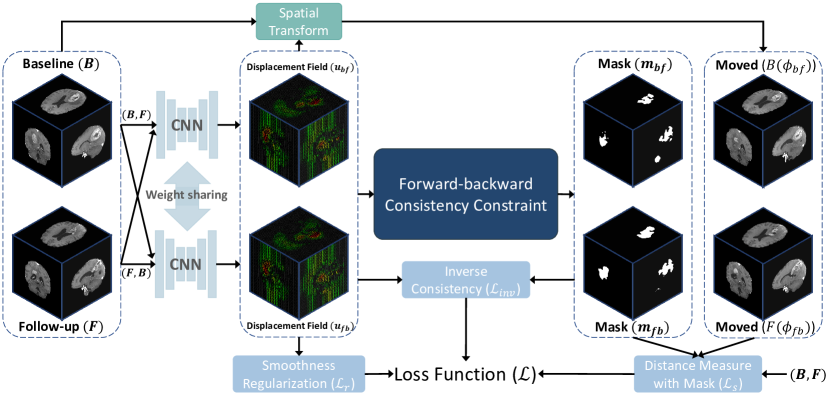
Accurate registration of pre-operative and post-recurrence brain MRI scans is crucial to the treatment plan and diagnosis of intracranial tumors, especially brain gliomas [7, 20]. To better interpret the location and extent of the tumor and its biological activity after resection, the dense correspondences between pre-operative and follow-up structural brain MRI scans of the patient first need to be established. However, deformable registration between the pre-operative and follow-up scans, including post-resection and post-recurrence, is challenging due to possible large deformations and absent correspondences caused by tumor’s mass effects [4], resection cavities, tumor recurrence and tissue relaxation in the follow-up scans. While recent deep learning-based deformable registration (DLDR) methods [2, 10, 9, 6, 13] have achieved remarkable registration performance in many medical applications, these registration approaches often ignored the absent correspondence problem in the pre-operative and post-recurrence images. To address this issue, we extend our deep learning-based method described in [17] by introducing affine pre-alignment and non-linear instance optimization as post-processing to our method. DIRAC leverages conditional Laplacian Pyramid Image Registration Networks (cLapIRN) [16] as the backbone network, jointly estimates the bidirectional deformation fields and explicitly locates regions with absent correspondence. By excluding the regions with absent correspondence in the similarity measure during training, DIRAC improves the target registration error of landmarks in pre-operative and follow-up images, especially for those near the tumour regions.
3 Methods
We propose a 3-step registration pipeline for pre-operative and follow-up brain MRI scans which consists of 1) a gradient descent-based affine registration method, 2) a deformable image registration method with absent correspondence (DIRAC), and 3) a non-linear instance optimization method. Let and be the pre-operative (baseline) scan and post-recurrence (follow-up) scan defined over a -D mutual spatial domain . Our goal is to establish a dense non-linear correspondence between the pre-operative scan and the post-recurrence scan of the same subject. In this paper, we focus on 3D registration, i.e., and .
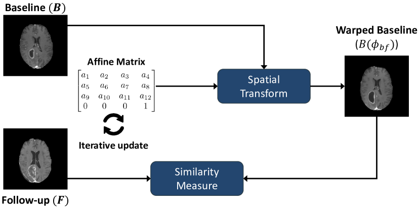
3.1 Affine Registration
Although all MRI scans provided by the challenge are rigidly registered to the same anatomical template [1], we found that there are large linear misalignments between the pre-operative and follow-up images in cases suffering from serious tumor mass effect. To factor out the possible linear misalignment between MRI scans and , we register T1-weighted and scans using the iterative affine registration method.
Figure 1 depicts the overview of the affine registration. The affine registration method starts by initializing two identity matrices as initial affine transformation and creating image pyramids with levels using trilinear interpolation for and . Then, we iteratively optimize the solutions by minimizing a suitable distance measure that quantifies alignment quality using the Adam optimizer [11] and a coarse-to-fine multi-resolution iteratively registration scheme. In this step, we use the Normalized Gradient Fields (NGF) distance measure [8]. Formally, the NGF is defined as:
| (1) |
where , and is an edge parameter controlling the level of influence of image gradients. The value of NGF is minimized when the gradients of and are aligned. The result with the minimal distance measure is selected as intermediate result.
3.2 Unsupervised Deformable Registration with Absent Correspondences Estimation
Assume that baseline scan and follow-up scan are affinely aligned in step 1 such that the main source of misalignment between and is non-linear, we then apply DIRAC [17] to further align and in an bidirectional manner. Since multi-parametric MRI sequences of each time-point, including T1 contrast-enhanced (T1ce), T2, T2 Fluid Attenuated (Flair) and T1-weighted (T1), are provided for each case in the BraTS-Reg challenge, we utilize all MRI modalities of the brain MRI scans in this step, i.e., and are 4-channel pre-operative and post-recurrence scans, respectively. Specifically, we parameterize the problem with a function cLapIRN [16], where is a set of learning parameters and represents the displacement field that transform to align with , i.e., and define similar anatomical locations for each voxel (except voxels with absent correspondence). Figure 2 illustrates the overview of DIRAC. DIRAC leverages the bidirectional displacement fields and a forward-backward consistency locate regions with absent correspondence and excludes them in the similarity measure during training phase.
| Parameters | Methods | ||
|---|---|---|---|
| Affine | DIRAC | Inst. Opt. | |
| Input MRI sequences | T1ce | T1,T1ce,T2,Flair | T1ce,T2 |
| Number of levels | 3 | 3 | 5 |
| Max. image dimension | (64, 64, 40) | (160, 160, 80) | (240, 240, 155) |
| Min. image dimension | (16, 16, 16) | (40, 40, 20) | (80, 80, 80) |
| Learning rate per level | [1e-2, 5e-3, 2e-3] | [1e-4, 1e-4, 1e-4] | [1e-2, 5e-3, 5e-3, 3e-3, 3e-3] |
| Max. iteration per level | [90, 90, 90] | - | [150, 100, 100, 100, 50] |
| Distance measure | NGF() | NCC-SP() | NCC() |
| Max. number of grid points | - | ||
| Min. number of grid points | - | ||
| Weight of Inverse consistency | - | [0.5, 0.5, 0.5] | [1.0, 2.0, 4.0, 8.0, 10.0] |
3.2.1 Forward-Backward Consistency
Given the deformation fields and , where is the identity transform and is the corresponding displacement vector field, we calculate the forward-backward error for to as:
| (2) |
Based on the observation that regions without true correspondences would have higher forward-backward error in solutions, we create a mask and mark whenever the forward-backward error is larger than the pre-defined threshold . The pre-defined threshold is defined as:
| (3) |
where is set to 0.015. Then, we create a binary mask to mark voxels with absent correspondence as follows:
| (4) |
where denotes an averaging filter of size and denotes a convolution operator with zero-padding .
3.2.2 Objective Function
The objective function of DIRAC is defined as follows:
| (5) |
where the masked dissimilarity measure and the masked inverse consistency loss are defined as:
| (6) |
and
| (7) |
In this step, we use masked negative local cross-correlation (NCC) with similarity pyramid [18] as the dissimilarity function. To encourage smooth solutions and penalize implausible solutions, we adopt a diffusion regularizer during training. We set and to 0.4 and 0.01, respectively. For more details, we recommend interested reader also refer to [17].
| Methods | MAE | MAE | Robustness |
|---|---|---|---|
| Initial | - | ||
| Ours |
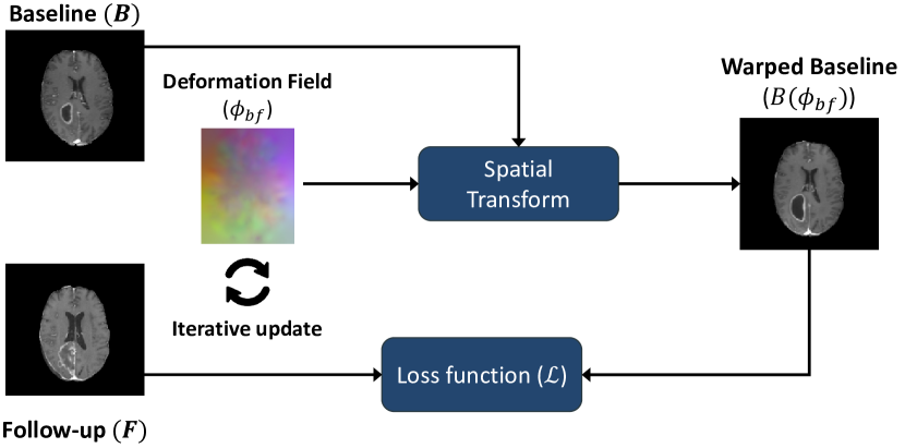
3.3 Non-rigid Instance Optimization
Due to insufficient amount of training data and discrepancy in distributions between training and test set, the learning-based method in step 2 may produce biased solutions, especially in cases with small initial misalignment. As such, we introduce a non-rigid instance optimization step to further improve the robustness and registration accuracy of solutions from the previous step. Figure 3 shows the overview of the non-rigid instance optimization. In the final step, the non-parametric deformation is controlled by the same objective function in 5, except we use NCC as the distance measure. The smoothness regularization coefficients for each level are set to [0.25, 0.3, 0.3, 0.35, 0.35], respectively. The displacement fields are discretized with trilinear interpolation defined on a uniform control point grid with a fixed number of points. We use an Adam optimizer together with multi-level continuation to avoid local minima.
3.4 Hyperparameters
The hyperparameters of our 3-step approach are summarized in Table 1.
| Case | Initial | Affine | A-DIRAC | A-DIRAC-IO | |||
|---|---|---|---|---|---|---|---|
| MAE | MAE | Robustness | MAE | Robustness | MAE | Robustness | |
| Case 141 | 13.50 | 4.26 | 1.00 | 1.94 | 1.00 | 1.62 | 1.00 |
| Case 142 | 14.00 | 6.12 | 0.88 | 3.07 | 1.00 | 1.88 | 1.00 |
| Case 143 | 16.00 | 8.98 | 1.00 | 2.63 | 1.00 | 1.14 | 1.00 |
| Case 144 | 15.00 | 9.52 | 0.88 | 3.10 | 1.00 | 2.56 | 1.00 |
| Case 145 | 17.00 | 5.14 | 1.00 | 2.09 | 1.00 | 1.13 | 1.00 |
| Case 146 | 17.00 | 5.74 | 1.00 | 1.84 | 1.00 | 1.94 | 1.00 |
| Case 147 | 1.50 | 2.03 | 0.45 | 2.17 | 0.60 | 1.64 | 0.55 |
| Case 148 | 3.50 | 2.90 | 0.75 | 1.71 | 0.90 | 1.43 | 0.90 |
| Case 149 | 9.00 | 2.22 | 1.00 | 1.94 | 1.00 | 1.56 | 1.00 |
| Case 150 | 4.00 | 3.65 | 0.53 | 2.87 | 0.63 | 1.27 | 0.74 |
| Case 151 | 3.00 | 2.13 | 0.5 | 1.39 | 0.75 | 1.18 | 0.85 |
| Case 152 | 5.00 | 2.11 | 0.95 | 1.45 | 0.84 | 1.42 | 0.95 |
| Case 153 | 2.00 | 2.04 | 0.33 | 1.44 | 0.67 | 1.80 | 0.67 |
| Case 154 | 2.00 | 2.61 | 0.25 | 2.02 | 0.4 | 1.98 | 0.55 |
| Case 155 | 2.00 | 3.09 | 0.21 | 2.43 | 0.37 | 1.70 | 0.53 |
| Case 156 | 7.00 | 2.84 | 1.00 | 2.29 | 1.00 | 1.45 | 1.00 |
| Case 157 | 10.00 | 4.90 | 0.90 | 2.67 | 1.00 | 1.66 | 1.00 |
| Case 158 | 4.50 | 3.48 | 0.40 | 1.39 | 0.80 | 1.13 | 1.00 |
| Case 159 | 6.00 | 7.28 | 0.36 | 2.25 | 1.00 | 2.28 | 1.00 |
| Case 160 | 4.00 | 2.55 | 0.7 | 2.29 | 0.80 | 1.94 | 0.90 |
| Mean | 7.80 | 4.18 | 0.70 | 2.15 | 0.84 | 1.64 | 0.88 |
| Std | 5.62 | 2.30 | 0.29 | 0.54 | 0.21 | 0.39 | 0.17 |
| Median | 5.50 | 3.28 | 0.81 | 2.13 | 0.95 | 1.63 | 1.00 |
4 Experiments
4.0.1 Implementation
Our proposed method is implemented with PyTorch 1.8 [19] and trained with an Nvidia Titan RTX GPU and an Intel Core (i7-4790) CPU. We build DIRAC on top of the official implementation of cLapIRN available in [12]. We adopt Adam optimizer [11] with a fixed learning rate and train it from scratch with the training data from the BraTS-Reg challenge.
4.0.2 Measurements
We quantitatively evaluate our method based on the average of the median absolute error (MAE) and robustness of anatomical landmarks. Specifically, the MAE is defined as:
| (8) |
where is the -th estimated anatomical landmark in the baseline scan and is the -th goundtruth landmark in the baseline scan. The robustness is defined as the ratio of landmarks with improved MAE after registration to all landmarks, following the definition in [1].
4.0.3 Results
For each case in the training and validation set of the BraTS-Reg challenge, we register the pre-operative image scan to the follow-up image scan and use the resulting deformation field to transform the manually defined landmarks in the follow-up scan. In total, there are 140 and 20 pairs of pre-operative and follow-up image scans in the training and validation set, respectively. We follow the evaluation pipeline of the BraTS-Reg challenge and report the average median absolute error MAE and robustness of the training and validation set in Tables 2 and 3.
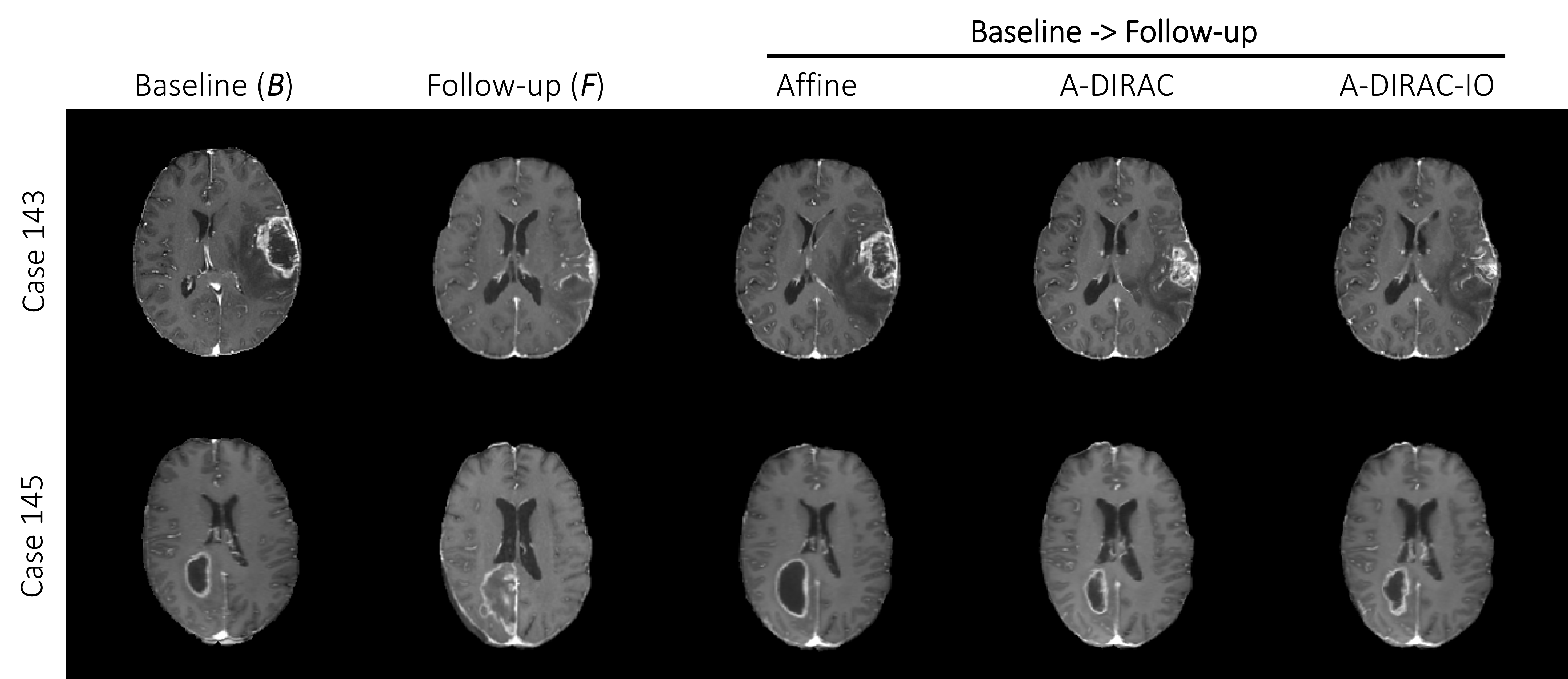
An example qualitative result is shown in Figure 4. The reduction of the registration error in the validation set in the pipeline is shown in Table 3. While the MRI scans are pre-registered to a common template, the average median error is reduced from 7.8 mm to 4.18 mm, indicating there exists a large linear misalignment between each case. Furthermore, the median error and robustness are consistently improved after each step, reaching to 1.64 mm average median error. Notably, our MAE is the lowest on the challenge’s validation leaderboard.
5 Conclusion
We proposed a 3-step registration method for pre-operative and follow-up brain tumor registration. The method was evaluated with the dataset provided by the BraTS-Reg challenge and ranked 1 place in the 2022 MICCAI BraTS-Reg challenge. By combining the pathological-aware deep learning-based method and instance optimization, we demonstrated the follow-up scan could be accurately registered to the pre-operative scan with an average median absolute error of 1.64mm. Compared to conventional methods, our method inherits the runtime advantage from deep learning-based approaches and does not require any manual interaction or supervision, demonstrating immense potential in the fully-automated patient-specific registration. We left the further analysis of our method and the comparison to existing methods for future work.
References
- [1] Baheti, B., Waldmannstetter, D., Chakrabarty, S., Akbari, H., Bilello, M., Wiestler, B., Schwarting, J., Calabrese, E., Rudie, J., Abidi, S., et al.: The brain tumor sequence registration challenge: Establishing correspondence between pre-operative and follow-up mri scans of diffuse glioma patients. arXiv preprint arXiv:2112.06979 (2021)
- [2] Balakrishnan, G., Zhao, A., Sabuncu, M.R., Guttag, J., Dalca, A.V.: An unsupervised learning model for deformable medical image registration. In: Proceedings of the IEEE conference on computer vision and pattern recognition. pp. 9252–9260 (2018)
- [3] Dalca, A.V., Balakrishnan, G., Guttag, J., Sabuncu, M.R.: Unsupervised learning for fast probabilistic diffeomorphic registration. In: International Conference on Medical Image Computing and Computer-Assisted Intervention. pp. 729–738. Springer (2018)
- [4] Dean, B.L., Drayer, B.P., Bird, C.R., Flom, R.A., Hodak, J.A., Coons, S.W., Carey, R.G.: Gliomas: classification with mr imaging. Radiology 174(2), 411–415 (1990)
- [5] Han, X., Shen, Z., Xu, Z., Bakas, S., Akbari, H., Bilello, M., Davatzikos, C., Niethammer, M.: A deep network for joint registration and reconstruction of images with pathologies. In: International Workshop on Machine Learning in Medical Imaging. pp. 342–352. Springer (2020)
- [6] Heinrich, M.P.: Closing the gap between deep and conventional image registration using probabilistic dense displacement networks. In: International Conference on Medical Image Computing and Computer-Assisted Intervention. pp. 50–58. Springer (2019)
- [7] Heiss, W.D., Raab, P., Lanfermann, H.: Multimodality assessment of brain tumors and tumor recurrence. Journal of Nuclear Medicine 52(10), 1585–1600 (2011)
- [8] Hodneland, E., Lundervold, A., Rørvik, J., Munthe-Kaas, A.Z.: Normalized gradient fields and mutual information for motion correction of dce-mri images. In: 2013 8th International Symposium on Image and Signal Processing and Analysis (ISPA). pp. 516–521. IEEE (2013)
- [9] Hu, X., Kang, M., Huang, W., Scott, M.R., Wiest, R., Reyes, M.: Dual-stream pyramid registration network. In: International Conference on Medical Image Computing and Computer-Assisted Intervention. pp. 382–390. Springer (2019)
- [10] Kim, B., Kim, J., Lee, J.G., Kim, D.H., Park, S.H., Ye, J.C.: Unsupervised deformable image registration using cycle-consistent cnn. In: International Conference on Medical Image Computing and Computer-Assisted Intervention. pp. 166–174. Springer (2019)
- [11] Kingma, D.P., Ba, J.: Adam: A method for stochastic optimization. arXiv preprint arXiv:1412.6980 (2014)
- [12] Mok, T.C., Chung, A.: Official implementation of conditional deep laplacian pyramid image registration network. https://github.com/cwmok/Conditional_LapIRN, accessed: 01.03.2021
- [13] Mok, T.C., Chung, A.: Fast symmetric diffeomorphic image registration with convolutional neural networks. In: Proceedings of the IEEE/CVF conference on computer vision and pattern recognition. pp. 4644–4653 (2020)
- [14] Mok, T.C., Chung, A.: Large deformation image registration with anatomy-aware laplacian pyramid networks. In: International Conference on Medical Image Computing and Computer-Assisted Intervention. pp. 61–67. Springer (2020)
- [15] Mok, T.C., Chung, A.: Conditional deep laplacian pyramid image registration network in learn2reg challenge. In: International Conference on Medical Image Computing and Computer-Assisted Intervention. pp. 161–167. Springer (2021)
- [16] Mok, T.C., Chung, A.: Conditional deformable image registration with convolutional neural network. In: International Conference on Medical Image Computing and Computer-Assisted Intervention. pp. 35–45. Springer (2021)
- [17] Mok, T.C., Chung, A.: Unsupervised deformable image registration with absent correspondences in pre-operative and post-recurrence brain tumor mri scans. In: International Conference on Medical Image Computing and Computer-Assisted Intervention. pp. 25–35. Springer (2022)
- [18] Mok, T.C., Chung, A.C.: Large deformation diffeomorphic image registration with laplacian pyramid networks. In: International Conference on Medical Image Computing and Computer-Assisted Intervention. pp. 211–221. Springer (2020)
- [19] Paszke, A., Gross, S., Chintala, S., et al.: Automatic differentiation in pytorch. In: NIPS-W (2017)
- [20] Price, S.J., Jena, R., Burnet, N.G., Carpenter, T.A., Pickard, J.D., Gillard, J.H.: Predicting patterns of glioma recurrence using diffusion tensor imaging. European radiology 17(7), 1675–1684 (2007)
- [21] de Vos, B.D., Berendsen, F.F., Viergever, M.A., Staring, M., Išgum, I.: End-to-end unsupervised deformable image registration with a convolutional neural network. In: Deep Learning in Medical Image Analysis and Multimodal Learning for Clinical Decision Support, pp. 204–212. Springer (2017)