Tracking nanoscale perturbation in active disordered media
Abstract
The disorder induced feedback makes random lasers very susceptible to any changes in the scattering medium. The sensitivity of the lasing modes to perturbations in the disordered systems have been utilized to map the regions of perturbation. A tracking parameter, that takes into account the cumulative effect of changes in the spatial distribution of the lasing modes of the system has been defined to locate the region in which a scatterer is displaced by a few nanometers. We show numerically that the precision of the method increases with the number of modes. The proposed method opens up the possibility of application of random lasers as a tool for monitoring locations of nanoscale displacement which can be useful for single particle detection and monitoring.
I Introduction
A random laser (RL) is an optical device that utilizes the disorder in the system for the optical feedback. Unlike conventional lasers, no well-defined cavities are present in RLs. The idea of feedback by multiple scattering was first proposed by Letokhov [1] and has been extensively used to realize random lasing in a variety of disordered systems [2, 3, 4, 5, 6, 7, 8]. Two types of RLs have been reported namely, coherent RLs and incoherent RLs, depending on whether the scattering induces the feedback in the field or the intensity, respectively [9]. The scattering strength determines the lasing characteristics such as the lasing threshold of the system, spatial confinement of the modes, etc. Based on the scattering strength, disordered systems can be broadly divided into two categories, namely, strongly scattering and weakly scattering systems. In the strongly scattering systems the lasing modes are localized well within the system and are identical to the quasi-bound (QB) states of the passive system [10, 11, 12], whereas, in weakly scattering systems the lasing modes extend all over the system [12, 13].
Unlike conventional lasers, RL emission is random in wavelength, omnidirectional [4] and has low spatial and temporal coherence [14, 15, 16]. These properties make them suitable for different applications like, imaging [17], displays and lighting [18], holography [19], etc., but it limits their use where specific wavelength or unidirectional emission is required. Spatial light modulators (SLMs) have been used to shape the pump intensity profile to control the emission and directionality of RLs making them useful for different applications [20, 21, 22, 23, 24, 25, 26]. As the feedback in RLs is provided by disorder-induced scattering, the lasing modes are very sensitive to any changes in the scattering medium. This makes RLs a natural candidate for designing sensors for various applications. The strong dependence of emission characteristics of RLs on the scattering properties of the medium have been utilized to assess nanoscale perturbations [27]. The monitoring of single nanoparticle perturbation enables to detect single virus, bacterium and biolmolecule. Random lasers have been used as a diagnostic tool for bio-imaging and bio sensing in various biological structures infiltrated with dye [28, 5, 29]. The nanoscale deformation and prefailure damage in bones can be detected by monitoring the shifts in the random lasing peaks [30]. In ex-vivo dye infiltrated human tissues, the changes in the emission spectrum have been observed in malignant tissues as compared to the healthy ones [31]. The cancerous tissues of different grades of malignancy can be differentiated as they exhibit different lasing spectra for same pump energy [32]. RLs have been proposed as an in-vivo tool to differentiate between skin, fat, muscle and nerve tissues during laser surgery [33].
In this work, RLs have been proposed as a tool to map the regions of nanoscale perturbation in several random media. A two dimensional (2D) active disordered system has been considered and nanoscale perturbations have been introduced in the medium. Using finite difference time domain (FDTD) method [34] the modes and the corresponding spatial field distributions for the system before and after the perturbation have been computed. In the past, RLs have been used to detect changes in the scattering medium [27]. In this work we go a step further and show numerically that it is also possible to identify the position of the perturbation with good precision. A small perturbation in the system leads to minute changes in the spectral position of the modes and their corresponding spatial field distributions, but the individual modes do not provide any information about the location of the perturbation. So, a tracking parameter is defined which takes into account the cumulative effect of changes in the modes, to map the region of perturbation. We find that its mapping converges to the defect location when the number of modes increases. This finding paves the way to single particle tracking in disordered systems. The theoretical explorations in this work provide an initial framework to utilize RLs in the field of diagnostics to monitor and track the growth of tumors in disordered biological systems.
II Numerical method and computational details
A 2D disordered system of size, has been considered. It consists of circular particles with radius, and refractive index, , randomly distributed in a background medium of refractive index, . The values of the refractive index have been chosen to mimic the presence of particles in 4-(Dicyanomethylene)-2-methyl-6-(4-dimethylaminostyryl)-4H-pyran (DCM) doped polyvinyl alcohol (PVA) thin films [35, 36, 37]. The background medium has been chosen as the active part of the system and modeled as a four level atomic system. The surface filling fraction of the scatterers is . In this study, 2D FDTD computation has been carried out using transverse magnetic fields with a grid resolution of , along and directions, respectively. In order to ensure the stability of the simulation, the time step chosen is, [38]. The parameters used for the active medium are mentioned in Ref. [10]. The system is pumped uniformly with a Gaussian pulse of central wavelength and pulse duration s at a pump level above the lasing threshold of system.
III Results and discussion
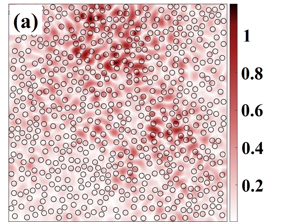
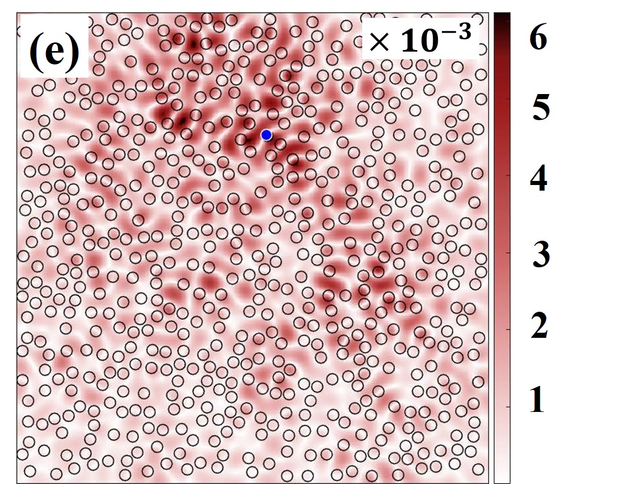
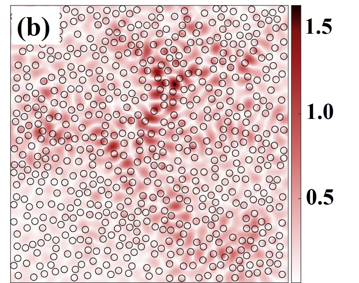
The 2D active, random system was pumped above the lasing threshold. The energy in the system was observed to grow exponentially, and after some strong relaxation oscillations, it eventually reaches a steady-state. The lasing modes of the system are calculated by the Fourier transform of the time records of the field after the system has reached the stationary state. Several distinct peaks are observed as shown in Fig. 1. The ten modes considered for further analysis are marked with arrows. The discrete peaks in the emission spectrum indicate lasing action with resonant feedback. The spatial field distribution of the modes is computed by taking the Fourier transform of the field recorded at each grid point. The spatial field distribution of the modes marked as (1-4) in Fig. 1 is shown in Figs. 2(a-d). It is observed that the modes are confined well within the system, indicating that the system is strongly scattering. The numerically computed scattering mean free path for the system using Mie scattering theory is, [39]. The localization length for the system is calculated by considering the field intensity profiles of modes averaged along or directions. The averaged intensity profile exhibits strong local fluctuations but its envelope decays exponentially whose characteristic length, gives the localization length of the modes. The average localization length calculated for the system is, . The scattering mean free path and the localization length also indicate that system is strongly scattering and the modes are confined well within the system, respectively.
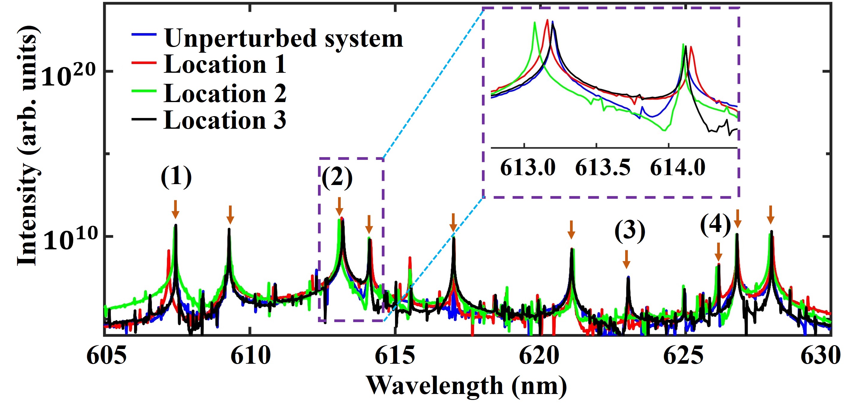
Next, in order to introduce a single nanoscale perturbation in the system, a randomly chosen scatterer was displaced by along an arbitrary direction. The numerical parameters limit the minimum and the maximum perturbation that can be introduced in the system. The minimum displacement cannot be smaller than the grid resolution and maximum displacement possible is dependent on the surface filling fraction of the system. In realistic systems, such limitations don’t exist and hence it is expected that even smaller perturbations can be detected. The effect of perturbation at different locations in the system, on the RL spectra and spatial field distribution of modes has been studied. The emission spectra of the system, when a single particle is perturbed at three different locations in the system are shown in Fig. 1. It is observed that the perturbation causes slight shift in the spectral positions of the random lasing modes, as shown in the inset. The modes 1-4 experience spectral shifts () of magnitude , , and , respectively. Further details on the correspondence between the magnitude of the spectral shifts and the mode field distributions are presented later.
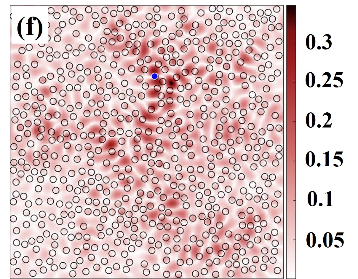
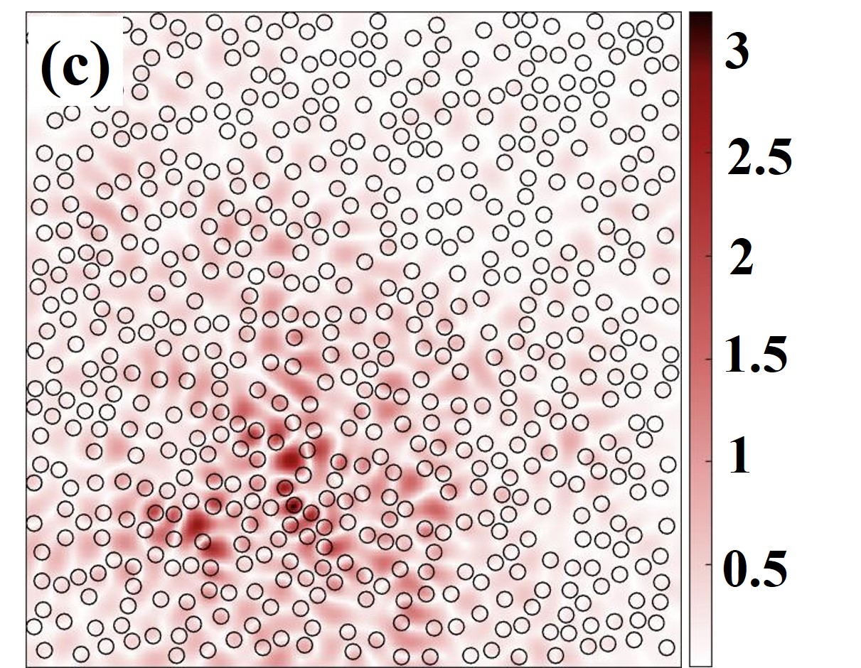
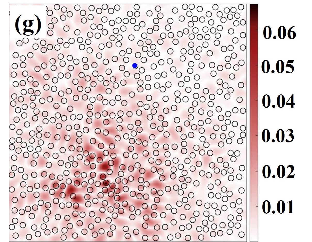
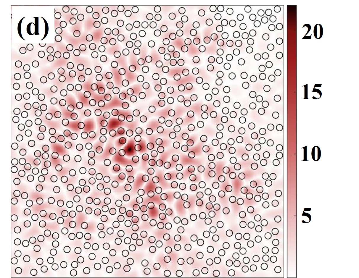
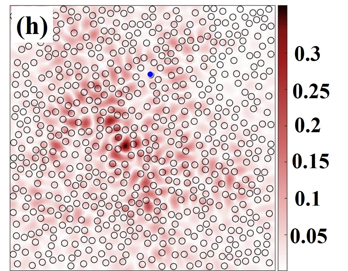
The perturbation also leads to changes in the spatial field distribution of modes as shown in Figs. 2(e-h), for the perturbation at location 1 (Fig. 1), wherein the perturbed particle is marked in blue. In order to quantify the changes in the system due to perturbation, 2D correlation coefficient () between the spatial field distribution of modes before and after perturbation is calculated, which is defined as:
| (1) |
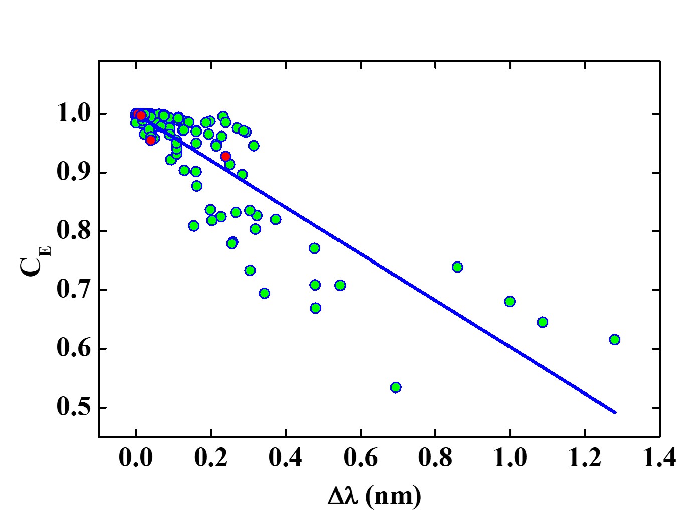
where, and are the field magnitudes of the modes at location in the system, before and after perturbation, respectively. and represent the mean field values of the corresponding modes. The value quantifies the similarity between the modes before and after the perturbation, and for the system shown in Fig. 2, it is found to be 0.92, 0.95, 0.99, and 0.99 for modes 1, 2, 3 and 4, respectively. The values indicate that the perturbation leads to more changes in modes 1 and 2 as compared to modes 3 and 4. It is evident from Figs. 2(e-f) that the perturbed scatterer is present in the region with high field value for modes 1 and 2 as compared to modes 3 and 4. Thus, a perturbation in the high field region of a mode leads to more changes in the lasing modes as compared to a perturbation in low field region. It is also observed that the shape of the spatial field profiles of modes do not change drastically after the perturbation. However, minute changes are observed in the distribution of the field and its magnitude. Moreover, the perturbation also leads to changes in the spectral location of the lasing modes. The spectral shift () in the modes is linearly related to value as shown in Fig. 3. For modes exhibiting small changes in their spectral position, the values are found to be 1, and as increases, decreases. Thus, with increasing spectral shift the changes in the field distribution of the corresponding modes become more prominent.
The sensitivity of lasing modes to nanoscale displacements has been utilized to monitor perturbations in the system. It is observed that the nanoscale alteration in the scatterer position leads to changes in the lasing modes and their spatial field distributions, and the amount of change varies for each mode. But, these changes in the individual modes do not provide any information about the position of the perturbed particle. Now, it is interesting to ask a question on whether one can identify the particle that has been perturbed, given the modes before and after the perturbation are known. Here, we show that it is possible to locate the position of the scatterer that has been perturbed with the help of the computed modes by defining a tracking parameter, TP as,
| (2) |
where and are the normalized field values of mode before and after the perturbation at position in the system, respectively. The tracking parameter is given by the product of change in the field distribution of the modes considered, due to perturbation. Here, the normalized field values have been considered as we are interested in how the field at a point in the system changes with respect to its neighbouring positions with perturbation.
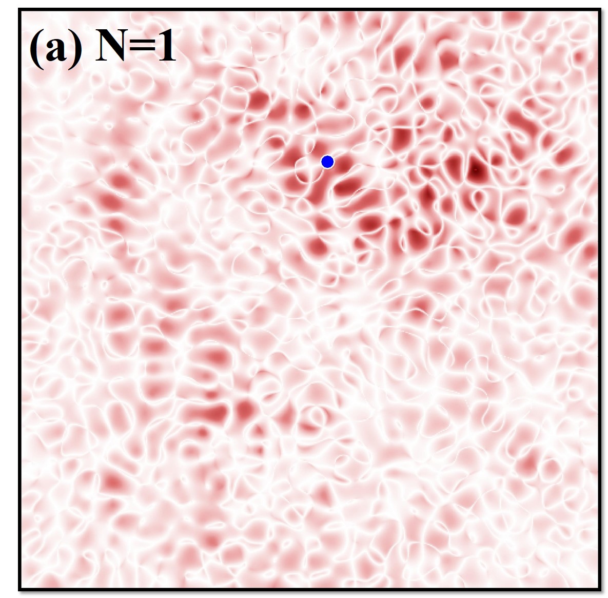
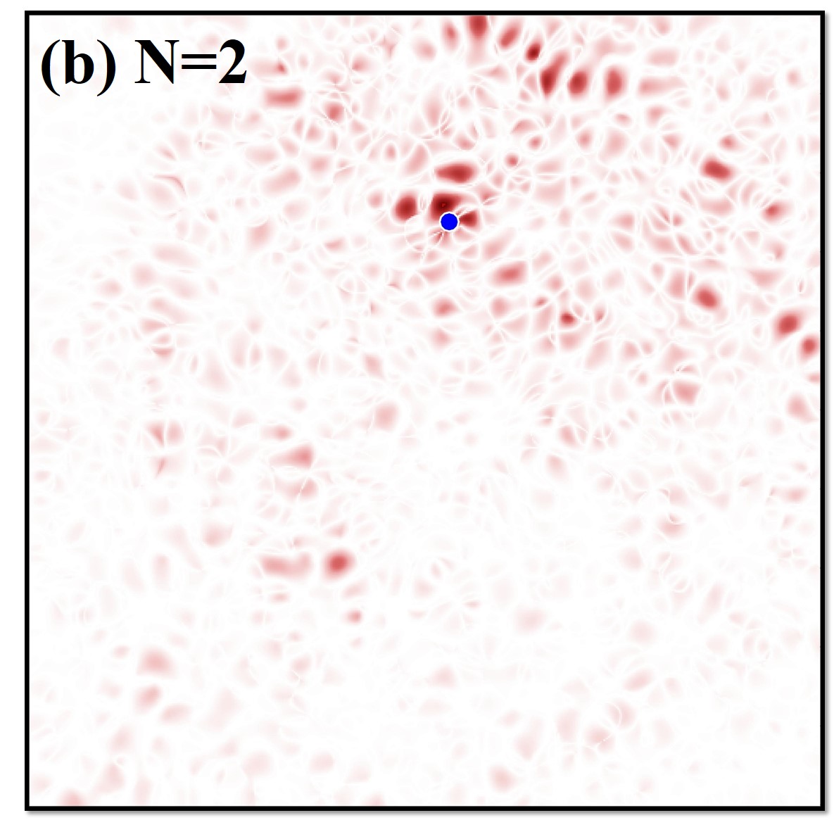
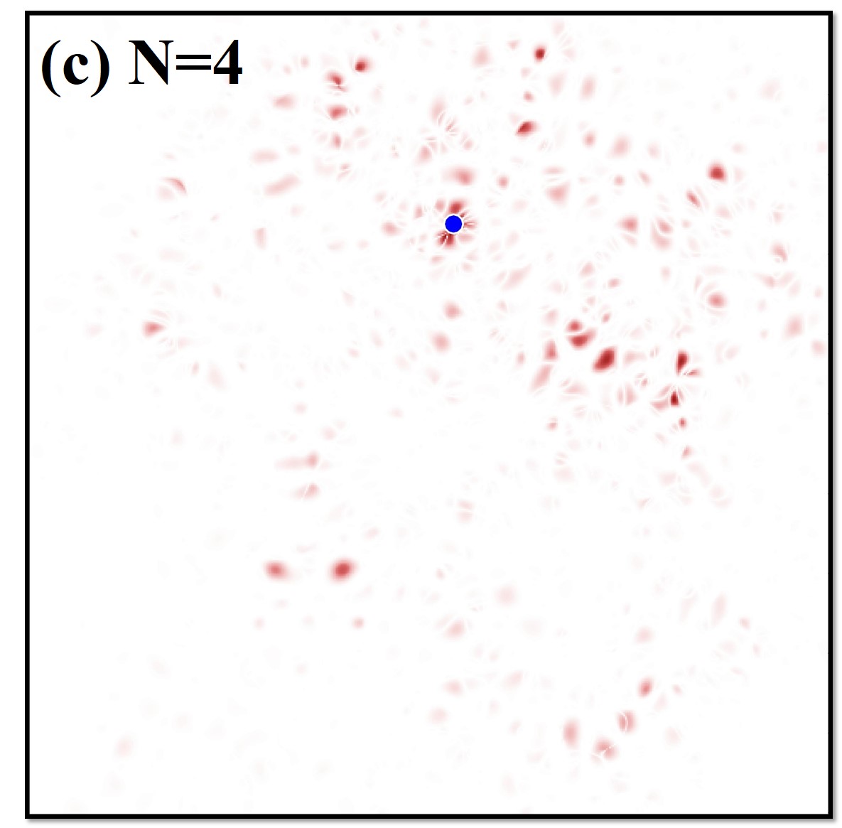
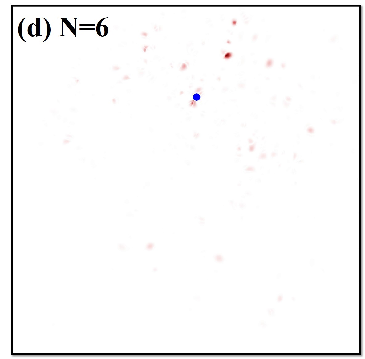
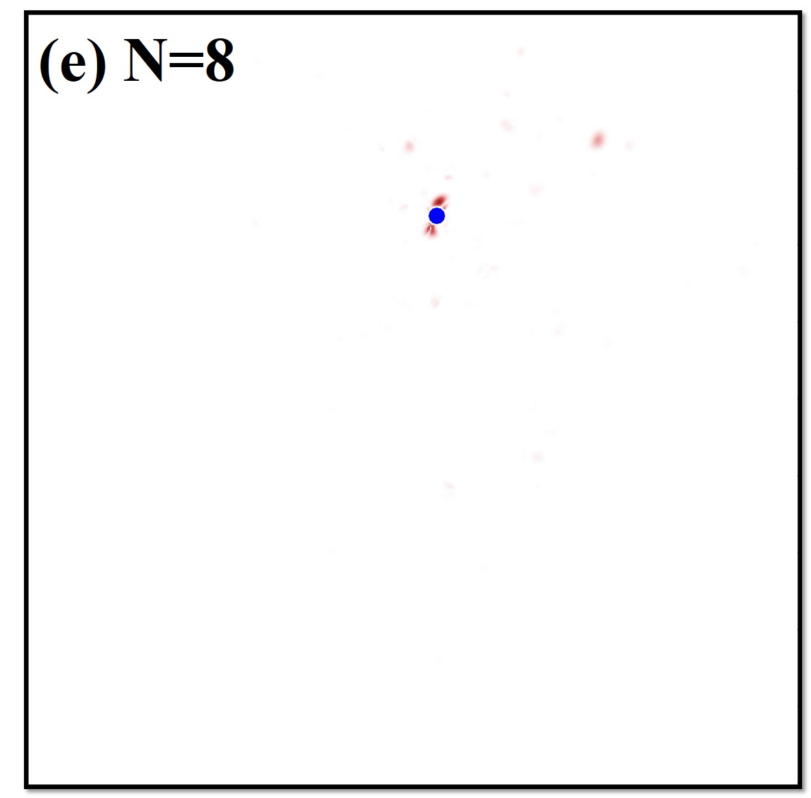
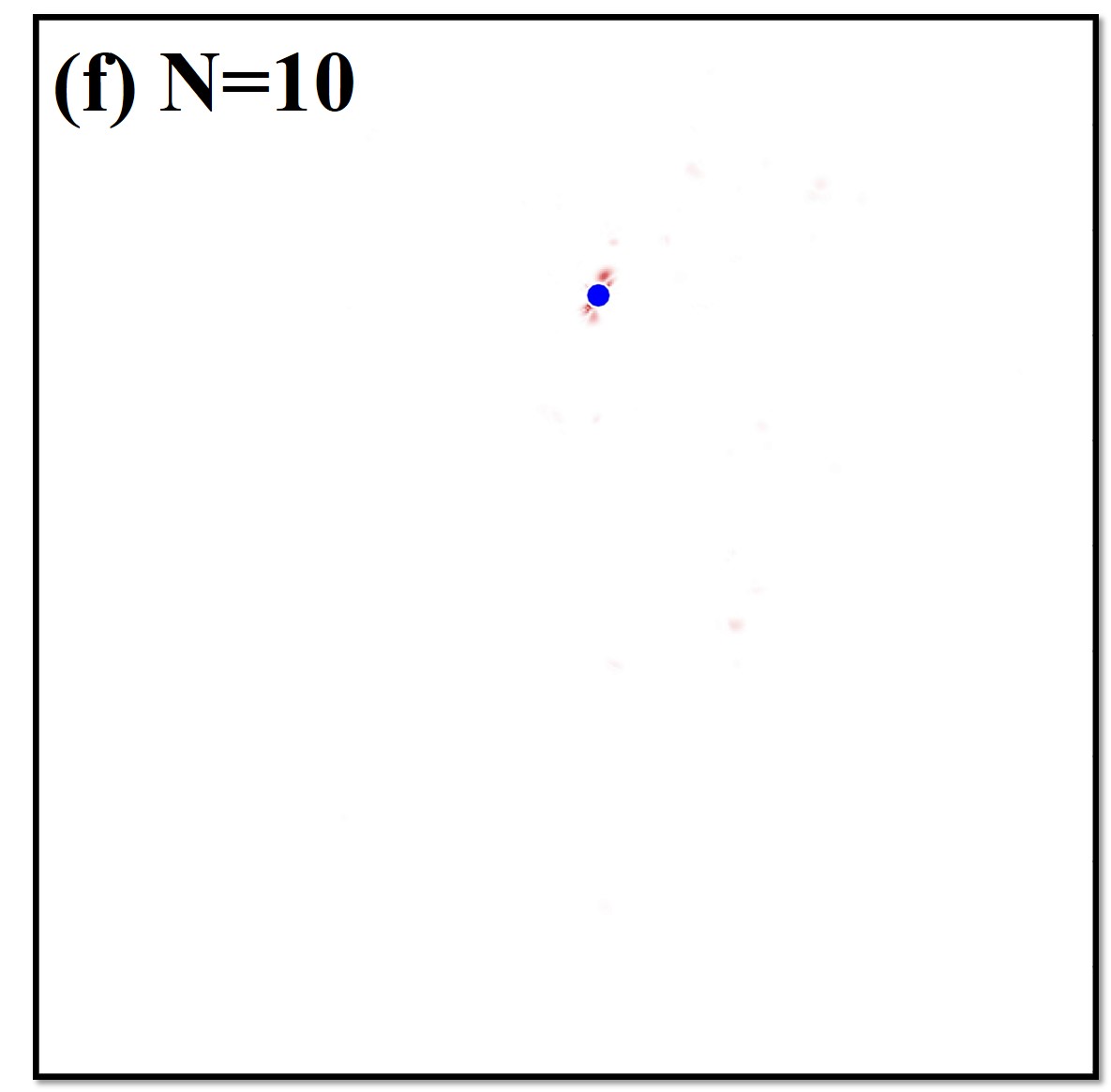
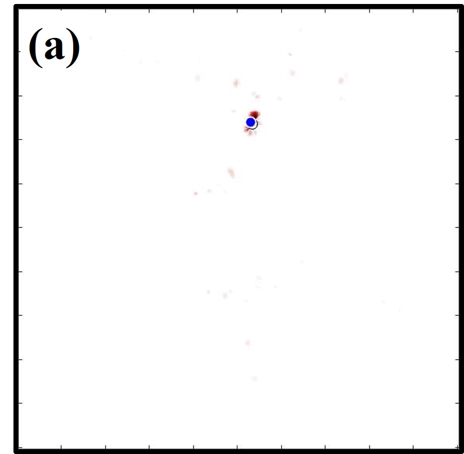
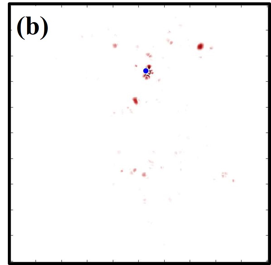
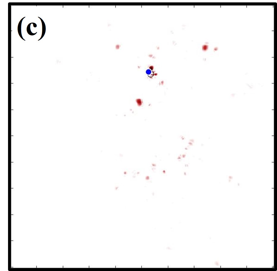
In Fig. 4(a), shows the impact of perturbation on a single mode of the system. This contrasts with Fig. 2 that shows that modes are rather preserved after perturbation. Thus, for N = 1, quantifies the impact of perturbation on a single mode and implies that a single mode is indeed sensitive to any small change in the system, but it fails to identify the location of perturbation. Further, when two modes are considered, a significant reduction in the region mapped by is observed as shown in Fig. 4(b). As the number of modes considered to evaluate is increased further, the mapped region reduces and concentrates around the perturbed particle as shown in Figs. 4 (c-f). Thus, provides a way to locate the perturbed particle. The accuracy of the localization of the perturbation increases with the number of modes considered to evaluate . Here, we were limited to ten modes, but by considering more modes (larger system, larger spectral range) the localization can be improved further.
Next, to understand the consistency of the proposed approach to locate perturbation, a single particle was displaced at different locations in the system along arbitrary directions by . Figs. 5 (a-d), show how the accuracy of the localization of the defect fluctuates from place to place in the system, when ten modes are considered to evaluate . It is observed that the perturbed particle lies within the mapped region for the perturbation at different locations in the system.
In order to quantify the accuracy to which the location of perturbation has been identified, a proximity parameter has been calculated as a function of number of modes considered. The proximity parameter, is defined as the root mean square of the distance of the perturbed particle from each point in the tracking parameter mapped region having a value of atleast times the maximum TP value. The value of is defined as follows,
| (3) |
Here, is the distance of perturbed particle at from the location in the region mapped with .
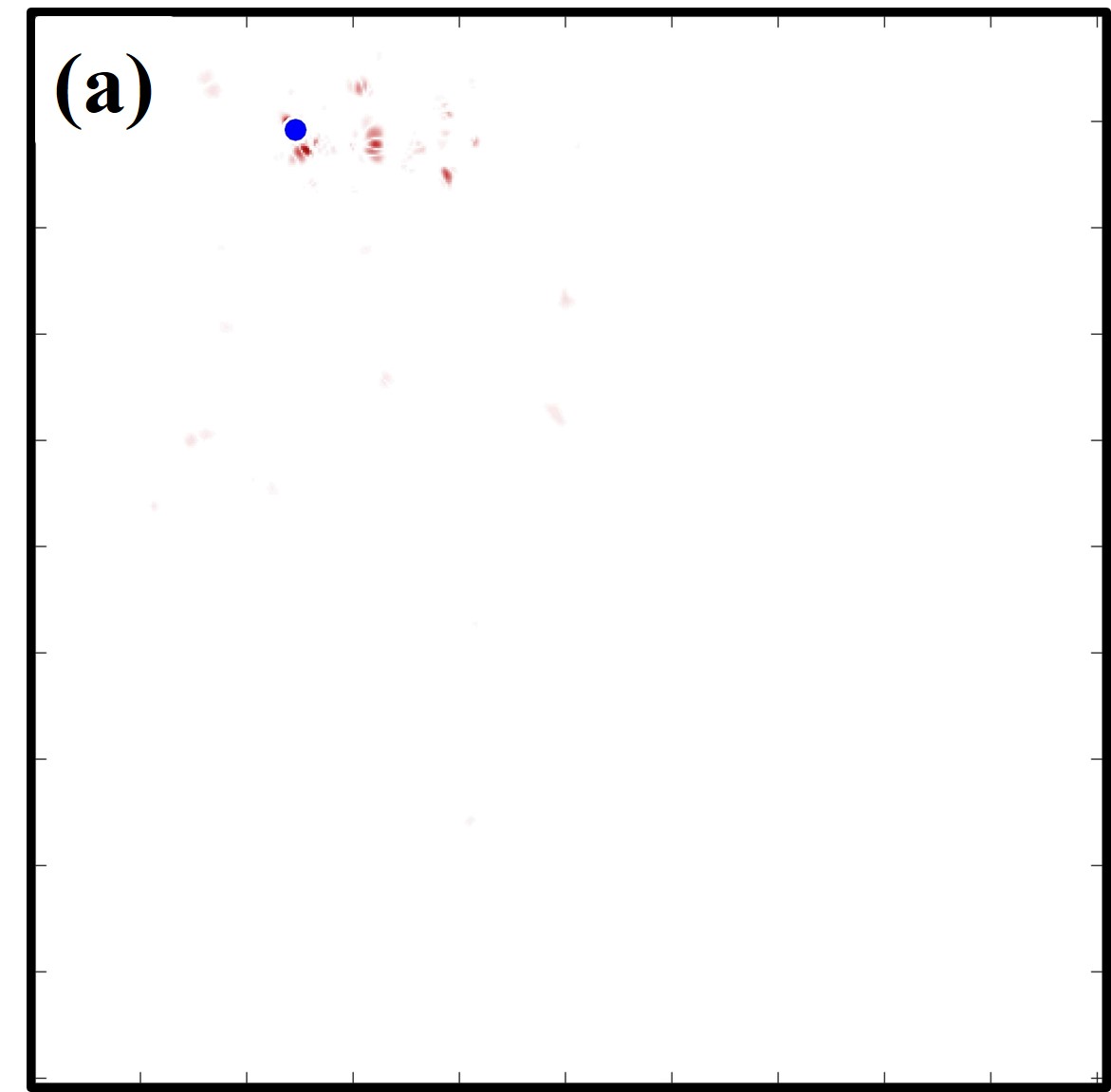
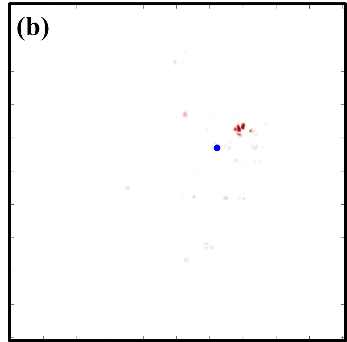
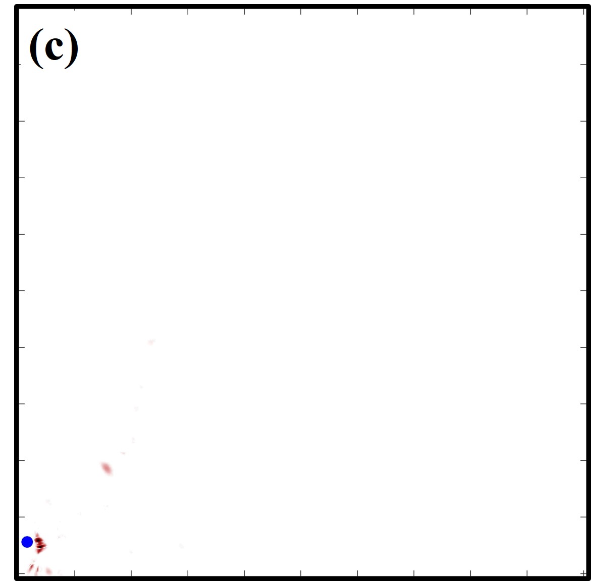
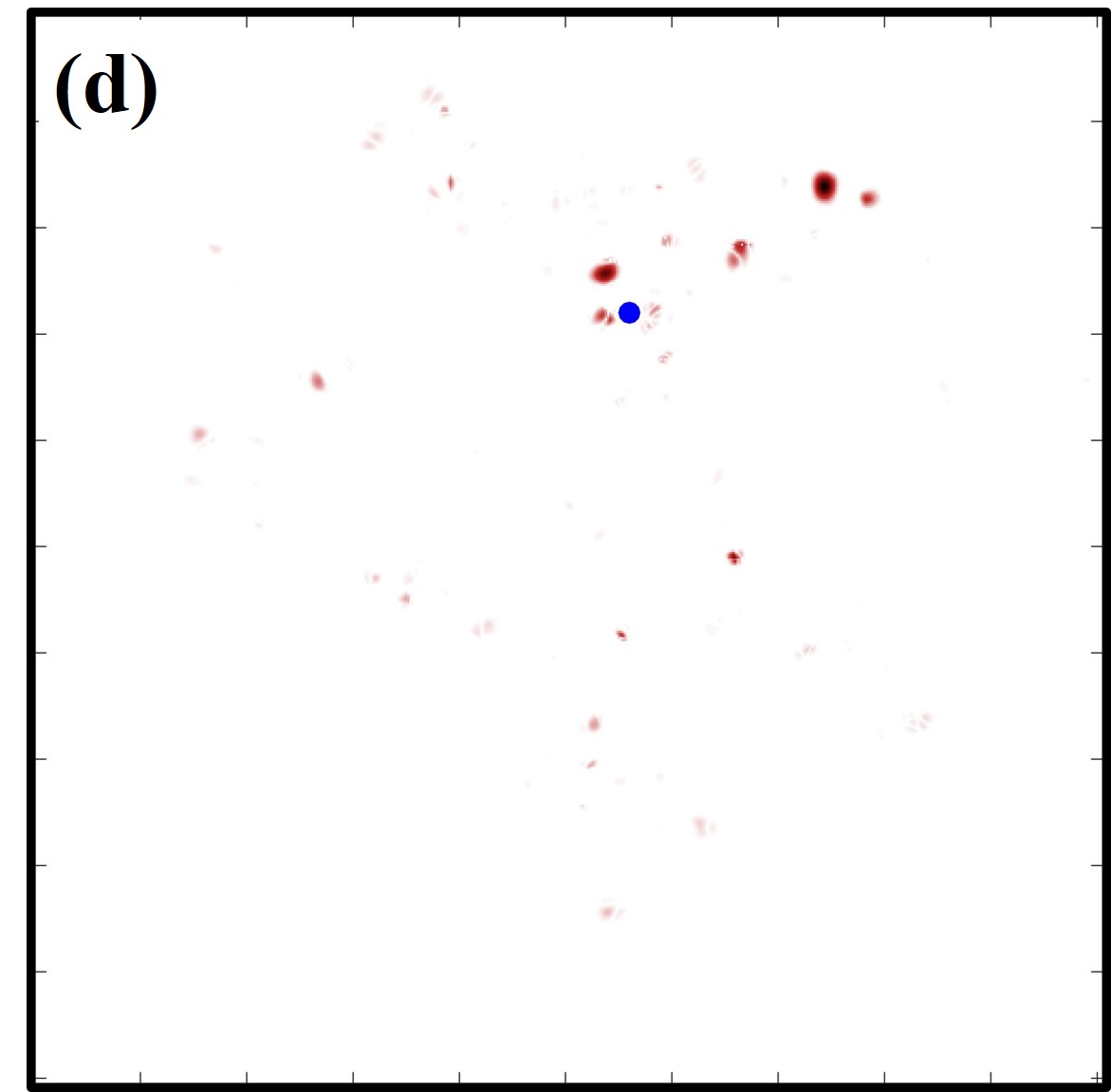
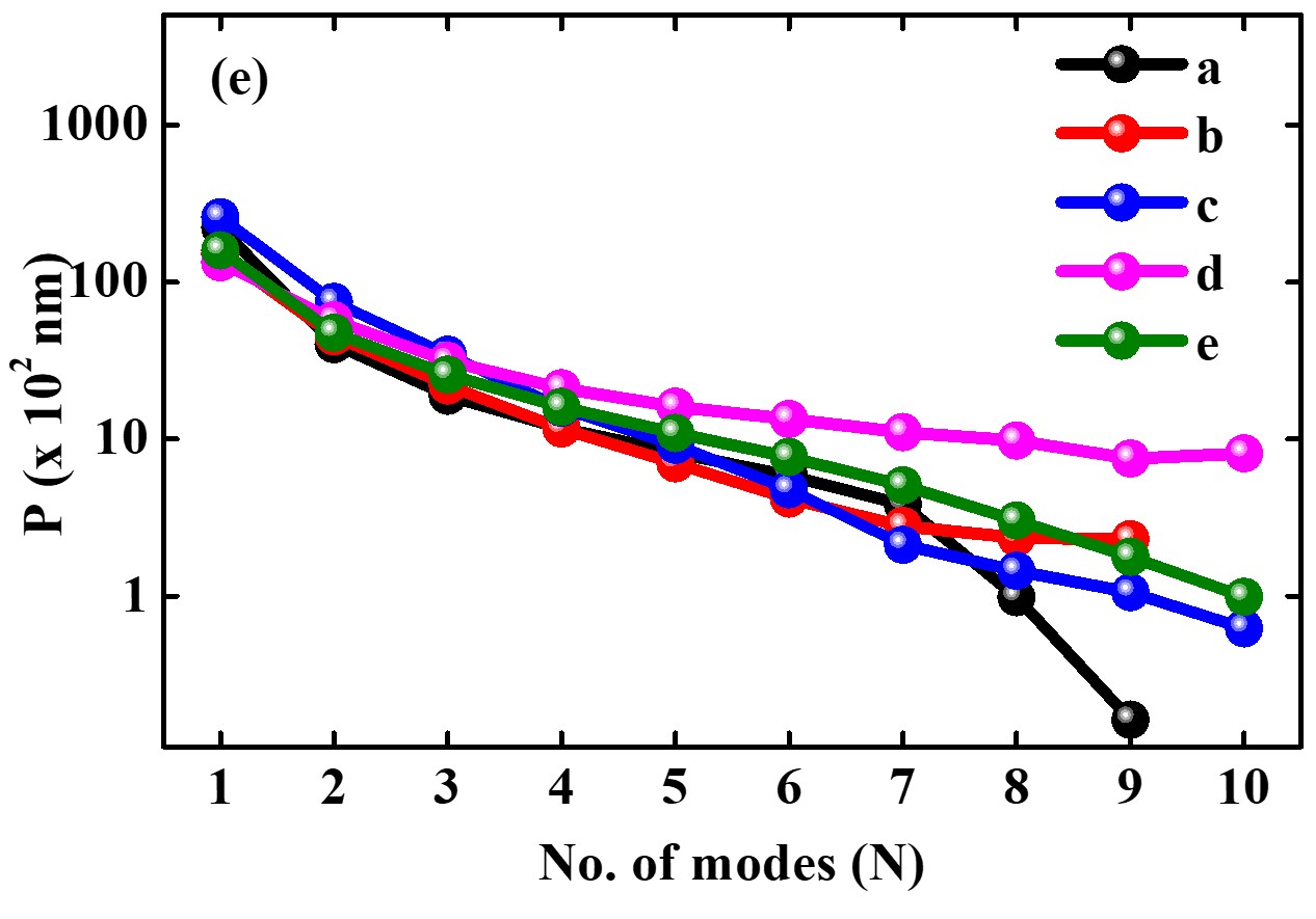
Fig. 5(e) shows how the value of changes when different number of modes are considered. The value of for systems in Figs. 5 (a-d) are marked as (a-d) in Fig. 5 (e). The plot (e) in Fig. 5 (e) corresponds to the system perturbed at location 1 in Fig. 1, for which the variation of for different number of modes considered is shown in Fig. 4. In Fig. 5(e), it is observed that as the number of modes considered increases, the proximity parameter value decreases, i.e. the region mapped with gives a more accurate estimate of the location of perturbation.
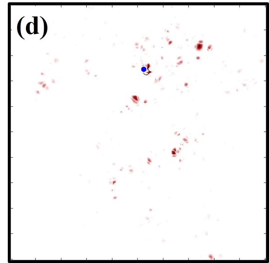
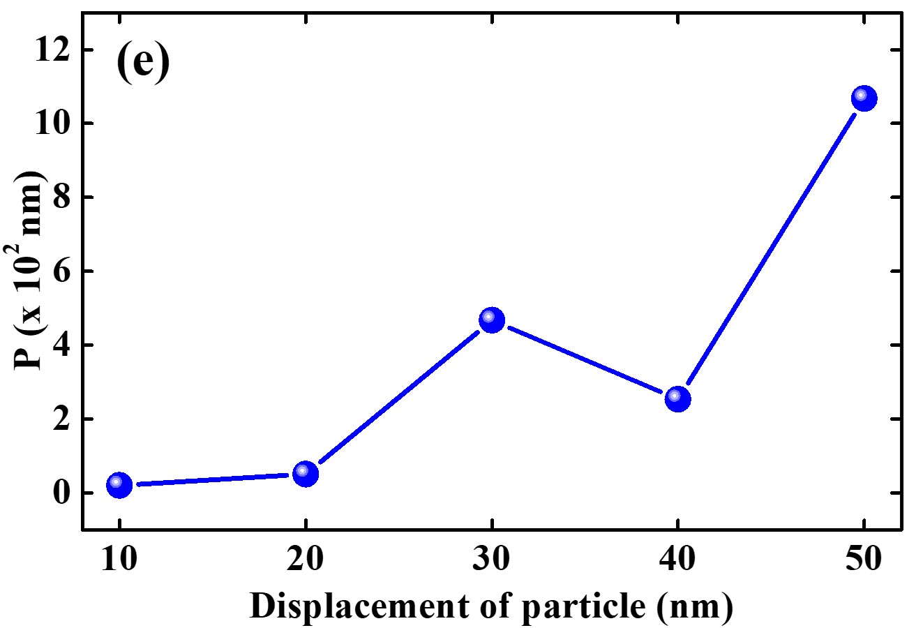
To investigate the applicability of the proposed method for larger displacements, the particle perturbed at location 1 in Fig. 1 is subjected to displacements of , along an arbitrary direction. It is observed that, for larger displacements, the changes in the mode locations and their corresponding spatial field distributions become more prominent. The region mapped with the help of and the displaced particle (marked in blue) are shown in Figs. 6 (a-d) when a single particle is perturbed by (a) , (b) , (c) and (d) . It is observed that as the displacement increases, the mapped regions becomes wider. This result is supported by the proximity parameter values in Fig. 6 (e). Effectively, the tracking parameter can be applicable to accurately identify single-step displacements of or smaller. Larger displacements can also be accurately mapped if they are carried out in multiple steps of .
IV Conclusion
In summary, a numerical study on the detection and localization of nanoscale perturbation in a 2D strongly scattering active disordered system has been presented. The modes and the corresponding spatial intensity distributions have been calculated by solving Maxwell’s equation combined with rate equations for a four level atomic system. A tracking parameter has been proposed to identify the region of nanoscale perturbation. It is shown that the tracking parameter can map the regions of perturbation very well for single nanoscale perturbations. The results presented in this paper demonstrate that nanoscale perturbations in a system can be tracked if the spatial field distribution of the modes before and after the perturbation are known. Thus, RLs have been proposed as a tool to track minute changes taking place in a disordered system. As of now, the tracking parameter can be evaluated with the help of tailored pump intensity profiles used for selective excitation of modes. It was demonstrated recently that localized modes of 1D random laser can be selected and mapped individually [40]. Our method can be easily tested in this system. It can prove to be useful in biomedical applications to track minute growth of tumor in cells. The imaging methods such as X-ray, CT scan etc. can be used to locate the region of diagnosis and then detailed monitoring of tumor can be carried out with the proposed method.
Acknowledgements.
The authors acknowledge Jonathan Andreasen, Georgia Tech Research Institute and Anirban Sarkar, National Institute of Technology, Calicut for fruitful discussions and help in computation. We acknowledge support from Science and Engineering Research Board (CRG/2020/002650) sponsored project. DST-FIST facility, Department of Physics, IIT Kharagpur is acknowledged for computational support. We acknowledge National Supercomputing Mission (NSM) for providing computing resources of ‘PARAM Shakti’ at IIT Kharagpur, which is implemented by C-DAC and supported by the Ministry of Electronics and Information Technology (MeitY), Department of Science and Technology (DST), Government of India. Israel Science Foundation (Grants No. 1871/15, 2074/15 and 2630/20), the United States-Israel Binational Science Foundation NSF/BSF (Grant No. 2015694 and Grant No 2021811) are acknowledged.References
- Letokhov [1968] V. Letokhov, Optical genration by a scattering medium with negative resonance absorption, Tech. Rep. (Foreiggn Technology Div Wrigth-Patterson AFB OHIO, 1968).
- Tulek and Vardeny [2010] A. Tulek and Z. Vardeny, Studies of random laser action in -conjugated polymers, Journal of Optics 12, 024008 (2010).
- Lü et al. [2015] J. Lü, T. Fan, and G. Chen, Random laser action in dye doped nanoporous polymeric film, Optics Communications 356, 17 (2015).
- Cao et al. [1999] H. Cao, Y. Zhao, S. Ho, E. Seelig, Q. Wang, and R. Chang, Random laser action in semiconductor powder, Physical Review Letters 82, 2278 (1999).
- Song et al. [2010a] Q. Song, S. Xiao, Z. Xu, J. Liu, X. Sun, V. Drachev, V. M. Shalaev, O. Akkus, and Y. L. Kim, Random lasing in bone tissue, Optics letters 35, 1425 (2010a).
- Lawandy et al. [1994] N. M. Lawandy, R. Balachandran, A. Gomes, and E. Sauvain, Laser action in strongly scattering media, Nature 368, 436 (1994).
- Shivakiran Bhaktha et al. [2012] B. Shivakiran Bhaktha, N. Bachelard, X. Noblin, and P. Sebbah, Optofluidic random laser, Applied Physics Letters 101, 151101 (2012).
- Ferjani et al. [2008] S. Ferjani, V. Barna, A. De Luca, C. Versace, and G. Strangi, Random lasing in freely suspended dye-doped nematic liquid crystals, Optics letters 33, 557 (2008).
- Cao [2003] H. Cao, Lasing in random media, Waves in random media 13, R1 (2003).
- Sebbah and Vanneste [2002] P. Sebbah and C. Vanneste, Random laser in the localized regime, Physical Review B 66, 144202 (2002).
- Jiang and Soukoulis [2002] X. Jiang and C. Soukoulis, Localized random lasing modes and a path for observing localization, Physical Review E 65, 025601 (2002).
- Andreasen et al. [2011] J. Andreasen, A. Asatryan, L. Botten, M. Byrne, H. Cao, L. Ge, L. Labonté, P. Sebbah, A. Stone, H. Türeci, et al., Modes of random lasers, Advances in Optics and Photonics 3, 88 (2011).
- Vanneste et al. [2007] C. Vanneste, P. Sebbah, and H. Cao, Lasing with resonant feedback in weakly scattering random systems, Physical review letters 98, 143902 (2007).
- Gouedard et al. [1993] C. Gouedard, D. Husson, C. Sauteret, F. Auzel, and A. Migus, Generation of spatially incoherent short pulses in laser-pumped neodymium stoichiometric crystals and powders, JOSA B 10, 2358 (1993).
- Noginov et al. [1999] M. Noginov, S. Egarievwe, N. Noginova, H. Caulfield, and J. Wang, Interferometric studies of coherence in a powder laser, Optical Materials 12, 127 (1999).
- Redding et al. [2011] B. Redding, M. A. Choma, and H. Cao, Spatial coherence of random laser emission, Optics letters 36, 3404 (2011).
- Redding et al. [2012] B. Redding, M. A. Choma, and H. Cao, Speckle-free laser imaging using random laser illumination, Nature photonics 6, 355 (2012).
- Chang et al. [2018] S.-W. Chang, W.-C. Liao, Y.-M. Liao, H.-I. Lin, H.-Y. Lin, W.-J. Lin, S.-Y. Lin, P. Perumal, G. Haider, C.-T. Tai, et al., A white random laser, Scientific reports 8, 1 (2018).
- Mallick and Sung [2020] S. P. Mallick and Z. Sung, Holographic image denoising using random laser illumination, Annalen der Physik 532, 2000323 (2020).
- Bachelard et al. [2012] N. Bachelard, J. Andreasen, S. Gigan, and P. Sebbah, Taming random lasers through active spatial control of the pump, Physical review letters 109, 033903 (2012).
- Hisch et al. [2013] T. Hisch, M. Liertzer, D. Pogany, F. Mintert, and S. Rotter, Pump-controlled directional light emission from random lasers, Physical review letters 111, 023902 (2013).
- Liew et al. [2014] S. F. Liew, B. Redding, L. Ge, G. S. Solomon, and H. Cao, Active control of emission directionality of semiconductor microdisk lasers, Applied Physics Letters 104, 231108 (2014).
- Liew et al. [2015] S. F. Liew, L. Ge, B. Redding, G. S. Solomon, and H. Cao, Pump-controlled modal interactions in microdisk lasers, Physical Review A 91, 043828 (2015).
- Ge [2015] L. Ge, Selective excitation of lasing modes by controlling modal interactions, Optics Express 23, 30049 (2015).
- Bachelard et al. [2014] N. Bachelard, S. Gigan, X. Noblin, and P. Sebbah, Adaptive pumping for spectral control of random lasers, Nature physics 10, 426 (2014).
- Kumar et al. [2021] B. Kumar, R. Homri, S. K. Maurya, M. Lebental, P. Sebbah, et al., Localized modes revealed in random lasers, Optica 8, 1033 (2021).
- Ho Choi and Kim [2012] S. Ho Choi and Y. L. Kim, Random lasing mode alterations by single-nanoparticle perturbations, Applied Physics Letters 100, 041101 (2012).
- Polson and Vardeny [2004] R. C. Polson and Z. V. Vardeny, Random lasing in human tissues, Applied physics letters 85, 1289 (2004).
- Siddique et al. [1995] M. Siddique, L. Yang, Q. Wang, and R. Alfano, Mirrorless laser action from optically pumped dye-treated animal tissues, Optics communications 117, 475 (1995).
- Song et al. [2010b] Q. Song, Z. Xu, S. H. Choi, X. Sun, S. Xiao, O. Akkus, and Y. L. Kim, Detection of nanoscale structural changes in bone using random lasers, Biomedical optics express 1, 1401 (2010b).
- Polson and Vardeny [2010] R. Polson and Z. Vardeny, Cancerous tissue mapping from random lasing emission spectra, Journal of Optics 12, 024010 (2010).
- Wang et al. [2017] Y. Wang, Z. Duan, Z. Qiu, P. Zhang, J. Wu, D. Zhang, and T. Xiang, Random lasing in human tissues embedded with organic dyes for cancer diagnosis, Scientific reports 7, 1 (2017).
- Hohmann et al. [2019] M. Hohmann, D. Dörner, F. Mehari, C. Chen, M. Späth, S. Müller, H. Albrecht, F. Klämpfl, and M. Schmidt, Investigation of random lasing as a feedback mechanism for tissue differentiation during laser surgery, Biomedical optics express 10, 807 (2019).
- Taflove [1995] A. Taflove, The finite-difference time-domain method., (1995).
- Sarkar et al. [2017] A. Sarkar, N. S. Ojha, and B. S. Bhaktha, Effect of photonic stop-band on the modes of a weakly scattering dcm-pva waveguide random laser, Applied Physics Letters 110, 251104 (2017).
- Choubey et al. [2020a] P. S. Choubey, S. Ghosh, S. K. Varshney, and S. B. BN, Origin of light scattering in dye doped polymeric waveguides and the dependence of excitation geometry on coherent random lasing, Journal of Physics D: Applied Physics 53, 245104 (2020a).
- Choubey et al. [2020b] P. S. Choubey, A. Sarkar, S. K. Varshney, and S. B. BN, Random laser spectroscopy and replica symmetry breaking phase transitions in a solvent-rich polymer thin film waveguide, JOSA B 37, 2505 (2020b).
- Yee [1966] K. Yee, Numerical solution of initial boundary value problems involving maxwell’s equations in isotropic media, IEEE Transactions on antennas and propagation 14, 302 (1966).
- Hulst and van de Hulst [1981] H. C. Hulst and H. C. van de Hulst, Light scattering by small particles (Courier Corporation, 1981).
- Kumar and Sebbah [2022] B. Kumar and P. Sebbah, Investigation of localized lasing modes of a one dimensional strongly scattering gain medium, in CLEO: QELS_Fundamental Science (Optica Publishing Group, 2022) pp. JW3B–66.