Currently at ]Department of Materials Science, LUMiNaD, University of Milano-Bicocca, Via Cozzi 55, 20125 Milan, Italy
Ultrafast generation of hidden phases
via energy-tuned electronic photoexcitation in magnetite
Abstract
Metal-insulator transitions (MIT) occurring in non-adiabatic conditions can evolve through high-energy intermediate states that are difficult to observe and control via static methods. By monitoring the out-of-equilibrium structural dynamics of a magnetite (Fe3O4) crystal via ultrafast electron diffraction, we show that MITs can evolve through different pathways by properly selecting the electronic excitation with light. Near-infrared (800 nm) photons inducing d-d electronic transitions is found to favor the destruction of the long-range zigzag network of the trimerons and to generate a phase separation between cubic-metallic and monoclinic-insulating regions. Instead, visible light (400 nm) further promotes the long-range order of the trimerons by stabilizing the charge density wave fluctuations through the excitation of the oxygen 2p to iron 3d charge transfer and, thus, fosters a reinforcement of the monoclinic insulating phase. Our experiments demonstrate that tailored light pulses can drive strongly correlated materials into different hidden phases, influencing the lifetime and emergent properties of the intermediate states.
I Introduction
The physical properties of strongly correlated materials are mainly defined by the complex interplay among the electronic, orbital, spin, and atomic degrees of freedom. At equilibrium, phase transitions follow an ergodic pathway within the material free-energy landscape, and the transition is characterized by a succession of thermodynamic equilibrium states between two global minima. Instead, using ultrashort laser pulses drive the transition out-of-equilibrium and induce a distinct pathway by transiently changing the coupling between the relevant degrees of freedom. Light-driven phase transitions reveal the presence of new intermediate (hidden) states of matter [1, 2, 3, 4, 5, 6, 7, 8, 9]. Such hidden phases are not only of interest from a fundamental point of view but also bear potential for ultrafast technological devices [9, 10].
Magnetite (Fe3O4) is a prototypical strongly-correlated system. It exhibits a complex interplay between the crystal structure [11], charge [12, 13, 14, 15] and orbital orders [16, 17], which leads to the emergence of an atypical thermodynamic MIT in the vicinity of 125 K, known as the Verwey transition (VT) [18]. It is found that the structural changes play a key role in VT [19, 20]. Above Verwey temperature (TV), magnetite has a cubic inverse spinel structure formally written Fe+3[Fe+2Fe+3]O4, the first Fe+3 (A-type) occupied the tetrahedral sites, whereas the [Fe+2Fe+3] (B-type) occupied the octahedral sites. Below the TV, the symmetry changes from the cubic to the monoclinic phase [11].
In the low-temperature (LT) phase, a new kind of bond dimerized state, the so-called trimeron, has been discovered and shown to form a long-range order [21, 22]. The trimeron unit results from multiple cooperative effects, including charge, orbital orderings, and strong electron-phonon coupling [23]. Therefore, trimerons are deemed to be the key actor of VT, which has been recently described microscopically as an order-disorder transition from a trimeron liquid with incommensurate fluctuations to a commensurate crystal below TV [24, 25, 26]. Optical experiments have shown that light offers the intriguing possibility to manipulate such charge fluctuations resulting in tuning the electron-phonon coupling [26] and suggesting that the light-induced transition can be very orbital-selective. Here, we directly visualize the out-of-equilibrium structural dynamics of a magnetite single crystal employing ultrafast electron diffraction (UED).
UED allows us to track the lattice evolution of magnetite across the photoinduced MIT. We show that, depending on the photon energy of the femtosecond optical excitation used in the experiment (1.55 eV vs. 3.10 eV), we trigger different electronic excitations, consequently leading to distinct nonequilibrium metastable structural states.
II Experimental Methods
The UED experiments were performed in reflection geometry [27, 28] with a grazing angle of 0.5∘ to 5∘. The light source is a Ti:sapphire laser amplifier with a central wavelength of 800 nm with a pulse duration of 45 fs at a repetition rate of 20 kHz.
High-quality magnetite with T 117 K exposing the flat and optically polished (110) surface was fixed on a cold finger attached to a five-axis manipulator with silver conductive paste and placed inside an ultrahigh vacuum chamber ( 10-9 mbar). The temperature range is controlled using an open cycle cryostat with helium liquid flow (see the SI).
III Results
We have first monitored the quasi-adiabatic transition induced by varying the temperature of magnetite (without photoexcitation). We investigate the structural changes by decreasing the temperature from 150 K down to 40 K and simultaneously following the quasi-static change of the diffraction pattern along the [110] direction where anomalies attributed to the trimerons have been recently observed [29].
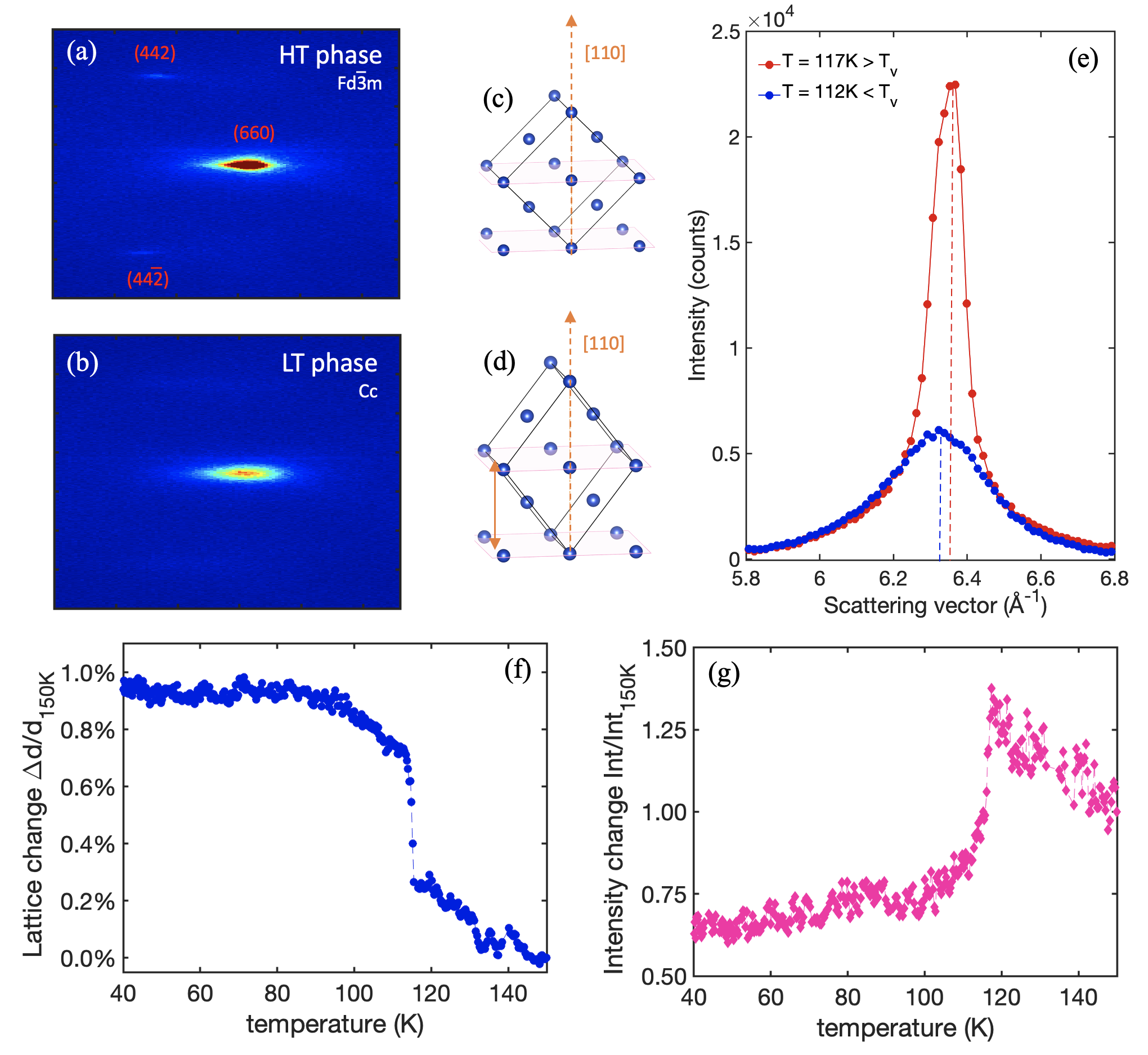
In Fig.1(a) and (b), we show the static diffraction patterns of magnetite measured above and below TV, respectively. Specifically, we monitor the changes of the (660) Bragg peak of the cubic phase and observe a significant modification of the peak position from which we extract the atomic interplanar expansion or compression (see SI) shown in Fig.1(f). This is accompanied by a drop in the diffraction intensity as illustrated in Fig.1(g). Such intensity drop results from the combination of two concomitant factors: i) the lowering of the structure factor when transitioning from a high-symmetry cubic phase to a lower symmetry monoclinic structure, and ii) the incoherent electron scattering process through multiple micro-sized structural domains (twins) that emerge in LT phase [30]. Across VT, at 117 K, the cubic lattice transforms into the monoclinic phase, which is evidenced by the expansion of the lattice along the [110] direction, as sketched in Fig.1(d). This specific expansion significantly changes the shear strain (see SI). In addition, it causes a significant softening in the shear elastic constant , as reported in ultrasound measurements [31]. Although our technique is moderately surface sensitive (5-10 nm), the agreement between the monitored position shift of the Bragg peak from our data and the reported softening in the elastic constant demonstrates that our observations are representative of bulk dynamics.
Ginzburg-Landau’s (GL) theory of phase transition such as VT in magnetite correlates transformation shear strains to an order parameter. Hence, we ascribe the measured shear strain () being strongly coupled to the order parameter (OP) , and retrieve its symmetry based on the framework of GL [32] and a fundamental group theory analysis (see SI). We found that has T2g symmetry with one nonzero component, i.e., = (, 0, 0).
Using detailed group theory calculations, several authors have identified the set of phonons, including , , and (T2g), as the structural OPs [33, 34, 35]. Furthermore, ab initio calculations have demonstrated the strong coupling between these structural OPs and the T2g orbital ordering within a trimeron [33]. Therefore, we conclude that the trimerons arrangement along the [110] direction is a conceivable OP candidate with T2g representation. The OP with T2g representation was suggested recently by electron diffraction measurements, where the authors consider an anomalous electronic nematic phase above Tv with a T2g representation which involves a different set of rotational symmetry breaking than the usual ones [36].
We obtain insights into the non-adiabatic MIT in magnetite by investigating the out-of-equilibrium response of the lattice, initially kept at 80 K (below TV), after photoexcitation. In Fig.2, we present the evolution of the (660) Bragg peak following ultrashort laser pulses at two different wavelengths, 800 nm and 400 nm at 2.9 mJ/cm2 and 1.2 mJ/cm2 incident fluence, respectively. The fluence used for 800 nm corresponds to the intermediate fluence regime [37].
Fig.2(a) shows the structural dynamics following 800 nm photoexcitation. The (660) atomic planes undergo a maximum compression of around -0.06 %. Based on our static data shown above (Fig.1), the compression of the monoclinic lattice along the [110] direction denotes the transformation toward a cubic structure, consistent with ref.[37]. Their recent out-of-equilibrium optical measurements have shown a photoinduced phase separation between insulating regions and metallic islands at LT through 800 nm laser pulses in a similar fluence regime. This observation is supported by pump-probe x-ray diffraction measurements [38], where the authors attribute the insulating state to the monoclinic regions and the metallic state to the cubic islands. Expanding the investigation range of previous measurements that were limited to the first 10 ps [36, 37, 38], our data unveil the complex establishment of the hidden phase which lasts approximately 50 ps and interestingly follows three compression stages. First, during the first 22 ps (N°1), an abrupt compression of -0.03 % occurs. In the second stage (N°2) between 22 ps and 27 ps, the lattice undergoes a minor contraction. Finally, the third step (N°3) emerges and adds an extra -0.03 % to the lattice compression. This multi-step process is characteristic of the presence of distinct dynamic processes, such as electron-phonon coupling and phonon-phonon interaction [39, 36]. Note that the electron-electron interaction is expected to occur on a faster timescale 300 fs [38], beyond the temporal resolution in these experiments. The relaxation process also evolves with multiple timescales. Qualitatively, the first recovery stage (N°4) occurs from 50 ps to 126 ps and reaches an intermediate compressed state close to -0.03 %, which interestingly corresponds to the value of the process N°3. Then, a second long process occurs towards the total recovery (N°5) to the equilibrium phase, which is still not reached after 1.3 ns (see SI). In a second data set with a slightly higher fluence (see SI), we confirm the multiple compression stage process and show that each stage’s duration and amplitude depend on the fluence used.
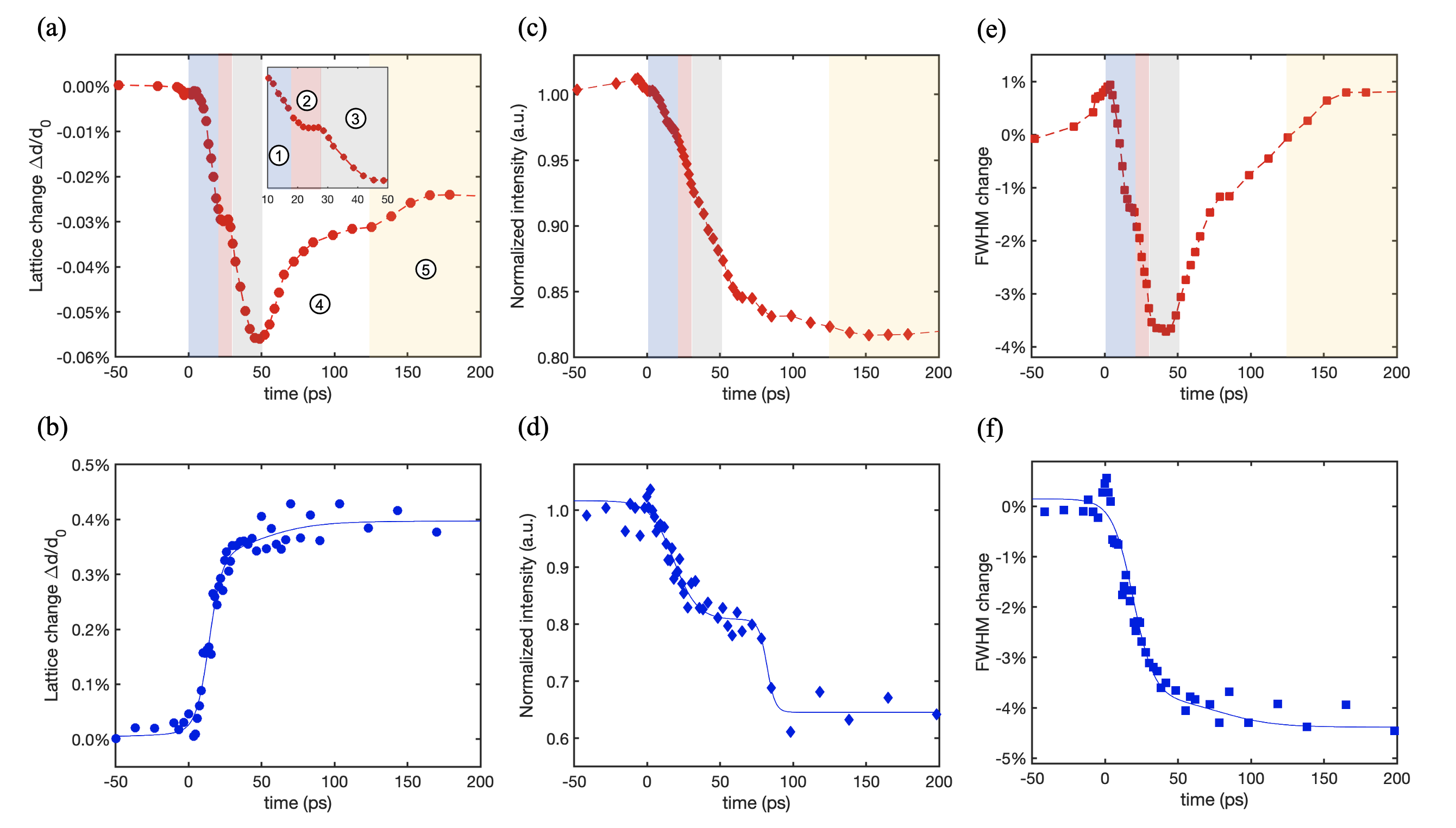
In Fig.2(c), the response of the Bragg peak intensity after the 800 nm photoexcitation shows a drop. Taking only into account the recovery towards the higher symmetry cubic high temperature phase after the 800 nm photoexcitation, we expect an increase in the intensity, which we do not observe. This observation suggests that the dominant process is the structural disorder caused by the motion of the atoms induced by the rise in the lattice temperature by ultrashort laser pulses [40], known as the induced Debye-Waller effect (see SI). Similarly to the peak position shift response, the Bragg peak intensity does not fully recover to its initial state after 1.3 ns (see SI). The long dynamics revealed by our data indicate the metastability of the induced 800 nm phase illustrating the complexity of the thermalization process in magnetite, involving multiple interplays of electron-electron, electron-phonon, and phonon-phonon scattering. Such lifetime is a signature of a hidden out-of-equilibrium phase which is supported by the unusual structural dynamics and the decrease in the intensity due to the phase separation between cubic and monoclinic islands, which is not an equilibrium state but rather a local minimum within the energy landscape of the excited configuration.
We extend our investigation for hidden phases in magnetite by changing the energy of the optical excitation to 3.10 eV (400 nm). In Fig.2(b), different from the 800 nm case, we observe that the 400 nm laser pulses induce a 0.4 % expansion of the lattice along the [110] direction (instead of a contraction), indicating a reinforcement of the monoclinic distortion. At 90 K, before excitation, the crystalline structure has a monoclinic angle = 90.236∘ [21]. The 400 nm induced expansion is mainly related to the variation of the tilting angle , where we expect a value 90.236∘. A quantitative value for the tilting angle can only be retrieved when monitoring the behavior of multiple Bragg peaks along different zone axes. Nevertheless, our data clearly show that the 400 nm optical excitation induces a lattice change that is opposite to the quasi-adiabatic lattice response from our equilibrium data presented above (Fig.1(f)), where the stabilization of the structure from 90 K down to 40 K shows no modification of the lattice parameters expected thermodynamically. Since the generated structure is not accessible thermally but only induced optically, we associate it with the emergence of a new hidden phase characterized by a monoclinic lattice with a tilting angle larger than the equilibrium value of 90.236∘. The 400 nm hidden phase is also completely different from the one established by the 800 nm light. The first is firmly monoclinic, whereas the second is a mixture of monoclinic and cubic separated regions. The 400 nm structural hidden phase takes around 50 ps to emerge with only one direct expansion process, as presented in Fig.2(b), which we relate mainly to electron-phonon interaction. This new state lasts up to 300 ps without any recovery to the initial state giving it a metastable character. We observe a significant drop in the intensity response (Fig.2(d)). For the 800 nm case, thermal effects and multiple scatterings from the mixed phase are the origins of the intensity drop. Although the decrease in intensity is consistent with the reduction of the structure factor for the (660) Bragg peak, it is surprising to observe a shrinking of the FWHM, indicating a higher homogeneity in the atomic planes, which we cannot associate with a thermal-like behavior. This suggests that the new hidden structural state possesses a larger structural long-range order related to a larger monoclinic angle.
IV Discussion
The formation of distinct metastable hidden phases through two different photon energies demonstrates the critical role played by electronic excitations in establishing such nonequilibrium phases in magnetite. At LT, magnetite is thermodynamically stabilized in the insulating phase, resulting from a commensurate long-range order along the [001] direction [21, 38] of the trimerons zigzag network [22, 41] with a coherent length of (385±10) nm [14]. Each trimeron unit couples linearly three FeB sites in the form Fe - Fe - Fe, in which the minority spin t2g electron is delocalized from the central Fe site into the nearest neighbors Fe, and each Fe site is shared between three trimerons. In addition to the Jahn-Teller distortion caused by the orbital ordering [42], the electron localization within a trimeron unit produces a structural distortion in the FeB sites, where the distance between the central Fe and its two Fe nearest neighbors gets shorter, leading to a monoclinic distortion [21]. This distortion has been confirmed by high-accuracy synchrotron x-ray structure refinements [43], where it is found that fourteen over the sixteen nonequivalent trimerons have shown a shorter FeB - FeB distance. When we excite magnetite using photon pulses with an energy of 1.55 eV (800 nm), LDA+U calculations [16] and optical conductivity measurements [37, 44] both agree in predicting the triggering of electronic d-d excitations. They correspond to an electron delocalization from an occupied t2g of Fe to an unoccupied t2g orbital of Fe following the configuration 3d3d 3d3d. The d-d excitations encompass another inter-site excitation corresponding to the transition from an occupied t2g of Fe to an unoccupied eg of Fe [16]. However, this excitation is only possible at an energy higher than 2 eV. Triggering the d-d excitation restores the mobility of the minority spin t2g electrons and causes the valency to change for both Fe and Fe and, hence, alternates their sites inside the trimeron (see SI). The direct consequence of this local electronic fluctuation is the destruction of the trimeron. According to out-of-equilibrium x-rays measurements, this destruction occurs in an ultrashort timescale fs [38]. The destruction of trimerons yields the suppression of the long-range zigzag order connected at Fe sites. When the trimeron breaks, the FeB - FeB distance returns to its initial value. Hence, the structure relaxes, giving rise to the emergence of cubic phase islands inside the remaining monoclinic regions forming a phase separation that we qualify as an 800 nm metastable hidden phase. Following the electronic excitation, the relaxation process back to the equilibrium configuration continues via electron-phonon and phonon-phonon scattering mechanisms. Electrons primarily interact with high-energy optical phonons, which then anharmonically decay toward acoustic modes via a three-phonon scattering process [45, 46, 47, 48]. For magnetite, we can interpret the newly observed two steps in the relaxation process as the strong electron-phonon coupling involving the conduction electrons coupling with the X3-driven mode (TO) for the first stage and the phonon-phonon coupling between X3 (TO) and (TA) modes for the second. This scenario is supported by recent UED data that have shown under similar photoexcitation (800 nm) that the X3 (TO) mode is preferably triggered via the electron-phonon coupling [36] in the first few picoseconds. In addition, inelastic neutron scattering has shown that the (TA) mode is the most susceptible across the cubic-monoclinic transformation [24].
In the equilibrium state, refined x-rays measurements show that in a high connectivity configuration with a maximum of three trimerons per Fe site, only four of the eight Fe sites participate, keeping the rest as inactive [21]. In addition, our static data in Fig.1 show that this thermodynamic maximum connectivity state is reached a few kelvins after TV, and no extra monoclinic distortion is observed from 90 K down to 40 K. When we excite the magnetite crystal with the 400 nm (3.10 eV) light, we trigger multiple electronic excitations (see SI). The most dominant excitation is the charge transfer from 2p bands of oxygen to 3d bands of FeB [44]. By activating the ligand-metal charge transfer, the oxygens of the octahedron FeO6 supply electrons to the Fe non-participating ions, which consequently become Fe. Hence, the proportion of Fe increases at the inactive trimerons B-sites, boosting t2g orbital ordering and creating extra trimerons, thus pushing beyond the limit of the thermodynamic maximum connectivity state leading to a new light-induced phase. This causes additional stress on the magnetite structure provoked by cooperative effects. One is the Jahn-Teller distortion due to the extending t2g orbital ordering. At the same time, the second contribution comes from the shortening of the atomic FeB - FeB distances induced by the charge localization within the newly formed trimerons (charge ordering) [21, 43]. Recently, a high-accuracy x-ray experiment has shown that chemically doped magnetite introduces B site-selective Fe2+ vacancies (which become Fe3+) and weakens the trimeron long range order [49]. The authors attribute the vanishing of the VT in doped magnetite to the absence of the trimeron network. In our case, the 400 nm photo-doping acts oppositely and provides additional electrons to the incomplete trimeron network. As our data clearly shows, this results in an expansion of the lattice along the [110] direction. It is reasonable to speculate that the photo-doping is site-selective, which orders and strengthens the trimeron crystal seen in the increase of the coherence length (Fig.2(f)). In this scenario, the trimeron network can be seen as an imperfect Wigner crystal with homogeneous vacancies (inactive Fe3+ B-sites). After 400 nm photo-doping, the electrons from the oxygens activate the missing sites and complete the trimeron crystal enhancing the connectivity, which ultimately induces a monoclinic distortion favored by a strong electron-phonon coupling.
V Conclusion
Our results reveal the emergence of two distinct metastable hidden phases in magnetite. Starting from the same LT equilibrium state, we drive magnetite into two different structural states using two different photon energies. The 800 nm light induces d-d excitations, which favor the destruction of the trimerons and their network in a percolating fashion, leading to a phase separation between monoclinic-insulating regions and cubic-metallic islands. The 400 nm light triggers charge transfer between oxygen and iron at B-sites, which are found to be prolific for the trimerons and their arrangement, enhancing their connectivity, which leads to a stronger monoclinic distortion inaccessible adiabatically. Our findings demonstrate the key role of the trimeron structural configuration in magnetite and show the ability to establish novel hidden phases in quantum materials via specific electronic excitations in a strongly correlated environment.
Acknowledgements.
The authors are grateful to J. Lorenzana, W. Tabis, A. Kozlowski and P. Piekarz for constructive discussions. This work was supported by the ERC consolidator grant ISCQuM No. 771346 , SNSF grant No.514725, NCCR-MUST No. 565194.References
- [1] D Fausti, R I Tobey, N Dean, S Kaiser, A Dienst, M C Hoffmann, S Pyon, T Takayama, H Takagi, and A Cavalleri. Light-Induced Superconductivity in a Stripe-Ordered Cuprate. Science, 331(6014):189–191, jan 2011.
- [2] V. Kiryukhin, D. Casa, J. P. Hill, B. Keimer, A. Vigliante, Y. Tomioka, and Y. Tokura. An X-ray-induced insulator–metal transition in a magnetoresistive manganite. Nature, 386(6627):813–815, apr 1997.
- [3] L. Stojchevska, I. Vaskivskyi, T. Mertelj, P. Kusar, D. Svetin, S. Brazovskii, and D. Mihailovic. Ultrafast switching to a stable hidden quantum state in an electronic crystal. Science, 344(6180):177–180, 2014.
- [4] K. Miyano, T. Tanaka, Y. Tomioka, and Y. Tokura. Photoinduced Insulator-to-Metal Transition in a Perovskite Manganite. Physical Review Letters, 78(22):4257–4260, jun 1997.
- [5] Hirohiko Ichikawa, Shunsuke Nozawa, Tokushi Sato, Ayana Tomita, Kouhei Ichiyanagi, Matthieu Chollet, Laurent Guerin, Nicky Dean, Andrea Cavalleri, Shin-ichi Adachi, Taka-hisa Arima, Hiroshi Sawa, Yasushi Ogimoto, Masao Nakamura, Ryo Tamaki, Kenjiro Miyano, and Shin-ya Koshihara. Transient photoinduced ’hidden’ phase in a manganite. Nature Materials, 10(2):101–105, feb 2011.
- [6] Frank Y. Gao, Zhuquan Zhang, Zhiyuan Sun, Linda Ye, Yu-Hsiang Cheng, Zi-Jie Liu, Joseph G. Checkelsky, Edoardo Baldini, and Keith A. Nelson. Snapshots of a light-induced metastable hidden phase driven by the collapse of charge order. Science Advances, 8(29):1–12, jul 2022.
- [7] Abdolnasser Zakery and Stephen R. Elliott. Optical Non-linearities in Chalcogenide Glasses and Their Applications, chapter CHAPTER 14, pages 433–486. 2021.
- [8] S. Koshihara, Y. Tokura, T. Mitani, G Saito, and T. Koda. Photoinduced valence instability in the organic molecular compound tetrathiafulvalene- p -chloranil (TTF-CA). Physical Review B, 42(10):6853–6856, oct 1990.
- [9] Alberto de la Torre, Dante M. Kennes, Martin Claassen, Simon Gerber, James W. McIver, and Michael A. Sentef. Colloquium: Nonthermal pathways to ultrafast control in quantum materials. Reviews of Modern Physics, 93(4):041002, oct 2021.
- [10] Feliciano Giustino, Jin Hong Lee, Felix Trier, Manuel Bibes, Stephen M Winter, Roser Valentí, Young-woo Son, Louis Taillefer, Christoph Heil, Adriana I Figueroa, Bernard Plaçais, Quansheng Wu, Oleg V Yazyev, Erik P A M Bakkers, Jesper Nygård, Pol Forn-Díaz, Silvano De Franceschi, J W McIver, L E F Foa Torres, Tony Low, Anshuman Kumar, Regina Galceran, Sergio O Valenzuela, Marius V Costache, Aurélien Manchon, Eun-Ah Kim, Gabriel R Schleder, Adalberto Fazzio, and Stephan Roche. The 2021 quantum materials roadmap. Journal of Physics: Materials, 3(4):042006, oct 2020.
- [11] M. Iizumi, T. F. Koetzle, G. Shirane, S. Chikazumi, M. Matsui, and S. Todo. Structure of magnetite (Fe3O4) below the Verwey transition temperature. Acta Crystallographica Section B Structural Crystallography and Crystal Chemistry, 38(8):2121–2133, aug 1982.
- [12] E. Nazarenko, J. E. Lorenzo, Y. Joly, J. L. Hodeau, D. Mannix, and C. Marin. Resonant X-Ray Diffraction Studies on the Charge Ordering in Magnetite. Physical Review Letters, 97(5):056403, aug 2006.
- [13] Richard J. Goff, Jon P. Wright, J. Paul Attfield, and Paolo G. Radaelli. Resonant x-ray diffraction study of the charge ordering in magnetite. Journal of Physics: Condensed Matter, 17(48):7633–7642, dec 2005.
- [14] J. E. Lorenzo, C. Mazzoli, N. Jaouen, C. Detlefs, D. Mannix, S. Grenier, Y. Joly, and C. Marin. Charge and Orbital Correlations at and above the Verwey Phase Transition in Magnetite. Physical Review Letters, 101(22):226401, nov 2008.
- [15] J. P. Wright, J. P. Attfield, and P. G. Radaelli. Long Range Charge Ordering in Magnetite Below the Verwey Transition. Physical Review Letters, 87(26):266401, dec 2001.
- [16] I. Leonov, A. N. Yaresko, V. N. Antonov, M. A. Korotin, and V. I. Anisimov. Charge and Orbital Order in Fe3O4. Physical Review Letters, 93(14):146404, sep 2004.
- [17] Horng-Tay Jeng, G. Y. Guo, and D. J. Huang. Charge-Orbital Ordering and Verwey Transition in Magnetite. Physical Review Letters, 93(15):156403, oct 2004.
- [18] E. J. W. Verwey. Electronic Conduction of Magnetite (Fe3O4) and its Transition Point at Low Temperatures. Nature, 144(3642):327–328, aug 1939.
- [19] D Ihle and B Lorenz. Electron correlation theory of Fe3O4. Philosophical Magazine B, 42(3):337–347, sep 1980.
- [20] Y. Yamada. Charge ordering and lattice instability in magnetite. AIP Conference Proceedings, 24(1):79–85, 1975.
- [21] Mark S. Senn, Jon P. Wright, and J. Paul Attfield. Charge order and three-site distortions in the Verwey structure of magnetite. Nature, 481(7380):173–176, jan 2012.
- [22] Mark S. Senn, Ingo Loa, Jon P. Wright, and J. Paul Attfield. Electronic orders in the Verwey structure of magnetite. Physical Review B, 85(12):125119, mar 2012.
- [23] Przemysław Piekarz, Dominik Legut, Edoardo Baldini, Carina A. Belvin, Tomasz Kołodziej, Wojciech Tabiś, Andrzej Kozłowski, Zbigniew Kakol, Zbigniew Tarnawski, José Lorenzana, Nuh Gedik, Andrzej M. Oleś, Jürgen M. Honig, and Krzysztof Parlinski. Trimeron-phonon coupling in magnetite. Physical Review B, 103(10):104303, mar 2021.
- [24] S. Borroni, G. S. Tucker, F. Pennacchio, J. Rajeswari, U. Stuhr, A. Pisoni, J. Lorenzana, H M Rønnow, and F. Carbone. Mapping the lattice dynamical anomaly of the order parameters across the Verwey transition in magnetite. New Journal of Physics, 19(10):103013, oct 2017.
- [25] S. Borroni, G. S. Tucker, U. Stuhr, J. Lorenzana, H. M. Rønnow, and F. Carbone. Energy domain versus time domain precursor fluctuations above the Verwey transition in magnetite. Physical Review B, 101(5):054303, feb 2020.
- [26] S. Borroni, E. Baldini, V. M. Katukuri, A. Mann, K. Parlinski, D. Legut, C. Arrell, F. van Mourik, J. Teyssier, A. Kozlowski, P. Piekarz, O. V. Yazyev, A. M. Oleś, J. Lorenzana, and F. Carbone. Coherent generation of symmetry-forbidden phonons by light-induced electron-phonon interactions in magnetite. Physical Review B, 96(10):104308, sep 2017.
- [27] Giulia Fulvia Mancini, Barbara Mansart, Saverio Pagano, Bas van der Geer, Marieke de Loos, and Fabrizio Carbone. Design and implementation of a flexible beamline for fs electron diffraction experiments. Nuclear Instruments and Methods in Physics Research Section A: Accelerators, Spectrometers, Detectors and Associated Equipment, 691:113–122, nov 2012.
- [28] Francesco Pennacchio, Giovanni M. Vanacore, Giulia F. Mancini, Malte Oppermann, Rajeswari Jayaraman, Pietro Musumeci, Peter Baum, and Fabrizio Carbone. Design and implementation of an optimal laser pulse front tilting scheme for ultrafast electron diffraction in reflection geometry with high temporal resolution. Structural Dynamics, 4(4):044032, jul 2017.
- [29] Jung-Fu Lin, Junjie Wu, Jie Zhu, Zhu Mao, Ayman H. Said, Bogdan M. Leu, Jinguang Cheng, Yoshiya Uwatoko, Changqing Jin, and Jianshi Zhou. Abnormal Elastic and Vibrational Behaviors of Magnetite at High Pressures. Scientific Reports, 4(1):6282, may 2015.
- [30] Takeshi Kasama, Nathan S. Church, Joshua M. Feinberg, Rafal E. Dunin-Borkowski, and Richard J. Harrison. Direct observation of ferrimagnetic/ferroelastic domain interactions in magnetite below the Verwey transition. Earth and Planetary Science Letters, 297(1-2):10–17, aug 2010.
- [31] H. Schwenk, S. Bareiter, C. Hinkel, B. Lüthi, Z. Kakol, A. Koslowski, and J.M. Honig. Charge ordering and elastic constants in Fe Zn O. The European Physical Journal B, 13(3):491–494, jan 2000.
- [32] L D Landau and E. M Lifschitz. Statistical Physics. Butterworth-Heineman, third edit edition, 1980.
- [33] Przemysław Piekarz, Krzysztof Parlinski, and Andrzej M. Oleś. Origin of the Verwey transition in magnetite: Group theory, electronic structure, and lattice dynamics study. Physical Review B, 76(16):165124, oct 2007.
- [34] L. V. Gasparov, D. B. Tanner, D. B. Romero, H. Berger, G. Margaritondo, and L. Forró. Infrared and Raman studies of the Verwey transition in magnetite. Physical Review B, 62(12):7939–7944, sep 2000.
- [35] Przemysław Piekarz, Krzysztof Parlinski, and Andrzej M. Oleś. Mechanism of the Verwey Transition in Magnetite. Physical Review Letters, 97(15):156402, oct 2006.
- [36] Wei Wang, Jun Li, Zhixiu Liang, Lijun Wu, Pedro M. Lozano, Alexander C. Komarek, Xiaozhe Shen, Alex H. Reid, Xijie Wang, Qiang Li, Weiguo Yin, Kai Sun, Yimei Zhu, Ian K. Robinson, Mark P. M. Dean, and Jing Tao. Verwey transition as evolution from electronic nematicity to trimerons via electron-phonon coupling. arxiv.org2202.08744, feb 2022.
- [37] F. Randi, I. Vergara, F. Novelli, M. Esposito, M. Dell’Angela, V. A. M. Brabers, P. Metcalf, R. Kukreja, H. A. Dürr, Daniele Fausti, M. Grüninger, and F. Parmigiani. Phase separation in the nonequilibrium Verwey transition in magnetite. Physical Review B, 93(5):054305, feb 2016.
- [38] S. de Jong, R. Kukreja, C. Trabant, N. Pontius, C. F. Chang, T. Kachel, M. Beye, F. Sorgenfrei, C. H. Back, B. Bräuer, W. F. Schlotter, J. J. Turner, O. Krupin, M. Doehler, D. Zhu, M. A. Hossain, A. O. Scherz, D. Fausti, F. Novelli, M. Esposito, W. S. Lee, Y. D. Chuang, D. H. Lu, R. G. Moore, M. Yi, M. Trigo, P. Kirchmann, L. Pathey, M. S. Golden, M. Buchholz, P. Metcalf, F. Parmigiani, W. Wurth, A. Föhlisch, C. Schüßler-Langeheine, and H. A. Dürr. Speed limit of the insulator–metal transition in magnetite. Nature Materials, 12(10):882–886, oct 2013.
- [39] Pablo Maldonado, Karel Carva, Martina Flammer, and Peter M. Oppeneer. Theory of out-of-equilibrium ultrafast relaxation dynamics in metals. Physical Review B, 96(17):174439, nov 2017.
- [40] Gustaaf Van.Tendeloo. 4D Electron Microscopy Imaging in Space and Time, By Ahmed.H. Zewail and John.M. Thomas. Angewandte Chemie International Edition, 50(13):2888–2888, 2011.
- [41] R. Řezníček, V. Chlan, H. Štěpánková, and P. Novák. Hyperfine field and electronic structure of magnetite below the Verwey transition. Physical Review B, 91(12):125134, mar 2015.
- [42] H. Y. Huang, Z. Y. Chen, R. P. Wang, F. M. F. de Groot, W. B. Wu, J. Okamoto, A. Chainani, A. Singh, Z. Y. Li, J. S. Zhou, H. T. Jeng, G. Y. Guo, Je-Geun Park, L. H. Tjeng, C. T. Chen, and D. J. Huang. Jahn-Teller distortion driven magnetic polarons in magnetite. Nature Communications, 8(1):15929, aug 2017.
- [43] Mark S. Senn, Jon P. Wright, James Cumby, and J. Paul Attfield. Charge localization in the Verwey structure of magnetite. Physical Review B, 92(2):024104, jul 2015.
- [44] S. K. Park, T. Ishikawa, and Y. Tokura. Charge-gap formation upon the Verwey transition in Fe3O4. Physical Review B, 58(7):3717–3720, aug 1998.
- [45] L. Perfetti, P. A. Loukakos, M. Lisowski, U. Bovensiepen, H. Eisaki, and M. Wolf. Ultrafast Electron Relaxation in Superconducting by Time-Resolved Photoelectron Spectroscopy. Physical Review Letters, 99(19):197001, nov 2007.
- [46] Fabrizio Carbone, Ding-Shyue Yang, Enrico Giannini, and Ahmed H. Zewail. Direct role of structural dynamics in electron-lattice coupling of superconducting cuprates. Proceedings of the National Academy of Sciences, 105(51):20161–20166, dec 2008.
- [47] Barbara Mansart, José Lorenzana, Andreas Mann, Ahmad Odeh, Mariateresa Scarongella, Majed Chergui, and Fabrizio Carbone. Coupling of a high-energy excitation to superconducting quasiparticles in a cuprate from coherent charge fluctuation spectroscopy. Proceedings of the National Academy of Sciences, 110(12):4539–4544, mar 2013.
- [48] Barbara Mansart, Mathieu J. G. Cottet, Thomas J. Penfold, Stephen B. Dugdale, Riccardo Tediosi, Majed Chergui, and Fabrizio Carbone. Evidence for a Peierls phase-transition in a three-dimensional multiple charge-density waves solid. Proceedings of the National Academy of Sciences, 109(15):5603–5608, apr 2012.
- [49] E. Pachoud, J. Cumby, G. Perversi, J. P. Wright, and J. P. Attfield. Site-selective doping of ordered charge states in magnetite. Nature Communications, 11(1):1671, dec 2020.