Sensing of magnetic field effects in radical-pair reactions using a quantum sensor
Abstract
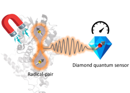
Magnetic field effects (MFE) in certain chemical reactions have been well established in the last five decades and are attributed to the evolution of transient radical-pairs whose spin dynamics are determined by local and external magnetic fields. The majority of existing experimental techniques used to probe these reactions only provide ensemble averaged reaction parameters and spin chemistry, hindering the observation of the potential presence of quantum coherent phenomena at the single molecule scale. Here, considering a single nitrogen vacancy (NV) centre as quantum sensor, we investigate the prospects and requirements for detection of MFEs on the spin dynamics of radical-pairs at the scale of single and small ensemble of molecules. We employ elaborate and realistic models of radical-pairs, considering its coupling to the local spin environment and the sensor. For two model systems, we derive signals of MFE detectable even in the weak coupling regime between radical-pair and NV quantum sensor, and observe that the dynamics of certain populations, as well as coherence elements, of the density matrix of the radical pair are directly detectable. Our investigations will provide important guidelines for potential detection of spin chemistry of bio-molecules at the single molecule scale, required to witness the hypothesised importance of quantum coherence in biological processes.
I Introduction
Investigating the fundamental role of spin interactions in magnetic field effects (MFE) in chemical reactions has a long history [1, 2, 3]. The study of spin-chemical effects provides important insights about structure, kinetics and magnetic properties of transient intermediate chemical species [4, 5] and are explored in various interdisciplinary applications including sensitivity enhancement in nuclear magnetic resonance (NMR) [6], quantum computing [7], avian magnetic compass [8], and solar energy conversion kinetics in photosynthetic systems [9].
It is astounding that spin interactions can have decisive effects on the fate of chemical reactions as the energy of spin transitions typically is orders of magnitude smaller than the thermal energy [4]. However, spin dependent MFEs in chemical systems, also known as the radical-pair mechanism (RPM) [2, 8, 10], rely on the creation of transient paramagnetic species in a non-equilibrium state called radicals, which are chemical species with an odd number of electrons. In the RPM, the radical pair (RP) is spatially separated but in a spin-correlated state and the recombination of the radicals back to the molecular precursor state is spin selective. The influence of an external magnetic field then occurs in terms of a modulation of the spin dynamics and consequently an alteration in the yield of products formed from the various spin states [11, 12].
The formulation of the RPM started in the late 1960s to explain non-equilibrium magnetic resonance spectra of chemical reactions of organic molecules [13, 14, 15, 16, 17, 18, 19], while the recent interest is fuelled by investigations of the RPM as the most plausible mechanism for MFE in biological reaction kinetics [20, 21]. These include flavoproteins related to DNA photolyases, the involvement of cryptochromes in circadian rhythms [22, 23] and their proposed role in animal magnetoreception [24]. MFEs have been recorded in cryptochromes [11] and seem to fulfil the structural and dynamical requirements of the RPM [8]. Considerable interest exists also in the role of radical pairs in chemical kinetics in photosynthetic reaction centres [9, 25, 26]. Flavoenzymes [27] - Flavin-based enzymes - are responsible for catalytic functions in diverse biological reactions [28] and the involvement of various RPs in these reactions is debated in recent discussions [29, 30].
Existing experimental techniques for in vitro probing of MFE in RP dynamics in chemical reactions rely on averaged signals collected from a large ensemble of molecules. These techniques include time-resolved electron paramagnetic resonance (TREPR) spectroscopy [31, 32], and optical methods based on absorption [33, 34, 35, 36] and fluorescence detection [37, 38, 39, 40, 41]. Studies of RP reactions conducted using these techniques can only provide ensemble averaged information of spin dynamics along with requiring a large quantity (few microliters) of potentially precious biological samples. It is therefore imperative to instead consider single-molecule detection techniques in order to reveal potential quantum coherent spin evolution otherwise hidden by ensemble averaging [42, 43, 44]. Specifically the single negatively charged nitrogen-vacancy (NV) center in diamond [45] has attracted significant interest as potential candidate for the detection of spin-chemical effects of RP reactions at the single molecule scale [43, 44]. This is achieved due to the excellent bio-compatibility of diamond [46, 47], the attainable nanometer scale spatial resolution and remarkable sensitivity to external electromagnetic fields of a single NV center [48, 49, 50, 51, 52].
In the present work, we discuss a realistic avenue for the detection of MFE associated with RPs at the scale of single and small ensembles () of molecules using a single NV center in diamond (Fig.1). We investigate both the weak and the strong coupling regime between the NV center and RPs. Compared to the simplified model employed by Finkler et al. [44] neglecting important spin interactions and considering only the strong coupling regime, we here use an elaborate model governing realistic RP spin dynamics. We include up to three nuclear spins per radical with maximum anisotropic hyperfine coupling along with dipolar and exchange coupling between the radicals. Applying standard sensing protocols, we derive measurable signals received by an NV centre and quantify magnetic field dependent RP dynamics. We show that in the weak coupling regime, the signal generated by an RP on a single-molecule is comfortably within the sensitivity limit of state-of-the-art shallow NV centers and dynamics of certain populations, as well as coherences, of the RP density matrix is directly accessible. Non-trivial features of RP spin dynamics can be observed depending on direction and magnitude of a bias magnetic field. Further, we observe that signal features tend to average out when we consider a small ensemble of RPs, highlighting the importance of single-molecule detection. In the strong coupling regime, we find that although there is an opportunity to probe various RP spin state populations individually, detection becomes challenging especially in larger bio-molecules due to the increased number of unavoidable and spectrally indistinguishable hyperfine interactions within the RP.
The article is organized as follows. In section II, we introduce the theoretical framework for the RP and interaction with the NV center. In section III, we describe the numerical simulations performed with realistic RP systems and present the main results. We conclude in section IV and discuss possible future directions.
II Description of the model
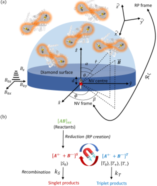
The general situation considered for the model is illustrated in Fig. 2 (a). Molecules hosting RPs are distributed above the diamond surface and an NV center is situated at a depth from the surface. We choose to work in the NV frame (shown in the figure) and the total Hamiltonian of the whole system in this frame is given as
| (1) |
where , , and are the Hamiltonians governing the dynamics of the NV center, the RP, and coupling between the NV center and the RP, respectively.
We consider the symmetric, negatively charged NV- center (hereafter called NV) that consists of ground (3A, total spin ), metastable (1A, ) and excited (3E, ) states [45]. The ground state triplet is split at zero magnetic field with GHz [48]. The and can be further split by application of an external magnetic field, thus making all the three states accessible by application of appropriate control fields. Upon optical excitation typically with 532nm light, the NV center might relax via a spin-dependent inter-system crossing [53], a process that allows for initialization in and spin-state dependent fluorescence contrast between and the states. These properties combined with an external magnetic field can be exploited to isolate or as a two-level system with effective spin . In addition, the NV center interacts with nuclear spins, predominantly (abundance of 99.6% [54]) the intrinsic 14N with total spin , hyperfine coupling tensor and quadrupolar coupling MHz [54], causing further splitting of the states. Due to the axial symmetry of the NV center, can be expressed in a diagonal form in the principal axis system of the NV axis with diagonal elements [54]. Taking the above details into account, the relevant part of the NV center Hamiltonian can be written as
| (2) |
where is the gyromagnetic ratio of an electron, and are, respectively, spin-operators of the NV center and 14N nuclear spin, and is the vector of the externally applied magnetic field on the NV center. The relatively large zero-field splitting allows to make the secular approximation where we can ignore all terms involving electron-nuclear spin flip-flops (containing ). Further, we assume and consequently choose (hereafter denoted ) as two-level system. Now the NV center Hamiltonian simplifies to
| (3) |
The RP consists of two radicals each containing an unpaired electron. A simplified reaction for the creation and recombination of radicals is shown in Fig. 2(b). The radicals can be generated either by electron transfer or chemical bond breaking from the oxidized state of the molecule. The reduction can be facilitated by various mechanisms, for example, by photoexcitation with light of appropriate wavelength (reduction of flavin based systems [55]) or by chemical means (Haber-Weiss reaction [56]). Depending on the internal molecular dynamics, the unpaired electrons in the radicals are born in either singlet () or triplet () states, where and denote the eigenstate of the Pauli matrix. After creation, the singlet and triplet states inter-convert among each other under the RP Hamiltonian, which for the RP situated at location see Fig. 2(a)) is given by [57]
| (4) |
where and are the spin-operators of the unpaired electrons of the and radical, respectively, and denotes the external magnetic field applied on the RP. The radicals couple to each other via exchange interaction (coupling constant ) and dipolar interaction (coupling tensor ). Each unpaired electron is further surrounded by a set of nuclei in the radical and interact with them via hyperfine coupling. The and are spin-operators (hyperfine coupling tensors) of the nuclei coupled to the unpaired electron in the and nuclei coupled to the unpaired electron in the radical, respectively. The magnitude of hyperfine coupling is typically in the range of 2.8 - 28 MHz [58] in organic radicals, whereas the strength of the dipolar and exchange interaction is dependent on the separation between the radicals [59]. Usually the dipolar and hyperfine coupling tensors are simulated or measured in a coordinate frame associated with the molecule that hosts the RP, hereafter referred to as the RP frame. The NV and RP frames are related by a rotation matrix , i.e., , where is the RP Hamiltonian in the RP frame.
To gain more insight into the inter-conversion dynamics of spin states of radicals, it is useful to work in the basis spanned by singlet and triplet states for the unpaired electrons. Using this choice of basis, we can divide into two parts . The first part is diagonal in the singlet-triplet basis for unpaired electrons irrespective of the basis chosen for the nuclei
| (5) |
where is the secular part of the dipolar coupling and is the distance between the radicals. The second part contains terms corresponding to the transverse magnetic field, non-secular dipolar coupling contribution and hyperfine interactions with the surrounding nuclei. The singlet and triplet states are anti-symmetric and symmetric under spin exchange, respectively, and the evolution under breaks this symmetry which results in inter-conversion between these states.
Due to interaction with the surrounding environment, after the creation, along with the inter-conversion dynamics, the spin-correlated RP recombine back to the equilibrium state. The rates of recombination depend on whether the RP is in the singlet (rate constant ) or the triplet state (rate constant ). Products are thus formed with spin-state dependent yield and can be altered by the application of a suitable magnetic field, a process that is called the radical-pair mechanism (RPM) [8]. The recombination dynamics can be modeled by treating the RP as an open quantum system with Lindbladian
| (6) |
Where is total number of nuclei in the RP and denotes the anticommutator.
The electron spin of the NV center and the RP is coupled via dipolar interaction and the corresponding Hamiltonian can be written as
| (7) |
where is the dipolar coupling tensor in the NV frame which depends on the direction of the applied magnetic field and the NV centre distance to the RP. Under the secular approximation and assumption , the coupling Hamiltonian simplifies to
| (8) |
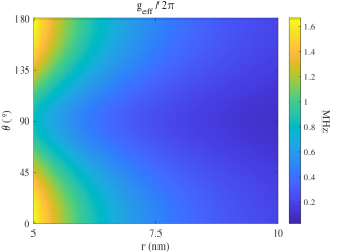
where , , , and . The trace norm represents the effective RP-NV coupling (Fig. 3) and can be determined using techniques developed in recent works for three-dimensional molecular localization using the NV center [60, 61, 62, 63]. Note that the coupling Hamiltonian can not be approximated to contain only the term [64, 44] as the strength of cross terms ( and ) may not be smaller compared to various other parameters in at low external magnetic fields.
Now, after establishing the general framework, the protocol to study RP dynamics using the NV center is as follows:
-
1.
Prepare the NV center and the RP in a known initial state . The initial state of the unpaired electrons in the RP is either the or state as described before, while the nuclei start in the maximally mixed state . The initial state of the NV center can be controlled and depends on the applied sensing scheme.
-
2.
Solve the quantum dynamics in the NV frame using the master equation: . Since the NV center has the capability of detecting magnetic signal directly originating from inter-conversion dynamics of the RP instead of relying on the product yield based detection, we can simplify the Lindbladian by assuming , thus giving [57]. Further, the rate may be allowed to include decoherence of the electron spins of NV center as well because its typical time-scale is similar to recombination rates in organic RPs which is in the order of tens of s.
-
3.
Trace out the RP to calculate the state of the NV center at any time : and investigate signatures of the RP evolution on the states of the NV center.
Depending on , and , the coupling between an NV center and an RP varies (Fig. 3). The resulting NV-RP dynamics can be divided in two dynamical regimes:
| (9) | ||||
| (10) |
where and is the dephasing time of the NV center. depends on factors including applied sensing sequence and magnetic field [65], proximity to surface impurities, presence of paramagnetic defects and nuclear spins around the NV center [45]. Typical values of range from a few microseconds to a few milliseconds depending mainly on the applied sensing sequence, the isotopic purity of the diamond sample used [66] and the surface termination [67], making both weak and strong coupling regimes achievable in practice.
III Simulations
In this section we present details and results of the simulations. We consider two RP systems, respectively with isotropic and anisotropic hyperfine coupling tensors. We investigate MFEs in RP systems in the weak as well as strong coupling regimes and discuss appropriate detection strategies. In the weak coupling regime, we study the MFE as a function of strength and direction of magnetic field due to a single as well as a small ensemble of RP. For the strong coupling regime, we point out potential challenges in the regime of single molecule detection that arise for larger bio-molecules.
III.1 RP systems
The two RP systems we investigate are:
- 1.
- 2.
III.2 Weak coupling regime
In the weak coupling regime, no observable splitting of the NV level occurs upon interaction with the RP and the effect of spin-dynamics of the RP on the NV center dynamics then appears only as a time-dependent classical magnetic field. The coupling Hamiltonian simplifies to
| (11) |
where with . The detectable magnetic signal generated from the RP situated at a location can now be calculated as (in units of Tesla):
| (12) |
where , is the number density of RPs, and is the sensing radius of the NV center (see Fig. 2(a)). The maximum signal is generated when , where is the identity matrix, i.e. when the RP frame is aligned with the NV frame:
| (13) |
The possibility of achieving such a condition is discussed in Appendix VI.1.
A simple protocol to detect the above signal is shown in Fig. 2(d). First, we initialize the NV center in and the RP in either the or state. An appropriate sensing sequence is then applied on the NV. The choice depends on the Fourier spectrum of the detectable signal, denoted by here. Finally, a projective measurement is made on the NV to yield information about the accumulated phase, which is proportional to , acquired during the sensing sequence.
As evident from Eq. (11), only specific combinations of RP density matrix elements corresponding to contribute to and are directly measurable by the NV center. Explicitly, in the singlet-triplet basis, the directly measurable elements are:
| (14) |
which measures the magnetization corresponding to the population difference of outer triplet states and
| (15) |
which measure the magnetization corresponding coherences between the outer and central triplet states. Thus the detection of magnetization corresponding to elements and can reveal involvement of quantum coherent phenomenon in RP dynamics in singlet-triplet basis. The full state tomography of density matrix of RP, however, requires conversion of various elements onto directly detectable ones (Eq. 14 and 15) and is beyond the scope of this work.
We consider RPs to be statistically distributed above the surface of the diamond sample. The coupling strength of an RP outside the sensing radius of three-four times the depth of the NV centre (cf. Fig.2(a)) decreases by an order of magnitude compared to RPs on the surface. The number of RPs falling in the sensing volume depends on the size of the host protein. For the case of proteins with many amino acids and large molecular weights (>Da, the average radius nm [70]), only one of them might fall in the sensing volume with high probability. On the other hand, for the case of smaller proteins (<50kDa, the average radius being 2-3 nm), tens of RPs can be accommodated in the sensing volume. The following two subsections discuss how the generated signal behaves in these two cases.
III.2.1 RP on single molecule
To study RP dynamics at the single-molecule level, we assume (therefore we drop notation for discussion in this subsection) and without loss of generality. We use three nuclei with maximum anisotropic hyperfine couplings for each of the radicals in (FAD∙- - TrPH∙+) : , and for FAD∙-, and , and for TrPH∙+) [58]. The used and couplings correspond to FAD∙- - TrPH∙+ RP in Drosophila melanogaster cryptochrome protein [57]. The recombination rate is assumed to be Hz which is of the order of the observed spin relaxation time in behavioural studies in migratory birds [71]. To calculate the total signal received (or phase accumulated by the NV center) at a given strength and direction of applied magnetic field, we define the time integrated signal per second as
| (16) |
In Fig. 5, we show the strength of the magnetic signal received by the NV from an RP on a single molecule at a distance nm. For such shallow NV centres, the achievable sensitivity is of the order of a few to a slowly alternating signal [64, 72]. For the case of , as we assume here, the detectable signal is only generated by and , which, respectively, corresponds to the dynamics of RP density matrix elements and .
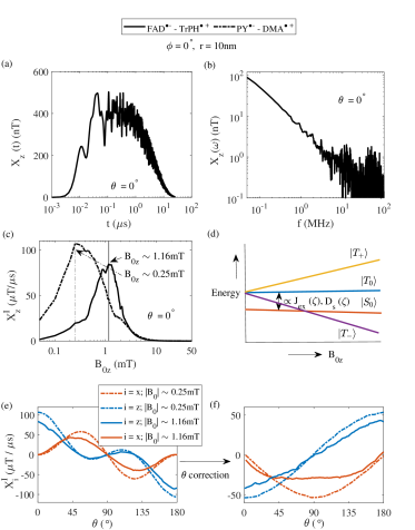
To determine the suitable NV sensing sequence to probe the generated signal, Fig. 5(a) plots the temporal evolution of for a bias magnetic field that maximizes the time integrated signal (see Fig. 5(c)). The unpaired electrons in the RP are initialized in the singlet-state whose population decays with time as it is converted into other density matrix elements under the evolution of , thus resulting in the build-up of . The generated signal (hundreds of nT) is within the sensitivity achieved for nm deep NV centres. The corresponding spectrum is shown in Fig. 5(b). Because we are interested in low frequency dynamics (kHz to tens of MHz) resulting from the dipolar and hyperfine coupling terms (typically 1-10mT) in , the maximum component of the signal in the frequency domain is limited to 500 MHz. The cut-off frequency of these oscillations observed is in the order of 10MHz. Sequences including Ramsey, spin-echo, and dynamical decoupling are thus suitable for probing such RP dynamics [65].
Variation with strength of the magnetic field To study the dependence of the signal on the strength of the applied magnetic field, we set , i.e., and so that there is no spin mixing of the NV center states keeping the two-level approximation valid and is varied from 0 to 50mT. The dependence of the time-integrated signal is plotted as a function of in Fig. 5(c).
The features of the time-integrated signal can be qualitatively explained by the dynamics of the RP under in singlet-triplet basis as shown in Fig. 5(d). At , all three triplet states are almost degenerate and the energy gap between and states is proportional to and . With the unpaired electrons in the RP initialized in the singlet state, singlet-triplet oscillations occur due to evolution of the RP under containing both a non-secular dipolar coupling contribution and hyperfine interactions with the surrounding nuclei (Eq. 4). However, for small , the RP only generates a very low signal as outer triplet states () remain close to degeneracy. As increases, the outer triplet states move apart and give rise to additional manifolds for singlet-triplet mixing [73, 74]. This opens the possibility of information (population and coherence) transfer among and states to only one of the outer triplet states, creating a population imbalance between outer triplet states and giving rise to an increase in . At a certain value, there is a maximum transfer between singlet-triplet oscillations to population difference of outer triplet states, giving rise to the peak-like features in (for example the peak at mT and mT, respectively, for FAD∙- - TrPH∙+ RP and PY∙- - DMA∙+ RP). This phenomenon, which is closely related to low field effects (LFE) in the context of singlet yield [75], occurs due to level crossing between and either of the outer triplet [76] states. A further increase in results in energetic isolation of the outer triplet states which causes steady decrease in the probability of information transfer between them, and and states, eventually vanishing at very high magnetic fields .
Variation with direction of the magnetic field In Fig. 5(e), we investigate the signal dependence on the direction of the applied magnetic field. Here we restrict the magnetic field to the plane by assuming as the following discussion holds for the general case. Although is nonzero for certain values of , its magnitude should be less than that required for mixing of the and levels of the NV center. We fix the magnetic field strength to mT and mT, respectively, for FAD∙- - TrPH∙+ RP and PY∙- - DMA∙+ RP where the maximum MFE is expected based on the results shown in Fig. 5(c). The dependence of the time-integrated signal on is plotted in Fig. 5(e). The shape of the signal received and ( since ) is dominated by the shape of and , respectively. To reveal the true dynamics of the RP with , we calculate the normalized signals instead in Fig. 5(f).
Once again, using the level dynamics under in the singlet-triplet basis, a qualitative explanation of various features of the generated signals is possible using the following arguments: (i) for a given magnetic field, close to essentially means presence of an extra channel for singlet-triplet mixing due to nonzero and and (ii) at close to , the outer triplet states are almost degenerate with maximum splitting occurring at close to or . The shape of is a direct consequence of argument (i) as nonzero transverse fields close to increase the probability of singlet-triplet as well as triplet-triplet mixing. The signal is a consequence of argument (ii) as higher splitting of outer triplet states opens new manifolds for information transfer between them and and states as described earlier.
For parameter values chosen here and described above, we do not observe the recently discovered spike feature [58] in the generated signals and presumably due to the high value of magnetic field used, and the non-zero exchange and dipolar coupling in the [77]. However, the spike feature can be observed when we consider a simpler RP system with only one nuclear spin in each RP in earth’s magnetic field as summarized in Appendix VI.2.
III.2.2 Ensemble of RPs
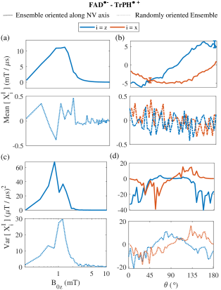
Assuming the minimum size of the protein to be 3-4 nm (small protein with molecular weight kDa [70]), RPs () might fall in the NV sensing volume. In Fig. 6 we plot the mean and variance of the generated signal as a function of strength and direction of applied magnetic field for an ensemble of RPs. We consider two types of ensembles: (i) All the RPs are oriented along the direction of the NV axis, i.e., (solid lines in Fig .6), and (ii) all RPs are randomly oriented with respect to the NV axis (dashed lines in Fig. 6), i.e. , where , , and , respectively, are rotation matrices about the , and axes with randomly chosen Euler angles , , and . For both cases, the distance between RPs and the NV were sampled from a uniform distribution in the range .
For the uniformly oriented ensemble (i), the mean of the generated signal is obviously amplified in contrast to the randomly oriented ensemble in (ii) where the features due to the RP spin dynamics are almost entirely removed because of random averaging of the signal over various realizations of . The variance of the generated signal as function of strength of the magnetic field is within the sensitivity range of a shallow NV center for both types of ensembles and the LFE behaviour akin to the single-molecule case (Fig. 5 (c)) is still observable after averaging. On the contrary, although there is an observable signal when is varied, the shape of the variation is erratic with no similarity to the single molecule case (Fig. 5 (f)). Therefore, although the ensemble (ii) is simply realized by drop casting a molecule sample on the surface of the diamond, detecting a MFE in this arrangement is more challenging. The situation might be compared to absorption or EPR studies where the size of the probed ensemble is very large (micro litres of sample) and hence MFE detection relies on statistical signals generated from a very small number of molecules. This limits the detection of MFE to a few percent [35] along with causing wastage of precious biological samples. Hence, it becomes important to appropriately position the proteins in the desired orientation with respect to the NV center to maximize the probability of successful detection of an MFE (Appendix VI.1).
III.3 Strong coupling regime
In the strong coupling regime, the energy levels of the NV and RP mix, giving rise to a level structure as illustrated in Fig. 7. Due to the structure of the NV center, the RP components of eigenstates in and are quantized along different axes determined by the strength of the coupling tensor. The total Hamiltonian in this regime can be reduced to
| (17) |

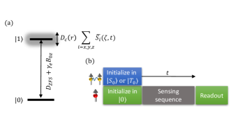
Depending on the magnitude of various couplings in , the and states of the NV center are further split into a maximum of levels, as shown in Fig. 7 (a). The level structure amounts to at maximum resonances in the magnetic resonance spectrum of the NV center, corresponding to transitions , where and are respectively eigenstates of and , and to . Monitoring the magnitude of these resonance peaks may facilitate a probing of the dynamics of the populations of using a simple sequence as shown in Fig. 7 (b).
The magnitude of the spin resonance peaks is proportional to the population difference of and states. Taking the contribution from all RPs into account, it can be calculated as
| (18) |
where is the projector corresponding to the state . There are a couple of important details about the detection of RP dynamics in this regime which should be pointed out:
-
(i)
Since we wish to study RP dynamics exclusively under an applied magnetic field, a pulsed sensing sequence (Fig 7 (b)) is preferred as it ensures that the NV center stays in the state during the time of evolution, implying no backaction of the NV on the RP. In a Ramsay type of sequence where the NV center is prepared in , the RP experiences an additional induced field from the state component [44] which is different for each RP, complicating the probing of MFEs.
-
(ii)
As the size of the bio-molecule increases, the number of unavoidable hyperfine interactions also grows. As a consequence, the spectral features will start to overlap, especially when the applied magnetic fields are comparable to the hyperfine couplings in (see Fig. 7 (c) for FAD∙- - TrPH∙+). In this case it becomes challenging to access the various electronic transitions of the RP separately. One could consider decoupling the nuclear spin bath [78, 79] during the readout time by driving the RP at a frequency larger than the strongest hyperfine coupling, however the short readout duration (typically ns for NV ceneter [45, 80]) will render the averaging inefficient.
In the light of the second argument, although the detection of RP dynamics in the strong coupling regime may be challenging, it offers an opportunity to track various populations of the RP density matrix individually and the possibility of quantum control of the RP using an NV center [44].
IV Conclusion and Outlook
In summary, we analysed the realistic prospects of MFE detection at the single molecule scale in RP reactions in bio-molecules using a single NV center in diamond. To realistically describe the bio-molecular spin dynamics, our RP model includes three nuclear spins per radical that have the largest hyperfine couplings. Including dipolar and exchange coupling between the two electrons forming the radical pair, the RP spin dynamics becomes correlated and can not be simulated by treating each of the radicals independently [43]. Depending on distance and orientation between NV and RP, two coupling regimes of dynamics can be considered requiring accordingly tailored NV sensing approaches. We find that in the weak coupling regime, RP dynamics can be seen as a classical magnetic field by the NV. This regime is most suitable for large bio-molecules with unavoidable and spectrally indistinguishable spin interactions. The signal generated even from a single RP is well within the sensitivity achievable by state of the art NV centers and distinct features of RP spin dynamics are thus observable at the single molecule and a small ensemble ( 100 molecules) scale.
The simulations performed in our work can be further improved by incorporating more nuclear spins and using the recently developed coherent state sampling method [81, 82] for efficient computation of spin dynamics. Adding to the recent theoretical works [42, 43, 44], we expect our analysis to pave the way towards experimental detection of MFE in bio-molecules at the single molecule level and unravel the importance of quantum coherent effects in biochemical processes [20, 21].
V Acknowledgments
We acknowledge financial support from the Novo Nordisk Foundation through the projects bioQ, bio-mag, and QuantBioEng, the Danish National Research Foundation (DNRF) through the center for Macroscopic Quantum States (bigQ, Grant No. DNRF0142), the Villum Foundation through the project bioCompass, and the EMPIR program co-financed by the Participating States and from the European Union’s Horizon 2020 research and innovation programme via the QADeT project (Grant No. 20IND05).
VI Appendix
VI.1 Orientation of RP with respect to NV
As described in the main text, the orientation of the molecule hosting the RP is crucial for obtaining maximum signal. Recent studies involving investigation of structure and motion of single proteins using NV centers have employed either statistical placement of proteins via diamond surface functionalization [83, 64, 84] or controlled alignment via attaching the protein to a solid host and then using a nanopositioning system, for example a tuning fork scanning probe of an atomic force microscope [85]. The latter method is preferred because, in the former case, there are two main disadvantages: (i) The RP might end up in an orientation where one or more coupling parameters () are zero, thereby reducing the coupling to the NV center (ii) The functionalization of the diamond surface may not be possible for all types of proteins.
VI.2 Spike features in the generated signal
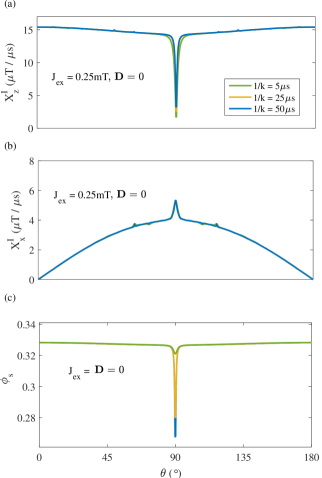
Here we consider a simple RP model system with only one nuclear spin in each radical, with hyperfine tensors and , to analyze which parameter ranges give rise to spike-like features of the generated signal as a function of the direction of the applied magnetic field. This feature has been suggested to be behind the precision of an avian magnetic compass [58].
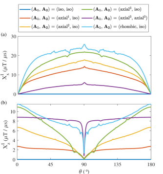
In Fig 8, we plot the generated signal and , respectively, in (a) and (b) for various types of hyperfine coupling tensors chosen by varying principal axis components set . For the anisotropic hyperfine coupling case, the principal axis system is that of the in FAD∙- [58]. A qualitative description of the seen behaviour can be provided by looking at the symmetry of the RP Hamiltonian. In the absence of dipolar coupling, there are three interactions in the RP Hamiltonian, namely, Zeeman, exchange and hyperfine. Zeeman (same magnetic field on all the spins) and exchange interactions are symmetrical with respect to electronic spin exchange while hyperfine interaction is symmetry breaking in general. When the hyperfine interaction is also assumed to be isotropic, the full Hamiltonian is symmetric, and singlet and triplet are eigenstates. As a result, there is no singlet-triplet oscillations possible for isotropic hyperfine coupling as the sub-spaces of different symmetry remain unconnected and as consequence, and remains zero. As the anisotropy is included in the Hamiltonian, a non-zero signal is generated along with appearance of spike-features with the most prominent around . Again, the amplitude of this spike is within the sensitivity achieved for nm deep NV centres. Now we analyze the behaviour of the generated signal and spike as a function of various parameters of the RP Hamiltonian.
The amplitude of the spike increases and it becomes narrower as the principal components and are increased, similar to the behaviour observed in the singlet yield in Ref. [58]. This observation hints that the origin of the spike might also be similar, i.e, an avoided crossing of energy levels as function of direction of the magnetic field. However, further investigations are required to draw firm conclusions.
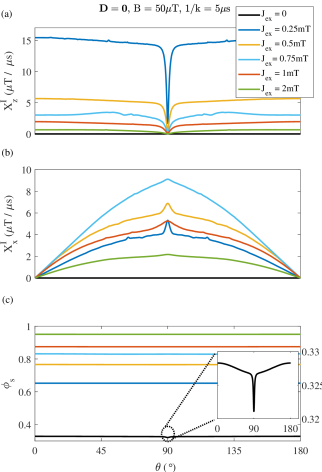
In Fig 9, we show the generated signal and along with the singlet yield [86] (c) with increasing value of . Using the symmetry argument, as is increased, the Hamiltonian eigenstate get closer to singlet and triplet states, and reduction (increase) in the signal (singlet yield) is observed. The spike feature in the generated signal at is most prominent when is comparable to the and principal component of the hyperfine coupling. This behaviour hints that the spike in the generated signal depends on exchange interaction (and also dipolar in general) as well as anisotropic hyperfine interaction. This is in contrast to the singlet yield where the spike depends strongly on hyperfine anisotropy as it disappears even for small , comparable to and parameters [77].
In Fig 10, we show the generated signal and along with the singlet yield [86] (c) with increasing value of RP lifetime . A contrasting behaviour is observed in the generated signal and singlet yield in addition to the former being relatively less sensitive to the lifetime of the radical. This observation seems to suggest that unpaired electrons prefer to stay in the singlet state compared to other populations and coherences of the RP. However, again, further studies are required in this direction to draw concrete conclusions which are beyond purview of current work.
References
- [1] Ulrich E Steiner and Thomas Ulrich. Magnetic field effects in chemical kinetics and related phenomena. Chemical Reviews, 89(1):51–147, 1989.
- [2] JR Woodward. Radical pairs in solution. Progress in Reaction Kinetics and Mechanism, 27(3):165–207, 2002.
- [3] Christiane R Timmel and Kevin B Henbest. A study of spin chemistry in weak magnetic fields. Philosophical Transactions of the Royal Society of London. Series A: Mathematical, Physical and Engineering Sciences, 362(1825):2573–2589, 2004.
- [4] Hisaharu Hayashi. Introduction to dynamic spin chemistry: magnetic field effects on chemical and biochemical reactions, volume 8. World Scientific, 2004.
- [5] PJ Hore, Konstantin L Ivanov, and Michael R Wasielewski. Spin chemistry, 2020.
- [6] Jung Ho Lee, Yusuke Okuno, and Silvia Cavagnero. Sensitivity enhancement in solution nmr: Emerging ideas and new frontiers. Journal of Magnetic Resonance, 241:18–31, 2014.
- [7] Brandon K Rugg, Matthew D Krzyaniak, Brian T Phelan, Mark A Ratner, Ryan M Young, and Michael R Wasielewski. Photodriven quantum teleportation of an electron spin state in a covalent donor–acceptor–radical system. Nature chemistry, 11(11):981–986, 2019.
- [8] Peter J Hore and Henrik Mouritsen. The radical-pair mechanism of magnetoreception. Annual review of biophysics, 45:299–344, 2016.
- [9] Michael T Colvin, Annie Butler Ricks, Amy M Scott, Amanda L Smeigh, Raanan Carmieli, Tomoaki Miura, and Michael R Wasielewski. Magnetic field-induced switching of the radical-pair intersystem crossing mechanism in a donor- bridge- acceptor molecule for artificial photosynthesis. Journal of the American Chemical Society, 133(5):1240–1243, 2011.
- [10] Christopher T Rodgers and Peter J Hore. Chemical magnetoreception in birds: the radical pair mechanism. Proceedings of the National Academy of Sciences, 106(2):353–360, 2009.
- [11] Emrys W Evans, Charlotte A Dodson, Kiminori Maeda, Till Biskup, CJ Wedge, and Christiane R Timmel. Magnetic field effects in flavoproteins and related systems. Interface focus, 3(5):20130037, 2013.
- [12] Alex R Jones. Magnetic field effects in proteins. Molecular Physics, 114(11):1691–1702, 2016.
- [13] R Kaptein and Luitzen J Oosterhoff. Chemically induced dynamic nuclear polarization iii (anomalous multiplets of radical coupling and disproportionation products). Chemical Physics Letters, 4(4):214–216, 1969.
- [14] R Kaptein and JL Oosterhoff. Chemically induced dynamic nuclear polarization ii:(relation with anomalous esr spectra). Chemical Physics Letters, 4(4):195–197, 1969.
- [15] Harold Roy Ward and Ronald G Lawler. Nuclear magnetic resonance emission and enhanced absorption in rapid organometallic reactions. Journal of the American Chemical Society, 89(21):5518–5519, 1967.
- [16] Richard W Fessenden and Robert H Schuler. Electron spin resonance studies of transient alkyl radicals. The Journal of Chemical Physics, 39(9):2147–2195, 1963.
- [17] Gerhard L Closs. Mechanism explaining nuclear spin polarizations in radical combination reactions. Journal of the American Chemical Society, 91(16):4552–4554, 1969.
- [18] Gerhard L Closs and AD Trifunac. Theory of chemically induced nuclear spin polarization. iii. effect of isotropic g shifts in the components of radical pairs with one hyperfine interaction. Journal of the American Chemical Society, 92(7):2183–2184, 1970.
- [19] SK Wong, DA Hutchinson, and JKS Wan. Chemically induced dynamic electron polarization. ii. a general theory for radicals produced by photochemical reactions of excited triplet carbonyl compounds. The Journal of Chemical Physics, 58(3):985–989, 1973.
- [20] Youngchan Kim, Federico Bertagna, Edeline M D’Souza, Derren J Heyes, Linus O Johannissen, Eveliny T Nery, Antonio Pantelias, Alejandro Sanchez-Pedreño Jimenez, Louie Slocombe, Michael G Spencer, et al. Quantum biology: An update and perspective. Quantum Reports, 3(1):80–126, 2021.
- [21] Adriana Marais, Betony Adams, Andrew K Ringsmuth, Marco Ferretti, J Michael Gruber, Ruud Hendrikx, Maria Schuld, Samuel L Smith, Ilya Sinayskiy, Tjaart PJ Krüger, et al. The future of quantum biology. Journal of the Royal Society Interface, 15(148):20180640, 2018.
- [22] Patrick Emery, Ralf Stanewsky, Jeffrey C Hall, and Michael Rosbash. A unique circadian-rhythm photoreceptor. Nature, 404(6777):456–457, 2000.
- [23] Satchidananda Panda, John B Hogenesch, and Steve A Kay. Circadian rhythms from flies to human. Nature, 417(6886):329–335, 2002.
- [24] Roswitha Wiltschko, Christine Nießner, and Wolfgang Wiltschko. The magnetic compass of birds: The role of cryptochrome. Frontiers in Physiology, 12:667000, 2021.
- [25] Adriana Marais, Ilya Sinayskiy, Francesco Petruccione, and Rienk van Grondelle. A quantum protective mechanism in photosynthesis. Scientific reports, 5(1):1–8, 2015.
- [26] Iannis K Kominis. Quantum measurement corrections to cidnp in photosynthetic reaction centers. New Journal of Physics, 15(7):075017, 2013.
- [27] Vivi Joosten and Willem JH van Berkel. Flavoenzymes. Current opinion in chemical biology, 11(2):195–202, 2007.
- [28] Panu Pimviriyakul and Pimchai Chaiyen. Overview of flavin-dependent enzymes. In The Enzymes, volume 47, pages 1–36. Elsevier, 2020.
- [29] Hanan L Messiha, Thanyaporn Wongnate, Pimchai Chaiyen, Alex R Jones, and Nigel S Scrutton. Magnetic field effects as a result of the radical pair mechanism are unlikely in redox enzymes. Journal of the Royal Society Interface, 12(103):20141155, 2015.
- [30] Darragh Crotty, Gary Silkstone, Soumya Poddar, Richard Ranson, Adriele Prina-Mello, Michael T Wilson, and JMD Coey. Reexamination of magnetic isotope and field effects on adenosine triphosphate production by creatine kinase. Proceedings of the National Academy of Sciences, 109(5):1437–1442, 2012.
- [31] Robert Bittl and Stefan Weber. Transient radical pairs studied by time-resolved epr. Biochimica et Biophysica Acta (BBA)-Bioenergetics, 1707(1):117–126, 2005.
- [32] Till Biskup. Time-resolved electron paramagnetic resonance of radical pair intermediates in cryptochromes. Molecular Physics, 111(24):3698–3703, 2013.
- [33] Kevin B Henbest, Kiminori Maeda, PJ Hore, Monika Joshi, Adelbert Bacher, Robert Bittl, Stefan Weber, Christiane R Timmel, and Erik Schleicher. Magnetic-field effect on the photoactivation reaction of escherichia coli dna photolyase. Proceedings of the National Academy of Sciences, 105(38):14395–14399, 2008.
- [34] Kiminori Maeda, Simon RT Neil, Kevin B Henbest, Stefan Weber, Erik Schleicher, Peter John Hore, Stuart R Mackenzie, and Christiane R Timmel. Following radical pair reactions in solution: a step change in sensitivity using cavity ring-down detection. Journal of the American Chemical Society, 133(44):17807–17815, 2011.
- [35] Kiminori Maeda, Alexander J Robinson, Kevin B Henbest, Hannah J Hogben, Till Biskup, Margaret Ahmad, Erik Schleicher, Stefan Weber, Christiane R Timmel, and Peter J Hore. Magnetically sensitive light-induced reactions in cryptochrome are consistent with its proposed role as a magnetoreceptor. Proceedings of the National Academy of Sciences, 109(13):4774–4779, 2012.
- [36] Dean MW Sheppard, Jing Li, Kevin B Henbest, Simon RT Neil, Kiminori Maeda, Jonathan Storey, Erik Schleicher, Till Biskup, Ryan Rodriguez, Stefan Weber, et al. Millitesla magnetic field effects on the photocycle of an animal cryptochrome. Scientific reports, 7(1):1–7, 2017.
- [37] Charlotte A Dodson, Christopher J Wedge, Masaaki Murakami, Kiminori Maeda, Mark I Wallace, and PJ Hore. Fluorescence-detected magnetic field effects on radical pair reactions from femtolitre volumes. Chemical Communications, 51(38):8023–8026, 2015.
- [38] Emrys W Evans, Jing Li, Jonathan G Storey, Kiminori Maeda, Kevin B Henbest, Charlotte A Dodson, PJ Hore, Stuart R Mackenzie, and Christiane R Timmel. Sensitive fluorescence-based detection of magnetic field effects in photoreactions of flavins. Physical Chemistry Chemical Physics, 17(28):18456–18463, 2015.
- [39] Daniel R Kattnig, Emrys W Evans, Victoire Déjean, Charlotte A Dodson, Mark I Wallace, Stuart R Mackenzie, Christiane R Timmel, and PJ Hore. Chemical amplification of magnetic field effects relevant to avian magnetoreception. Nature Chemistry, 8(4):384–391, 2016.
- [40] Victoire Déjean, Marcin Konowalczyk, Jamie Gravell, Matthew J Golesworthy, Catlin Gunn, Nils Pompe, Olivia Foster Vander Elst, Ke-Jie Tan, Mark Oxborrow, Dirk GAL Aarts, et al. Detection of magnetic field effects by confocal microscopy. Chemical science, 11(30):7772–7781, 2020.
- [41] Noboru Ikeya and Jonathan R Woodward. Cellular autofluorescence is magnetic field sensitive. Proceedings of the National Academy of Sciences, 118(3):e2018043118, 2021.
- [42] Noboru Ikeya, Egor A Nasibulov, Konstantin L Ivanov, Kiminori Maeda, and Jonathan R Woodward. Single-molecule spectroscopy of radical pairs, a theoretical treatment and experimental considerations. Molecular Physics, 117(19):2604–2617, 2019.
- [43] Haibin Liu, Martin B Plenio, and Jianming Cai. Scheme for detection of single-molecule radical pair reaction using spin in diamond. Physical Review Letters, 118(20):200402, 2017.
- [44] Amit Finkler and Durga Dasari. Quantum sensing and control of spin-state dynamics in the radical-pair mechanism. Physical Review Applied, 15(3):034066, 2021.
- [45] Marcus W Doherty, Neil B Manson, Paul Delaney, Fedor Jelezko, Jörg Wrachtrup, and Lloyd CL Hollenberg. The nitrogen-vacancy colour centre in diamond. Physics Reports, 528(1):1–45, 2013.
- [46] Romana Schirhagl, Kevin Chang, Michael Loretz, and Christian L Degen. Nitrogen-vacancy centers in diamond: nanoscale sensors for physics and biology. Annu. Rev. Phys. Chem, 65(1):83–105, 2014.
- [47] David A Simpson. Quantum probes for biology: Unlocking single molecule dynamics. Nano Today, 24:7–9, 2019.
- [48] Gopalakrishnan Balasubramanian, IY Chan, Roman Kolesov, Mohannad Al-Hmoud, Julia Tisler, Chang Shin, Changdong Kim, Aleksander Wojcik, Philip R Hemmer, Anke Krueger, et al. Nanoscale imaging magnetometry with diamond spins under ambient conditions. Nature, 455(7213):648–651, 2008.
- [49] Jeronimo R Maze, Paul L Stanwix, James S Hodges, Seungpyo Hong, Jacob M Taylor, Paola Cappellaro, Liang Jiang, MV Dutt, Emre Togan, AS Zibrov, et al. Nanoscale magnetic sensing with an individual electronic spin in diamond. Nature, 455(7213):644–647, 2008.
- [50] Jacob M Taylor, Paola Cappellaro, Lilian Childress, Liang Jiang, Dmitry Budker, PR Hemmer, Amir Yacoby, Ronald Walsworth, and MD Lukin. High-sensitivity diamond magnetometer with nanoscale resolution. Nature Physics, 4(10):810–816, 2008.
- [51] Florian Dolde, Helmut Fedder, Marcus W Doherty, Tobias Nöbauer, Florian Rempp, Gopalakrishnan Balasubramanian, Thomas Wolf, Friedemann Reinhard, Lloyd CL Hollenberg, Fedor Jelezko, et al. Electric-field sensing using single diamond spins. Nature Physics, 7(6):459–463, 2011.
- [52] Florian Dolde, Marcus W Doherty, Julia Michl, Ingmar Jakobi, Boris Naydenov, Sebastien Pezzagna, Jan Meijer, Philipp Neumann, Fedor Jelezko, Neil B Manson, et al. Nanoscale detection of a single fundamental charge in ambient conditions using the nv- center in diamond. Physical review letters, 112(9):097603, 2014.
- [53] Michael Lurie Goldman, MW Doherty, Alp Sipahigil, Norman Ying Yao, SD Bennett, NB Manson, Alexander Kubanek, and Mikhail D Lukin. State-selective intersystem crossing in nitrogen-vacancy centers. Physical Review B, 91(16):165201, 2015.
- [54] S Felton, AM Edmonds, ME Newton, PM Martineau, D Fisher, DJ Twitchen, and JM Baker. Hyperfine interaction in the ground state of the negatively charged nitrogen vacancy center in diamond. Physical Review B, 79(7):075203, 2009.
- [55] Karno Schwinn, Nicolas Ferré, and Miquel Huix-Rotllant. Uv-visible absorption spectrum of fad and its reduced forms embedded in a cryptochrome protein. Physical Chemistry Chemical Physics, 22(22):12447–12455, 2020.
- [56] Marcin Kruszewski. Labile iron pool: the main determinant of cellular response to oxidative stress. Mutation Research/Fundamental and Molecular Mechanisms of Mutagenesis, 531(1-2):81–92, 2003.
- [57] Thomas P Fay, Lachlan P Lindoy, David E Manolopoulos, and PJ Hore. How quantum is radical pair magnetoreception? Faraday discussions, 221:77–91, 2020.
- [58] Hamish G Hiscock, Susannah Worster, Daniel R Kattnig, Charlotte Steers, Ye Jin, David E Manolopoulos, Henrik Mouritsen, and PJ Hore. The quantum needle of the avian magnetic compass. Proceedings of the National Academy of Sciences, 113(17):4634–4639, 2016.
- [59] Olga Efimova and PJ Hore. Role of exchange and dipolar interactions in the radical pair model of the avian magnetic compass. Biophysical Journal, 94(5):1565–1574, 2008.
- [60] Nan Zhao, Jan Honert, Bernhard Schmid, Michael Klas, Junichi Isoya, Matthew Markham, Daniel Twitchen, Fedor Jelezko, Ren-Bao Liu, Helmut Fedder, et al. Sensing single remote nuclear spins. Nature nanotechnology, 7(10):657–662, 2012.
- [61] Jonathan Zopes, K Sasaki Cujia, Kazunari Sasaki, Jens M Boss, Kohei M Itoh, and Christian L Degen. Three-dimensional localization spectroscopy of individual nuclear spins with sub-angstrom resolution. Nature communications, 9(1):1–8, 2018.
- [62] Jonathan Zopes, Konstantin Herb, KS Cujia, and Christian L Degen. Three-dimensional nuclear spin positioning using coherent radio-frequency control. Physical Review Letters, 121(17):170801, 2018.
- [63] Abdelghani Laraoui, Daniela Pagliero, and Carlos A Meriles. Imaging nuclear spins weakly coupled to a probe paramagnetic center. Physical Review B, 91(20):205410, 2015.
- [64] Fazhan Shi, Qi Zhang, Pengfei Wang, Hongbin Sun, Jiarong Wang, Xing Rong, Ming Chen, Chenyong Ju, Friedemann Reinhard, Hongwei Chen, et al. Single-protein spin resonance spectroscopy under ambient conditions. Science, 347(6226):1135–1138, 2015.
- [65] Christian L Degen, F Reinhard, and Paola Cappellaro. Quantum sensing. Reviews of modern physics, 89(3):035002, 2017.
- [66] N Mizuochi, P Neumann, F Rempp, J Beck, V Jacques, P Siyushev, K Nakamura, DJ Twitchen, H Watanabe, S Yamasaki, et al. Coherence of single spins coupled to a nuclear spin bath of varying density. Physical review B, 80(4):041201, 2009.
- [67] Sorawis Sangtawesin, Bo L Dwyer, Srikanth Srinivasan, James J Allred, Lila VH Rodgers, Kristiaan De Greve, Alastair Stacey, Nikolai Dontschuk, Kane M O’Donnell, Di Hu, et al. Origins of diamond surface noise probed by correlating single-spin measurements with surface spectroscopy. Physical Review X, 9(3):031052, 2019.
- [68] H-J Werner, Zan Schulten, and Klaus Schulten. Theory of the magnetic field modulated geminate recombination of radical ion pairs in polar solvents: application to the pyrene–n, n-dimethylaniline system. The Journal of Chemical Physics, 67(2):646–663, 1977.
- [69] Christopher T Rodgers, Stuart A Norman, Kevin B Henbest, Christiane R Timmel, and PJ Hore. Determination of radical re-encounter probability distributions from magnetic field effects on reaction yields. Journal of the American Chemical Society, 129(21):6746–6755, 2007.
- [70] Harold P Erickson. Size and shape of protein molecules at the nanometer level determined by sedimentation, gel filtration, and electron microscopy. Biological procedures online, 11(1):32–51, 2009.
- [71] Dmitry Kobylkov, Joe Wynn, Michael Winklhofer, Raisa Chetverikova, Jingjing Xu, Hamish Hiscock, PJ Hore, and Henrik Mouritsen. Electromagnetic 0.1–100 khz noise does not disrupt orientation in a night-migrating songbird implying a spin coherence lifetime of less than 10 s. Journal of the Royal Society Interface, 16(161):20190716, 2019.
- [72] Michael Sean Grinolds, Sungkun Hong, Patrick Maletinsky, Lan Luan, Mikhail D Lukin, Ronald Lee Walsworth, and Amir Yacoby. Nanoscale magnetic imaging of a single electron spin under ambient conditions. Nature Physics, 9(4):215–219, 2013.
- [73] Brian Brocklehurst. Spin correlation in the geminate recombination of radical ions in hydrocarbons. part 1.—theory of the magnetic field effect. Journal of the Chemical Society, Faraday Transactions 2: Molecular and Chemical Physics, 72:1869–1884, 1976.
- [74] Brian Brocklehurst and Keith A McLauchlan. Free radical mechanism for the effects of environmental electromagnetic fields on biological systems. International journal of radiation biology, 69(1):3–24, 1996.
- [75] Alan M Lewis, Thomas P Fay, David E Manolopoulos, Christian Kerpal, Sabine Richert, and Christiane R Timmel. On the low magnetic field effect in radical pair reactions. The Journal of Chemical Physics, 149(3):034103, 2018.
- [76] U Till and PJ Hore. Radical pair kinetics in a magnetic field. Molecular Physics, 90(2):289–296, 1997.
- [77] Hamish G Hiscock, Henrik Mouritsen, David E Manolopoulos, and PJ Hore. Disruption of magnetic compass orientation in migratory birds by radiofrequency electromagnetic fields. Biophysical journal, 113(7):1475–1484, 2017.
- [78] Gijs De Lange, Toeno Van Der Sar, Machiel Blok, Zhi-Hui Wang, Viatcheslav Dobrovitski, and Ronald Hanson. Controlling the quantum dynamics of a mesoscopic spin bath in diamond. Scientific reports, 2(1):1–5, 2012.
- [79] Erik Bauch, Connor A Hart, Jennifer M Schloss, Matthew J Turner, John F Barry, Pauli Kehayias, Swati Singh, and Ronald L Walsworth. Ultralong dephasing times in solid-state spin ensembles via quantum control. Physical Review X, 8(3):031025, 2018.
- [80] M Steiner, P Neumann, J Beck, F Jelezko, and J Wrachtrup. Universal enhancement of the optical readout fidelity of single electron spins at nitrogen-vacancy centers in diamond. Physical Review B, 81(3):035205, 2010.
- [81] Alan M Lewis, Thomas P Fay, and David E Manolopoulos. An efficient quantum mechanical method for radical pair recombination reactions. The Journal of Chemical Physics, 145(24):244101, 2016.
- [82] Thomas P Fay, Alan M Lewis, and David E Manolopoulos. Spin-dependent charge recombination along para-phenylene molecular wires. The Journal of Chemical Physics, 147(6):064107, 2017.
- [83] Igor Lovchinsky, AO Sushkov, E Urbach, Nathalie P de Leon, Soonwon Choi, Kristiaan De Greve, R Evans, R Gertner, E Bersin, C Müller, et al. Nuclear magnetic resonance detection and spectroscopy of single proteins using quantum logic. Science, 351(6275):836–841, 2016.
- [84] AO Sushkov, N Chisholm, I Lovchinsky, M Kubo, PK Lo, SD Bennett, David Hunger, Alexey Akimov, Ronald Lee Walsworth, H Park, et al. All-optical sensing of a single-molecule electron spin. Nano letters, 14(11):6443–6448, 2014.
- [85] Pengfei Wang, Sanyou Chen, Maosen Guo, Shijie Peng, Mengqi Wang, Ming Chen, Wenchao Ma, Rui Zhang, Jihu Su, Xing Rong, et al. Nanoscale magnetic imaging of ferritins in a single cell. Science advances, 5(4):eaau8038, 2019.
- [86] Christiane R Timmel, U Till, Brian Brocklehurst, Keith A Mclauchlan, and Peter J Hore. Effects of weak magnetic fields on free radical recombination reactions. Molecular Physics, 95(1):71–89, 1998.