Zero-Shot 3D Drug Design by Sketching and Generating
Abstract
Drug design is a crucial step in the drug discovery cycle. Recently, various deep learning-based methods design drugs by generating novel molecules from scratch, avoiding traversing large-scale drug libraries. However, they depend on scarce experimental data or time-consuming docking simulation, leading to overfitting issues with limited training data and slow generation speed. In this study, we propose the zero-shot drug design method Desert (Drug dEsign by SkEtching and geneRaTing). Specifically, Desert splits the design process into two stages: sketching and generating, and bridges them with the molecular shape. The two-stage fashion enables our method to utilize the large-scale molecular database to reduce the need for experimental data and docking simulation. Experiments show that Desert achieves a new state-of-the-art at a fast speed.111Code is available at https://github.com/longlongman/DESERT. †† Work was done when Siyu Long was a research intern at Bytedance AI Lab.
1 Introduction
Drug design is a crucial step in the drug discovery cycle, which is the inventive process of finding new drugs based on a biological target (usually a protein pocket) [1, 2, 3]. However, seeking appropriate drugs for a particular target is quite challenging due to the enormous space of drug candidates (almost ). Traditional drug design approaches usually employ virtual screening [4, 5, 6] and molecular dynamics [7, 8] to traverse in a large scaled drug library, which is time-consuming and could not produce novel drug candidates. Recently, a line of work proposes to realize drug design by generating drug molecules from scratch using deep generative models [9, 2, 10], which is quite promising due to the fast speed and the ability of de-novo drug design.
Most of current drug generation model are developed upon 1D (SMILES) [11, 12, 13] or 2D (molecular graph) [14, 15, 16, 17, 18, 19] molecular structures, which heavily rely on expensive experimental data for supervised training while ignoring the 3D interaction information between the drug and the pocket. In a word, they attempt to find a molecule that maximizes the score given by a bio-activity predictor trained on experimental data. They employ different optimization methods, such as Generative Adversarial Network (GAN) [20], Bayesian Optimization (BO) [21, 22, 9], Reinforcement Learning (RL) [23, 24, 20, 25, 26, 27], Evolutionary and Genetic Algorithms (EA/GA) [28, 29, 30, 31, 32], and Markov Chain Monte Carlo (MCMC) [33, 34], in the molecular space for obtaining desired drug molecule under certain constraints. However, we argue that a purely data-driven approach of drug design is practically limited, since in most cases, the quantity of experimental drug-pocket pairs hardly enables supervised training of de-novo drug design. Generally, most protein pockets lack bio-activity data, and learning on noisy and deficient data may lead to severe overfitting problems.
Recently, several drug design models have been proposed to directly generate drug molecules in the 3D space, the realistic space of drug-target interaction. Generating in such space is very promising for the potential of leveraging some prior knowledge (e.g., physical knowledge) instead of entirely counting on data-driven methodology. Specifically, \AtNextCite\@nocounterrmaxnames[35] [35] and \AtNextCite\@nocounterrmaxnames[2] [2] propose efficient 3D generative models to learn the atom density conditioned on protein pockets, with GAN and auto-regressive models, respectively. Nevertheless experimental data are still obligatory in their models for achieving satisfactory results. Even more noteworthy is GEKO [10], which combines the ideas of 3D generation and physical simulation to obtain state-of-the-art drug design performance without the help of large-scale experimental data. Intuitively, GEKO performs geometric editing in the 3D molecule space guided by docking simulation (physical knowledge) [36, 37]. However, GEKO may suffer from two concerns: a) the frequent invocation of docking is very time consuming, which significantly slows down the speed of the drug design model [38, 39]. b) docking may not always be accurate enough, especially in some complex settings [40, 41]. In such a case, being heavily dependent on the docking accuracy could hurt the generalization of the proposed drug design model.
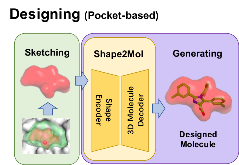
In this paper, we propose the zero-shot approach Desert, namely Drug dEsign by SkEtching and geneRaTing. Motivated by the idea of structure determines properties [42, 43, 44, 45], Desert is built on the assumption that molecular shape determines bio-activity between drug molecules and its target pocket. In other words, we suppose that a drug candidate would have satisfactory bio-activity to a target pocket if their shapes are complementary (see Figure 2).
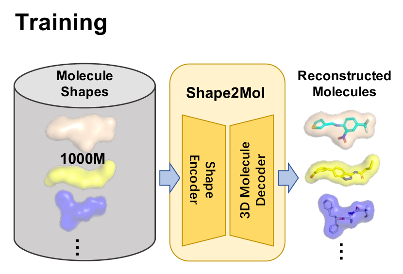
With such prior, as shown in Figure 2, Desert splits the whole drug design process into two stage: sketching and generating, which employs the molecular shape as the bridge of the two stages. Such splitting makes Desert enjoy two advantages: a) Desert does not heavily rely on docking simulation, which only optionally uses docking for post-process and thus avoids the aforementioned disadvantages of GEKO. b) Desert abandons the expensive experimental data. Specifically, in the sketching stage, we only need to sample some reasonable shapes complementary to the target pocket. In the generating stage, Desert proposes to employ a generative pre-trained model (from shape to concrete molecular) to fill the shape obtained in the last sketching stage. Notably, the generative pre-trained model is only trained on the ZINC database, which contains 1000M pairs of molecules and their corresponding shapes. This process does not rely on experimental data, making Desert work in a zero-shot fashion. 222The generation stage of Desert can also be equipped with chemistry priors, we conduct some experiments in Appendix 2.2.
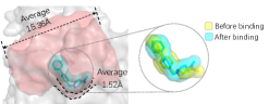
Note that Desert is not baseless. Besides the idea that structure determines properties (an important concept of Structural Biochemistry), we also have preliminary results to verify our assumption. Figure 2 shows that after binding to a pocket, the root-mean-square deviation (RMSD) is not too shabby (Å) compared with molecular conformation generation methods such as CGCF [46] (Å) and ETKDG [47] (Å). The results suggest that the molecular shape is stable when molecules bind to proteins. We also show in Figure 2 that a ligand often attaches tight to a pocket, which means their shape is complementary.
In summary, our contributions are three-fold: (1) We propose Desert, a generative method for de-novo 3D shape-based drug design in the zero-shot setting. (2) Desert trains a pre-trained model from massive unbound molecules 333For clarity, the following terms will be used throughout this study: “bound drug/molecule” (or “unbound drug/molecule”) refers to the drug/molecule that is bound (or unbound) to proteins [48]., eliminating the constraints of labeled data. (3) Desert achieves a new state-of-the-art result (the docking score improves and over the best supervised method on two datasets) at a fast speed (about times faster than GEKO444We calculate the speed by measuring the time from given a pocket to getting 100 molecules.).
2 Proposed Method: Desert
In this section, we describe the proposed method in detail. Inspired by the two aforementioned preliminary studies (see Figure 2 and Figure 2), Desert designs drugs for proteins in a two-stage fashion and employs the shape as a bridge since previous work has shown the feasibility of designing drug by molecular shape [49, 50, 51, 52, 53].
Specifically, Desert designs drugs by first sampling appropriate shapes complementary to the target pocket and then mapping the shapes to specific molecules. In Section 2.1, we first introduce the overall picture of how Desert works in a zero-shot setting. Then we pose two challenges: how to sketch the reasonable molecular shapes and how to generate corresponding molecules based on the shapes. We put forward solutions in Section 2.2 and Section 2.3, respectively.
2.1 Zero-Shot Pipeline
In this section, we focus on how Desert designs drugs in the zero-shot setting. Briefly speaking, Desert produces molecules in a two-stage fashion: sampling the shape of the desired drug first (sketching) and generating molecules conditioned on the resulting shape (generating).
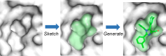
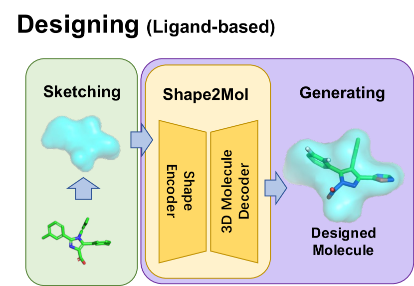
Zero-Shot Sketching There are mainly two cases when Desert needs to sample molecular shapes. In the zero-shot case where no reference protein ligands are available, Desert samples reasonable shapes from protein pockets (see Figure 3(c)) based on biological observations. Besides, Desert can also reuse the shape of a ligand to design a novel one (see Figure 3(b)). Details are listed in Section 2.2.
Zero-Shot Generating Desert generates molecules through a pre-trained generative model, namely Shape2Mol, which can convert a given shape into diverse molecules (see Figure 3(a)). In this procedure, we utilize massive unbound molecules to train the model. Thus no information about proteins is needed. Details of this model are presented in Section 2.3.
2.2 Sketching Molecular Shapes
The sketching stage is responsible for deciding what desired molecules look like. In this section, we show how we design two heuristic methods to sketch the shapes of desired molecules.
There are mainly two cases when we sketch a molecule shape based on whether the ligand is provided. When the ligand is available, the sketching process can be trivial since molecules with similar shapes have similar properties. It is reasonable to directly use the ligand’s shape as the shape of the desired molecule, which we call Ligand-based Sketching. If the ligand of the pocket is unavailable, the challenge is how to obtain a shape that has a high potential to bind to a pocket. 555The pocket can be generated by CAVITY [54] or f-pocket [55]. In this study, all protein pockets are generated by CAVITY. We name this Pocket-based Sketching, and our main idea is to sample a region with an appropriate size complementary to the surface of the pocket.
Our idea for Pocket-based Sketching is based on two main observations:
-
1.
Ligands mainly lie in the area close to the pocket surface. Figure 2 shows that the shape of satisfactory ligand is tightly complementary to the pocket.
-
2.
Pockets are usually much larger than ligands, suggesting that directly utilizing the shape of pockets to design molecules is inappropriate.
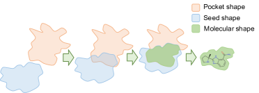
To this end, we present an algorithm (see the Appendix 1.1) to obtain the desired molecule shape, which is of the appropriate size and complementary to the pocket surface. We achieve this goal by finding another shape (namely seed shape) that intersects with the pocket, where the intersection has a similar size to a molecule. We also show a 2D illustration in Figure 4.
2.3 Generating 3D Molecules by Shape2Mol
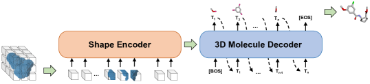
In this section, we introduce Shape2Mol, an encoder-decoder network mapping a shape to diverse and high-quality 3D molecules. There are plenty of unbound molecule data, e.g., 1000M molecules in the ZINC database, making it possible to learn a large-scale pre-trained generative model from shape to molecule.
Concretely, we formulate the problem as an image-to-sequence generation, where the shape is voxelized as a 3D image (see 2.3.1), and the 3D molecule is converted to be a sequence (see 2.3.2). Our generative approach is capable of modeling any complicated molecule structure and the linearization makes large-scale pre-training easier to implement.
2.3.1 Encoder: Voxelized Shape
The shape encoder is a 3D extension of the ViT [56], where we use 3D patches instead of 2D patches in the original ViT. Let denote the set of all atoms, a molecule can be constructed as a collection of atoms and their corresponding coordinates:
Given a molecule , we transform its shape into a 3D image with a voxelization function :
where denotes the Van der Waals radii [57], is a perturbed noise which helps prevent overfitting [58].
2.3.2 Decoder: Linearized Molecule
The molecule decoder in our model is similar to the Transformer decoder in machine translation [59]. The main difference is that the decoding object here is a 3D molecule instead of a 1D sequence. To address this, we propose a 3D molecule decoder, which handles a 3D molecule as a sequence of tuples. The sequence object eases the implementation of a pre-trained model. To obtain the object, we first cut a molecule into pieces, then convert it to a sequence.
Tokenization We cut a molecule into pieces so that the generative process can be easily factorized. Our principles are three folds: (i) preserving the functional groups since they are vital for determining molecule properties [60], (ii) avoiding too large size of the vocabulary to ease the pre-training process [61], (iii) no circles exist in the segmented molecules since a tree structure is simpler to handle than a graph. Our method is simple yet efficient: first tokenizing the molecules with BRCIS [62] and then cutting all single bonds attached to a ring. More details about some pilot experiments can be found in Appendix 1.2.
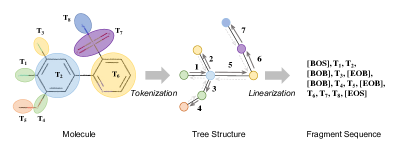
Linearization For the network output, we propose to utilize a linearized sequence to represent the target molecule graph, which is not only convenient for training, but also has the strong power to represent any complicated tree structure. We first select an fragment whose degree is as the root of the tree (e.g., T1, T3, T5, and T8 in Figure 6). Then we traverse the tree in the depth-first-traverse style[63]. Whenever we enter or leave a branch, we will add two special symbols [BOB] and [EOB] (beginning/ending of a branch), respectively.
More particularly, we use a tuple to represent a fragment , where is an indicator function [64] denoting its index in the vocabulary, is the translation vector [65], is the rotation quaternion [66]. In order to stabilize the training process, we further discretize the continuous variable and into and , respectively. Taking the translation vector as an example, we convert it into a binary vector , which satisfies:
where is the max translation length, is the bin size.
2.4 Training & Decoding
Training Given the output probabilities of the model , which denote the probability of a fragment, a discreterized translation vector, and a discreterized rotation vector, respectively. We calculate the corresponding cross-entropy loss and use their sum as the final loss function.
| (1) |
where is the length of fragment sequence.
We use 100M unbound molecules sampled from the lead-like subset of ZINC as the training data. The Transformer’s dimension is 1024, and both encoder and decoder have 12 stacked Transformer layers. When training Shape2Mol, we set the dropout rate as 0.1, batch size 2048, train step 300K and use AdamW [67] with learning rate 5e-4, weight decay 1e-2, and warmup step 4000 as the optimizer. The model is developed by ParaGen 666https://github.com/bytedance/ParaGen and trained on 32 Telsa V100 GPU cards for 2 weeks. Following [35], we also randomly rotate and translate the input shape for invariance.
Decoding We design a decoding strategy to provide diverse and high-quality candidate molecules for a given protein. To achieve diversity, we employ the sampling method Nucleus [68] to generate multiple fragment sequences. Then we convert the sequences back to molecules with a greedy algorithm (see Appendix 1.2), which connects the fragments by greedily enumerating the nearest pair of breakpoints. 777Whenever we cut a chemical bond in tokenizing, we mark the atoms of the chemical bond as breakpoints. Finally, we do some post-processing operation to further improve the diversity and quality. We remove the duplicate molecules and leverage the docking simulation to drop molecules that do not pass the affinity threshold.
When testing Shape2Mol, we set the threshold of Nucleus sampling to 0.95. For each protein pocket, we sketch 200 shapes. For each shape, we generate 1000 molecules. More details of Shape2Mol can be found in the Appendix 1.3.
3 Results and Discussions
3.1 Experiments
Data We evaluate the performance of our method on drug design by using a total of 12 proteins (PDB IDs: 1FKG, 2RD6, 3H7W, 3VRJ, 4CG9, 4OQ3, 4PS7, 5E19, 5MKU, 3FI2, 4J71), which is a combination of the test data used in \AtNextCite\@nocounterrmaxnames[35] [35] and \AtNextCite\@nocounterrmaxnames[23] [23]. Among the proteins, only JNK3 and GSK3 (PDB IDs: 3FI2, 4J71) have sufficient labeled data to train well-performing bioactivity predictors. We denote the set of these two proteins as Set B and the rest as Set A. Because 1D/2D methods need bioactivity predictors to design drugs for given proteins, we only evaluate them on Set B while evaluating 3D-based methods on both sets.
Baselines We compare Desert with some baselines for drug design. Based on the resource needed for designing drugs, we further divide these baselines into three groups: Guided methods need docking simulation or extra bioactivity predictors to provide supervision signals. Supervised methods rely on labeled data to train their models. Retrieved methods directly search the database for desired molecules.
Evaluation Following \AtNextCite\@nocounterrmaxnames[10] [10], we evaluate the performance of methods from two aspects. (1) the molecular space covered by designed results. (2) the capacity to provide highly active molecules. As a high-quality molecular space should contain diverse, novel molecules with pharmaceutical potential, we use five metrics to evaluate the molecular space: Uniqueness (Uniq), Novelty (Nov), Diversity (Div), Success rate (Succ), and Product (Prod). To evaluate the ability to provide highly active molecules, we compare the distributions of and use Median Vina Score (Median) to quantify the distribution. More details about these metrics can be found in Appendix 2.1.
Detail of Generation We describe how different methods generate molecules for comparison. For each protein, every method needs to generate 100 molecules for comparison. For 3D methods, we further use the local minimization module in Vina [36] to optimize the generated structures. For 1D/2D methods, we first use RDKit to generate the 3D conformer of their results. For method Screen, we sample 1K and 200K molecules from ZINC and return 100 molecules with the highest as the generated results.
3.2 Main Results
| Targets | Method | Uniq | Succ | Nov | Div | Prod | Median | |
| (%) | (%) | (%) | (kcal/mol) | |||||
| Set A | Guided | GEKO [10] | 100.0 | 55.7 | 100.0 | 0.912 | 0.51 | -9.58 |
| Supervised | liGAN [35] | 100.0 | 0.4 | 100.0 | 0.924 | 0.00 | -5.84 | |
| 3D SBDD [2] | 69.7 | 13.6 | 98.9 | 0.839 | 0.08 | -8.83 | ||
| Retrieved | Screen (1K) | 100.0 | 25.6 | 100.0 | 0.892 | 0.23 | -7.46 | |
| Screen (200K) | 100.0 | 64.0 | 100.0 | 0.889 | 0.57 | -8.66 | ||
| Ours | Desert-Ligand | 100.0 | 65.3 | 87.0 | 0.786 | 0.41 | -8.89 | |
| Desert-Pocket | 100.0 | 61.1 | 100.0 | 0.908 | 0.57 | -9.62 | ||
| Set B | Guided | JT-VAE [9] | 100.0 | 13.0 | 100.0 | 0.907 | 0.12 | -8.35 |
| RationaleRL [23] | 100.0 | 27.0 | 35.0 | 0.884 | 0.08 | -7.75 | ||
| GA + D [30] | 39.0 | 24.0 | 87.0 | 0.852 | 0.06 | -7.22 | ||
| GraphAF [26] | 97.0 | 0.5 | 100.0 | 0.946 | 0.00 | -4.22 | ||
| MolDQN [27] | 76.5 | 0.0 | 100.0 | 0.742 | 0.00 | -5.52 | ||
| MolEvol [69] | 99.5 | 40.5 | 63.5 | 0.742 | 0.17 | -8.19 | ||
| MARS [33] | 86.0 | 31.5 | 93.0 | 0.805 | 0.22 | -7.68 | ||
| GEKO [10] | 100.0 | 57.0 | 100.0 | 0.910 | 0.52 | -9.19 | ||
| Supervised | liGAN [35] | 99.8 | 0.2 | 100.0 | 0.923 | 0.00 | -5.34 | |
| 3D SBDD [2] | 99.9 | 5.2 | 100.0 | 0.853 | 0.05 | -6.39 | ||
| Retrieved | Screen (1K) | 100.0 | 3.0 | 100.0 | 0.891 | 0.03 | -6.94 | |
| Screen (200K) | 100.0 | 32.0 | 100.0 | 0.882 | 0.28 | -7.95 | ||
| Ours | Desert-Ligand | 100.0 | 18.0 | 100.0 | 0.913 | 0.17 | -7.34 | |
| Desert-Pocket | 100.0 | 61.0 | 100.0 | 0.907 | 0.55 | -9.32 | ||
Table 1 shows the results of Desert and baselines. All values are averaged over target proteins. Our main findings are listed as follows:
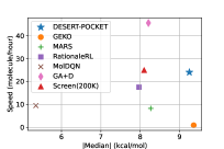
I.The zero-shot Desert achieves the SOTA result at a fast speed. Desert’s performance is strong compared with GEKO, an MCMC-based model with a huge sampling space. Compared with GEKO, which works in a trial-and-error way, Desert makes a more clever choice by pruning the space with its biological knowledge regarding the shape and quickly finds a good solution with limited hints from a teacher. We compare the generation speed in Figure 8.
II.The shape helps Desert produce high quality molecules. The molecular space of Screen is the ZINC database, while that of Desert is generated by the pre-trained model Shape2Mol, which is aware of the pocket’s shape. According to the table, both Desert-Ligand and Desert-Pocket show observably better performance than their counterparts, i.e., Screen (1K) and Screen (200K).
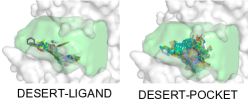
III. More comprehensive exploration of protein pockets benefits performance. Instead of using the molecular shapes of reference ligands as input, Desert-Pocket comprehensively explores the protein pockets by sampling multiple molecular shapes from them. Therefore, it has the potential to obtain diverse, high-quality molecules that bind to protein pockets in different regions. While Desert-Ligand only considers one region, i.e., the region that ligands lie in, limiting the exploration of protein pockets. We show a case in Figure 8.
IV. Unsupervised methods have larger potential than the supervised counterparts. As labeled data is inadequate, e.g., scPDB 888scPDB is a high-quality labeled dataset for 3D drug design. only has 16,034 entries, the supervised methods easily collapse to the main molecule pattern in their dataset. When generating molecules with them, we often get the same molecules, e.g., 3D SBDD can only generate 16 unique molecules for protein 4OQ3. Desert utilizes massive unbound molecules, which leads to the learned space being denser. Combing with an appropriate sampling method, it can generate diverse molecules.
3.3 Comparison with Related Shape-based Models
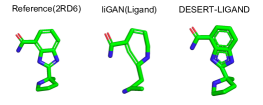
In this section, we study the shape faithfulness and structure rationality compared with previous shape-based models. In Table 2, we use Shape Tanimoto 999Shape Tanimoto [70, 71, 72] measures the similarity between the input and generated molecules. , where and are two molecular shapes. to evaluate the faithfulness and use Free Energy to quantify the rationality [73]. We compares Desert-Ligand with liGAN (Ligand), a variant of liGAN [35] which utilizes the existing ligands. Although liGAN (Ligand) achieves high performance on Shape Tanimoto, its atom-based decoding strategy does not guarantee the correct relative position between atoms. Therefore, liGAN shows higher Free Energy, indicating that unrealistic structures may appear. Figure 9 shows a case where liGAN produces an ill ring structure.
| Method | Shape Tanimoto | Free Energy |
|---|---|---|
| (kcal/mol) | ||
| Random | 0.325 | / |
| Real | / | 167.28 |
| liGAN (Ligand) | 0.869 | 289.55 |
| Desert-Ligand | 0.875 | 188.54 |
3.4 Ablation Study of Generating
In this section, we evaluate several designs of the generating stage in our Desert method, which relate to the pre-trained model and decoding strategy. All results are based on an extra test set from ZINC and a smaller version of Shape2Mol. 101010The extra test set contains 10K molecules. The smaller Shape2Mol has 512 model dimension and 6 layers of encoder and decoder. We use a greedy decoding strategy and remove post-processing.
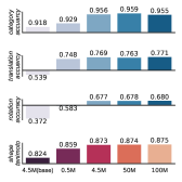
Pre-training Configuration We evaluate the model quality on different pre-training configurations (mainly focusing on the size of the model and training data). The results in Figure 3.4 show: (1) Larger model achieves better performance. \AtNextCite\@nocounterrmaxnames[74] [74] observes similar phenomenons in natural language modeling. (2) Performance saturation occurs when the dataset is of moderate size. As the map from radii to atoms is easy to learn, the model can capture it with a moderate dataset. \AtNextCite\@nocounterrmaxnames[75] [75] reports a similar result that a large dataset does not necessarily lead to better quality.
More Ablation We also do ablation study about some model variants (including discretization and robust training) and decoding strategies. We further study chemical information driven design and atom-based pre-training. We refer the reader for more details to the Appendix 2.2.
| Metric | 3D SBDD | 3D SBDD | Desert-Pocket | Desert-Pocket |
|---|---|---|---|---|
| Med. | w/o post-processing | w post-processing | w/o post-processing | w post-processing |
| Vina Score | -6.069 | -7.584 | -6.148 | -9.410 |
| (kcal/mol) | ||||
| QED | 0.522 | 0.501 | 0.614 | 0.549 |
| SA | 0.672 | 0.623 | 0.612 | 0.616 |
| Diversity | 0.873 | 0.826 | 0.926 | 0.908 |
3.5 Ablation Study of Sketching
In this section, we study the sketching stage in our Desert method, which includes the effect of sampling space size and seed shape on the method’s performance. All the results are based on the same Desert-Pocket method in Section 3.2 and are calculated on Set B.
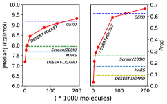
Sampling Space Size In Figure 11, we evaluate the performance of Desert with respect to sampling space size, i.e., the total number of generated molecules before post-processing. The results show: (1) Increasing sampling space size leads to better performance. With a larger sampling space, Desert finds more molecular shapes complementary to pockets, leading to a performance rise. (2) The shape can effectively prune the sampling space for screening. Instead of directly searching molecular space, Desert achieves a similar performance by pruning the space from 200K to 10K with molecular shapes.
More Ablation We also do ablation study about the usage of different seed shape. We refer the reader for more details to the Appendix 2.3.
3.6 Apply Desert to More Protein Targets
To test the generalization ability of our method more widely, we also apply Desert to the test data from \AtNextCite\@nocounterrmaxnames[2] [2], which contains protein targets. As shown in Table 3, based on the idea of structure determines properties, Desert-Pocket generalizes well on different target proteins in both setting, i.e., with/without post-processing. Supervised methods, like 3D SBDD, hindered by scarce training data, can not generate diverse molecules. In contrast, training on massive unbound and drug-like molecules, Desert easily generates diverse and promising molecules. Moreover, sketching molecular shapes based on given pockets also contributes to the better binding affinity of Desert. However, DESERT gives a lower SA score than 3D SBDD. We assume that it is because the generated molecules of DESERT tend to be structurally complicated, which leads to a slightly worse synthesis score.
4 Conclusions
In this study, we propose a zero-shot drug design method Desert, which splits the drug design process into two stages: sketching and generating. Desert bridges the two stages with the molecular shape and utilize a large-scale molecular database to reduce the dependence on experimental data and docking simulation. Experiments show that Desert achieves a new state-of-the-art at a fast speed.
Acknowledgements
We would like to thank the anonymous reviewers for their insightful comments. Both Hao Zhou and Xinyu Dai are the corresponding authors. This work is jointly supported by Guoqiang Research Institute General Project, Tsinghua University (No. 2021GQG1012) and National Science Foundation of China (No. 61936012 and 61976114).
References
- [1] Xiliang Zheng, LinFeng Gan, Erkang Wang and Jin Wang “Pocket-based drug design: exploring pocket space” In The AAPS journal 15.1 Springer, 2013, pp. 228–241
- [2] Shitong Luo, Jiaqi Guan, Jianzhu Ma and Jian Peng “A 3D Generative Model for Structure-Based Drug Design” In Advances in Neural Information Processing Systems 34, 2021
- [3] Pavol Drotár et al. “Structure-aware generation of drug-like molecules” In arXiv preprint arXiv:2111.04107, 2021
- [4] Leonardo G Ferreira, Ricardo N Dos Santos, Glaucius Oliva and Adriano D Andricopulo “Molecular docking and structure-based drug design strategies” In Molecules 20.7 Multidisciplinary Digital Publishing Institute, 2015, pp. 13384–13421
- [5] Xuan-Yu Meng, Hong-Xing Zhang, Mihaly Mezei and Meng Cui “Molecular docking: a powerful approach for structure-based drug discovery” In Current computer-aided drug design 7.2 Bentham Science Publishers, 2011, pp. 146–157
- [6] Nafisa M Hassan, Amr A Alhossary, Yuguang Mu and Chee-Keong Kwoh “Protein-ligand blind docking using QuickVina-W with inter-process spatio-temporal integration” In Scientific reports 7.1 Nature Publishing Group, 2017, pp. 1–13
- [7] Marco De Vivo, Matteo Masetti, Giovanni Bottegoni and Andrea Cavalli “Role of molecular dynamics and related methods in drug discovery” In Journal of medicinal chemistry 59.9 ACS Publications, 2016, pp. 4035–4061
- [8] Jacob D Durrant and J Andrew McCammon “Molecular dynamics simulations and drug discovery” In BMC biology 9.1 BioMed Central, 2011, pp. 1–9
- [9] Wengong Jin, Regina Barzilay and Tommi Jaakkola “Junction tree variational autoencoder for molecular graph generation” In International conference on machine learning, 2018, pp. 2323–2332 PMLR
- [10] Yuwei Yang et al. “Knowledge Guided Geometric Editing for Unsupervised Drug Design”, 2022 URL: https://openreview.net/forum?id=91muTwt1_t5
- [11] Seokho Kang and Kyunghyun Cho “Conditional molecular design with deep generative models” In Journal of chemical information and modeling 59.1 ACS Publications, 2018, pp. 43–52
- [12] Marwin HS Segler, Thierry Kogej, Christian Tyrchan and Mark P Waller “Generating focused molecule libraries for drug discovery with recurrent neural networks” In ACS central science 4.1 ACS Publications, 2018, pp. 120–131
- [13] Matt J Kusner, Brooks Paige and José Miguel Hernández-Lobato “Grammar variational autoencoder” In International conference on machine learning, 2017, pp. 1945–1954 PMLR
- [14] Qi Liu, Miltiadis Allamanis, Marc Brockschmidt and Alexander Gaunt “Constrained graph variational autoencoders for molecule design” In Advances in neural information processing systems 31, 2018
- [15] Tengfei Ma, Jie Chen and Cao Xiao “Constrained generation of semantically valid graphs via regularizing variational autoencoders” In Advances in Neural Information Processing Systems 31, 2018
- [16] Bidisha Samanta et al. “Nevae: A deep generative model for molecular graphs” In Journal of machine learning research. 2020 Apr; 21 (114): 1-33 Journal of Machine Learning Research, 2020
- [17] Wengong Jin, Kevin Yang, Regina Barzilay and Tommi Jaakkola “Learning multimodal graph-to-graph translation for molecular optimization” In arXiv preprint arXiv:1812.01070, 2018
- [18] Wengong Jin, Regina Barzilay and Tommi Jaakkola “Composing molecules with multiple property constraints” In arXiv preprint arXiv:2002.03244, 2020
- [19] Ari Seff et al. “Discrete object generation with reversible inductive construction” In Advances in Neural Information Processing Systems 32, 2019
- [20] Nicola De Cao and Thomas Kipf “MolGAN: An implicit generative model for small molecular graphs” In arXiv preprint arXiv:1805.11973, 2018
- [21] Rafael Gómez-Bombarelli et al. “Automatic chemical design using a data-driven continuous representation of molecules” In ACS central science 4.2 ACS Publications, 2018, pp. 268–276
- [22] Robin Winter et al. “Efficient multi-objective molecular optimization in a continuous latent space” In Chemical science 10.34 Royal Society of Chemistry, 2019, pp. 8016–8024
- [23] Wengong Jin, Regina Barzilay and Tommi Jaakkola “Multi-objective molecule generation using interpretable substructures” In International conference on machine learning, 2020, pp. 4849–4859 PMLR
- [24] Mariya Popova, Olexandr Isayev and Alexander Tropsha “Deep reinforcement learning for de novo drug design” In Science advances 4.7 American Association for the Advancement of Science, 2018, pp. eaap7885
- [25] Jiaxuan You et al. “Graph convolutional policy network for goal-directed molecular graph generation” In Advances in neural information processing systems 31, 2018
- [26] Chence Shi et al. “Graphaf: a flow-based autoregressive model for molecular graph generation” In arXiv preprint arXiv:2001.09382, 2020
- [27] Zhenpeng Zhou et al. “Optimization of molecules via deep reinforcement learning” In Scientific reports 9.1 Nature Publishing Group, 2019, pp. 1–10
- [28] Jan H Jensen “A graph-based genetic algorithm and generative model/Monte Carlo tree search for the exploration of chemical space” In Chemical science 10.12 Royal Society of Chemistry, 2019, pp. 3567–3572
- [29] Sungsoo Ahn, Junsu Kim, Hankook Lee and Jinwoo Shin “Guiding deep molecular optimization with genetic exploration” In Advances in neural information processing systems 33, 2020, pp. 12008–12021
- [30] AkshatKumar Nigam, Pascal Friederich, Mario Krenn and Alán Aspuru-Guzik “Augmenting genetic algorithms with deep neural networks for exploring the chemical space” In arXiv preprint arXiv:1909.11655, 2019
- [31] R Vasundhara Devi, S Siva Sathya and Mohane Selvaraj Coumar “Evolutionary algorithms for de novo drug design–A survey” In Applied Soft Computing 27 Elsevier, 2015, pp. 543–552
- [32] Jan H Jensen “A graph-based genetic algorithm and generative model/Monte Carlo tree search for the exploration of chemical space” In Chemical science 10.12 Royal Society of Chemistry, 2019, pp. 3567–3572
- [33] Yutong Xie et al. “Mars: Markov molecular sampling for multi-objective drug discovery” In arXiv preprint arXiv:2103.10432, 2021
- [34] Tianfan Fu et al. “Mimosa: Multi-constraint molecule sampling for molecule optimization” In arXiv preprint arXiv:2010.02318, 2020
- [35] Tomohide Masuda, Matthew Ragoza and David Ryan Koes “Generating 3D Molecular Structures Conditional on a Receptor Binding Site with Deep Generative Models” In CoRR abs/2010.14442, 2020 arXiv: https://arxiv.org/abs/2010.14442
- [36] Oleg Trott and Arthur J Olson “AutoDock Vina: improving the speed and accuracy of docking with a new scoring function, efficient optimization, and multithreading” In Journal of computational chemistry 31.2 Wiley Online Library, 2010, pp. 455–461
- [37] Jerome Eberhardt, Diogo Santos-Martins, Andreas F Tillack and Stefano Forli “AutoDock Vina 1.2. 0: New docking methods, expanded force field, and python bindings” In Journal of Chemical Information and Modeling 61.8 ACS Publications, 2021, pp. 3891–3898
- [38] Francois Berenger, Ashutosh Kumar, Kam YJ Zhang and Yoshihiro Yamanishi “Lean-Docking: Exploiting Ligands’ Predicted Docking Scores to Accelerate Molecular Docking” In Journal of Chemical Information and Modeling 61.5 ACS Publications, 2021, pp. 2341–2352
- [39] Ondrej Vavra et al. “CaverDock: a molecular docking-based tool to analyse ligand transport through protein tunnels and channels” In Bioinformatics 35.23 Oxford University Press, 2019, pp. 4986–4993
- [40] David Ramı́rez and Julio Caballero “Is it reliable to use common molecular docking methods for comparing the binding affinities of enantiomer pairs for their protein target?” In International journal of molecular sciences 17.4 Multidisciplinary Digital Publishing Institute, 2016, pp. 525
- [41] Kun-Yi Hsin, Samik Ghosh and Hiroaki Kitano “Combining machine learning systems and multiple docking simulation packages to improve docking prediction reliability for network pharmacology” In PloS one 8.12 Public Library of Science San Francisco, USA, 2013, pp. e83922
- [42] Alan R Katritzky et al. “Computational Chemistry Approaches for Understanding how Structure Determines Properties” In Zeitschrift für Naturforschung B 64.6 Verlag der Zeitschrift für Naturforschung, 2009, pp. 773–777
- [43] RM Kell and PB Stickney “How Structure Determines Properties of Plastics”, 1964
- [44] Alan R Katritzky and Dan C Fara “How chemical structure determines physical, chemical, and technological properties: An overview illustrating the potential of quantitative structure- property relationships for fuels science” In Energy & Fuels 19.3 ACS Publications, 2005, pp. 922–935
- [45] Milan Rambukwella et al. “Ligand Structure Determines Nanoparticles’ Atomic Structure, Metal-Ligand Interface and Properties” In Frontiers in chemistry Frontiers, 2018, pp. 330
- [46] Minkai Xu et al. “Learning neural generative dynamics for molecular conformation generation” In arXiv preprint arXiv:2102.10240, 2021
- [47] Sereina Riniker and Gregory A Landrum “Better informed distance geometry: using what we know to improve conformation generation” In Journal of chemical information and modeling 55.12 ACS Publications, 2015, pp. 2562–2574
- [48] Stephan T Stern, Marilyn N Martinez and David M Stevens “When is it important to measure unbound drug in evaluating nanomedicine pharmacokinetics?” In Drug Metabolism and Disposition 44.12 ASPET, 2016, pp. 1934–1939
- [49] Miha Skalic, José Jiménez, Davide Sabbadin and Gianni De Fabritiis “Shape-based generative modeling for de novo drug design” In Journal of chemical information and modeling 59.3 ACS Publications, 2019, pp. 1205–1214
- [50] Ashutosh Kumar and Kam YJ Zhang “Advances in the development of shape similarity methods and their application in drug discovery” In Frontiers in chemistry 6 Frontiers, 2018, pp. 315
- [51] Camila Cardoso Santos et al. “Drug screening using shape-based virtual screening and in vitro experimental models of cutaneous Leishmaniasis” In Parasitology 148.1 Cambridge University Press, 2021, pp. 98–104
- [52] Mu Gao and Jeffrey Skolnick “A comprehensive survey of small-molecule binding pockets in proteins” In PLoS computational biology 9.10 Public Library of Science San Francisco, USA, 2013, pp. e1003302
- [53] Rob LM Van Montfort and Paul Workman “Structure-based drug design: aiming for a perfect fit” In Essays in biochemistry 61.5 Portland Press Ltd., 2017, pp. 431–437
- [54] Yaxia Yuan, Jianfeng Pei and Luhua Lai “Binding site detection and druggability prediction of protein targets for structure-based drug design” In Current pharmaceutical design 19.12 Bentham Science Publishers, 2013, pp. 2326–2333
- [55] Vincent Le Guilloux, Peter Schmidtke and Pierre Tuffery “Fpocket: an open source platform for ligand pocket detection” In BMC bioinformatics 10.1 BioMed Central, 2009, pp. 1–11
- [56] Alexey Dosovitskiy et al. “An image is worth 16x16 words: Transformers for image recognition at scale” In arXiv preprint arXiv:2010.11929, 2020
- [57] A van Bondi “van der Waals volumes and radii” In The Journal of physical chemistry 68.3 ACS Publications, 1964, pp. 441–451
- [58] Stephan Zheng, Yang Song, Thomas Leung and Ian Goodfellow “Improving the robustness of deep neural networks via stability training” In Proceedings of the ieee conference on computer vision and pattern recognition, 2016, pp. 4480–4488
- [59] Ashish Vaswani et al. “Attention is all you need” In Advances in neural information processing systems 30, 2017
- [60] Elena S Salmina, Norbert Haider and Igor V Tetko “Extended functional groups (EFG): an efficient set for chemical characterization and structure-activity relationship studies of chemical compounds” In Molecules 21.1 MDPI, 2015, pp. 1
- [61] Alexis Conneau et al. “Unsupervised cross-lingual representation learning at scale” In arXiv preprint arXiv:1911.02116, 2019
- [62] Jörg Degen, Christof Wegscheid-Gerlach, Andrea Zaliani and Matthias Rarey “On the Art of Compiling and Using’Drug-Like’Chemical Fragment Spaces” In ChemMedChem: Chemistry Enabling Drug Discovery 3.10 Wiley Online Library, 2008, pp. 1503–1507
- [63] Mohit Iyyer, John Wieting, Kevin Gimpel and Luke Zettlemoyer “Adversarial Example Generation with Syntactically Controlled Paraphrase Networks” In Proceedings of the 2018 Conference of the North American Chapter of the Association for Computational Linguistics: Human Language Technologies, Volume 1 (Long Papers) New Orleans, Louisiana: Association for Computational Linguistics, 2018, pp. 1875–1885 DOI: 10.18653/v1/N18-1170
- [64] “Indicator function” In Wikipedia Wikimedia Foundation URL: https://en.wikipedia.org/wiki/Indicator_function
- [65] “Translation (geometry)” In Wikipedia Wikimedia Foundation URL: https://en.wikipedia.org/wiki/Translation_(geometry)
- [66] “Quaternion” In Wikipedia Wikimedia Foundation URL: https://en.wikipedia.org/wiki/Quaternion
- [67] Ilya Loshchilov and Frank Hutter “Decoupled weight decay regularization” In arXiv preprint arXiv:1711.05101, 2017
- [68] Ari Holtzman et al. “The curious case of neural text degeneration” In arXiv preprint arXiv:1904.09751, 2019
- [69] Binghong Chen et al. “Molecule Optimization by Explainable Evolution” In International Conference on Learning Representations, 2020
- [70] David Ryan Koes and Carlos J Camacho “Shape-based virtual screening with volumetric aligned molecular shapes” In Journal of computational chemistry 35.25 Wiley Online Library, 2014, pp. 1824–1834
- [71] Thomas S Rush, J Andrew Grant, Lidia Mosyak and Anthony Nicholls “A shape-based 3-D scaffold hopping method and its application to a bacterial protein- protein interaction” In Journal of medicinal chemistry 48.5 ACS Publications, 2005, pp. 1489–1495
- [72] Sunghwan Kim, Evan E Bolton and Stephen H Bryant “PubChem3D: shape compatibility filtering using molecular shape quadrupoles” In Journal of cheminformatics 3.1 Springer, 2011, pp. 1–14
- [73] Roland J-M Pellenq et al. “A realistic molecular model of cement hydrates” In Proceedings of the National Academy of Sciences 106.38 National Acad Sciences, 2009, pp. 16102–16107
- [74] Jacob Devlin, Ming-Wei Chang, Kenton Lee and Kristina Toutanova “Bert: Pre-training of deep bidirectional transformers for language understanding” In arXiv preprint arXiv:1810.04805, 2018
- [75] Yinhan Liu et al. “Roberta: A robustly optimized bert pretraining approach” In arXiv preprint arXiv:1907.11692, 2019
Checklist
-
1.
For all authors…
-
(a)
Do the main claims made in the abstract and introduction accurately reflect the paper’s contributions and scope? [Yes]
-
(b)
Did you describe the limitations of your work? [Yes]
-
(c)
Did you discuss any potential negative societal impacts of your work? [No]
-
(d)
Have you read the ethics review guidelines and ensured that your paper conforms to them? [Yes]
-
(a)
-
2.
If you are including theoretical results…
-
(a)
Did you state the full set of assumptions of all theoretical results? [N/A]
-
(b)
Did you include complete proofs of all theoretical results? [N/A]
-
(a)
-
3.
If you ran experiments…
-
(a)
Did you include the code, data, and instructions needed to reproduce the main experimental results (either in the supplemental material or as a URL)? [Yes]
-
(b)
Did you specify all the training details (e.g., data splits, hyperparameters, how they were chosen)? [Yes]
-
(c)
Did you report error bars (e.g., with respect to the random seed after running experiments multiple times)? [No]
-
(d)
Did you include the total amount of compute and the type of resources used (e.g., type of GPUs, internal cluster, or cloud provider)? [Yes]
-
(a)
-
4.
If you are using existing assets (e.g., code, data, models) or curating/releasing new assets…
-
(a)
If your work uses existing assets, did you cite the creators? [Yes]
-
(b)
Did you mention the license of the assets? [No]
-
(c)
Did you include any new assets either in the supplemental material or as a URL? [No]
-
(d)
Did you discuss whether and how consent was obtained from people whose data you’re using/curating? [Yes]
-
(e)
Did you discuss whether the data you are using/curating contains personally identifiable information or offensive content? [No]
-
(a)
-
5.
If you used crowdsourcing or conducted research with human subjects…
-
(a)
Did you include the full text of instructions given to participants and screenshots, if applicable? [N/A]
-
(b)
Did you describe any potential participant risks, with links to Institutional Review Board (IRB) approvals, if applicable? [N/A]
-
(c)
Did you include the estimated hourly wage paid to participants and the total amount spent on participant compensation? [N/A]
-
(a)
See pages - of pdf/camera_ready_appendix.pdf