Nonequilibrium Modeling of the Elementary Step in PDZ3 Allosteric Communication
Abstract
ABSTRACT: While allostery is of paramount importance for protein signaling and regulation, the underlying dynamical process of allosteric communication is not well understood. PDZ3 domain represents a prime example of an allosteric single-domain protein, as it features a well-established long-range coupling between the C-terminal -helix and ligand binding. In an intriguing experiment, Hamm and coworkers employed photoswitching of the -helix to initiate a conformational change of PDZ3 that propagates from the C-terminus to the bound ligand within 200 ns. Performing extensive nonequilibrium molecular dynamics simulations, the modeling of the experiment reproduces the measured timescales and reveals a detailed picture of the allosteric communication in PDZ3. In particular, a correlation analysis identifies a network of contacts connecting the -helix and the core of the protein, which move in a concerted manner. Representing a one-step process and involving direct -ligand contacts, this cooperative transition is considered as elementary step in the propagation of conformational change.
![[Uncaptioned image]](/html/2209.07829/assets/x1.png)
Introduction
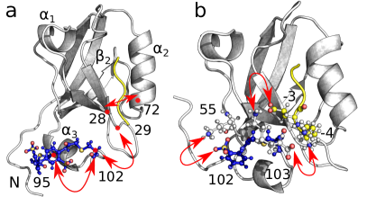
Allostery is one of the most important mechanisms of biomolecular regulation and signal transduction. Wodak19 ; Gunasekaran04 ; Bahar07 ; Cui08 ; Changeux12 ; McLeish13 ; Motlagh14 ; Tsai14 ; Thirumalai19 While commonly this term is meant to describe the communication between distal domains of large macromolecules, it has been suggested that even relatively small single-domain proteins exhibit allosteric properties. Gunasekaran04 PDZ domains, for example, are well-established and structurally conserved protein interaction modules involved in the regulation of multiple receptor-coupled signal transduction processes, but at the same time have also been studied extensively as isolated model systems of allosteric communication. Fuentes04 ; Petit09 ; Lee10 ; Ye13 They share a common fold, which consists of two -helices and six -strands, with the second -helix and the second -strand forming the canonical ligand binding groove (Fig. 1), and generally bind the C-terminus of their targets.
Notably, the NMR study of Petit et al. Petit09 showed that the removal of a short -helix at the C-terminal of PDZ3 reduces ligand affinity by a factor of 21, thus revealing an allosteric communication between ligand binding and C-terminal dynamics. In this way, PDZ3 can be regarded as one of the smallest allosteric proteins, in the sense that both the active site (the -helix) and the allosteric site (the binding pocket) are clearly defined. Notwithstanding numerous experimental and computational studies of PDZ domains (see Refs. Fuentes04, ; Petit09, ; Lee10, ; Ye13, ; Lockless99, ; Ota05, ; Kong09, ; Gerek11, ; Ishikura15, ; Kumawat17, ; Stock18, ; Faure22, for a rather incomplete selection), however, it is still not well understood how exactly a local perturbation propagates through the protein in space and time to a distal site. This is due to the typically small local structural changes of an allosteric signal which are challenging to observe in experiments, Brueschweiler09 and also because of the timescale limitations of molecular dynamics (MD) simulations.Chen07 ; Vesper13 ; Pontiggia15 ; Zheng18
To facilitate a real-time study of the underlying allosteric transition in PDZ domains, Hamm and coworkers implemented an azobenzene photoswitch that impulsively perturb the protein (e.g., by causing the unbinding of the ligand), and monitored the subsequent structural evolution of the system via time-resolved vibrational spectroscopy. Buchli13 ; Bozovic20 ; Bozovic20a ; Bozovic21 ; Bozovic22 As a main result, they found for PDZ2 that the conformational rearrangement of the photoswitchable protein occurs on multiple timescales from pico- to microseconds in a highly nonexponential manner. Stock18 Accompanying MD studies of the nonequilibrium dynamics reproduced these findings and revealed a quite complex structural reorganization of the system. Buchenberg14 ; Buchenberg17 Most recently, Bozovic et al.Bozovic21 employed photoswitching of the -helix to initiate a conformational change of PDZ3 that propagates from the -helix to the binding pocket of the ligand. They found a timescale of 4 to 6 ns for the enforced unfolding of the -helix, as well as 200 ns for the time it takes for the allosteric signal to reach the ligand.
To aid the interpretation of these experiments and to unravel the atomistic mechanism of the putative allosteric transition in PDZ3, in this work we perform extensive all-atom explicit-solvent MD simulations of photoswitchable PDZ3. Aiming to directly model the experiment of Bozovic et al.,Bozovic21 we employ a potential-energy surface switching methodNguyen06 to collect s-long nonequilibrium trajectories of the photoinduced conformational change of PDZ3. In excellent agreement with experiment, we find that the initial unfolding of the -helix occurs on a 5 ns timescale and causes a population shift of protein-attached and protein-separated conformations of the -helix, which in turn triggers a long-range conformational transition involving the ligand on a timescale of about 300 ns.
Results
Similarity of photoswitchable and wild-type PDZ3
In the experiment of Bozovic et al.,Bozovic21 the azobenzene photoswitch was attached to the side-chains of residues 95 and 102 of the -helix. In this way, the end-to-end distance of azobenzene in its stretched trans configuration accommodates a stable -helix, while the twisted cis configuration destabilizes the helix. To verify that the photoswitchable PDZ3 represents a suitable model for the wild-type protein, we performed s-long equilibrium MD simulations of the cis and the trans systems, based on a crystal structureDoyle96 (PDB entry 1TP5) and using the GROMACS v2020 software packageGROMACS20 and the Amber99SB*ILDN force field,Hornak06 ; Best09 ; LindorffLarsen10 see SI Methods. Calculating the time-dependent root mean square displacement (RMSD) matrix of the cis and the wild-type trajectories, Fig. S1 shows that the Cα-RMSD of the full protein never exceeds 2.65 Å throughout the s time evolution shown. With an average of Å, the RMSD can be regarded as a sufficient similarity between photoswitchable and wild-type protein. As an further validation of the simulation model, we calculated the side-chain methyl groups fluctuationsHoffmann20 as measured by the NMR order parameters of Petit et al.,Petit09 see SI Methods. Comparing values obtained from the simulation of cis PDZ3 and the wild-type protein as well as from experiment and wild-type simulation, we obtain good overall agreement (Fig. S1).
Structural changes of PDZ3 upon photoswitching
To get a first impression of the photoinduced conformational transition of PDZ3, we follow Buchenberg et al.Buchenberg17 and consider selected interresidue Cα-distances that account for various aspects of the structural rearrangement. As a first example, Fig. 2 shows the distance between residues Azo95 and Azo102 bridged by the photoswitch, which reports on the length of the -helix (Fig. 1a). The distribution of reveals a shift of about 3 Å upon switching from the cis to the trans equilibrium state, which clearly reflects the photoinduced stretching and partial unfolding of .
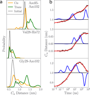
To study the time evolution of the system following cis to trans photoswitching, we sampled 100 statistically independent structures from the cis equilibrium simulations, performed at time a potential-energy surface switching method,Nguyen06 and calculated s-long nonequilibrium trajectories. By performing an ensemble average over these trajectories, we obtain the time-dependent mean value of some observable . Using a log-scale representation of the time axis ranging from 10 ps to 1 s, Fig. 2b shows the photoinduced evolution of , which is found to increase on several timescales. To facilitate the interpretation, we perform a timescale analysis using a maximum entropy methodLorenz-Fonfria06
| (1) |
where the amplitudes of the multiexponential fit yield the timescale spectrum of the evolution. We used 10 equally distributed timescales per decade and a regularization parameter . The analysis of reveals that the sub-ps photoisomerization of azobenzeneNaegele97 causes an elongation of the -helix on timescales of and ns. A structural analysis (Fig. S2) shows that the short timescale reflects the initial strtching of , which is in perfect agreement with the experimental result (4 to 6 ns) of Bozovic et al.Bozovic21 The long timescale accounts for the structural relaxation of the -helix due to the subsequent rearrangement of PDZ3 to be discussed below.
As an indicator of how the initial stretching of the -helix affects the environment of the distal ligand of PDZ3, Fig. 2 shows the distance between Val28 in the -strand and His72 in the -helix, which accounts for the width of the binding pocketBuchenberg17 (Fig. 1a). While the distributions of in cis and trans equilibrium states are rather similar, we find a clear increase of on timescales of and ns. This supposed discrepancy between distributions and time evolution reflects the fact that the nonequilibrium simulations were started close to the initial structure (dotted lines in Fig. 2), and therefore exhibit a short-time relaxation to the appropriate conformational distribution.
Finally, we consider the distance between Azo102 and Gly29 in the - loop as an observable that reports on the attachment of the -helix to the core of the protein (Fig. 1a). Since the alignment and the separation of to and from the protein core is believed to represent the origin of the change in ligand binding affinity,Petit09 ; Bozovic20a can be considered as a simple descriptor of the allosteric transition. Figure 2a shows that the distribution of is rather broad and exhibits a significant shift (nm) of its maximum upon cis-to-trans switching. When we roughly associate distances 1 nm with structures where the -helix is attached to the core, and larger distances with an unattached -helix, suggests a decrease of 68 to 32% of attached structures when we change from cis to trans. Rather than a simple one-to-one relation of cis and trans states with an attached and separated -helix, respectively, we thus find a coexistence of both structures in both states, which undergo a population shift upon cis to trans switching.
In line with its large structural heterogeneity, the time evolution of the -core distance is found to be quite complex. first increases on several timescales (, 1 and 10 ns), before it starts to decrease on a ns timescale. While the fast timescales again reflect the short-time relaxation of the initially prepared state, the long timescale seems to indicate the subsequent allosteric transition of PDZ3. To summarize, we have identified a timescale of 5 ns associated with the initial stretching of the -helix, an intermediate timescale (tens of ns) that reflects the short-time relaxation of the initially prepared structural distribution, and a slow timescale (ns) whose structural origin is to be explained next.
Correlation analysis of interresidue contacts
To identify internal coordinates that change significantly during the conformational transition, we follow recent workErnst17 ; Post22a and focus on interresidue contact distances. This is for two reasons. First, although contact distances report only on near-order interactions, long-distance changes of the structure result as a consequence and are therefore included as well. Moreover, while side-chain dihedral angles may also mediate allosteric couplings,Bowman12 ; Post22a analyses including them showed that they are not of relevance for PDZ3. Assuming that a contact is formed if the distance between the closest non-hydrogen atoms of residues and is shorter than 4.5 Å,Ernst15 we identified 403 interresidue contacts (see SI Methods). To discriminate collective motions underlying functional dynamics from uncorrelated motion, we calculated (the modulus of) the linear correlation matrix of these coordinates and rearranged this matrix in an approximately block-diagonal form. Following Diez et al., Diez22 this is achieved via a community detection technique called Leiden clustering,Traag19 employing the Python package MoSAICDiez22 and a Leiden resolution parameter .
Figure 3a shows the resulting block-diagonal correlation matrix , which reveals eight main blocks or clusters. Within such a cluster, the coordinates are highly correlated (i.e., on average ), while the correlation between different clusters is low (i.e., ). On the other hand, we find that 56 % of all coordinates (shown in the lower right square) correlate only weakly with few other coordinates. (This is, e.g., the case for stable contacts and contacts on the protein surface that form and break frequently.) These contact distances are therefore classified as noise and can be omitted in the further analysis. Finally, there are coordinates (or mini-clusters with coordinates) that are not a member of a main cluster but still exhibit moderate correlation with some of the main clusters; they are shown in between the main clusters.
To illustrate the coordinates contained in the main clusters, Fig. 3b shows the corresponding contact distances inserted into the structure of PDZ3. (See Table S1 for a list of the coordinates of all clusters.) Most interestingly, we find that cluster 1 contains contacts between the -helix and the ligand as well as the core of the protein. Causing the (un)alignment of to the protein core, which in turn induces the change in ligand binding,Petit09 ; Bozovic20a these coordinates are the key to describe the allosteric transition in PDZ3. The remaining clusters, on the other hand, are found to describe interactions in different sections of PDZ3, whose motions are mostly uncorrelated to the coordinates of cluster 1. For example, cluster 2 connects residue Phe100 of to the - loop, and cluster 3 and cluster 6 accounts for motions within the - and - loops, respectively.
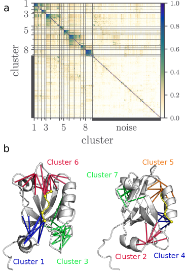
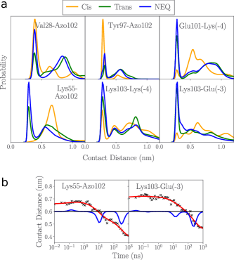
Hence the Leiden correlation analysis leaves us with 11 (instead of initially 403) coordinates comprised in cluster 1, which form a network of highly correlated interresidue contacts (Fig. 3b). The network includes 6 hydrophobic contacts, 2 hydrogen bonds, and 3 salt bridges, which connect the -helix (mostly residues Azo102 and Lys103) to ligand residues Glu(-3) and Lys(-4) and various protein residues. Selecting six representative contacts (see Fig. S4 for the other cases), Fig. 4a shows the corresponding distance distributions in the cis and trans equilibrium states. For all contacts we find a coexistence between contact-formed states (with a well-defined peak at distances Å) and contact-broken states (with a broad distribution of longer distances). We note that in cis the -helix is mostly stabilized by nonpolar contacts with the protein (e.g., Val28 and Tyr97 connect to Azo102), while in trans polar contacts with the ligand dominate. The latter include two salt bridges between C-terminal Lys103 and the ligand residues Lys(-4) and Glu(-3), as well as a hydrogen bond between the backbone of Glu101 and the side-chain of Lys(-4). Moreover, we find the salt bridge Azo102-Lys55 between and the protein core, which exists almost exclusively in trans. That is, during the allosteric transition, (at least) four new contacts are formed, including three salt bridges and one hydrogen bond.
As representative examples of contacts that are formed during the photoinduced transition, Fig. 4b shows the time evolution of the salt bridges Lys55-Azo102 and Lys103-Glu(-3). In both cases, the formation occurs on two timescales of about 10 ns and 300 ns. Similar results are also obtained for the other contact distances of cluster 2 (Fig. S4). While the 10 ns timescale reflects the above discussed short-time relaxation of the initially prepared structural distribution, the long timescale (already found in Fig. 2b) can now unambiguously be associated with the allosteric transition. Assuming that the experimentally measured infrared response of the ligand is related to changes of protein-ligand contacts, the 300 ns timescale compares well to experimental ligand response time of 200 ns obtained by Bozovic et al.Bozovic21
Cooperative mechanism of the long-range communication
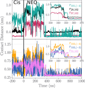
The above correlation analysis indicates that the allosteric transition in PDZ3 is mediated by a network of 11 highly correlated interresidue contacts (Fig. 3b). This raises the question if the transition proceeds sequentially (i.e., involving intermediate states) or rather in a concerted manner. To address this point, Fig. 5 shows the time evolution of the above discussed contact distances along a single nonequilibrium trajectory (i.e., without performing an ensemble average). Starting in the cis equilibrium state for times -200 ns , we find that the two cis-stabilizing contacts are formed (i.e., distances and fluctuate with small amplitude around 3 Å), while the trans-stabilizing contacts are broken such that the corresponding distances , , and fluctuate with large amplitudes. Upon cis-to-trans photoswitching at , contacts 28-102 and 97-102 are broken, too, within tens of nanosecond. As a consequence, all distances fluctuate wildly, reflecting the large-amplitude motion of the detached -helix. At ns this motion localizes abruptly, when all six contacts are formed within 5–7 ns, i.e., virtually simultaneously on the transition timescale of hundreds of nanoseconds. While contact distances , still exhibit residual fluctuations, the other contacts are tightly closed.
Inspecting the other nonequilibrium trajectories, Fig. S5 shows that the overall picture is qualitatively similar to the above example. Of the s-long trajectories, 44 remain in the large-amplitude state, while the other 56 undergo a concerted conformational transition, with 7 trajectories showing multiple transitions. Hence we have shown that the allosteric transition in PDZ3 proceeds in a concerted manner. That is, all contacts change almost simultaneously, similar to the cooperative mechanism that was observed in the functional dynamics of T4 lysozyme.Post22a Starting in the relatively ordered cis state, the system becomes disordered upon cis-to-trans photoswitching and returns to an ordered state via a cooperative transition. In this way, the system follows a order-disorder-order transition, which was also observed for a photoswitchable PDZ2 domain.Buchenberg17
Comparing the distance distributions from the nonequilibrium and the trans simulations (Fig. 4a), we notice that within s the system has not yet reached the trans equilibrium state, because most contacts are less likely in trans than during the nonequilibrium process. Similarly as shown for a photoswitchable PDZ2 domain,Buchenberg17 we thus find that the final step of the allosteric transition is the slow relaxation into the new equilibrium state. Interestingly, a visual inspection of the experimental data (Fig. 3f in Ref. Bozovic21, ) also seems to indicate weak changes of the transient infrared spectrum at long times s.
Discussion and conclusions
In their beautiful experiment, Bozovic et al.Bozovic21 implemented an azobenzene photoswitch in the -helix of PDZ3, which allows to change between the cis state (-helix folded) and the trans state (-helix unfolded). The underlying idea of the experiment is that the unfolding of causes the separation of from the protein core, which in turn causes a change in ligand binding affinity, Petit09 ; Bozovic20a thus establishing an allosteric coupling between and the distal ligand. Using time-resolved infrared spectroscopy, Bozovic et al. observed a timescale of 4 to 6 ns associated with the enforced unfolding of the -helix, as well as a response of the ligand within 200 ns.
Our simulation results reproduce these timescales quite accurately. We obtain a timescale of 5 ns associated with the initial stretching of the -helix (Fig. 2b), as well as a timescale of ns that reflects the reordering of the contact network between the -helix and the core of the protein as well as the ligand (Fig. 4b). Because the transition leads to a change of contacts formed by the ligand, it eventually can be measured via vibrational spectroscopy of the ligand. Hence we have shown that the measured timescale of 200 ns can be clearly assigned to elementary contact changes in the protein. This also aids the interpretation of previous transient infrared experiments on photoswitchable proteins, Buchli13 ; Bozovic20 which too reported a typical timescale of several hundreds of nanoseconds for this process.
To identify the intramolecular interactions mediating the allosteric transition in PDZ3, we have employed a recently proposed correlation analysis of interresidue contact distances.Diez22 It reveals a network of highly correlated -protein contacts (Fig. 3b) that mediate a cooperative contact rearrangement in PDZ3 (Fig. 5). Involving only a one-step contact changing process, the cooperative transition can be considered as a minimal solution to transduce the information on the state of the -helix to the ligand, and represents therefore the elementary step in the allosteric communication in PDZ3. Cooperative contact rearrangements also cause large global free energy barriers that offer a plausible explanation why the often tiny structural changes in allostery can be stabilized in spite of the presence of thermal fluctuations and noise.
Allosteric phenomena in PDZ domains (and in particular in PDZ3) have been debated for long.Lockless99 ; Ota05 ; Kong09 ; Gerek11 ; Ishikura15 ; Kumawat17 ; Stock18 ; Faure22 Nonetheless, our simulation of a specific experiment on PDZ3 has revealed several new and in part unexpected findings. To begin with, this concerns the existence of direct contacts between the -helix and the ligand (Fig. 4a). While the centers of mass of and the ligand are on average separated by about 1.5 nm, the long side-chains of Lys103, Azo102, Glu(3) and Lys(-4) nevertheless facilitate such contacts. They are formed in particular during the cooperative transition (Fig. 5), but may also occur in the trans state. Our results are in contrast to the NMR study of Petit et al.,Petit09 which excluded the possibility of direct -ligand contacts. However, they used a different ligand (NYKQTSV instead of KETWV used here) and their PDZ3 was one amino acid shorter than ours (i.e, it misses Lys103 which is involved in several contacts). We also note that direct -ligand contacts where discussed for the slightly longer ligand KKETWVMurciano-Calles14 and for a PDZ3 domains with a longer C-terminus. Chi12 ; Mostarda12 Hence we conclude that the existence of direct contacts in PDZ3 are specific to the considered version of protein and ligand.
Recognizing that direct contacts between the putative active site (the -helix) and the allosteric site (the binding pocket) may cause at least in part the observed binding affinity change, one may question if the notion of allostery is still adequate here. On the other hand, we expect that the main finding, i.e., that allosteric communication is mediated through a concerted conformational transition that involves numerous simultaneous contact changes, prevails also in the absence of direct contacts and therefore represents a general result. For example, Post et al.Post22a found a quite similar scenario for the allosteric coupling in T4 lysozyme.
A further complication revealed by our analysis is that there is no simple one-to-one relation of the cis and trans states of PDZ3 with the attachment and the separation of , respectively. Rather the calculations of the distribution of the Cα-distance (Fig. 2a) and the -ligand contact distances (Fig. 4a) reveal the coexistence of attached and separated structures in both states. We obtain a population shift of 68 to 32% of attached structures when we change from cis to trans, while we find on average 51% during our nonequilibrium simulations (Fig. S3). Interestingly, this dynamical heterogeneity of occurs also in the wild-type system, where the attached structures persist 45% of the time (Fig. S3). Unfortunately, existing NMR Petit09 or mutational Faure22 studies do not include the -helix, which could confirm this effect.
Allosteric interactions have been commonly described by network models, which may be based on statistical inference, Lockless99 correlations, Sethi09 forces, Stacklies11 energy transfer, Ishikura15 electrostatic interactionsKumawat17 and many variants of thereof. While in this work we also have constructed a correlation matrix reflecting allosteric interactions, our work differs from common network approaches in two important aspects. First, instead of calculating all interresidue correlations using Cartesian Cα-coordinates, we directly focus on interresidue contacts that change during the allosteric transition. For the considered conformational rearrangement in PDZ3, the block diagonalization of the contact distances yielded a single cluster containing highly correlated -protein contacts (Fig. 3b), indicating that only a small part of the protein is involved in the allosteric transition.
Second, rather than describing allostery via the interpretation of some network model, we have directly simulated the allosteric transition using nonequilibrium MD, and employed the correlation analysis merely for the interpretation of the simulations. While such an approach is desirable, we admit that it will be limited to rather small allosteric systems. On the other hand, by a comparison to our MD simulations, we are now in a position to test the validity and potential of various network models, which may then facilitate the study of larger proteins.
Acknowledgments
The authors thank Andrew L. Lee for providing experimental order parameters, and Peter Hamm and his group as well as Georg Diez, Emanuel Dorbath, Daniel Nagel and Matthias Post for numerous instructive and helpful discussions. This work has been supported by the Deutsche Forschungsgemeinschaft (DFG) via the Research Unit FOR 5099 ”Reducing complexity of nonequilibrium” (project No. 431945604). The authors acknowledge support by the High Performance and Cloud Computing Group at the Zentrum für Datenverarbeitung of the University of Tübingen and the Rechenzentrum of the University of Freiburg, the state of Baden-Württemberg through bwHPC and the DFG through Grant Nos. INST 37/935-1 FUGG (RV bw16I016) and INST 39/963-1 FUGG (RV bw18A004).
Supporting Information Available:
Methods, 1 table, and 5 figures.
References
- (1) S. J. Wodak et al., Allostery in its many disguises: From theory to applications, Structure 27, 566 (2019).
- (2) K. Gunasekaran, B. Ma, and R. Nussinov, Is allostery an intrinsic property of all dynamic proteins?, Proteins 57, 433 (2004).
- (3) I. Bahar, C. Chennubhotla, and D. Tobi, Intrinsic dynamics of enzymes in the unbound state and relation to allosteric regulation, Curr. Opin. Struct. Biol 17, 633 (2007).
- (4) Q. Cui and M. Karplus, Allostery and cooperativity revisited, Prot. Sci. 17, 1295 (2008).
- (5) J.-P. Changeux, Allostery and the Monod-Wyman-Changeux model after 50 years, Ann. Rev. Biophys. 41, 103 (2012).
- (6) T. C. B. McLeish, T. L. Rodgers, and M. R. Wilson, Allostery without conformational change: modelling protein dynamics at multiple scales, Phys. Biol. 10, 056004 (2013).
- (7) H. N. Motlagh, J. O. Wrabl, J. Li, and V. J. Hilser, The ensemble nature of allostery, Nature (London) 508, 331 (2014).
- (8) C. J. Tsai and R. Nussinov, A Unified View of ”How Allostery Works”, PLoS Comput. Biol. 10 (2014).
- (9) D. Thirumalai, C. Hyeon, P. I. Zhuravlev, and G. H. Lorimer, Symmetry, rigidity, and allosteric signaling: From monomeric proteins to molecular machines, Chem. Rev. 119, 6788 (2019).
- (10) E. Fuentes, C. Der, and A. Lee, Ligand-dependent dynamics and intramolecular signaling in a PDZ domain, J. Mol. Biol. 335, 1105 (2004).
- (11) C. M. Petit, J. Zhang, P. J. Sapienza, E. J. Fuentes, and A. L. Lee, Hidden dynamic allostery in a PDZ domain, Proc. Natl. Acad. Sci. USA 106, 18249 (2009).
- (12) H.-J. Lee and J. J. Zheng, Pdz domains and their binding partners: structure, specificity, and modification., Cell communication and signaling : CCS 8, 8 (2010).
- (13) F. Ye and M. Zhang, Structures and target recognition modes of PDZ domains: recurring themes and emerging pictures, Biochem. J. 455, 1 (2013).
- (14) S. W. Lockless and R. Ranganathan, Evolutionarily conserved pathways of energetic connectivity in protein families, Science 286, 295 (1999).
- (15) N. Ota and D. A. Agard, Intramolecular signaling pathways revealed by molecular anisotropic thermal diffusion, J. Mol. Biol. 351, 345 (2005).
- (16) Y. Kong and M. Karplus, Signaling pathways of PDZ2 domain: A molecular dynamics interaction correlation analysis, Proteins 74, 145 (2009).
- (17) Z. N. Gerek and S. B. Ozkan, Change in Allosteric Network Affects Binding Affinities of PDZ Domains: Analysis through Perturbation Response Scanning, PLoS Comput. Biol. 7 (2011).
- (18) T. Ishikura, Y. Iwata, T. Hatano, and T. Yamato, Energy exchange network of inter-residue interactions within a thermally fluctuating protein molecule: A computational study, J. Comput. Chem. 36, 1709 (2015).
- (19) A. Kumawat and S. Chakrabarty, Hidden electrostatic basis of dynamic allostery in a PDZ domain, Proc. Natl. Acad. Sci. USA 114, E5825 (2017).
- (20) G. Stock and P. Hamm, A nonequilibrium approach to allosteric communication, Phil. Trans. B 373, 20170187 (2018).
- (21) A. J. Faure, J. Domingo, J. M. Schmiedel, C. Hidalgo-Carcedo, G. Diss, and B. Lehner, Mapping the energetic and allosteric landscapes of protein binding domains, Nature 604, 175 (2022).
- (22) S. Brüschweiler, P. Schanda, K. Kloiber, B. Brutscher, G. Kontaxis, R. Konrat, and M. Tollinger, Direct observation of the dynamic process underlying allosteric signal transmission, J. Am. Chem. Soc. 131, 3063 (2009).
- (23) J. Chen, R. I. Dima, and D. Thirumalai, Allosteric communication in dihydrofolate reductase: Signaling network and pathways for closed to occluded transition and back, J. Mol. Biol. 374, 250 (2007).
- (24) M. D. Vesper and B. L. de Groot, Collective dynamics underlying allosteric transitions in hemoglobin, PLoS Comp. Biol. 9, e1003232 (2013).
- (25) F. Pontiggia, D. Pachov, M. Clarkson, J. Villali, M. Hagan, V. Pande, and D. Kern, Free energy landscape of activation in a signalling protein at atomic resolution, Nat. Commun. 6, 7284 (2015).
- (26) Y. Zheng and Q. Cui, Multiple pathways and time scales for conformational transitions in apo-adenylate kinase, J. Chem. Theory Comput. 14, 1716 (2018).
- (27) O. Bozovic, J. Ruf, C. Zanobini, B. Jankovic, D. Buhrke, P. J. M. Johnson, and P. Hamm, The Speed of Allosteric Signaling Within a Single-Domain Protein., J. Phys. Chem. Lett. 12, 4262 (2021).
- (28) B. Buchli, S. A. Waldauer, R. Walser, M. L. Donten, R. Pfister, N. Bloechliger, S. Steiner, A. Caflisch, O. Zerbe, and P. Hamm, Kinetic response of a photoperturbed allosteric protein, Proc. Natl. Acad. Sci. USA 110, 11725 (2013).
- (29) O. Bozovic, C. Zanobini, A. Gulzar, B. Jankovic, D. Buhrke, M. Post, S. Wolf, G. Stock, and P. Hamm, Real-time observation of ligand-induced allosteric transitions in a PDZ domain, Proc. Natl. Acad. Sci. USA 117, 26031 (2020).
- (30) O. Bozovic, B. Jankovic, and P. Hamm, Sensing the allosteric force, Nat. Commun. 11, 5841 (2020).
- (31) O. Bozovic, B. Jankovic, and P. Hamm, Using azobenzene photocontrol to set proteins in motion, Nat. Rev. Chem. 6, 112 (2022).
- (32) S. Buchenberg, V. Knecht, R. Walser, P. Hamm, and G. Stock, Long-range conformational transition in a photoswitchable allosteric protein: A molecular dynamics simulation study, J. Phys. Chem. B 118, 13468 (2014).
- (33) S. Buchenberg, F. Sittel, and G. Stock, Time-resolved observation of protein allosteric communication, Proc. Natl. Acad. Sci. USA 114, E6804 (2017).
- (34) P. H. Nguyen and G. Stock, Nonequilibrium molecular dynamics simulation of a photoswitchable peptide, Chem. Phys. 323, 36 (2006).
- (35) D. A. Doyle, A. Lee, J. Lewis, E. Kim, M. Sheng, and R. MacKinnon, Crystal structures of a complexed and peptide-free membrane protein-binding domain: Molecular basis of peptide recognition by PDZ, Cell 85, 1067 (1996).
- (36) E. Lindahl, M. J. Abraham, B. Hess, and D. van der Spoel, Gromacs 2020, Xenodo (2020).
- (37) V. Hornak, R. Abel, A. Okur, B. Strockbine, A. Roitberg, and C. Simmerling, Comparison of multiple Amber force fields and development of improved protein backbone parameters, Proteins 65, 712 (2006).
- (38) R. B. Best and G. Hummer, Optimized molecular dynamics force fields applied to the helix-coil transition of polypeptides, J. Phys. Chem. B 113, 9004 (2009).
- (39) K. Lindorff-Larsen, S. Piana, K. Palmo, P. Maragakis, J. L. Klepeis, R. O. Dror, and D. E. Shaw, Improved side-chain torsion potentials for the amber ff99sb protein force field., Proteins 78, 1950 (2010).
- (40) F. Hoffmann, F. A. A. Mulder, and L. V. Schäfer, Predicting NMR relaxation of proteins from molecular dynamics simulations with accurate methyl rotation barriers, J. Chem. Phys. 152, 084102 (2020).
- (41) V. A. Lórenz-Fonfría and H. Kandori, Transformation of time-resolved spectra to lifetime-resolved spectra by maximum entropy inversion of the Laplace transform, Appl. Spectrosc. 60, 407 (2006).
- (42) T. Nägele, R. Hoche, W. Zinth, and J. Wachtveitl, Femtosecond photoisomerization of cis-azobenzene, Chem. Phys. Lett. 272, 489 (1997).
- (43) M. Ernst, S. Wolf, and G. Stock, Identification and validation of reaction coordinates describing protein functional motion: Hierarchical dynamics of T4 Lysozyme, J. Chem. Theory Comput. 13, 5076 (2017).
- (44) M. Post, B. Lickert, G. Diez, S. Wolf, and G. Stock, Cooperative protein allosteric transition mediated by a fluctuating transmission network, J. Mol. Bio. 434, 167679 (2022).
- (45) G. R. Bowman and P. L. Geissler, Equilibrium fluctuations of a single folded protein reveal a multitude of potential cryptic allosteric sites, Proc. Natl. Acad. Sci. USA 109, 11681 (2012).
- (46) M. Ernst, F. Sittel, and G. Stock, Contact- and distance-based principal component analysis of protein dynamics, J. Chem. Phys. 143, 244114 (2015).
- (47) G. Diez, D. Nagel, and G. Stock, Correlation-based feature selection to identify functional dynamics in proteins, J. Chem. Theory Comput. 18, 5079 – 5088 (2022).
- (48) V. Traag, L. Waltman, and N. van Eck, From Louvain to Leiden: guaranteeing well-connected communities, Sci. Rep. 9, 5233 (2019).
- (49) J. Murciano-Calles, C. Corbi-Verge, A. M. Candel, I. Luque, and J. C. Martinez, Post-translational modifications modulate ligand recognition by the third PDZ domain of the MAGUK protein PSD-95, PLOS ONE 9, 1 (2014).
- (50) C. N. Chi, A. Bach, K. Stromgaard, S. Gianni, and P. Jemth, Ligand binding by PDZ domains, Biofactors 38, 338 (2012).
- (51) S. Mostarda, D. Gfeller, and F. Rao, Beyond the binding site: The role of the – loop and extra-domain structures in PDZ domains, PLOS Comp. Bio. 8, 1 (2012).
- (52) A. Sethi, J. Eargle, A. A. Black, and Z. Luthey-Schulten, Dynamical networks in tRNA:protein complexes, Proc. Natl. Acad. Sci. USA 106, 6620 (2009).
- (53) W. Stacklies, C. Seifert, and F. Graeter, Implementation of force distribution analysis for molecular dynamics simulations, BMC Bioinform. 12, 101 (2011).