STUDIES OF THE LINEARITY AND STABILITY OF SILICON DRIFT DETECTORS FOR KAONIC ATOMS X-RAY SPECTROSCOPY††thanks: Presented at the 4th Jagiellonian Symposium on Advances in Particle Physics and Medicine, Kraków, Poland, 10-15 July 2022.
Abstract
The SIDDHARTA-2 experiment at the DANE collider aims to perform precision measurements of kaonic atoms X-ray spectroscopy for the investigation of the antikaon-nucleon strong interaction. To achieve this goal, novel large-area Silicon Drift Detectors (SDDs) have been developed. These devices have special geometry, field configuration and readout electronics that ensure excellent performance in terms of linearity and stability. The paper presents preliminary results for the linearity determination and stability monitoring of the SDDs system during the measurement of kaonic deuterium carried out in the summer of 2022.
DOI:10.5506/APhysPolBSupp…
1 Introduction
The Silicon Drift Detectors (SDDs) [1, 2] combine the silicon p–n junction response to ionizing radiation with an innovative electronic field design [3]. These devices play a key role in high-precision X-ray spectroscopy of light exotic atoms [4], due to their low electronic noise, good energy resolution and high rate capability [5]. In particular, their excellent capabilities are exploited to extract the shift () and width () of atomic levels caused by the strong antikaon-nucleon interaction [6]. Experimental studies of light kaonic atoms provide a unique tool to investigate the non-perturbative quantum chromodynamics (QCD) in the strangeness sector [7, 8].
After the successful data taking campaign of the SIDDHARTA (Silicon Drift Detectors for Hadronic Atom Research by Timing Application) experiment in 2009, resulting in the most precise measurement of the kaonic hydrogen () fundamental level shift () and width () [9], the SIDDHARTA-2 collaboration implemented several updates of the apparatus to perform the analogous, more challenging kaonic deuterium () transition measurement [10]. The experimental facility is located at the DANE collider [11] of the Istituto Nazionale di Fisica Nucleare - Laboratori Nazionali di Frascati (INFN-LNF) in Italy.
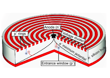
The SDD detector, used in the experiment, is composed of a fully depleted n- silicon cylindrical bulk, double sided by p+ silicon doped electrodes [1, 2]. On one side, a layer of p+ concentric ring strips are biased with a negative voltage through an internal divider, while on the opposite side, a metalized non-structured p+ layer forms the radiation entrance window (see Fig. 1). The electrons produced by the absorbed radiation are focused by the radial drift field to the small n+ anode, placed in the center of the structured side and connected to the dedicated front-end electronics.
New monolithic large-area SDD arrays have been developed by Fondazione Bruno Kessler, together with Politecnico di Milano, INFN-LNF in Italy and the Stefan Meyer Institute (SMI) in Austria, to perform high precision kaonic deuterium spectroscopy [3, 7]. The 450 m thick SDD array consists of a matrix of square cells (0.64 cm2 area for each single unit), mounted on an alumina carrier. The signals coming from the detectors are buffered by a CMOS low-noise charge sensitive preamplifier (CUBE [13]), directly connected to the n+ anode, then processed by a dedicated analog SDD front-end readout ASIC (SFERA [14]).
The SIDDHARTA-2 experimental apparatus [6, 10] consists of 48 SDD arrays, resulting in a total of 384 readout channels, and a luminometer built with plastic scintillators, based on J-PET technology [15, 16, 17, 18, 19]. Studies of the linearity and stability of the SDD system are mandatory to achieve the planned high precision measurement of kaonic deuterium X-rays by keeping the systematic error within a few eV.
2 Linearity characterization
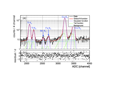
The SDD linearity studies were performed with an X-ray tube exciting a target made of high purity titanium and copper strips [12]. Fig. 2(a) shows a typical spectroscopic response of a single SDD unit at C, irradiated by fluorescence emission from the target strips, activated by the X-ray tube operated at 25 kV and 50 A. Together with the titanium (Ti) and copper (Cu) lines, the Mn Kα and Fe Kα (produced by the excitation of other components of the setup) are also visible.
The spectral response of silicon detectors is predominantly described by a Gaussian function for each X-ray peak, with a low energy tail due to the incomplete charge collection at the anode, and electron-hole recombination. Thus, the shape of the peaks was modeled taking into account the intrinsic Gaussian statistical spread and the tail effect [20, 21, 22].
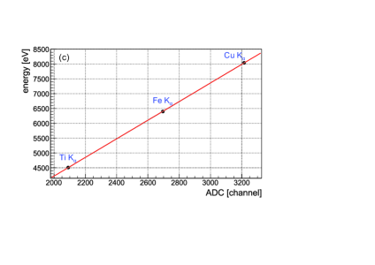
The fit function (red curve in Fig. 2(a)) reproduces the fluorescence lines coming from the target. The various contributions to the overall fit function are also shown. A constant plus an exponential function describes the background. Fig. 2(b) presents the fit residuals. The Ti Kα, Fe Kα and Cu Kα peaks were exploited to evaluate the linearity of the detectors, due to their higher signal-to-background ratio. The positions of these peaks were plotted as a function of the tabulated energy values [23], and a straight line was used to interpolate the points (see Fig. 2(c)). The slope of the fit function (in eV/channel) gives the gain parameter value of the spectrum.
The final energy spectrum is shown in Fig. 3(a). The difference between each Kα experimental calibrated point () with respect to its corresponding theoretical value () was used for the evaluation of the system linearity:
| (1) |
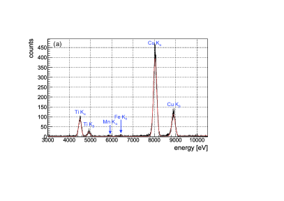
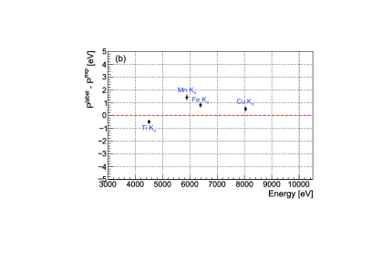
The distribution in Fig. 3(b) shows that the peak energies differ from the theoretical values by less than 1.5 eV, setting the linear response of the SDD below .
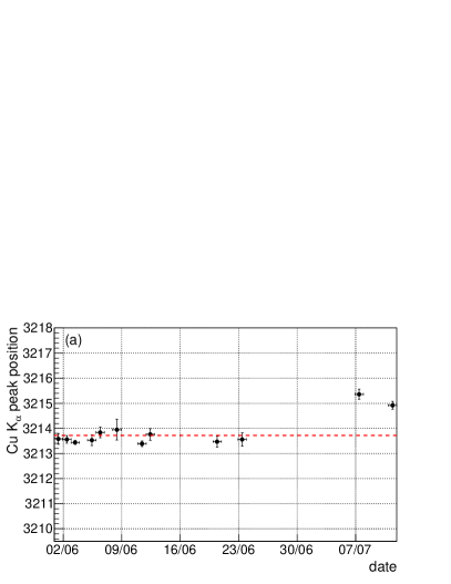
3 Stability
Another important aspect of the SDD system that needs to be monitored during high-precision measurements is its long-term stability, since it is a possible source of systematic error in the experiment. During the kaonic deuterium test measurement period, which took place in June-July 2022, 12 calibration runs were performed. For each SDD, fluctuations over time of the Cu Kα peak position were used to control the stability (see Fig. 4(a)). During the measurements in June 2022 the fluctuations of the Cu Kα position were about 0.5 channels, which corresponds to 1.5 eV, and are comparable with the residuals evaluated during the analysis in the Section 2. However, the deviation of the peak position in the last two runs of July was larger (within channels, corresponding to eV), which is related to more unstable beams condition during the measurement.
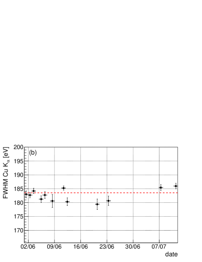
The energy resolution reflects its accuracy in determining the energy of the incoming radiation. For the SDD, the Full Width at Half Maximum (FWHM) of the peak depends on the energy of the incoming photons, and on the electronic noise of the system [24]. Fig. 4(b) shows the FWHM (in eV) of the Cu Kα line, as a function of time, for a typical SDD unit. The width of the peak varies by about eV.
Both results indicate very good stability of the SDDs throughout the experiment, thus providing an excellent opportunity to accurately determine the position of the kaonic deuterium peak, as well as the peak width.
Conclusions
The SIDDHARTA-2 collaboration has developed a new Silicon Drift Detector (SDD) system for the measurement of the kaonic deuterium X-ray transitions to the ground state. The experimental setup has been installed at the DANE collider of the INFN-LNF. The linearity and stability of the SDDs were monitored during the kaonic deuterium test run, and its performance is described in this work. The detectors are linear and stable within a few eV in the energy region of interest. Following the 2022 kaonic deuterium test run, a full measurement will be performed in 2023 to extract for the first time the strong interaction effects in this exotic atom.
Acknowledgments
We thank C. Capoccia from LNF-INFN and H. Schneider, L. Stohwasser, and D. Pristauz-Telsnigg from Stefan-Meyer-Institut for their fundamental contribution in designing and building the SIDDHARTA-2 setup. We thank as well the DANE staff for the excellent working conditions and permanent support. Part of this work was supported by the Austrian Science Fund (FWF): [P24756-N20 and P33037-N]; the Croatian Science Foundation under the project IP-2018-01-8570; the EU STRONG-2020 project (Grant Agreement No. 824093); the EU Horizon 2020 research and innovation programme under the Marie Skłodowska-Curie Grant Agreement No. 754496; the Japan Society for the Promotion of Science JSPS KAKENHI Grant No. JP18H05402; the Polish National Agency for Academic Exchange (Grant No. PPN/BIT/2021/1/00037); the SciMat and qLIFE Priority Research Areas budget under the program Excellence Initiative - Research University at the Jagiellonian University.
References
- [1] P. Lechner “Silicon drift detectors for high resolution room temperature X-ray spectroscopy” In Nucl. Instrum. Meth. Phys. Res. A 377.2, 1996, pp. 346 DOI: 10.1016/0168-9002(96)00210-0
- [2] P. Lechner “Silicon drift detectors for high count rate X-ray spectroscopy at room temperature” In Nucl. Instrum. Meth. Phys. Res. A 458.1, 2001, pp. 281 DOI: 10.1016/S0168-9002(00)00872-X
- [3] M. Miliucci “Large area silicon drift detectors system for high precision timed x-ray spectroscopy” In Meas. Sci. Technol. 33.9, 2022, pp. 095502 DOI: 10.1088/1361-6501/ac777a
- [4] M. Bazzi “Performance of silicon-drift detectors in kaonic atom X-ray measurements” In Nucl. Instrum. Meth. Phys. Res. A 628.1, 2011, pp. 264 DOI: 10.1016/j.nima.2010.06.332
- [5] M. Miliucci “Silicon drift detectors system for high-precision light kaonic atoms spectroscopy” In Meas. Sci. Technol. 32.9, 2021, pp. 095501 DOI: 10.1088/1361-6501/abeea9
- [6] Marco Miliucci “Silicon Drift Detectors‘ Spectroscopic Response during the SIDDHARTA-2 Kaonic Helium Run at the DANE Collider” In Condens. Matter. 6, 2021, pp. 47 DOI: 10.3390/condmat6040047
- [7] Catalina Curceanu “The modern era of light kaonic atom experiments” In Rev. Mod. Phys. 91, 2019, pp. 025006 DOI: 10.1103/RevModPhys.91.025006
- [8] Catalina Curceanu “Kaonic Atoms to Investigate Global Symmetry Breaking” In Symmetry 12.4, 2020, pp. 547 DOI: 10.3390/sym12040547
- [9] M. Bazzi “A new measurement of kaonic hydrogen X-rays” In Phys. Lett. B 704.3, 2011, pp. 113 DOI: 10.1016/j.physletb.2011.09.011
- [10] Marlene Tüchler “Main Features of the SIDDHARTA-2 Apparatus for Kaonic Deuterium X-Ray Measurements” In EPJ Web Conf. 262, 2022, pp. 01016 DOI: 10.1051/epjconf/202226201016
- [11] C. Milardi “Preparation activity for the SIDHARTA-2 run at DANE” In Proc. 9th International Particle Accelerator Conference (IPAC’18), 2018, pp. 334 DOI: 10.18429/JACoW-IPAC2018-MOPMF088
- [12] F. Sgaramella “The SIDDHARTA-2 calibration method for high precision kaonic atoms X-ray spectroscopy measurements” In arXiv:2201.12101, 2022 URL: https://arxiv.org/abs/2201.12101
- [13] L. Bombelli “”CUBE”, A low-noise CMOS preamplifier as alternative to JFET front-end for high-count rate spectroscopy” In IEEE Nucl. Sc. Symp. Conf. Record, 2011, pp. 1972 DOI: 10.1109/NSSMIC.2011.6154396
- [14] R. Quaglia “Development of arrays of Silicon Drift Detectors and readout ASIC for the SIDDHARTA experiment” In Nucl. Instrum. Meth. Phys. Res. A 824, 2016, pp. 449 DOI: https://doi.org/10.1016/j.nima.2015.08.079
- [15] M. Skurzok “Characterization of the SIDDHARTA-2 luminosity monitor” In JINST 15.10, 2020, pp. P10010 DOI: 10.1088/1748-0221/15/10/p10010
- [16] S. Niedźwiecki “J-PET: A NEW TECHNOLOGY FOR THE WHOLE-BODY PET IMAGING” In Acta Phys. Pol. B 48.10, 2017, pp. 1567 DOI: 10.5506/APhysPolB.48.1567
- [17] P. Moskal “Simulating NEMA characteristics of the modular total-body J-PET scanner an economic total-body PET from plastic scintillators” In Phys. Med. Biol. 66.17, 2021, pp. 175015 DOI: 10.1088/1361-6560/ac16bd
- [18] P. Moskal “Testing CPT symmetry in ortho-positronium decays with positronium annihilation tomography” In Nat. Commun. 12, 2021, pp. 5658 DOI: 10.1038/s41467-021-25905-9
- [19] P. Moskal “Positronium imaging with the novel multiphoton PET scanner” In Sci. Adv. 7.42, 2021, pp. eabh4394 DOI: 10.1126/sciadv.abh4394
- [20] J. L. Campbell “X-ray spectrometers for PIXE” In Nucl. Instrum. Methods Phys. Res. B 49, 1990, pp. 115 DOI: 10.1016/0168-583X(90)90227-L
- [21] J. Campbell and J. Maxwell “A cautionary note on the use of the Hypermet tailing function in X-ray spectrometry with Si(Li) detectors” In Nucl. Instrum. Methods Phys. Res. B 129, 1997, pp. 297 DOI: 10.1016/S0168-583X(97)00229-2
- [22] M. V. Gysel, P. Lemberge and P. V. Espen “Implementation of a spectrum fitting procedure using a robust peak model” In X-Ray Spectrom. 32, 2003, pp. 434 DOI: 10.1002/xrs.666
- [23] H. Winick “X-Ray Data Booklet” Berkeley: LBNL, 2009
- [24] M. Miliucci “Energy Response of Silicon Drift Detectors for Kaonic Atom Precision Measurements” In Condens. Matter. 4.1, 2019, pp. 31 DOI: 10.3390/condmat4010031