Combining Density-Functional Theory with Low-Temperature, Polarized Terahertz Spectroscopy of Single Crystals Explicates the Fundamental Modes of L-Alanine
Abstract
Density-functional theory may be used to predict both the frequency and the dipole moment of the fundamental oscillations of molecular crystals. Suitably polarized photons at those frequencies excite such oscillations. Thus, in principle, terahertz spectroscopy may confirm the calculated fundamental modes of amino acids. However, reports to date have multiple shortcomings: (a) material of uncertain purity and morphology and diluted in a binder material is employed; (b) consequently, vibrations along all crystal axes are excited simultaneously; (c) data is restricted to room temperature, where resonances are broad and the background dominant; (d) comparison with theory has been unsatisfactory (in part because the theory assumes zero temperature). Here, we overcome all four obstacles, in reporting detailed low-temperature polarized THz spectra of single-crystal l-alanine, assigning vibrational modes using density-functional theory, and comparing the calculated dipole moment vector direction to the electric field polarization of the measured spectra. Our direct and detailed comparison of theory with experiment corrects previous mode assignments for l-alanine, and reveals unreported modes, previously obscured by closely-spaced spectral absorptions. The fundamental modes are thereby determined.


Alanine (C3H7NO2) is an amino acid which is naturally occurring in the human body. It is the simplest amino acid to demonstrate chirality. Alanine was one of the earliest amino acids, fundamental to early life for metabolic processes and protein formation [2, 3]. In modern life, alanine is essential to many peptide and protein structures, whose hydrogen bonds and van der Waals interactions mediate essential biochemical functions. Recently, the simplest amino acid, glycine, has been found to form in interstellar space through a non-energetic mechanism, and it is suspected that the next-simplest amino acid, alanine, may also [4]. l-Alanine forms a molecular crystal. Each molecule is in a zwitterionic form, where the amine and carboxylic groups are ionized. As is evident in the molecular crystal structure shown in Fig. 1(a), this charge distribution gives rise to a dense and complex network of hydrogen bonds [1].
Low-energy hydrogen bonds, such as those which constitute crystalline l-alanine, are associated in energy with electromagnetic radiation in the terahertz (THz) region. For this reason, terahertz spectroscopy has served as an effective probe of the intermolecular interactions of a wide range of biomolecules [5, 6, 7, 8, 9, 10] and pharmaceuticals [11, 12] and is an appropriate technique to characterize the interactions in l-alanine. Terahertz spectroscopy has been used to assign vibrational modes, elucidate the mechanics of protein formation and function [13], and unravel molecular dynamics [14]. More generally, terahertz physics is an emerging field of wide application [15]. The terahertz roadmap [16] predicts numerous advances, which have recently spanned terahertz axions [17], metamaterials [18], and field-induced ferroelectricity [19].
Returning now to molecular crystals, the precise origins of the terahertz modes are poorly understood. They are usually inferred from the mode energy and crystal structure. In contrast to the bulk of experimental data reported, which does not use polarized radiation, polarized (‘anisotropic’) THz spectroscopy distinguishes vibrational modes with respect to crystal symmetry via the orientation of the dipole moments [20]. This technique also permits the observation of modes that would otherwise be obscured by closely spaced absorption bands [21, 22]. Thus, polarized THz spectroscopy allows for a much more precise mode assignment, yielding better insight into the physical origin of the observed modes.
We now focus on alanine. While it has been intensely studied in the low-energy spectral region [23, 24, 10, 25, 26, 27, 7, 28, 29, 30, 31, 32], many questions relating to its molecular dynamics are still to be answered with precision, due to inadequate theory and experiment. This is due to five main factors.
First, density-functional theory (DFT) calculations, while prolific [33, 34, 35, 9, 36], in many cases only agree poorly with experimental spectra. The difficulty may be often traced to the use of functionals that do not model intermolecular hydrogen bonds well. As mentioned above, such bonds are largely responsible for the low-energy vibrational modes in alanine. This issue has only recently been identified [37] and will be fully resolved here.
Secondly, the precise characterization of l-alanine in the terahertz region is hampered by the lack of high-quality samples. Previous studies have commonly used blends containing l-alanine in a binding medium for pellets [9, 25, 27]. The single-crystal approach of this work removes the potential of observing extrinsic features arising from the binder or interactions between the binding media and the material under study. Moreover, single crystals scatter less light than pellets.
Thirdly, the use of crystals permits the modes associated with different crystallographic axes to be excited by linearly polarized radiation. Polarized THz spectroscopy provides direct information to compare to DFT modeling concerning the dipole moment direction.
Fourthly, the polarized data allows us to separate modes which were previously obscured.
Fifthly, by measuring at low temperatures, where resonances are sharper, we have been able to observe previously unidentified modes with unparalleled clarity.
We now outline our theoretical approach. Further details are provided in the Supplemental Material. [38] DFT was used to calculate the vibrational modes in l-alanine. The CRYSTAL17 package was used [39]. Initial atomic coordinates from Lehmann et al. [1] were the seed for full geometric optimization of both atomic positions and the unit cell.
Zwitterionic amino-acids have proven very difficult to model with DFT methods to the accuracy required for obtaining THz spectra comparable to experiment. Key to our modeling is the choice of an appropriate DFT functional. The B97-3c functional was chosen on three grounds: it includes an appropriate basis set, is fast and accurate in modeling non-covalent interactions that are important for this crystal. The B97-3c functional has been successful in modeling other molecular crystals [40, 41, 42]. It uses van der Waals interactions, and short-range basis set corrections. Modified def2-TZVP basis sets [43] are used, specifically mTZVP [43, 44]. The B97-3c functional is a low-cost Generalized Gradient Approximation (GGA) functional 2–3 times faster than the standard GGA functional when used with the mTZVP basis set. Even though faster, it has been shown to perform with an accuracy level better than the standard GGA functionals for light, main group elements [40].
Since accurate modeling of the experimental terahertz spectrum of l-alanine has proved to be difficult [9, 33, 45], very tight convergence criteria were employed for the full geometry optimization. The details are given in the Supplemental Material [38]. The infrared absorption spectrum was calculated in the harmonic approximation for the final converged geometry. The intensities of the absorptions were calculated with the Berry phase approach [46, 47].


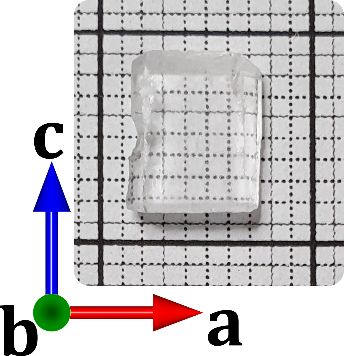
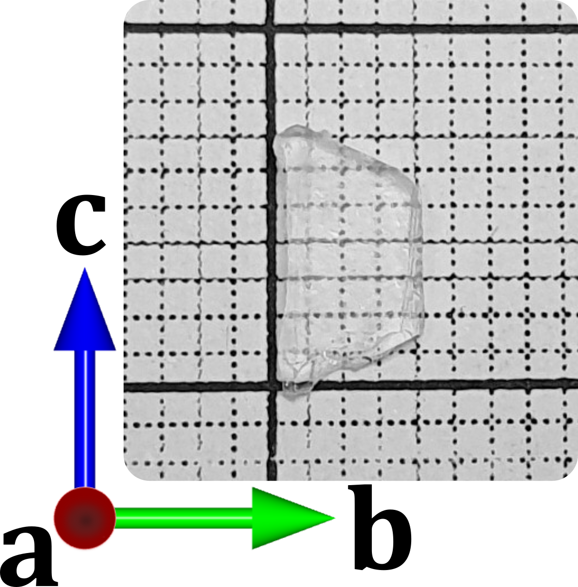
We now characterize the samples. Single crystals were grown using the solvent evaporation method [48]. The crystals used for measurements are shown in Fig. 1(b). Two crystals were used, one with an -face and one with a -face, to allow all three principal crystallographic axes to be probed. For optimum anisotropic measurements, it is best to ensure that only one crystallographic axis is probed in any one measurement. Ideally the sample should be monocrystalline. The morphology of the samples was determined using elastic neutron scattering on the Taipan thermal triple-axis spectrometer [49] at the Australian Nuclear Science and Technology Organisation (ANSTO), and agrees with the accepted growth morphology [50]. In checking the orientation, the 0 4 0 reflection peaks in the l-alanine single crystals were found to have a low mosaic spread, of 0.8, as may be seen in the sample rotation scan in Fig. 1(c). This is (only just) larger than the instrumental resolution of 0.66. Figure 1(c) also indicates a good fit of the data to a Gaussian curve, which is expected for a crystalline reflection peak; moreover, there are no obvious additional peaks. This is also true for - scans performed on the crystal samples. The same holds for the scans done on the 1 2 0 reflection peak (as seen in Supplemental Fig. S3 [38]). The good Gaussian fits and the relatively narrow mosaic spreads suggests the samples are highly crystalline, and confirms that they are suitable for polarization measurements.
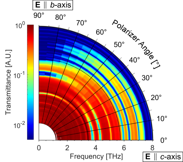
We now present our results. Anisotropic THz spectra of l-alanine were measured using synchrotron radiation at the Australian Synchrotron. The light was polarized using a rotatable wire-grid polarizer. The polarized experimental transmittance spectra of l-alanine are shown in Fig. 2. The a-axis and c-axis spectra were measured with the sample having the face perpendicular to the -axis (left-hand sample in Fig. 1(b)). The b-axis spectrum was measured on the sample having the face perpendicular to the -axis (right-hand sample in Fig. 1(b)). The frequency positions and absorbance intensities from our l-alanine DFT calculations are also shown on the same plots for electric dipoles excited by crystalline vibrations along the direction of the each of the three axes.
| -axis | -axis | -axis | |||||
|---|---|---|---|---|---|---|---|
| Experiment | DFT | Experiment | DFT | Experiment | DFT | ||
| 2.74 | 2.76 | 2.25 | 2.18 | 2.94 | 2.84 | ||
| 3.12 | 3.16 | 2.63 | 2.65 | 3.34 | 3.58 | ||
| 3.26 | 3.33 | 3.30 | 3.41 | 3.79 | 3.85 | ||
| 4.25 | 4.28 | 4.71 | 4.68 | 4.31 | 4.25 | ||
| 5.46 | 4.96 | 5.08 | 4.93 | ||||
| 6.33 | 6.51 | 6.44 | |||||
| 7.08 | 7.11 | ||||||
The polarized spectra of l-alanine coincide well with the unpolarized spectrum (Fig. 2; also Fig. S4 and the discussion in Supplemental Material [38]). All features seen in the unpolarized spectrum appear distinctly in one of the polarized spectra. Furthermore, calculated modes from DFT also align well with the experimental spectra in Fig. 2, with the the same number and approximately equivalent positions of the calculated modes and experimental absorption bands. Additionally, Fig. 3 shows the evolution of absorption bands as the incident polarization angle is varied. All absorption features fade systematically as the electric field polarization is rotated away from the axis along which the absorption is associated.
With the incident electric field of the THz radiation polarized in the -direction, absorption bands are observed at 2.74, 3.12, and 3.26 THz, with a broad absorption band at 4.25 THz, which is not completely resolved as it reaches the noise floor. These experimental results are in good agreement with the DFT calculation when the vibration-induced dipole moment direction is taken into consideration. For dipole moments along the -direction, DFT calculation predicts vibrational modes at 2.76, 3.16, 3.33, and 4.28 THz. Additional modes are calculated at 5.46 and 6.33 THz, beyond the experimental frequency cutoff of approximately 4.5 THz.
In the -direction, absorption bands are observed at 2.25, 2.63, and 3.30 THz. A broad absorption appears at 4.71 THz and is absorptive enough to reach the noise floor. The observations agree with the DFT calculations of modes at 2.18, 2.65, 3.41, and 4.68 THz. A further mode is calculated at 4.96 THz. While not associable with an additional unique experimental absorption beyond that at 4.71 THz, it may be merged with the 4.71 THz absorption, given it is broad and it is incompletely resolved. An additional calculated mode at 6.51 THz lies above the cutoff frequency of the -axis polarization at 5.70 THz.
The -direction polarization shows absorption bands at 2.94, 3.34, 3.79, 4.31, 5.08, 6.44, and 7.08 THz. DFT calculates modes at 2.84, 3.58, 3.85, 4.25, 4.93, and 7.11 THz. These are all in good agreement with the experimental absorptions, with the exception of the observed absorption at 6.44 THz. However, this feature does not have the same Lorenzian-like profile as the rest of the absorption bands assigned, and could reasonably be a non-resonant feature in origin. The frequency cutoff along the -axis is approximately 7.5 THz.
Previous work using unpolarized light [37] assigns the same DFT modes. However, it does so to different experimental modes in five cases, namely the 3.41, 3.58, 4.24, 4.28, and 4.96 THz modes. This clearly shows the improved reliability of studies using polarized spectra. An additional DFT mode at 7.11 THz is identified here which was not presented in the earlier work.
As mentioned above, the polarization data exhibits frequency cutoffs of approximately 4.5 THz for the -axis, 5.7 THz for the -axis, and 7.5 THz for the -axis, which is the same for the anisotropic spectra. These results are consistent with and extend a previous report on the reflection THz spectrum of l-alanine [23] which shows strong, broad reflection beyond 4.8 THz perpendicular to the -axis, but no significant broad, intense reflections parallel to the -axis. Thus, it is not absorption, but rather strong reflection which most likely affects the frequency cutoff in the - and -directions.
In summary, our DFT modeling of dipoles excited along specific crystal directions agrees very well with our experimental polarization results. This lends confidence to the physical basis of the DFT calculations. Specifically, the success of the B97-3c functional in providing very accurate mode calculations has verified that weak hydrogen and van der Waals bonds are critical to the origin of the fundamental modes of l-alanine. Experimentally, through the use of polarized THz spectroscopy, closely-spaced absorption bands have been now separated, confirming an additional mode at 3.30 THz (with a dipole moment along the -axis). Comparison with the DFT modelling has also extended the assignment of modes made in previous work [37]. Anisotropic theory combined with polarized experiments thus resolves the fundamental modes of l-alanine.
This research used the THz/Far-IR beamline at the Australian Synchrotron, part of ANSTO, the Australian Nuclear Science and Technology Organisation. We thank D. Appadoo and R. Plathe for assistance. Numerical modeling was assisted by the National Computational Infrastructure (NCI), supported by the Australian Government. We thank AINSE Ltd for financial assistance (Award-PGRA) to enable this research, also supported by Australian Research Council DP160101474.
References
- Lehmann et al. [1972] M. S. Lehmann, T. F. Koetzle, and W. C. Hamilton, Journal of the American Chemical Society 94, 2657 (1972).
- Higgs and Pudritz [2009] P. G. Higgs and R. E. Pudritz, Astrobiology 9, 483 (2009).
- Kubyshkin and Budisa [2019] V. Kubyshkin and N. Budisa, International Journal of Molecular Sciences 20 (2019).
- Ioppolo et al. [2021] S. Ioppolo, G. Fedoseev, K. Chuang, H. Cuppen, A. Clements, M. Jin, R. Garrod, D. Qasim, V. Kofman, E. van Dishoeck, and H. Linnartz, Nature Astronomy 5, 197 (2021).
- Chen et al. [2013] T. Chen, Z. Li, and W. Mo, Spectrochimica Acta Part A: Molecular and Biomolecular Spectroscopy 106, 48 (2013).
- Kleine-Ostmann et al. [2008] T. Kleine-Ostmann, R. Wilk, F. Rutz, M. Koch, H. Niemann, B. Güttler, K. Brandhorst, and J. Grunenberg, ChemPhysChem 9, 544 (2008).
- Shen et al. [2007] S. C. Shen, L. Santo, and L. Genzel, International Journal of Infrared & Millimeter Waves 28, 595 (2007).
- Williams et al. [2011] M. R. C. Williams, A. B. True, A. F. Izmaylov, T. A. French, K. Schroeck, and C. A. Schmuttenmaer, Physical Chemistry Chemical Physics 13, 11719 (2011).
- Yi et al. [2017] W. Yi, J. Yu, Y. Xu, F. Wang, Q. Yu, H. Sun, L. Xu, Y. Liu, and L. Jiang, Instrumentation Science & Technology 45, 423 (2017).
- Wang et al. [2009] W.-N. Wang, H.-Q. Li, Y. Zhang, and C.-L. Zhang, Acta Physico-Chimica Sinica 25, 2074 (2009).
- Lepodise [2019] L. M. Lepodise, Spectrochimica Acta Part A: Molecular and Biomolecular Spectroscopy 217, 35 (2019).
- Xie et al. [2019] L. Xie, C. Wang, M. Chen, B.-B. Jin, R. Zhou, Y. Huang, S. Hameed, and Y. Ying, Spectrochimica Acta Part A: Molecular and Biomolecular Spectroscopy 222, 117179 (2019).
- He et al. [2008] Y. He, P. I. Ku, J. R. Knab, J. Y. Chen, and A. G. Markelz, Physical Review Letters 101, 178103 (2008).
- Hutereau et al. [2020] M. Hutereau, P. A. Banks, B. Slater, J. A. Zeitler, A. D. Bond, and M. T. Ruggiero, Physical Review Letters 125, 103001 (2020).
- Lewis [2013] R. A. Lewis, Terahertz Physics (Cambridge University Press, Cambridge, UK, 2013).
- Dhillon et al. [2017] S. S. Dhillon, M. S. Vitiello, E. H. Linfield, A. G. Davies, M. C. Hoffmann, J. Booske, C. Paoloni, M. Gensch, P. Weightman, G. P. Williams, E. Castro-Camus, D. R. S. Cumming, F. Simoens, I. Escorcia-Carranza, J. Grant, S. Lucyszyn, M. Kuwata-Gonokami, K. Konishi, M. Koch, C. A. Schmuttenmaer, T. L. Cocker, R. Huber, A. G. Markelz, Z. D. Taylor, V. P. Wallace, J. A. Zeitler, J. Sibik, T. M. Korter, B. Ellison, S. Rea, P. Goldsmith, K. B. Cooper, R. Appleby, D. Pardo, P. G. Huggard, V. Krozer, H. Shams, M. Fice, C. Renaud, A. Seeds, A. Stöhr, M. Naftaly, N. Ridler, R. Clarke, J. E. Cunningham, and M. B. Johnston, Journal of Physics D: Applied Physics 50, 043001 (2017).
- Liu et al. [2022] J. Liu, K. Dona, G. Hoshino, S. Knirck, N. Kurinsky, M. Malaker, D. W. Miller, A. Sonnenschein, M. H. Awida, P. S. Barry, K. K. Berggren, D. Bowring, G. Carosi, C. Chang, A. Chou, R. Khatiwada, S. Lewis, J. Li, S. W. Nam, O. Noroozian, and T. X. Zhou (BREAD Collaboration), Phys. Rev. Lett. 128, 131801 (2022).
- He et al. [2021] J. He, X. He, T. Dong, S. Wang, M. Fu, and Y. Zhang, Journal of Physics D: Applied Physics 55, 123002 (2021).
- Li et al. [2019] X. Li, T. Qiu, J. Zhang, E. Baldini, J. Lu, A. M. Rappe, and K. A. Nelson, Science 364, 1079 (2019).
- Hoshina et al. [2011] H. Hoshina, Y. Morisawa, H. Sato, H. Minamide, I. Noda, Y. Ozaki, and C. Otani, Physical Chemistry Chemical Physics 13, 9173 (2011).
- Deng et al. [2021] Y. Deng, J. A. McKinney, D. K. George, K. A. Niessen, A. Sharma, and A. G. Markelz, ACS Photonics 8, 658 (2021).
- Singh et al. [2012] R. Singh, D. K. George, J. B. Benedict, T. M. Korter, and A. G. Markelz, The Journal of Physical Chemistry A 116, 10359 (2012).
- Mita et al. [2019] Z. Mita, H. Watanabe, and S. Kimura, Infrared Physics & Technology 96, 7 (2019).
- Laman et al. [2008] N. Laman, S. Harsha, D. Grischkowsky, and J. S. Melinger, Biophysical Journal 94, 1010 (2008).
- Liu et al. [2019] Y. Liu, T. Zhou, and J.-C. Cao, Infrared Physics & Technology 96, 17 (2019).
- Darkwah et al. [2013] J. Darkwah, G. Smith, I. Ermolina, and M. Mueller-Holtz, International Journal of Pharmaceutics 455, 357 (2013).
- Ponseca et al. [2010] C. Ponseca, O. Kambara, S. Kawaguchi, K. Yamamoto, and K. Tominaga, Journal of Infrared, Millimeter, and Terahertz Waves 31, 799 (2010).
- Taulbee et al. [2009] A. R. Taulbee, J. A. Heuser, W. U. Spendel, and G. E. Pacey, Analytical Chemistry 81, 2664 (2009).
- Nishizawa et al. [2006] J. Nishizawa, T. Sasaki, K. Suto, T. Tanabe, T. Yoshida, T. Kimura, and K. Saito, International Journal of Infrared and Millimeter Waves 27, 779 (2006).
- Matei et al. [2005] A. Matei, N. Drichko, B. Gompf, and M. Dressel, Chemical Physics 316, 61 (2005).
- Yamaguchi et al. [2005] M. Yamaguchi, F. Miyamaru, K. Yamamoto, M. Tani, and M. Hangyo, Applied Physics Letters 86, 053903 (2005).
- Barthes et al. [2002] M. Barthes, A. F. Vik, A. Spire, H. N. Bordallo, and J. Eckert, The Journal of Physical Chemistry A 106, 5230 (2002).
- Jiang et al. [2016] L. Jiang, J. Yu, C. Li, W. Yi, Y. Xu, and Y. Liu, in 2016 41st International Conference on Infrared, Millimeter, and Terahertz waves (IRMMW-THz) (2016) pp. 1–2.
- Guo and Wei-Ning [2012] W. Guo and W. Wei-Ning, Acta Physico-Chimica Sinica 28, 1579 (2012).
- Zheng and Fan [2012] Z.-P. Zheng and W. Fan, Journal of Biological Physics 38, 405 (2012).
- Zhang et al. [2015] F. Zhang, H.-W. Wang, K. Tominaga, and M. Hayashi, The Journal of Physical Chemistry A 119, 3008 (2015).
- Sanders et al. [2021] T. J. Sanders, J. L. Allen, J. Horvat, and R. A. Lewis, The Journal of Chemical Physics 154, 244311 (2021).
- [38] See Supplemental Material at [link], for details about the calculations and instrumentation, comparison of -axis spectra, triple-axis neutron scattering for the 1 2 0 reflection, and a comparison to the unpolarized spectrum.
- Dovesi et al. [2018] R. Dovesi, A. Erba, R. Orlando, C. M. Zicovich-Wilson, B. Civalleri, L. Maschio, M. Rérat, S. Casassa, J. Baima, S. Salustro, and B. Kirtman, WIREs Computational Molecular Science 8, e1360 (2018).
- Brandenburg et al. [2018] J. G. Brandenburg, C. Bannwarth, A. Hansen, and S. Grimme, The Journal of Chemical Physics 148, 064104 (2018).
- Grimme et al. [2010] S. Grimme, J. Antony, S. Ehrlich, and H. Krieg, The Journal of Chemical Physics 132, 154104 (2010).
- Katsyuba et al. [2019] S. A. Katsyuba, E. E. Zvereva, and S. Grimme, The Journal of Physical Chemistry A 123, 3802 (2019).
- Weigend and Ahlrichs [2005] F. Weigend and R. Ahlrichs, Physical Chemistry Chemical Physics 7, 3297 (2005).
- Grimme et al. [2015] S. Grimme, J. G. Brandenburg, C. Bannwarth, and A. Hansen, The Journal of Chemical Physics 143, 054107 (2015).
- Tulip and Clark [2004] P. R. Tulip and S. J. Clark, The Journal of Chemical Physics 121, 5201 (2004).
- Pascale et al. [2004] F. Pascale, C. M. Zicovich-Wilson, F. López Gejo, B. Civalleri, R. Orlando, and R. Dovesi, Journal of Computational Chemistry 25, 888 (2004).
- Zicovich-Wilson et al. [2004] C. M. Zicovich-Wilson, F. Pascale, C. Roetti, V. R. Saunders, R. Orlando, and R. Dovesi, Journal of Computational Chemistry 25, 1873 (2004).
- Srinivasan et al. [2011] T. P. Srinivasan, R. Indirajith, and R. Gopalakrishnan, Journal of Crystal Growth 318, 762 (2011).
- Danilkin and Yethiraj [2009] S. A. Danilkin and M. Yethiraj, Neutron News 20, 37 (2009).
- Massimino et al. [2011] F. Massimino, M. Bruno, M. Rubbo, and D. Aquilano, Crystal Research and Technology 46, 789 (2011).