Membrane viscosity signature in thermal undulations of curved fluid bilayers
Abstract
Membrane viscosity is usually assumed to only affect short-wavelength undulations of lipid bilayers. Here, we show that fluctuation dynamics about a curved shape such as a quasi-spherical vesicle is sensitive to the membrane viscosity even at long wavelengths, if the Saffman-Debrück length is larger than the radius of curvature. The theory predicts a relaxation rate of for a spherical harmonic mode , a drastic change from the classic result . Accordingly, the anomalous diffusion exponent governing the Dynamic Structure Factor (DSF) becomes 1/2 instead of the commonly used 2/3 [Zilman and Granek, Phys. Rev. Lett. (1996)]. Flickering spectroscopy of the shape fluctuations of giant vesicles made of phospholipid/cholesterol mixtures confirm the theoretical results and, for the first time, demonstrate the effect of membrane viscosity in the bilayer thermal undulations. The new DSF scaling implies that the data analysis in methods that utilize DSF of liposomes, e.g., neutron spin echo, need to be reassessed.
Cells and cellular organelles are enveloped by membranes whose main structural component is a fluid lipid bilayer Lipowsky-Sackmann:1995 ; ACIS:2014 . Biological membranes are dynamic structures that undergo constant remodeling, e.g., budding, fission, and fusion Monzel:2016 ; Bassereau_2018 ; Betz:2019 . The rate of conformational changes, and thus the time scale of biomembrane reorganization, is controlled by dissipation in the surroundings fluids and the membrane Yeung:1995 ; Seifert-Langer:1993 ; Seifert:1999 . An undulation with wavenumber of a planar tensionless interface with bending rigidity relaxes exponentially with a rate determined by the solvent viscosity brochard.1975 ; Levine-MacKintosh:2002 if the viscous fluid flow in the bulk is the main dominant mechanism. Dissipation can also occur internally in the membrane due to relative motion of the two leaflets (bilayer slip) and lateral lipid flow Yeung:1995 ; Seifert-Langer:1993 ; Watson:2010 ; Watson:2011 . Bending the bilayer causes stretching and compression of the outer and inner monolayers. The resulting density difference can relax by lateral redistribution of the lipids in each monolayer that is opposed by the inter-monolayer friction Seifert-Langer:1993 ; Miao:2002 . The undulation dynamics is governed by the unrelaxed bending modulus for deformations measured at the bilayer midplane in the absence of any slip; here is the monolayer compression modulus and is the monolayer thickness. This dynamics plays a prominent role in the high-curvature, submicron short-wavelength undulations of lipid bilayers probed by scattering techniques such as neutron spin echo (NSE)Nagao:2017 .
However, even if lipid density is relaxed, viscous dissipation due to lateral lipid flow can affect undulation dynamics if the membrane rest state is not planar Olla:2000 ; Rochal:2005 ; Henle:2010 ; woodhouse_goldstein_2012 ; Rahimi:2013 ; Rahimi_thesis ; Sigurdsson:2016 ; Vlahovska:2019 ; Sahu:2020 , since in-plane (shear) and out-of-plane (bending) displacements are coupled Rochal:2005 . For a quasi-spherical vesicle, whose shape is described in terms of fluctuating spherical harmonics modes , the relaxation rate of a mode amplitude is predicted to be Olla:2000 ; Rochal:2005 ; Vlahovska:2019
| (1) |
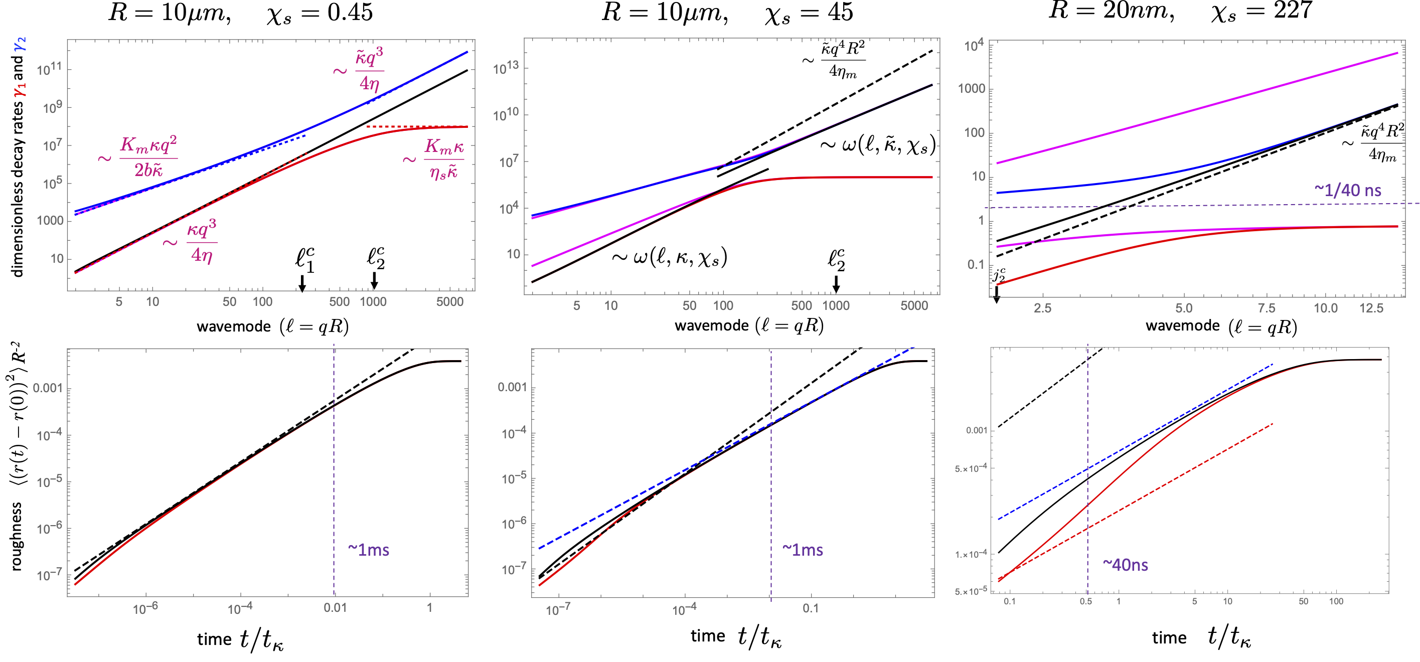
where is a dimensionless membrane viscosity parameter, the ratio of the Saffman-Delbrück length () to the vesicle radius, and is the reduced membrane tension. Setting reduces Eq. [1] to the result for a non-viscous area-incompressible membrane Milner-Safran:1987 ; SJW:1984 (an area-compressible membrane has been considered in Komura-Seki:1993 ). For , Eq. [1] shows that a new regime emerges in the relaxation spectrum that is membrane viscosity dominated, for , followed by the classical bending dominated regime, for , where . Even a moderate monolayer viscosity, , would affect the long wavelength undulations that are comparable with the radius of curvature of the equilibrium shape and thus could be observable in the thermally-excited shape fluctuations of giant vesicles, m.
In this work, we provide the first experimental evidence of the monolayer viscosity signature in the shape fluctuations of a giant vesicle. Furthermore, we shed light on the relative importance of the two dissipative mechanisms in the membrane – bilayer slip and monolayer viscosity – by generalizing the Seifert-Langer model of planar membrane undulations Seifert-Langer:1993 to a vesicle. The unified theory (details provided in the Appendix supports the flickering spectroscopy analysis of giant vesicles, while also providing a framework to analyze the dynamics of thermal undulations in broader time regime and wavelength spectrum relevant to the highly curved submicron cellular structures and experimental methods using liposomes such as NSE.

Flickering spectroscopy of giant vesicles: For the fluctuation analysis, a time series of the equatorial contour of a giant quasi-spherical vesicle (m) is recorded using phase-contrast microscopy and high speed camera (details about the methods and materials are given in the Appendix). The quasi-circular contour is decomposed in Fourier modes, . The corresponding autocorrelation function (ACF) picks up all the terms of the expansion of in spherical harmonics leading to an ACF in the form
| (2) |
where is the time-averaged mean squared amplitude of the spherical harmonic modes. It follows that at long times, , the ACF relaxes exponentially , while for short times, , all modes contribute and the ACF decay is non-exponential.
For the experiments, giant unilamellar vesicles (GUVs) were electroformed from pure dioleoylphosphatidylcholine (DOPC), stearoyloleoylphosphatdylcholine (SOPC), and mixtures of dipalmitoylphosphatidylcholine (DPPC) and cholesterol (Chol). DOPC and SOPC viscosities are 4.1 nPa.s.m and 9.75.8 nPa.s.m, respectively Faizi:2022 , corresponding to dimensionless surface viscosities for a typical 10 m GUV, too small to have a detectable effect on vesicle shape fluctuations. To achieve a lipid bilayer with high viscosity, we choose DPPC:Chol mixtures because they are in the liquid-ordered phase and thus expected to be very viscous WANG20162846 ; Faizi:2022 . Membrane viscosity measured with the electrodeformation method Faizi:2022 yielded 57.612.6 nPa.s.m for DPPC:Chol (1:1), 83.614.3 nPa.s.m for DPPC:Chol (6:4), and 1450928 nPa.s.m for DPPC:Chol (7:3), spanning a range of dimensionless viscosities, . To ensure good statistics for the ACF, images of the fluctuating contour are acquired at 50-500 fps for 5-10 mins.
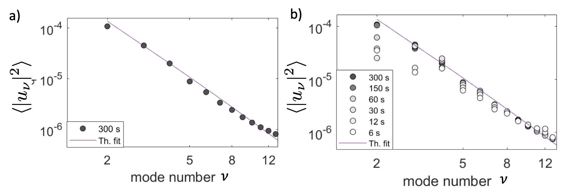
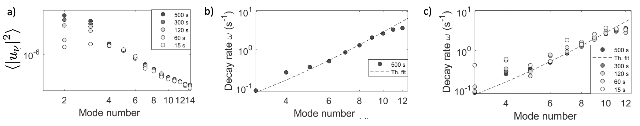
The ACF depends on three bilayer material parameters: bending rigidity, tension, and membrane viscosity. Flickering spectroscopy allows to obtain the values for the bilayer elastic properties, bending rigidity and tension, from the time-averaged amplitude of the shape fluctuations (Eq. [2] at ). This leaves the membrane viscosity as the only unknown in the relaxation rate obtained from the long-time single-exponential decay of the ACF. The power spectrum shown in Figure 1b follows bending dominated scaling, . Rescaling the spectrum by the bending rigidity collapses the data, see Figure 1c, and confirms that the static spectrum is controlled solely by bending rigidity. Figure 1d-e show the typical ACFs, calculated by standard time-averaging and normalized to 1 at , for two different bilayer compositions, SOPC and DPPC:Chol (1:1) (the ACFs for modes 3-8 can be found in the Appendix, Figure 7 and 8). The DOPC and SOPC bilayers follow the relaxation of a bilayer with negligible viscosity, . However, the ACF of DPPC:Chol bilayers decay much slower. With known bending rigidity and tension, one parameter fit of the single exponential yields values for the membrane viscosity similar to those obtained from the electrodeformation method (see Table 1 in the Appendix). The decay rates increase with the mode number according to Eq. [1] with , see Figure 1f.
Unified Theory: The analysis used to interpret our flickering spectroscopy experiments assumes that the bending fluctuations occur at relaxed lipid densities of the the monolayers. To confirm the validity of this assumption, we generalize the Seifert-Langer model, originally developed for a planar bilayer membrane Seifert-Langer:1993 , to describe the fluctuation dynamics of a quasi-spherical vesicle that encompasses both inter-monolayer slippage that relaxes lateral density fluctuations, and membrane shear. The theory provides a unified description of the dynamics of a quasi-spherical bilayer at all length- and time-scales.
The density difference between the monolayers is expanded in spherical harmonics, .The average density relaxes on a much faster scale compared to the shape and density difference modes Miao:2002 . In terms of spherical harmonic modes, the Hamiltonian Seifert-Langer:1993 becomes
| (3) |
where
| (4) |
The evolution equations for the shape and density modes are Watson:2011
| (5) |
where and
where is the dimensionless bilayer slip coefficient, and are the dimensionless membrane shear and dilatational viscosities. The characteristic time for bending relaxation is .
The Seifert-Langer’s result for a tensionless planar membrane is recovered assuming , , and setting and , where is the 2D wavenumber, . The solution to Eq. [5] yields for the time correlations of the shape modes
with relaxation rates
| (6) |
and amplitudes
| (7) |
For a planar tensionless membrane, Watson:2011 . For a quasi-spherical vesicle, given by Eq. [1].
Flickering spectroscopy of GUVs typically resolves only modes up to and the temporal resolution is in the order of miliseconds. In this low wave-number regime, the fast mode, , relaxes quickly while the bending mode relaxation rate is smaller by orders of magnitude, see Figure 10 in the Appendix. As a result, vesicle shape fluctuations are well described by the simplified model of a structurless, viscous bilayer with spherical mode decay rate given by Eq. [1]; the time-correlation functions computed from the full and simplified models are indistinguishable. The long-wavelength shape undulations are sensitive to membrane viscosity, if their wavelength are comparable to the Saffman-Delbrück’s length, , as in lipid bilayer membranes in the liquid-ordered state, studied here, or polymersomes Faizi:2020 .
It is instructive to consider the asymptotic behavior of the mode amplitude mean-squared displacement in the flickering spectroscopy experiment, (see Appendix for the complete expressions): (i) for , and (ii) for , where . The “late-short-time” regime suggests an effective stretched exponential relaxation of the ACF from its static value, . The anomalous diffusion – or stretching – exponent of , is identical to the one governing semi-flexible polymers obeying the worm-like chain model. This results from a combination of two effects: (i) the effective dimensionality of the phase space (of the Helfrich Hamiltonian) at the equator being reduced from two to one, as for semi-flexible polymers Farge:1993 ; Granek:1997 , and (ii) suppressed long range (solvent mediated) hydrodynamic interaction due to dominant dissipation by membrane flow.
Scattering techniques such as neutron spin echo NSE Nagao:2017 , dynamic light scattering (DLS) Freyssingeas:1997 , and X-ray photon correlation spectroscopy (XPCS) Falus:2005 and some flickering experiments Betz:2012 ; Helfer:2001a measure the single-point mean-squared displacement (MSD). The transverse (i.e. radial) membrane segment MSD at long times, for which the Seifert-Langer slippage mode is completely relaxed and undulations decay is controlled by the equilibrium bending modulus , in the time range exhibits subdiffusive behavior with exponent . The dimensionless MSD obeys
| (8) |
Instead, is the well known prediction for non-viscous membranes Zilman-Granek:1996 ; Zilman-Granek:2002 (see the Appendix for details). It follows that the scattering from vesicles in this time range and large scattering wavenumbers, , would still exhibit a stretched exponential DSF, , but with a modified stretching exponent and relaxation rate , changing from and to and
| (9) |
Note that this prediction for the DSF does not include intricate finite-size effects associated with the scattering from a spherical shell. In addition, since is dependent, polydispersity is expected to affect the decay profile.

However, for the short times and short wavelengths detected by NSE, our unified theory shows that it is not a good approximation to simply replace in the above predictions by , as in the case of a planar bilayer Watson:2011 . Figure 2 demonstrates that the MSD is not governed by a single power-law. Indeed, Ref. Watson:2011 did speculate that accounting for the spherical bilayer geometry may invalidate the Zilman-Granek power-law, , to interpret NSE data of liposome suspensions.
Conclusions and open questions: In summary, we show that membrane viscosity plays a prominent role in the fluctuation dynamics of quasi-spherical vesicles if the Saffman-Delbrück length is comparable or larger than the vesicle radius , that is . This can occur if either the membrane viscosity is large, , as in diblock-copolymer bilayers or lipid bilayers in the liquid-ordered phase such as DPPC:Chol, or vesicle size is small, , as in submicron lipid liposomes. The MSD and decay rates for the latter case are shown in Figure 2. The results suggests that the Seifert-Langer model is a poor approximation of the fluctuation dynamics in this case. First, the membrane viscosity significantly reduces the decay rate of the slow mode, see Figure 2a. Second, the MSD is also dampened and may not exhibit a clean power on the typical time scales of a scattering experiment. Accordingly, force-fitting the MSD with the Zilman-Granek power-law approximation would result in overestimation of the unrelaxed bending rigidity , see Figure 3. Similar error would emerge if flickering spectroscopy dynamics is interpreted with the equilibrium bending modulus alone.
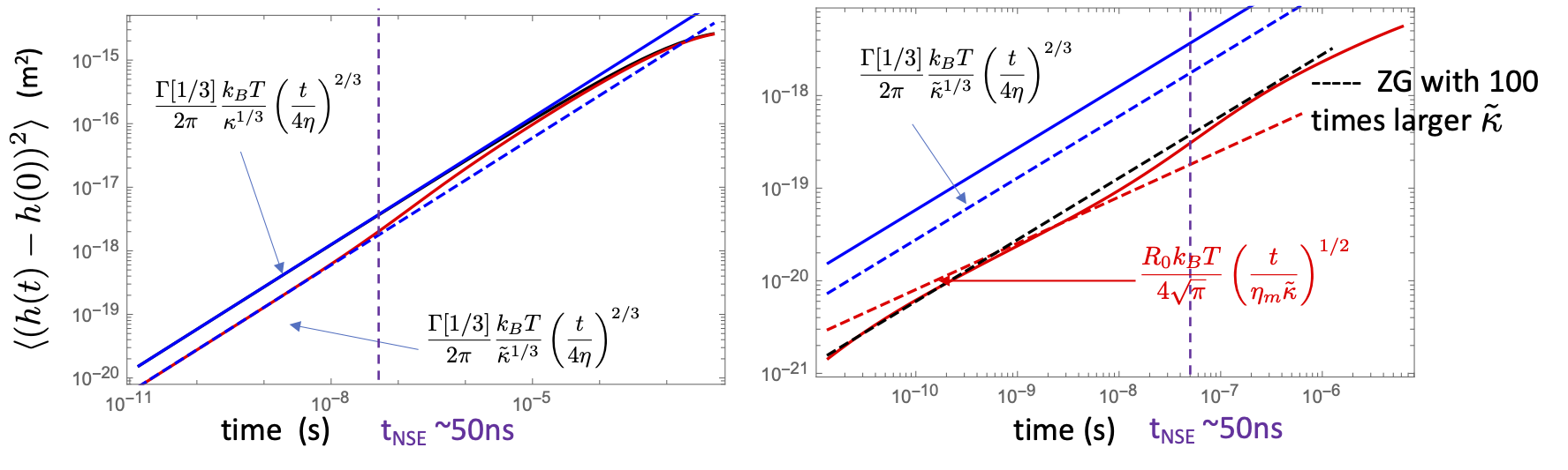
The failure of the Seifert-Langer model to describe liposome fluctuations suggests a reexamination of the interpretation of data obtained by dynamic scattering methods. Figure 2 suggests that in NSE experiment of DPPC:Chol system of the same membrane compositions as studied here and of vesicles of radii nm – leading to 100 times or more larger values than those obtained here (for m) – the whole time range observed in NSE is affected by membrane viscosity and the MSD does not obey a simple power-law. A similar conclusion can be reached if we adopt values of membrane viscosity of other systems Faizi:2022 , even DOPC and DOPC:Chol systems Chakraborty21896 for which can become for a 50nm liposome, see Figure 3 for the MSD is such system.
We hope our findings will stimulate further studies into the effect of surface viscosity on the dynamics of membranes and other complex interfaces.
P.M.V and H.A.F acknowledge financial support by NIGMS award 1R01GM140461. This research was also supported in part by the National Science Foundation under Grant NSF PHY-1748958.
Appendix A Experimental methods and additional data
A.1 Vesicle preparation
The classical electroformation method angelova.1987 is used to prepare giant unilamellar vesicles (GUVs) from stearoyloleoylphosphatdylcholine (SOPC), dioleoylphosphatidylcholine (DOPC), dipalmitoylphosphatidylcholine (DPPC) and cholesterol (Chol). The lipids are purchased from Avanti Polar Lipids (Alabaster, AL). The stock solutions are diluted in chloroform to obtain a final concentration of 4 mM. Initially, 7-8 l of lipid solution is spread on the conductive side of two Indium tin oxide (ITO, Delta Technologies) glass slides with a 10 l gas tight syringes (Hamilton, USA). The slides are placed inside vacuum to evaporate any leftover solvents for at least 3 hours. Afterwards, a 2 mm Teflon spacer is sandwiched between the two glass slides and the chamber is filled with 100 mM sucrose solution. The conductive side of the slides are connected to AC signal generator Agilent 33220A (Agilent, Germany) at a voltage of 1.8 and 10 Hz. The connected chamber is placed inside an oven at C for 2 hours. This procedure results in 10-50 m sized GUVs. The vesicle suspension is aspirated from the chamber and diluted in 110 mM glucose. The vesicles are visualized under the microscope.
A.2 Microscopy and video recording
The equatorial fluctuations of a GUV is visualized in phase contrast mode with Axio Observer A1 microscope (Zeiss, Germany). The microscope objective used is Plan-Apochromat 100x/1.4 Oil Ph3 M27 (FWD=0.17mm), with Immersol 518 F oil. Focal depth (FD) or FWHM (full width half maximum) of phase contrast imaging for our setting is determined using the standard formula FD . For a wavelength of transmission light 550 nm, the calculated FD is 281 nm.
A.3 Electrodeformation: membrane viscosity
We implement the transient electrodeformation of GUVs to measure membrane viscosity Faizi:2022 . To summarize, the harvested GUVs are diluted in 510 mM Glucose solution without any salt in an Eppendorf electrofusion chamber (Eppendorf, Germany) containing two Pt electrodes spaced 500 m apart. The method involves measuring the initial deformation rate of a vesicle as AC electric field is applied at a particular frequency. High speed imaging of the increase of the vesicle aspect ratio, , is done at 1-2 kfps. The linear slope of the aspect ratio as a function of time depends on membrane viscosity as
| (10) |
where is the dimensionless membrane viscosity , is the viscosity the solution inside and outside the vesicle, is the electric field strength amplitude and is forcing field function detailed in Faizi et al. Faizi:2022 . The apparent viscosities are measured at different frequencies in the range 0.1-1 kHz. The zero-frequency viscosity is obtained by extrapolating a linear fit of the viscosity vs frequency data. Electric field of 8 kV/m produces a good range of data in the linear initial slope. The viscosity compares well with the data obtained from flickering spectroscopy, see Table 1.
A.4 Flickering spectroscopy: bending rigidity
Membrane bending rigidity is determined from the equilibrium spectrum of the vesicle shape fluctuations. The flickering spectroscopy method takes advantage of non-invasive data collection and well developed statistical analysis. The details of the method can be found in Refs. Gracia:2010 ; Faizi:2019 ; Faizi:2020 . In essence, a time series of the fluctuating equatorial cross-section of a vesicle is recorded. The nearly-circular contour is represented in Fourier modes, . The mean squared amplitude of the fluctuating amplitudes depends on the membrane elastic properties, bending rigidity and the tension , see Eq. [20]. The contour dynamics is recorded with high-speed camera. The integration time effect of the camera is minimized by acquiring images at a low shutter speed of 200 s. Images are acquired with SA1.1 high speed camera (Photron) at 50-500 fps for 5-10 mins for a total of 0.1-0.5 million images. Only vesicles with low tension value in the range N/m are chosen. This results in a small cross over mode given by above which the shape fluctuations are dominated by bending rigidity. The ellipsoidal mode ( =2) has been ignored from the analysis as it is weighted with most excess area which leads to fluctuations with an increased amplitude.
| Composition | Phase state | Bending Rigidity | Viscosity , ED | Viscosity , FA | |
|---|---|---|---|---|---|
| () | (nPa.s.m) | (nPa.s.m) | |||
| DOPC | 21.7 | 4.1 | NA | 0.4 | |
| SOPC | 25.7 | 9.7 | NA | 1.0 | |
| DPPC:Chol (1:1) | 124.4 | 57.6 | 89.726.3 | 5.8 | |
| DPPC:Chol (6:4) | 152.6 | 83.6 | 10647 | 8.4 | |
| DPPC:Chol (7:3) | 189.6 | 1450 | 1777682 | 145 | |
Figure 1a in the main text shows the equilibrium spectrum for bilayers with different compositions, SOPC, DOPC, DPPC Chol (1:1) and DPPC:Chol (7:3). Working with floppy vesicles (low tension) leads to only a bending dominated fluctuations following power law. DPPC:Chol systems exhibit higher bending rigidity () compared to SOPC bilayer (), likely due to thicker bilayers and ordered bilayer structure. DPPC exists as a gel state below its phase transition temperature C. The phase diagram of the DPPC Cholesterol system suggests a strong effect of the cholesterol on bilayer thickness, lipid order and diffusivity WANG20162846 . Incorporation of cholesterol in a DPPC bilayer breaks the rigid gel structure into the liquid-ordered phase. This leads to increased fluidity and membrane softening. The tension-free or low-tension state of the vesicles can be confirmed by normalizing the spectrum with the respective bending rigidity values. Figure 1c in the main text demonstrates a perfect collapse of the data confirming that the shape fluctuations depend only on bending rigidity. The collapse also confirms that the equilibrium fluctuations are independent of membrane’s viscosity or dissipative properties. Equilibrium techniques alone cannot demonstrate the effect of transport properties such as membrane viscosity. The measured bending rigidities are listed in Table 1.

Timescales: Resolving the dynamics with robust statistics depends on two time scales. The first is , the duration of the recording. The second is the time step-size in the temporal space, , which experimentally corresponds to the frames per second (fps) or acquisition speed of the movie. can be predicted from the slowest relaxation mode given by:
| (11) |
For robust statistics, a factor of ten accurately resolved the dynamics, . This was confirmed by achieving experimental convergence in the results.
Similarly, is determined by the highest experimentally resolved mode . Again we kept a factor of 10 to ensure enough statistics.
| (12) |
Figure 4 demonstrates the importance of collecting long enough data set in order to ensure good statistics for the equilibrium spectrum. Longer wavelength (smaller mode numbers) take long time to explore their temporal configuration. Figure 4a represents a typical SOPC fluctuation equilibrium spectrum taken over 300 seconds. If the same data is analyzed for shorter time, e.g., the first 150 seconds only, the same spectrum is recovered. This implies that experimental convergence has been achieved. However, trimming the data to the first 60 seconds, or shorter, results in artifacts starting from lower mode number spectrum and creeping to higher ones. The spectrum can be misinterpreted with a higher tension value. This illustrates the importance of Eq 11.
Similarly, for time correlations analysis of the same vesicle as in Fig. 4a, Figure 6 illustrates the importance of Eq. [11] and Eq. [12]. Analyzing the data for a trimmed data set results in artifacts in data interpretation at lower modes as shown in Figure 6b with a higher membrane tension. Similarly, analyzing the data set at lower fps or lower temporal resolution affects higher mode number data as shown in 6b. It can been seen that the artifacts are exacerbated for membranes with higher membrane viscosity like DPPC:Chol (1:1) with as shown by time correlation analysis in Figure 6.
A.5 Autocorrelation functions for modes 3-8 of SOPC and DPPC: Chol (1:1) bilayers


Appendix B Flickering spectroscopy: theoretical basis
Fluctuation spectroscopy analyzes the thermally-driven membrane undulations of giant unilamellar vesicles. In essence, a time series of vesicle contours in the focal plane (the equator of the quasi-spherical vesicle) is recorded. The quasi-circular contour is decomposed in Fourier modes,
| (13) |
where is the radius of an equivalent sphere with the volume of the GUV and is the mode number. In practice, is the maximum number of experimentally resolved modes. The Fourier coefficient for the -th mode is then given by
| (14) |
as all the other terms integrate to zero. In the above equation, we have inserted the definition of the spherical harmonic,
| (15) |
where are the associated Legendre polynomials.
B.1 Time-averaged mean squared amplitude of the membrane thermal undulations
The mean squared amplitude of is given by
| (16) |
In terms of the spherical harmonic mode amplitudes, the Helfrich Hamiltonian is
| (17) |
showing that indeed all modes are decoupled from each other. Equipartition theorem then dictates
| (18) |
Eq. [16] therefore simplifies to
| (19) |
or, explicitly,
| (20) |
B.2 Autocorrelation function of the equatorial plane Fourier modes: Asymptotic behavior for tensionless membranes
The auto-correlation function (ACF) of the equatorial plane Fourier modes is given by
| (21) |
where for we shall use here Eq. [1] (see explanation in the main text for the its validity for GUVs dynamics).
Let us consider the short and long time asymptotic behaviors of the ACF. At times much longer than the longest relaxation time of Eq. (21), , the ACF reduces to a single exponential relaxation
| (22) |
For short times, , we consider the behavior of the series terms, and approximately evaluate the series by transforming it to an integral. For , we have from Eq. (15) , and the associate Legendre polynomials behave as Gradshteyn-Ryzhik:1980
| (23) |
such that for and we obtain
or
| (24) |
These lead to
| (25) |
and the sum in Eq. (21) becomes
| (26) |
Transforming the sum to an integral leads to
| (27) |
(The prefactor of results from the fact that only half of series terms in Eq. (25) are non-zero.) Finally, we can use in Eq. (27) the large limits of and , with the tension being neglected, and assuming
| (28) |
and
| (29) |
We now wish replace the lower bound of the integral in Eq. (27) by 0, however, since the integral diverges as the lower bound approaches , we make use of the identity
| (30) |
Note that the second integral is essentially half of the MSD of the -th Fourier mode, .
“Scaling” the integral in Eq. (30) (i.e. changing the variable of integration), we obtain the two “short times” regimes, commencing by a solvent viscosity dominated regime which is followed by a membrane viscosity dominated regime. The crossover time separating the two regimes is
| (31) |
For the earlier regime of “short times”, , where is the shortest relaxation time, , we get the solvent dissipation regime
| (32) |
For the late regime of “short times”, , we obtain the membrane dissipation regime
| (33) |
presenting a non-exponential relaxation of the ACF from its static value; effectively (to first order) it is a stretched exponential decay with stretching exponent . This anomalous diffusion (or ACF stretching) exponent, describing the evolution of the MSD of the equatorial Fourier modes, is identical to the one governing rod-like semi-flexible polymers obeying the worm-like chain model. This is interesting and can be rationalized by the following argument. First, by looking at the Fourier-modes of deformations at the equator, the effective dimensionality of the Helfrich bending energy phase space is reduced from two to one, as for semi-flexible polymers Farge:1993 ; Granek:1997 . Second, the membrane viscosity dominated dissipation suppresses the long-range solvent hydrodynamic interaction, which again leads to a similar effect as in semi-flexible polymers; in the latter, the solvent mediated hydrodynamic interaction reduces to merely a marginal, logarithmic, effect.
Appendix C Transverse MSD of a membrane segment for tensionless membranes: Implications for the dynamic structure factor
In Refs. Zilman-Granek:1996 ; Zilman-Granek:2002 ; Granek:1997 it was shown that pure bending undulations that are dissipated by solvent viscosity, produce a transverse subdiffusion of a membrane segment with an MSD that grows in time as . At large scattering wavenumbers that are sensitive to single membrane dynamics, this subdiffusion leads to stretched exponential relaxation, , of the dynamic structure factor (DSF) of membrane phases. We now discuss how these dynamics are modified when membrane viscosity is included.
C.1 Mean Squared Displacement (MSD)
The dimensionless membrane segment MSD at an arbitrary 3D angle , , is given by (the MSD with physical dimensions is given by )
| (34) |
Using Jackson:1998
| (35) |
(which may be verified by using Eq. 15), we arrive at
| (36) |
which is independent of the angle as expected.
We now assume again vanishing tension. For times , where and are the shortest and longest relaxation times (respectively), and , we may use the asymptotic of both the spectrum of modes () and of the relaxation frequency , as given by Eq. (29), and use . We may also replace the sum in Eq. (36 ) by an integral, with the lower and upper integration limits replaced by and , respectively. This leads to
| (37) |
Note that the latter expression is identical to the one derived for flat membranes using standard 2D Fourier modes Zilman-Granek:1996 ; Zilman-Granek:2002 , yet for flat membranes by definition.
Eq. (37) leads to two “short time” regimes, a solvent viscosity dominated regime that is followed by a membrane viscosity dominated regime. We find
| (38) |
Importantly, , which allows for a very wide membrane viscosity dominated regime if .
C.2 Dynamic Structure Factor
Since for unilamellar vesicles within the radius range nm, on which most NSE experiments are performed, additional finite size and spherical geometry effects might be important, we limit the discussion below to large or giant unilamellar vesicles, where is in the range of a few hundreds of nm to m.
Following Refs.Zilman-Granek:1996 ; Zilman-Granek:2002 , the membrane segment MSD controls the DSF relaxation. Excluding the effect of center-of-mass diffusion, which comes in the form of a multiplying factor, the main DSF relaxation is well captured by . It follows that in principle the Zilman-Granek (ZG), bulk viscosity dominated, decay, should be followed by a new, membrane viscosity dominated, decay, as follows
| (39) |
where is the ZG relaxation rate (possibly with replacing Watson:2011 ), and the new, membrane viscosity controlled, relaxation rate, is given by
| (40) |
Given that can be extremely short for viscous membrane vesicles with nm, it is quite possible that the whole NSE time range is controlled by membrane viscosity. As mentioned above, further efforts are required to include other important effects associated with small vesicle sizes.
Appendix D Fluctuation dynamics of a quasi-spherical vesicle: outline of theory
D.1 Surface viscous stresses
The membrane is treated as being composed of two coupled monolayers, see Figure 9.
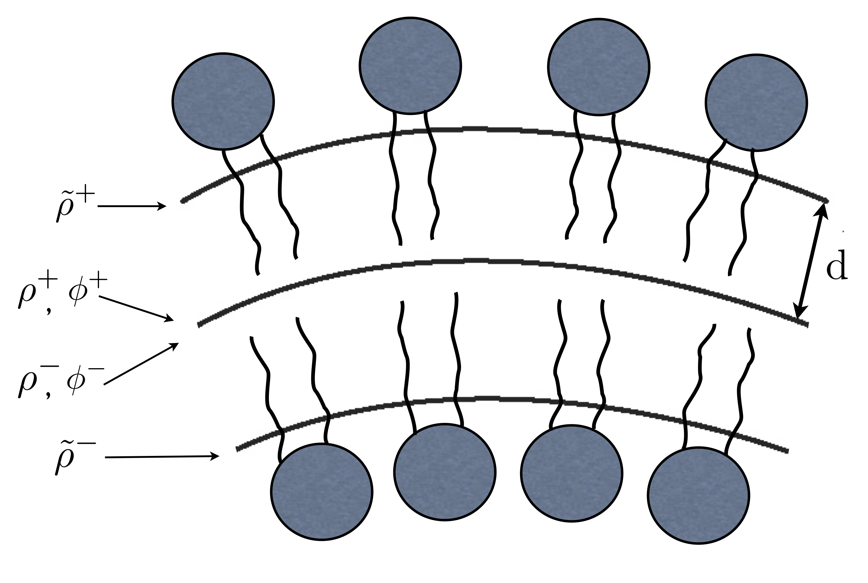
Vesicle shape and monolayer densities are expanded in spherical harmonics
where the sum denotes . and . The average density relaxes on a much faster scale compared to the shape and density difference modes, hence we solve the problem with .
We extended the model of Ref. Miao:2002 to include membrane viscosity in the dynamics of the shape fluctuation of a quasi-spherical vesicle. We use the same notation and the reader is referred to the paper for details of the solution. We include in the stress balance the tractions due to viscous dissipation in the monolayers described by the Boussinesq-Scriven law Edwards-Brenner-Wasan:1991 , which for a quasi-spherical vesicle are Vlahovska:2016a
| (41) |
where the amplitudes are
| (42) |
The tractions are added to the boundary conditions Eq.(49)-(51) ((55), (59) and (60) in terms of spherical harmonics amplitudes) in Ref. Miao:2002 . Following the solution procedure in Ref. Miao:2002 leads to the evolution equations Eq. 5 in the main text.
D.2 Limiting behaviors of the theoretical decay rates
D.2.1 Planar bilayer:
The general expressions for the decay rates and are complicated, but there are approximations in some limits relative to the crossover wavenumbers Watson:2011 :
At low wavenumbers, ,
In the range of wavenumbers
At high wavenumbers, ,
D.2.2 Quasi-spherical vesicle:
Figure 10 illustrates the relaxation dynamics for and in the case of a typical unsaturated phospholipid.
References
- [1] R. Lipowsky and E. Sackmann. Structure and dynamics of membranes. Elsevier, Amsterdam, 1995.
- [2] Special issue in honour of Wolfgang Helfrich, Advances in Colloid and Interface Science Vol 208. 2014.
- [3] C Monzel and K Sengupta. Measuring shape fluctuations in biological membranes. Journal of Physics D: Applied Physics, 49(24):243002, may 2016.
- [4] Patricia Bassereau, Rui Jin, Tobias Baumgart, Markus Deserno, Rumiana Dimova, Vadim A Frolov, Pavel V Bashkirov, Helmut GrubmÃŒller, Reinhard Jahn, H Jelger Risselada, Ludger Johannes, Michael M Kozlov, Reinhard Lipowsky, Thomas J Pucadyil, Wade F Zeno, Jeanne C Stachowiak, Dimitrios Stamou, Artù Breuer, Line Lauritsen, Camille Simon, Cécile Sykes, Gregory A Voth, and Thomas R Weikl. The 2018 biomembrane curvature and remodeling roadmap. Journal of Physics D: Applied Physics, 51(34):343001, jul 2018.
- [5] Hervé Turlier and Timo Betz. Unveiling the active nature of living-membrane fluctuations and mechanics. Annual Review of Condensed Matter Physics, 10(1):213–232, 2019.
- [6] E. Evans and A. Yeung. Unexpected dynamics in shape fluctuations of bilayer vesicles. J. Phys. II, 5:1501–1523, 1995.
- [7] U. Seifert and S.A. Langer. Viscous modes of fluid bilayer membranes. Europhys. Lett., 23:71–76, 1993.
- [8] U. Seifert. Fluid membranes in hydrodynamic flow fields: Formalism and an application to fluctuating quasispherical vesicles. Eur. Phys. J. B, 8:405–415, 1999.
- [9] Brochard, F. and Lennon, J.F. Frequency spectrum of the flicker phenomenon in erythrocytes. J. Phys. France, 36(11):1035–1047, 1975.
- [10] A.J. Levine and F.C. MacKintosh. Dynamics of viscoelastic membranes. Phys. Rev. E, 66:061606, 2002.
- [11] M. C. Watson and F. L. H. Brown. Interpreting membrane scattering experiments at the mesoscale: The contribution of dissipation within the bilayer. J. Chem. Phys., 98:L9–L11, 2010.
- [12] M. C. Watson, Y. Peng, Y. Zheng, and F. L. H. Brown. The intermediate scattering function for lipid bilayer membranes: From nanometers to microns. J. Chem. Phys., 135:194701, 2011.
- [13] L. Miao, M. A. Lomholt, and J. Kleis. Dynamics of shape fluctuations of quasi-spherical vesicles revisited. Eur. Phys. J. E., 9:143–160, 2002.
- [14] Michihiro Nagao, Elizabeth G. Kelley, Rana Ashkar, Robert Bradbury, and Paul D. Butler. Probing elastic and viscous properties of phospholipid bilayers using neutron spin echo spectroscopy. The Journal of Physical Chemistry Letters, 8(19):4679–4684, 2017. PMID: 28892394.
- [15] P. Olla. The behavior of closed inextensible membranes in linear and quadratic shear flows. Physica A, 278:87–106, 2000.
- [16] S. B. Rochal, V. L. Lorman, and G. Mennessier. Viscoelastic dynamics of spherical composite vesicles. Phys. Rev. E, 71:021905, 2005.
- [17] Mark L. Henle and Alex J. Levine. Hydrodynamics in curved membranes: The effect of geometry on particulate mobility. Phys. Rev. E, 81:011905, Jan 2010.
- [18] Francis G. Woodhouse and Raymond E. Goldstein. Shear-driven circulation patterns in lipid membrane vesicles. Journal of Fluid Mechanics, 705:165–175, 2012.
- [19] Mohammad Rahimi, Antonio DeSimone, and Marino Arroyo. Curved fluid membranes behave laterally as effective viscoelastic media. Soft Matter, 9:11033–11045, 2013.
- [20] Mohammad Rahimi. Shape dynamics and lipid hydrodynamics of bilayer membranes: modeling, simulation and experiments. PhD thesis, Universitat Politecnica de Catalunya, 2013.
- [21] Jon Karl Sigurdsson and Paul J. Atzberger. Hydrodynamic coupling of particle inclusions embedded in curved lipid bilayer membranes. Soft Matter, 12:6685–6707, 2016.
- [22] Petia M. Vlahovska. Electrohydrodynamics of drops and vesicles. Annu. Rev. Fluid Mech., 51: 305–330, 2019.
- [23] Amaresh Sahu, Alec Glisman, Joël Tchoufag, and Kranthi K. Mandadapu. Geometry and dynamics of lipid membranes: The scriven-love number. Phys. Rev. E, 101:052401, May 2020.
- [24] S. T. Milner and S. A. Safran. Dynamical fluctuations of droplet microemulsions and vesicles. Phys. Rev. A, 36:4371–4379, 1987.
- [25] M. B. Schneider, J. T. Jenkins, and W. W. Webb. Thermal fluctuations of large quasi-spherical bimolecular phospholipid-vesicles. Journal de Physique, 45(9):1457–1472, 1984.
- [26] S. Komura and K. Seki. Dynamical fluctuations of spherically closed fluid membranes. Physica A, 192:27–46, 1993.
- [27] Hammad A. Faizi, Rumiana Dimova, and Petia M. Vlahovska. A vesicle microrheometer for high-throughput viscosity measurements of lipid and polymer membranes. BIOPHYSICAL JOURNAL, 121(6):910–918, MAR 15 2022.
- [28] Yin Wang, Paraskevi Gkeka, Julian E. Fuchs, Klaus R. Liedl, and Zoe Cournia. Dppc-cholesterol phase diagram using coarse-grained molecular dynamics simulations. Biochimica et Biophysica Acta (BBA) - Biomembranes, 1858(11):2846–2857, 2016.
- [29] Hammad A. Faizi, Cody J. Reeves, Vasil N. Georgiev, Petia M. Vlahovska, and Rumiana Dimova. Fluctuation spectroscopy of giant unilamellar vesicles using confocal and phase contrast microscopy. Soft Matter, 16:8996–9001, 2020.
- [30] Emmanuel Farge and Anthony C. Maggs. Dynamic scattering from semiflexible polymers. Macromolecules, 26(19):5041–5044, 1993.
- [31] R Granek. From semi-flexible polymers to membranes: Anomalous diffusion and reptation. J. Physique II, 7(12):1761–1788, DEC 1997.
- [32] E Freyssingeas and D Roux. Quasi-elastic light scattering study of highly swollen lamellar and ‘’sponge” phases. JOURNAL DE PHYSIQUE II, 7(6):913–929, JUN 1997.
- [33] P. Falus, M. A. Borthwick, and S. G. J. Mochrie. Fluctuation dynamics of block copolymer vesicles. Phys. Rev. Lett., 94:016105, Jan 2005.
- [34] T. Betz and C. Sykes. Time resolved membrane fluctuation spectroscopy. Soft Matter, 8:5317–5326, 2012.
- [35] E. Helfer, S. Harlepp, L. Bourdieu, J. Robert, F. C. MacKintosh, and D. Chatenay. Microrheology of biopolymer-membrane complexes. Phys. Rev. Lett., 85:457–460, 2000.
- [36] A. G. Zilman and R. Granek. Undulations and dynamic structure factor of membranes. Phys. Rev. Lett., 77:4788–4791, Dec 1996.
- [37] Anton G. Zilman and Rony Granek. Membrane dynamics and structure factor. Chemical Physics, 284(1):195–204, 2002. Strange Kinetics.
- [38] Saptarshi Chakraborty, Milka Doktorova, Trivikram R. Molugu, Frederick A. Heberle, Haden L. Scott, Boris Dzikovski, Michihiro Nagao, Laura-Roxana Stingaciu, Robert F. Standaert, Francisco N. Barrera, John Katsaras, George Khelashvili, Michael F. Brown, and Rana Ashkar. How cholesterol stiffens unsaturated lipid membranes. Proceedings of the National Academy of Sciences, 117(36):21896–21905, 2020.
- [39] Miglena I. Angelova and Dimiter S. Dimitrov. Liposome electroformation. Faraday Discuss. Chem. Soc., 81:303–311, 1986.
- [40] R. S. Gracia, N. Bezlyepkina, R. L. Knorr, R. L. Lipowsky, and R. Dimova. Effect of cholesterol on the rigidity of saturated and unsaturated membranes: fluctuation and electrodeformation analysis of giant vesicles. Soft Matter, 6:1472 – 1482, 2010.
- [41] Hammad A. Faizi, Shelli L. Frey, Jan Steinkuehler, Rumiana Dimova, and Petia M. Vlahovska. Bending rigidity of charged lipid bilayer membranes. Soft Matter, 15(29):6006–6013, AUG 7 2019.
- [42] I. S. Gradshteyn and I. M. Ryzhik. Table of Integrals, Series, and Products: pp. 1003, Sec. 8.72, item 3, ref. (MO 92). Academic Press, New York, 1980.
- [43] J. D. Jackson. Classical electrodynamics. Wiley, New York, 1998.
- [44] D. A. Edwards, H. Brenner, and D. T. Wasan. Interfacial Transport Processes and Rheology. Butterworth-Heinemann, Boston, 1991.
- [45] Petia M. Vlahovska. Dynamics of membrane bound particles: capsules and vesicles. In C. Duprat and H.A. Stone, editors, Low-Reynolds-Number Flows: Fluid-Structure Interactions. Royal Society of Chemistry Series RSC Soft Matter, 2016.