MIDLARXIV
\jmlrpages2
\jmlryear2022
\midlauthor\NameHaleh Akrami\midljointauthortextContributed equally\nametag1 \Emailakrami@usc.edu
\NameWenhui Cui\midlotherjointauthor\nametag1 \Emailwenhuicu@usc.edu
\NameAnand A. Joshi\nametag1 \Emailajoshi@usc.edu
\NameRichard M. Leahy\nametag1 \Emailleahy@sipi.usc.edu
\addr1 University of Southern California, Los Angeles, CA, USA
Learning from imperfect training data using a robust loss function: application to brain image segmentation
Abstract
Segmentation is one of the most important tasks in MRI medical image analysis and is often the first and the most critical step in many clinical applications. In brain MRI analysis, head segmentation is commonly used for measuring and visualizing the brain’s anatomical structures and is also a necessary step for other applications such as current-source reconstruction in electroencephalography and magnetoencephalography (EEG/MEG). Here we propose a deep learning framework that can segment brain, skull, and extra-cranial tissue using only T1-weighted MRI as input. In addition, we describe a robust method for training the model in the presence of noisy labels.
keywords:
brain, segmentation, robustness, machine-learning1 Introduction
Image segmentation is an important task in medical image processing. Deep learning methods can be used for this purpose but in practice, especially in large datasets, training data inevitably contain mislabeled data, anomalies, or outliers. Training based on noisy labeled datasets can result in output quality loss because deep neural networks (DNNs) can overfit to noisy labels. This issue is particularly severe in the field of medical image analysis where the quality of annotation is strongly dependent on the skill, experience, and time expended by the annotators.
Here our goal is MRI head segmentation when only a T1-weighted image is available. Head segmentation (including gray and white matter, CSF, skull, scalp, and other extra-cranial tissue) is commonly used for measuring and visualizing the brain’s anatomical structures and is also a necessary step for other applications, for example in generating head-models for use in current-source reconstruction in electroencephalography and magnetoencephalography (EEG/MEG) [Huang and Parra(2015)].
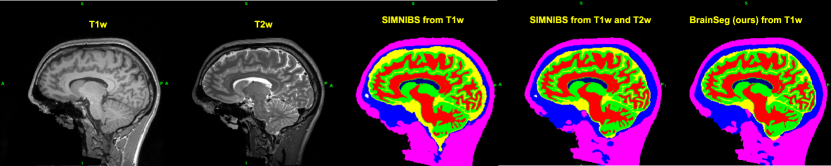
T1-weighted (T1-W) images are commonly available and employ the most cost-effective and fastest pulse sequences used to create accurate head segmentations. However, adding T2-weighted (T2-W) images can help better segmentation of the skull due to superior contrast, particularly between CSF and skull [Nielsen et al.(2018)Nielsen, Madsen, Puonti, Siebner, Bauer, Madsen, Saturnino, and Thielscher]. Our goal was to develop a fast head segmentation using only the T1-W modality. We investigated three scenarios: (i) First, we generated head segmentations (”software-generated labels”, (SGLs) using SimNIBS (\urlhttps://simnibs.github.io) where the input was T1-W and T2-W images. We then trained a CNN using only T1-W as input to predict these labels. (ii) We obtained SGLs using only T1-W data and trained a CNN using T1-W as input to predict these labels. (iii) We repeated (ii) using a robust loss function to compensate for the poor quality of training labels for some classes due to the absence of T2-W images.
Robust loss functions offer a statistically principled approach to the noisy label problem. By applying a power-law function, beta-cross entropy (BCE) loss can mitigate the effect of noise in the training data [Akrami et al.(2022)Akrami, Joshi, Li, Aydöre, and Leahy]. Minimizing BCE is equivalent to minimizing beta-divergence, which is the robust counterpart of KL-divergence. In contrast to this earlier work, which focused on anomaly detection, here we focus on image segmentation.
Our key contributions are as follows: (1) training a state-of-the-art neural network, TransUNet [Chen et al.(2021)Chen, Lu, Yu, Luo, Adeli, Wang, Lu, Yuille, and Zhou], for head segmentation. This method is very fast at inference time compared to traditional software-generated segmentations (less than 1 min. for the neural network approach vs. 5-10 hrs for traditional software); (2) Investigating using of a robust loss in the absence of good quality training data, such as lack of good quality segmentations for training due to the absence of T2-W modality data.
2 Method
We used the CamCan dataset (\urlhttps://www.cam-can.org ) that consists of T1-W and T2-W MRI images. We used SimNIBS to segment the head into 9 classes: 0=background (outside head), 1=WM, 2=GM, 3=CSF, 4= bone, 5= skin, 6= cavities, 7 = eyes, 8=ventricles. We split the data 60%/20%/20% for training/validation/test sets. We trained a TransUNet [Chen et al.(2021)Chen, Lu, Yu, Luo, Adeli, Wang, Lu, Yuille, and Zhou] using 2D slices as input and resized to 256*256 for 10 epochs. (i) In the first experiment, we generated SGLs using T1-W and T2-W images as input to SimNIBS to segment the head. We then used T1-W as input and T1-W+T2-W SGLs as the ground-truth labels for training the TransUNet (T1-W+T2-W TransUNet). (ii) We obtained T1-W SGLs by using T1-W images only as input to SimNIBS; then, we used T1-W as input and T1-W SGLs as noisy labels for training the TransUNet with a cross-entropy loss (T1-W TransUNet) (iii) finally we repeated experiment (ii) but with the robust BCE loss (T1-W TransUNet Robust) . The BCE loss is expressed as:
| (1) |
where is the input variable, is the response variable, and is a hyper-parameter; is the probability output of a deep neural network (DNN) classifier, and is the number of classes. BCE minimizes -divergence between the posterior and empirical distributions when the posterior is a categorical distribution [Akrami et al.(2022)Akrami, Joshi, Li, Aydöre, and Leahy]. Using L’Hôpital’s rule, it can be shown that BCE is equivalent to CE for where -divergence also converges to KL-divergence. -divergence is the robust counterpart of KL-divergence using a power-law function. We tuned parameter using the validation data. We warmed up the robust model for two epochs using the cross-entropy loss.
fig:lables
3 Results and discussion
To measure the performance of our method, we calculated the Dice score between each segmentation and T1-W+T2-W SGL labels (table 1). The result shows our T1-W+T2-W TransUNet model can be used to predict head segmentation when only a T1-W image is available during inference. The dice score of bone/skull for this model is improved compared to T1-W SGL segmentation. Further, as we expected we find an improvement in the dice score for the robust model trained with T1-W only SGL labels compared to the non-robust model for these labels. The dice score between T1-W+T2-W SGL and T1-W SGL represents the level of inaccuracy in each label. The degradation of dice coefficients for the eye labels when using the robust loss is probably due to the low occurrence of these voxels in the training data leading to their interpretation as outliers. A hybrid CE/BCE loss would address this issue.
| Model | background | WM | GM | CSF | bones | skin | cavities | eyes | ventricles |
|---|---|---|---|---|---|---|---|---|---|
| T1-W SGL | 0.99 | 0.99 | 0.98 | 0.82 | 0.84 | 0.94 | 0.73 | 0.75 | 0.99 |
| T1-W+T2-W TransUnet | 0.99 | 0.92 | 0.90 | 0.81 | 0.89 | 0.94 | 0.71 | 0.78 | 0.94 |
| T1-W TransUnet | 0.99 | 0.93 | 0.90 | 0.74 | 0.83 | 0.93 | 0.68 | 0.74 | 0.94 |
| T1-W TransUnet Robust | 0.99 | 0.90 | 0.87 | 0.78 | 0.85 | 0.94 | 0.72 | 0.68 | 0.94 |
References
- [Akrami et al.(2022)Akrami, Joshi, Li, Aydöre, and Leahy] Haleh Akrami, Anand A Joshi, Jian Li, Sergül Aydöre, and Richard M Leahy. A robust variational autoencoder using beta divergence. Knowledge-Based Systems, 238:107886, 2022.
- [Chen et al.(2021)Chen, Lu, Yu, Luo, Adeli, Wang, Lu, Yuille, and Zhou] Jieneng Chen, Yongyi Lu, Qihang Yu, Xiangde Luo, Ehsan Adeli, Yan Wang, Le Lu, Alan L Yuille, and Yuyin Zhou. Transunet: Transformers make strong encoders for medical image segmentation. arXiv preprint arXiv:2102.04306, 2021.
- [Huang and Parra(2015)] Yu Huang and Lucas C Parra. Fully automated whole-head segmentation with improved smoothness and continuity, with theory reviewed. PloS one, 10(5):e0125477, 2015.
- [Nielsen et al.(2018)Nielsen, Madsen, Puonti, Siebner, Bauer, Madsen, Saturnino, and Thielscher] Jesper D Nielsen, Kristoffer H Madsen, Oula Puonti, Hartwig R Siebner, Christian Bauer, Camilla Gøbel Madsen, Guilherme B Saturnino, and Axel Thielscher. Automatic skull segmentation from mr images for realistic volume conductor models of the head: Assessment of the state-of-the-art. Neuroimage, 174:587–598, 2018.