Free-electron-light interactions in nanophotonics
Abstract
When impinging on optical structures or passing in their vicinity, free electrons can spontaneously emit electromagnetic radiation, a phenomenon generally known as cathodoluminescence. Free-electron radiation comes in many guises: Cherenkov, transition, and Smith-Purcell radiation, but also electron scintillation, commonly referred to as incoherent cathodoluminescence. While those effects have been at the heart of many fundamental discoveries and technological developments in high-energy physics in the past century, their recent demonstration in photonic and nanophotonic systems has attracted a lot of attention. Those developments arose from predictions that exploit nanophotonics for novel radiation regimes, now becoming accessible thanks to advances in nanofabrication. In general, the proper design of nanophotonic structures can enable shaping, control, and enhancement of free-electron radiation, for any of the above-mentioned effects. Free-electron radiation in nanophotonics opens the way to promising applications, such as widely-tunable integrated light sources from x-ray to THz frequencies, miniaturized particle accelerators, and highly sensitive high-energy particle detectors. Here, we review the emerging field of free-electron radiation in nanophotonics. We first present a general, unified framework to describe free-electron light-matter interaction in arbitrary nanophotonic systems. We then show how this framework sheds light on the physical underpinnings of many methods in the field used to control and enhance free-electron radiation. Namely, the framework points to the central role played by the photonic eigenmodes in controlling the output properties of free-electron radiation (e.g., frequency, directionality, and polarization). We then review experimental techniques to characterize free-electron radiation in scanning and transmission electron microscopes, which have emerged as the central platforms for experimental realization of the phenomena described in this Review. We further discuss various experimental methods to control and extract spectral, angular, and polarization-resolved information on free-electron radiation. We conclude this Review by outlining novel directions for this field, including ultrafast and quantum effects in free-electron radiation, tunable short-wavelength emitters in the ultraviolet and soft x-ray regimes, and free-electron radiation from topological states in photonic crystals.
I Introduction
The interaction of free electrons with light and matter is a century-old field of research, that has had profound implications in electron microscopy, radiation sources, and high-energy particle detection. At the heart of this field lies a few fundamental discoveries, unveiling various conditions in which free electrons can convert part of their energy into photons. Crooke (1879); Cherenkov (1934); Ginzburg and Frank (1945); Smith and Purcell (1953); Ritchie (1957) On the other hand, over the past two decades, nanophotonics has emerged as a platform to control photonic modes at the nanoscale, by patterning materials at scales comparable to the photon wavelength. The recent merger of these two fields has spurred new applications and fundamental discoveries.Luo et al. (2003); Bashevoy et al. (2006); Van Wijngaarden et al. (2006); Bashevoy et al. (2007); Vesseur et al. (2007); Hofmann et al. (2007); Adamo et al. (2009, 2010); Barnard et al. (2011); Adamo et al. (2012); Kaminer et al. (2017a); Liu et al. (2017a); Massuda et al. (2017a); Yang et al. (2018a); Sapra et al. (2020); Shentcis et al. (2020); Kfir et al. (2020); Wang et al. (2020a); Nussupbekov et al. (2021); Roques-Carmes et al. (2022a); García De Abajo (2010); Polman, Kociak, and García de Abajo (2019); Talebi (2017); Coenen, Hoedt, and Polman (2016); Christopher et al. (2020) This Review is dedicated to (1) providing a general understanding of free-electron-light interactions mediated by nanophotonic structures; (2) highlighting recent theoretical and experimental developments in the field; (3) outlining future prospects for fundamental research and novel applications.
How can charged particles emit light? This fundamental question has driven much development in theoretical and experimental physics in the twentieth century. Perhaps the original interest in this question can be traced back to early discoveries in radioactivity, where luminescence from liquids was used to detect the presence of radioactive substances. Marie Curie (1904); Ginzburg (1996) Later on, emission processes such as the Cherenkov effect were extensively used to track and detect particles. Akopov et al. (2002); Adams et al. (2001); Nakamura (2003); Kleinknecht (1982) Therefore, the understanding of radiation processes from charged particles, such as free electrons, has evolved in tandem with some of the most profound discoveries of modern physics, such as quantum electrodynamics and particle detection within and beyond the standard model. Nakamura (2003); Papanestis (2014) Concepts from free-electron radiation have also permeated throughout many fields of physics, from nonlinear optics to gravitational physics. Carusotto and Rousseaux (2013) Other forms of spontaneous emission induced by free-electron (de)acceleration – which are not covered in this Review – have received a lot of interest, especially in the context of free-electron lasers. Pellegrini, Marinelli, and Reiche (2016)
Much more recently, nanophotonics has become a paramount framework and technology, enabling, among other things, the design of novel light sources, detectors, and devices controlling the polarization, spectral, and angular distribution of light. Joannopoulos et al. (2011); Novotny and Hecht (2009) A hallmark of nanophotonics is the design of nanostructured materials (metasurfaces, Yu and Capasso (2014) photonic crystals, Joannopoulos et al. (2011); Yablonovitch (1994) resonators, Vahala (2003); Armani et al. (2003) etc.) to tailor the interaction of light with matter, either by shaping light propagation at the nanoscale, or by controlling emission from atoms and molecules.
Free electrons and other types of charged high-energy particles usually carry large kinetic energies compared to the energies of optical photons often controlled with nanophotonics, and can in principle emit photons with any energy below the kinetic energy of the electron (including at optical frequencies). The perspective of enhancing and controlling free-electron radiation with nanophotonics thus applies to wide spectral ranges.
There has been a recent surge of interest in research at the intersection of free-electron physics and nanophotonics. García De Abajo (2010); Polman, Kociak, and García de Abajo (2019) If this research is successful, nanophotonics-enhanced free-electron light sources could cover the entire electromagnetic spectrum, with controllable polarization, spectral, spatial, and angular properties. This perspective is all the more attractive for regions of the electromagnetic spectrum where sources are scarce, inefficient, bulky, and/or expensive (such as THz, deep ultraviolet (UV), and x-rays), enabling novel lab-on-chip applications. Unveiling novel regimes of free-electron radiation in nanophotonic systems would also open the way to enhanced beam diagnosis and detection tools, such as Cherenkov and scintillation detectors which are ubiquitous in many domains of modern science and engineering. Akopov et al. (2002); Adams et al. (2001); Nakamura (2003); Kleinknecht (1982); Gektin and Korzhik (2017); Shaffer, Pratt, and Grimm (2017) Such detectors could, for instance, leverage various effects in nanophotonics to significantly increase their sensitivity and strongly discriminate signals from various incident particles. Lin et al. (2021, 2018a)
The inverse effects have also attracted a great amount of attention: in nanostructures absorbing energy from powerful lasers, particles can be accelerated, forming the basis for highly compact particle accelerators that may even one day fit on a chip. Niedermayer, Boine-Frankenheim, and Egenolf (2017); Black et al. (2019); Sapra et al. (2020) Nanophotonic particle accelerators exhibit much higher damage threshold and acceleration gradient than conventional linear accelerators and their compact form factor opens the perspectives of point-of-care radiation medicine and table-top high-energy electron microscopes. One should expect other areas of nanophotonics, such as topological photonics and design optimization (via inverse design and topology optimization), to have an equivalently important impact on nanophotonics-enhanced free-electron physics. In this context, we wish to provide a unified picture of free-electron radiation in nanophotonic structures, highlighting physical processes to control and enhance radiation, thereby enabling some of the applications mentioned above.
In all of the above-mentioned applications, recent works Karnieli et al. (2021); Ben Hayun et al. (2021); Wong et al. (2021); García De Abajo and Di Giulio (2021); Di Giulio et al. (2021); Kaminer et al. (2016a); Kfir et al. (2021); Rivera and Kaminer (2020); Adiv et al. (2021) have highlighted the possibility of shaping the quantum properties of free electrons and the emitted radiation. This branch of work could also open the way to novel sources of quantum light with controllable properties.
Previous works reviewed experimental results in electron-light interactions, providing frameworks to calculate electron energy loss spectroscopy (EELS) and cathodoluminescence (CL) in classical and quantum regimes. García De Abajo (2010) More recent reviews highlighted spectroscopy techniques combining the unprecedented combination of high space, energy, and time resolution enabled by electron beams, with a focus on quantum and ultrafast effects. Polman, Kociak, and García de Abajo (2019)
In this Review, we highlight the role of nanophotonics in free-electron physics and electron-light interactions. We show how one can control and enhance the interaction of electron beams with photonic modes for various types of free-electron radiation physics. We first give a high-level overview of several types of free-electron radiation processes, followed by a historical timeline of the field of free-electron physics, and an outline of some of the recent achievements enabled in this field by nanophotonics. In section II.1, we then revisit the typology of free-electron radiation with a general formalism accounting for most types of coherent (sections II.2.1, II.2.2, II.2.3, and II.2.4) and incoherent cathodoluminescence (section II.4). With building blocks of the formalism outlined in section II, we revisit several types of free-electron radiation as a form of interaction between a free electron and specific photonic eigenmodes in a nanophotonic structure. We also connect our formalism to recent works on calculating bounds for free-electron radiation and energy loss in nanophotonics (section II.3). In section III, we review experimental methods and considerations to observe and quantify such effects (section III.1), and describe nanophotonic techniques to control (section III.2) and enhance (section III.3) (coherent and incoherent) cathodoluminescence. We then outline several exciting perspectives at the intersection of nanophotonics and free-electron physics in section IV. We conclude this Review in section V by summarizing our main findings, progress in the field, and future applications of this active field of research.
I.1 Free-electron-light interaction mediated by nanophotonic structures
Free electrons can emit light in many different ways. Radiation generally occurs when the electron (with charge and propagating at velocity ) interacts with a structure or medium supporting photonic modes such that energy-momentum conservation is satisfied. In general, one can predict which modes are excited by a free electron by considering the phase-matching condition: Schächter (1997)
| (1) |
where is the photon frequency, and its wavevector. This condition requires that the electron velocity and the mode phase velocity coincide. From this formula, many situations in free-electron radiation can be readily analyzed. For example, one immediate consequence of Eq. (1) is that in free space, free-electron radiation from uniformly moving particles is prohibited. It would require the electron to move at the phase velocity of light in vacuum. Unless stated otherwise, in the following, we will consider point electrons propagating in rectilinear motion defined by in three-dimensional space. This is in contrast to sheet electrons, which can be considered in two-dimensional problems as a mathematical convenience that reduces computational complexity. In the latter case, the transverse component of the momentum in Eq. (1) can be neglected.
Nanophotonic structures with various geometries and symmetries enable the control of the dispersion relation . In particular, because of the ability of these structures to reduce the phase velocity of light, they enable radiation in situations where unstructured materials may not. In periodic media, Bloch modes generally have an infinite Fourier series of components at wavevectors given by , where lies in the first Brillouin Zone (BZ) and is a reciprocal lattice vector. This means that Eq. (1) can be satisfied at arbitrarily small velocities , which is why Smith-Purcell radiation (SPR) – an effect we discuss extensively in this Review – has no low-velocity cutoff. Another important consequence of Eq. (1) is that for structures that break translation symmetry (e.g., a photonic crystal defect cavity or a plasmonic nanoparticle), photonic modes have all possible wavevectors, allowing photon emission into any localized mode. Another example in which translation-symmetry-breaking is important is in transition radiation, discussed below, where the interface between two materials is chiefly responsible for the emitted light.
A typical interaction of an electron beam with a sample is shown in Fig. 1. A beam of electrons interacts with a sample in two distinct manners, corresponding to the grazing-angle and impact interaction zones. We now proceed to discuss each of these two zones separately.
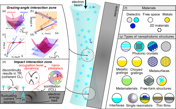
I.1.1 Grazing-angle interaction zone
In the grazing-angle interaction zone, the phase-matching condition from Eq. (1) allows us to predict which photonic modes are excited by free electrons, given prior knowledge of the photonic modes, as shown in Fig. 1(a-d). This general principle accounts for the excitation of localized and extended modes, in various photonic environments.
More specifically, as shown in Fig. 1(a-d), electrons passing in the vicinity of photonic structures can transfer some of their energy into available modes. Examples of such interactions include Cherenkov radiation from free electrons propagating in a uniform medium (Fig. 1(a)); excitation of guided, radiative, and bound states in the continuum (BIC) modes in photonic crystals (PhC) (Fig. 1(b)); excitation of surface plasmon polaritons (SPP) localized at metal-dielectric interfaces (Fig. 1(c)); and excitation of a superposition of Bloch modes in a two-dimensional PhC (Fig. 1(d)).
In a bulk medium, free electrons propagating faster than the phase velocity of light in the medium can excite photonic modes (such that the velocity component of the photon phase velocity is equal to the electron velocity, as per Eq. (1)). In a non-dispersive dielectric, such an electron thereby emits Cherenkov radiation, a "shock wave" of light.Cherenkov (1934); Ginzburg (1940, 1996); Cox (1944) In Fig. 1(a), the free-electron plane (defined as ) intersects the light cone (defining bulk medium plane waves) only above a certain threshold velocity (where is the reduced velocity). At a given frequency (isofrequency plane shown in black in Fig. 1(a)), the intersection of the two surfaces (consisting of two points at ) defines the angle of emission (see section II.2.1 for more details).
When propagating in periodic media, free electrons can spontaneously emit photons in the form of Bloch modes.Smith and Purcell (1953) The folded free-electron line (for sheet electrons) or plane (for point electrons) can intersect photonic bands at various locations in the BZ. Two such scenarios are shown in Fig. 1(b,d) for the case of a one-dimensional PhC slab (interacting with a sheet electron) and a two-dimensional PhC (interacting with a point electron). In both cases, free electrons can excite modes from several bands at the same time, resulting in complex emission processes. In PhC slabs, guided modes and leaky resonances can be excited, and even modes with diverging quality factors (so-called bound states in the continuum (BIC)Hsu et al. (2013)). Emission patterns in the two-dimensional case can exhibit even richer physics, given that radiation arises as a superposition of multiple modes with various group velocities.Luo et al. (2003)
Other types of spatially-extended modes, such as SPPs, phonon polaritons, and other surface waves can be excited by free electrons flying in their vicinity (for SPPs, typically, parallel to the interface between a metal and a dielectric). The excitation of SPPs with free electrons was originally proposed by Ritchie. Ritchie (1957) The typical dispersion relation converging asymptotically to (where is the plasma frequency) intersects the electron dispersion at various () as a function of the electron velocity. Such as dispersion relation is shown in Fig. 1(c) for the case of a sheet electron. Similar analysis can be performed to account for the excitation of high- resonances in, e.g. optical beads exhibiting whispering-gallery modes.Kfir et al. (2020); Müller et al. (2021)
I.1.2 Impact interaction zone
In the impact interaction zone, one way in which electrons can radiate is transition radiation (TR), which occurs when an electron crosses the interface between two media with distinct electromagnetic properties. From an electromagnetic perspective, TR originates in the continuity relation of the fields at the interface. García De Abajo (2010) This effect might still be explained in terms of the excitation of photonic modes by a free-electron current source, using the image charge formalism (see section II.2.3).
Beyond the simple TR effect, the more general family of impact interaction effects appear in almost all experiments, whenever some fraction of the incoming electrons penetrates into the sample. In this impact interaction zone, the electron kinetic energy can be transferred to the material, allowing subsequent emission processes.
Those emission processes can be understood as arising from fluctuating current sources associated with bound polarization (e.g., from excited electrons in defects, excitons, etc.). Such bound polarization is qualitatively similar to electrons in atoms or molecules, and thus their light emission (and nanophotonic shaping perspective) is understood from the perspective of bound-electron radiation engineering, which is a dominant paradigm in nanophotonics currently. Therefore, in this process, the phase-matching condition does not describe the coupling of free electrons with light. However, photonic engineering can still be used to enhance and control radiation.
This type of radiation is commonly referred to as incoherent cathodoluminescence (ICL). Since this process is equivalent to what is referred to as scintillation in other fields of physics (e.g. as observed with x-rays, -rays, Hall and Giaccia (2012) and particles Birks (1967)), we will refer to this effect as "electron scintillation" as well, to highlight their common physical origin and similarity. Scintillation is a complex “multi-physics” process spanning several disparate length, time, and energy scales. The key steps in scintillation can be summarized as follows: (1) ionization of electrons in the sample by the pump electrons followed by production and diffusion of secondary electrons Klein (1968); (2) establishment of a non-equilibrium steady-state of bound electrons Wurfel (1982); Greffet et al. (2018); and (3) recombination, leading to light emission (when the recombination is radiative). The final step of light emission is particularly complex in nanophotonics environments, as it results from fluctuating, spatially-distributed dipoles with a non-equilibrium distribution function. The final step also hints at the feasibility of enhancing and controlling ICL with nanophotonics. Moreover, such ICL is often ubiquitously present in experimental studies of CL, and much analysis is typically devoted to attributing signals to CL versus ICL/scintillation. Brenny, Coenen, and Polman (2014)
We note that there exist additional free-electron radiation processes such as bremsstrahlung ("braking" radiation) that arises from abrupt deceleration. We do not focus on these processes in this Review because their enhancement or shaping with nanophotonics structures has not been (yet) demonstrated. In contrast, free-electron radiation effects such as parametric X-ray and coherent bremsstrahlung are discussed below to highlight their connections to the wider family of diffraction radiation phenomena that can be affected by nanophotonics. We do however discuss connections between the previously-mentioned acceleration/deceleration effects (in both interaction zones) and other forms of "diffraction radiation"Potylitsyn et al. (2010) such as parametric x-ray emission and coherent bremsstrahlung.
I.2 Fundamental discoveries in free-electron physics
The fundamental physics of radiation by free-electrons (without nanophotonics) has been known for decades in the context of macroscopic electrodynamics and high-energy physics. The fundamental physics of electron-light interaction has a long history, dating back to the early 1900’s and has resulted in several cornerstone discoveries in fundamental physics.
Perhaps the most celebrated of those effects is Cherenkov radiation (CR), given its many analogues in other systems,Carusotto and Rousseaux (2013) applications in nonlinear optics, Yuan et al. (2011); Chang, Chen, and Kärtner (2010); Skryabin et al. (2003); Zhang et al. (2013); Belkin and Capasso (2015) high-energy particle detectors, Nakamura (2003) dosimetry, medical imaging and therapy.Shaffer, Pratt, and Grimm (2017) The original observation of CR was reported by CherenkovCherenkov (1934) and VavilovVavilov (1934) from secondary (Compton) electrons in a liquid irradiated by -rays. Shortly thereafter, the observation was confirmed by a series of observations and theoretical predictions by Cherenkov, Vavilov, Frank, and Tamm.Frank and Tamm (1937); Cherenkov (1937, 1938) Cherenkov, Frank, and Tamm were awarded the Nobel Prize in Physics in 1958 for the "discovery and the interpretation of the Cherenkov effect" (a few years after Vavilov had passed away). Tamm insisted in his Nobel lectureIE Tamm (1959) that the effect should rather be named the "Vavilov-Cherenkov effect", to highlight the contribution of Vavilov. Ginzburg later noted (regretfully) that the name "Vavilov" had been dropped in most instances.Ginzburg (1996) CR is discussed in greater depth in section II.2.1.
TR was originally proposed by Ginzburg and Tamm in 1945.Ginzburg and Frank (1945, 1946) The original observation was reported by Goldsmith and Jelly in 1959 in the visible by bombarding metallic surfaces with 1 MeV protons. Significant important contributions to the field of CR and TR were reported in the few decades following their original discovery, such as TR calculations from metallic thin filmsGaribyan (1960) discovery of the anomalous Doppler effect in the Cherenkov cone,Frank (1942); Ginzburg and Frank (1947); Lin et al. (2018a) and quantum recoil corrections to the Cherenkov effect. Ginzburg (1940); Cox (1944); Kaminer et al. (2016a) Both techniques became mainstay technologies in high-energy particle detector experiments.Akopov et al. (2002); Adams et al. (2001); Nakamura (2003); Kleinknecht (1982) Free-electron injection into a metal can also lead to the generation of SPPs. This fundamental discovery was first proposed by Ritchie in 1957 Ritchie (1957) and experimentally observed for the first time in 2006. Bashevoy et al. (2006)
Scintillation (from various high-energy particles, such as x-rays, free electrons, and -particles) was originally discovered as a diagnosis and detection tool in early works on gemology and radioactivity. Early works by Hittorf (reported in Ref.Urbain (1909)) and later CrookesCrooke (1879) reported ICL from various stones, including diamonds. The first reported scintillator detector was invented by Crookes in 1903 to detect -particles, following original observations of light emission from phosphorescent powders in cathode-ray tubes in 1879.Crooke (1879) Developments in optical amplification devices made scintillator detectors widely available for applications in radiology, electron microscopy, and high-energy particle detection.Gektin and Korzhik (2017) Interestingly, the first x-ray images following Röntgen’s discovery were not performed with scintillators, but rather radiation-sensitive photographic film,Röntgen (1896) which required very long exposure and acquisition times. Scintillators became the workhorse detection technique in x-ray imaging around the 1990’s with the emergence of digital detectors.Lecoq (2016) ICL, or equivalently free-electron scintillation, has remained a technique of interest in gemologyRemond, Phillips, and Roques-Carmes (2000) and semiconductor physics,Yacobi and Holt (1986) with applications in cathode-ray tube instruments.Garlick (1950) ICL, scintillation and their applications to nanophotonics are discussed in section II.4.
All of the original observations discussed up to this point were performed with bulk media and high-energy electrons. The first occurrence of free-electron radiation in structured media was done in 1953 by Smith and Purcell who observed visible radiation from 300 keV electrons flying above a metallic diffraction grating.Smith and Purcell (1953) The effect, now coined as SPR is also sometimes referred to as a form of "diffraction radiationPotylitsyn et al. (2010)". SPR has found direct applications in microwave electronicsTsimring (2006) and is considered as a promising platform for non-invasive particle beam diagnosis.Lampel (1997) SPR is discussed in greater depth in section II.2.2.
The core of this Review is to discuss recent developments in nanophotonics which have enabled a plethora of new effects and a new framework to understand free-electron emission. Specifically, we discuss how the interplay of free-electron physics (and more generally high-energy physics in the case of scintillation) has enabled the control and enhancement of the above-mentioned emission effects.
I.3 Recent milestones enabled by nanophotonics
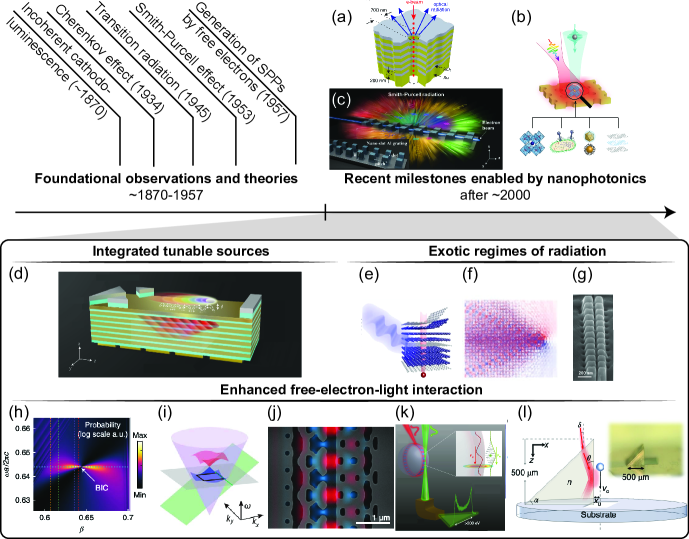
A historical timeline of free-electron radiation, from the discovery of its fundamental building blocks to recent effects enabled by nanophotonics, is shown in Fig. 2. The first wave of discoveries in the field of free-electron radiation happened in the years between 1870-1953. In this period, the fundamental mechanisms were first observed and explained. What we have seen in the past 10-20 years is a second wave of discoveries, mostly driven by the possibilities and new concepts emerging from the field of nanophotonics. 111This Review focuses on spontaneous emission from free electrons, which is why we do not display major achievements like the first operation of a free-electron laser,Deacon et al. (1977) or work on dielectric laser acceleratorsEngland et al. (2014) in the timeline (though we discuss their connection with nanophotonics in section IV).
Around the turn of the 21 century, advances in nanofabrication triggered a renewal of interest in understanding light propagation in patterned materials on the scale of optical wavelengths.Novotny and Hecht (2009) In particular, the birth of PhCs enabled an abundance of techniques to control photonic properties in engineered materials.Joannopoulos et al. (2011); Yablonovitch (1994) The field of nanophotonics recently found applications in free-electron physics, as a way to control and enhance radiation from free electrons. Alternatively, free electron beams can be used as diagnostic tools to probe photonic properties of nanostructures.García De Abajo (2010); Polman, Kociak, and García de Abajo (2019)
Perhaps the most obvious, yet much awaited application of nanophotonics in free-electron physics is the miniaturization of free-electron-driven radiation sources.Ishizuka et al. (2001); Roques-Carmes et al. (2019); Massuda et al. (2017a); Liu et al. (2017a); Adamo et al. (2009, 2012, 2010) Specifically, nanophotonic structures have been shown to allow visible radiation from relatively slow electrons (, which can be generated and accelerated on chip-scale distances), and to eliminate emission threshold in CR.Liu et al. (2017a); Vick et al. (2018); Zhang et al. (2021) This effort has also been bolstered by advances in integrated free-electron sources such as field emitter arrays.Guerrera and Akinwande (2016a, b); Temple (1999)
More generally, nanophotonics offers a convenient platform to control and enhance radiation by engineering the interaction of electron beams with photonic modes.Yang et al. (2018a, 2021); Remez et al. (2017); Chuang and Kong (1984); Coenen, Vesseur, and Polman (2011); Yang et al. (2018b) Some regimes of electron emission forbidden in most macroscopic mediaVeselago (1968) are realizable in some specific nanophotonic structures, such as backward CR in PhC.Luo et al. (2003); Lin et al. (2018a)
One of the most promising advantages of free-electron radiation is its wide tunability and the available wavelength ranges, from microwave to x-ray radiation. This tunability is achieved via structural and electron beam engineering. This is in contrast with wavelength tunability in, e.g. laser sources, which typically requires the sometimes painstaking development of new materials emitting at the wavelength of interest. Specifically, nanophotonic structures pumped by free-electron beams have been shown to emit photons in hard-to-reach regimes, such as UV,Ye et al. (2019); Vick et al. (2018); Watanabe et al. (2009); Watanabe and Taniguchi (2011); Watanabe, Taniguchi, and Kanda (2004) soft x-ray,Shentcis et al. (2020) THz,Urata et al. (1998) and mm-wave.Goldstein et al. (1998) Free electrons provide a versatile platform to access parts of the electromagnetic spectrum where few sources are available, utilizing the radiation control and enhancement techniques mentioned above.
Given the nanometer scale spatial resolution of electron beams in most electron microscopes, free-electron radiation has been considered as a spectroscopic probe to study nanophotonic structures.Coenen and Haegel (2017); García De Abajo (2010); Polman, Kociak, and García de Abajo (2019) Free electrons can also interact with nanophotonic structures over extended interaction lengths, thereby achieving stronger coupling strengths, enabling regimes of multi-photons stimulated emission and absorption by a single electron, Dahan et al. (2020) and electron acceleration in integrated dielectric laser accelerators. Sapra et al. (2020)
Some of the above-mentioned applications have been enabled by recent advances in ultrafast electron microscopy, where electron-beam and optical excitation of the sample can be modulated in time down to attosecond pulses,Morimoto and Baum (2018); Priebe et al. (2017); Vanacore et al. (2018); Kozák, Schönenberger, and Hommelhoff (2018) thereby unveiling quantum properties of electron-light interactions.Feist et al. (2015); Di Giulio, Kociak, and de Abajo (2019); Lim et al. (2021) In particular, in photon-induced near-field electron microscopyBarwick, Flannigan, and Zewail (2009); Park, Lin, and Zewail (2010); Garcia De Abajo, Asenjo-Garcia, and Kociak (2010) (PINEM), one can probe near-field non-equilibrium properties of physical systems with unprecedented time and spatial resolution. The recent introduction of nanophotonics in PINEM has enabled the implementation of cavity quantum electrodynamicsWang et al. (2020a); Kfir et al. (2020) with free electrons, the generation of electron vortex beams,Vanacore et al. (2019) the coherent control of electron beam statistics,Dahan et al. (2021) electron beam modulation with silicon photonics, Henke et al. (2021) coincidence electron-photon detection, Feist et al. (2022); Varkentina et al. (2022) and strong coupling in the single-photon–single-electron regime.Adiv et al. (2022)
| Acronym | Meaning |
|---|---|
| CR | Cherenkov effect |
| SPR | Smith-Purcell radiation |
| TR | transition radiation |
| CL | cathodoluminescence |
| ICL | incoherent cathodoluminescence |
| PhC | photonic crystal |
| BIC | bound state in the continuum |
| SPP | surface plasmon polariton |
| BZ | Brillouin zone |
| UV | ultraviolet |
| NIR | near-infrared |
| PINEM | photon-induced near-field electron microscopy |
| EELS | electron energy-loss spectroscopy |
II Typology of free-electron radiation
In this section, we describe the basic organizing principles of this Review, which help to sort out the different effects under a general formalism that highlights the role and prospects of nanophotonics in this field.
II.1 Coherent vs. incoherent cathodoluminescence
All of the effects in the grazing-angle interaction and impact interaction zones have a common physical origin: they result from the coherent interaction between the photonic eigenmodes of the structure and a current source (describing the particle trajectory). The current distribution can be equivalently described in time and frequency domain. In the following, we consider a trajectory of the form (with the initial position at taken to zero without loss of generality):
| (2) | ||||
| (3) |
where is the electron charge. The particle propagates along the linear trajectory defined by the velocity vector . The unit vector parallel to is and the orthogonal space is denoted as (where is the dimensionality of space). Given the mismatch in energies between the incoming electron ( keV in most settings) and the emitted photon energy (few eV’s in the visible to near-infrared (NIR)), one can often safely neglect the quantum emission recoil, and therefore consider the trajectory to be unaffected by radiation processes.
This current source can be plugged into Maxwell’s equations in free space, resulting in an evanescent near field, John David Jackson (1999) which can be scattered by the structureGarcía De Abajo (2010); Polman, Kociak, and García de Abajo (2019). This observation also led to the application of fundamental bounds on free-electron radiation and energy loss (derived in section II.3 and Ref.Yang et al. (2018a)). Said differently, the current source (representing the free electron) is performing work on the system and some of which results in radiation.
The current distribution in Eq. (2) can be modeled in Maxwell’s equations in several ways, depending on the type of numerical solver. For instance, in finite-difference time-domain solvers, it can be modeled as an array of dipoles "turned on" sequentially at a speed corresponding to the flight of the electron.Roques-Carmes et al. (2019); Massuda et al. (2017a) Alternatively, it can be directly injected as a line current in frequency domain (with Bloch periodic boundary conditions to model periodic systems). Yang et al. (2018a); Szczepkowicz, Schächter, and England (2020)
Coherent CL effects are often considered in opposition to ICL, which originates from stochastic energy losses in the material. Because of the stochastic nature of the process, emitted photons lose their coherence with respect to the incoming electron. Instead of the deterministic current distribution from Eq. (2), one models ICL as radiation from a stochastic current distribution, whose current-current correlations are prescribed as:
| (4) |
with , and is a normalization time. In this spectral function, is the occupation factor of microscopic state with energy , represents the matrix element of the current density operator , and . The current density matrix and the occupation functions can depend on position, as they depend on the electron energy loss density.
Direct calculation of radiation from the stochastic current source described in Eq. (4) is a computationally expensive problem, as is known in the context of thermal emission.Chan, Soljačić, and Joannopoulos (2006) Such calculations would indeed require the sampling of a three-dimensional current distribution, whose correlations partially depend on the microscopics and electron energy loss dynamics. Therefore, it is strongly beneficial to resort to more efficient numerical methods, leveraging electromagnetic reciprocity, to make such calculations tractable in three-dimensions.Roques-Carmes et al. (2022a)
The distinction between coherent and incoherent CL is also linked to the final quantum state in which the sample is left after radiation.García De Abajo (2010) In the following, we show how CR, TR, SPR, and other coherent CL effects arise from the coherent interaction of the current distribution from Eq. (2) with photonic eigenmodes. We also show how the stochastic current distribution from Eq. (4) can radiate ICL in arbitrary nanophotonic environments, and computational techniques to calculate it efficiently.
II.2 A unifying picture of coherent cathodoluminescence in arbitrary nanophotonic environments
Considering the current source in Eq. (2) as a source in Maxwell’s equations, one can calculate radiation from a moving free electron in arbitrary nanophotonic media. We expand the Green dyadic tensor – relating currents to fields linearly as – over its set of eigenmodesNovotny and Hecht (2009) :
| (5) |
where . The eigenmodes are normalized such that:. The main assumption in deriving this equation are small losses and weak dispersion, which are valid in most cases we consider in this work, and can be relaxed further by using the dyadic Green tensor directlyRivera and Kaminer (2020) (without referring to a mode expansion).
The total energy radiated by a dipole can be calculated as an integral in frequency domain,Kremers, Chigrin, and Kroha (2009) and we get the following expression:
| (6) |
Eq. (6) already highlights the main physical parameters relevant to understanding all effects in coherent CL, and we therefore refer to it as the "master equation". The prefactor is always proportional to , which underlines possible radiation enhancements by considering highly-charged particles (such as heavy ionsRoques-Carmes et al. (2018)). More generally, the dependence offers a mechanism to distinguish between particles with elementary charge (such as electrons, protons, etc.) and nuclei with charge (where is the atomic number). For instance, even CR from fully ionized helium is four times stronger than that of hydrogen isotopes and elementary charges. Ginzburg (1996) In heavier materials, the larger discrepancy can be used to "count" the energy of each incoming particle. Kleinknecht (1982)
The rest of the expression is summed over all photonic modes indexed by which may, in principle, contribute to radiation. The photonic mode-electron overlap highlights the importance of the spatial overlap between the photonic mode and the electron beam (since is evaluated at the fixed location perpendicular to the axis of propagation of the electron). Radiation might also be enhanced by considering extended interactions over lengths such that remains large over the polarization parallel to the electron trajectory.
Further physical considerations can be made to evaluate the mode-electron overlap. The mode profile has a given polarization distribution, but only the polarization along the beam propagation contributes to emission. The spatial dependence of the field profile results in evanescent coupling strengths in many scenarios, as will be made evident for excitation of SPPs and electromagnetic bounds on coherent CL (sections II.2.4 and II.3).
The spectral dependence of the emitted energy is encoded in , such that in the limit of small losses (where Im denotes the imaginary part). Therefore, radiation in eigenfrequencies of the photonic structure are strongly enhanced.
The term labeled "phase-matching" in the master equation Eq. (6) only contributes partially to the phase-matching condition described in Eq. (1) and is complemented by a phasor of the form from the mode-electron overlap term, which for instance appears in systems exhibiting translational invariance (see below). Their combination yields the phase-matching relation from Eq. (1). To further outline the physical importance of phase-matching, we consider a specific mode with longitudinal wavevector , such that . This yields a version of the master equation where the phase-matching condition is evident:
| (7) |
where is the sinc function, and is the length of interaction. The phase-matching term becomes a Dirac delta function for large interaction lengths , thereby highlighting the critical importance of phase-matching in physical settings with extended interactions. Compared to Eq. (6), this equation also highlights the spectral dependence of the radiation, which is determined by the photonic eigenfrequencies . This compact form also hints at a geometric method to calculate the emitted power by (1) considering the intersection of the band structure dispersion with the electron plane ; (2) weighting each intersection by the mode-electron overlap term in Eq. (7). We described in section I.1 how such a method can be used to predict radiation in various radiation events occuring in the grazing-angle interaction zone in the introduction, for CR in bulk media and PhCs, SPR, and excitation of SPPs.
To gain further physical insight with this formalism, we must consider specific photonic environments, which will be described by various eigenmode distributions, and are discussed in the next sections.
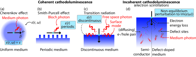
II.2.1 Cherenkov radiation
CR in its simplest embodiment occurs in a homogeneous dielectric medium (of index ) and consists in the spontaneous emission of plane waves by a charged particle (see Fig. 3(a)). Its description, in the language of the previous section and the master equation, can be understood in terms of plane wave eigenmodes of a medium of index . The dispersion relation is . Plugging this expression into the master equation Eq. (6), we can get the famous Frank-Tamm formula Frank and Tamm (1937) for the spectral density per unit propagation length, shown in the Appendix A.1. Emission is only allowed for superluminal electrons, which is equivalent to the phase-matching condition from Eq. (1) in a bulk medium.
Quantum corrections to this formalism can be introduced, taking into account recoil,Ginzburg (1940); Cox (1944) non-perturbative effects with heavy ions,Roques-Carmes et al. (2018) the particle’s spin and orbital angular momentum,Kaminer et al. (2016a) reduced dimensionality,Adiv et al. (2022) or emission from hot electrons in two-dimensional materials.Kaminer et al. (2016b)
II.2.2 Smith-Purcell radiation
SPR is a natural extension of CR to periodic media as the spontaneous emission of Bloch photonsRivera and Kaminer (2020) (see Fig. 3(b)), which we use as eigenmodes in the expansion from Eq. (5). We first consider the case of a one-dimensional periodic structure (period ) along the direction of electron propagation. Eigenmodes can be described by the band index and reciprocal lattice vector in the first BZ, such that , where are coefficients of the mode’s Fourier expansion.
We then get the following expression for the energy spectral density:
| (8) |
The function in Eq. (8) gives us a geometric way of predicting which Bloch modes are excited by the electron beam, by considering the intersection of the band structure with . This method generalizes well to higher dimensions where the electron "line" becomes a plane in the band structure representation (see Fig. 1(b,d) for some example band structures). The phase-matching condition also sets the dispersion relation, known as the Smith-Purcell relation:
| (9) |
where is the photon wavelength, and the Bloch mode index. The emission angle is measured, as in the case of CR, with respect to the electron propagation direction.
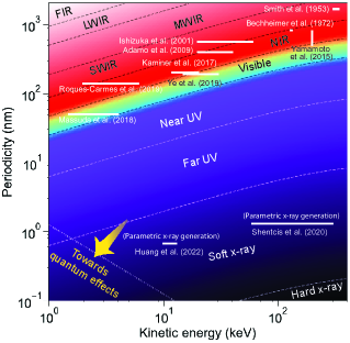
This simple relation enables us to make quick predictions of the radiation spectrum (in the absence of resonant enhancement), knowing the structure periodicity along the electron trajectory. Several observations of SPR from various periodicities and electron energies are shown in Fig. 4, on a background corresponding to the wavelength predicted by Eq. (9) with and . The original observation from Smith and Purcell was performed with . The relatively large grating pitch they used should have resulted in radiation in the short-wave infrared at normal emission direction, but they measured visible radiation at a shallower angle .
Early work on SPR was essentially focused on metallic structures.Salisbury (1970) More recent work used electrons in similar energy ranges, pushing radiation into the near-UV regime with higher diffraction orders.Yamamoto, Javier García De Abajo, and Myroshnychenko (2015) With the goal of integrating Smith-Purcell emitters into optoelectronic devices, recent effort has been targeted at reducing the electron kinetic energy and periodicity, to retain emission in the NIR to UV regimes222We note that SPR was also observed with relativistic electrons.Doucas et al. (1992). This effort has been enabled by recent progress in nanofabrication, namely the capability of fabrication sub-100 nm periodic structures with electron-beam lithography.Massuda et al. (2017a) Some works even reported emission from electron beams generated on-chip by field emittersIshizuka et al. (2001) and/or with integrated all-silicon structures.Roques-Carmes et al. (2019) The prospect of generating radiation deeper in the UV remains an exciting perspective, with recent work demonstrating SPR in the UV with and leveraging higher-order diffraction modes.Ye et al. (2019) Beyond the technological promise of integrated and tunable UV emitters, short-wavelength SPR also exhibits quantum recoil effects in low-energy and short-period settings.Tsesses, Bartal, and Kaminer (2017) While demonstrations of short-wavelength SPR have been limited to the near UV, other free-electron radiation effects, such as coherent bremsstrahlung and parametric x-ray radiation, being similar in some respects to SPR, are amenable to generation of x-ray radiation. In these effects, the electron flies in a "structured" medium consisting of a crystalline lattice of atoms, such that the period is on the ångström scale. Those techniques offer an interesting platform to generate x-ray photons with moderately relativistic electrons.Baryshevsky et al. (2006); Baryshevsky and Feranchuk (1983); Überall (1956); Korobochko, Kosmach, and VI Mineev (1965); Wong and Kaminer (2021); Tan et al. (2021) We highlight recent workShentcis et al. (2020); Balanov, Gorlach, and Kaminer (2021) to reveal the similarities and differences between SPR and the previously-mentioned x-ray emission techniques.
Many theoretical studies focused on SPR and CR in 2D, where the structures are assumed to be invariant along the third dimension and the electron beam is assumed to be a "sheet" beam. In higher dimensions, the point-like nature of the electron must be considered, and the photonic band structure gives greater flexibility in tuning the coupling between the electron and photonic modes. Considering the case of a three-dimensional periodic PhC, we get the following expression, as previously reported in Ref.Kremers, Chigrin, and Kroha (2009):
| (10) |
where is the contour defined by . This formula shows that the radiated power is proportional to the Fourier coefficient , describing the coupling of the current density with the electromagnetic field at the electron location. Also, the emitted power is proportional to the inverse of the transverse group velocity. This suggests a path to strongly enhance emission from electrons using engineered bands in PhCs.Kremers, Chigrin, and Kroha (2009); Yang et al. (2018a, 2021)
II.2.3 Transition radiation
To describe transition radiation within the same framework, we consider the simplified case of a charge impinging at normal incidence on a perfect conductor (see Fig. 3(c)) and resort to the introduction of an image charge with opposite charge and velocity. The current distribution is modified accordingly and its emission in free space is considered. Doing so, we get the spectral distribution shown in Appendix A.2, first derived by Ginzburg and Tamm.Ginzburg (1940)
This relation can be extended to interfaces between two media with finite permittivities,Ginzburg (1940); VL Ginzburg and VN Tsytovich (1984) radiation into waveguides,KA Barsukov (1960) and metallic thin films.Garibyan (1960) Resonant transition radiation, the coherent interference of multiple TR emission in a multilayered medium, has also been considered as a promising platform for high-energy particle detectorsLin et al. (2018a, 2021) and x-ray emission.Yamada, Hosokawa, and Takenaka (1999) Though considered as an extension of TR at a single interface, we note that resonant TR has a dispersion relation similar to that of SPR.Pardo and André (2000)
II.2.4 Coherent excitation of surface plasmon polaritons (SPPs) by free electrons
SPPs can be excited by free electrons Keller (1994); Bashevoy et al. (2006); Liu et al. (2012); Garciía De Abajo (2013); Gong et al. (2014); Lin et al. (2017); Liu et al. (2017a); Adiv et al. (2022) either when impinging on metals or when grazing the interface, with first observations reported in Ref. Bashevoy et al. (2006) and Ref. Adiv et al. (2022), respectively. This phenomenon is observed even in the absence of corrugation at the surface, and can be understood with our formalism, when considering SPP eigenmodes. For concreteness, we consider the case of an interface between two media and , with the electron propagating in . The lower branch of the SPP mode is shown in Fig. 1(c) for a Drude-like metal (where is the plasma frequency). The energy emitted by the free electron per unit length is derived and shown in Appendix A.3.
As expected from the master equation Eq. (6), the exponential decay of the field at the interface results in a factor , arising from the mode-electron overlap evaluated along the electron trajectory. As with other effects in the grazing-angle interaction zone, one can predict which SPP modes are excited by an electron with velocity reduced by identifying the intersection of the dispersion relation with the electron lineLiu et al. (2012) . Analogues of this effect have been observed in systems supporting Dyakonov surface wavesHu et al. (2022) and hyperbolic dispersion.Ye et al. (2019)
II.3 Fundamental bounds for coherent cathodoluminescence
Another perspective that the field of nanophotonics brought to research on free-electron radiation is the idea of setting electromagnetic bounds from first principles. Nanophotonics research led to the formulation of universal bounds on various photonic processes, Chao et al. (2021) such as scattering and absorption, Miller et al. (2016) focusing, Shim, Chung, and Miller (2020) Raman scattering, Michon et al. (2019) and near-field optical response. Shim et al. (2019) The same approach was applied in Ref. Yang et al. (2018a) to propose a universal bound on free-electron radiation and energy loss. Such bounds represent the maximal amount of power that could be scattered or absorbed by an optimal structure excited by free electrons and enclosed in a given volume.
Coherent CL can be interpreted as a scattering problem, and is therefore amenable to recent work on electromagnetic bounds.Yang et al. (2018a) Intuitively, the scattering problem can be bounded by a convex optimization problem, whose solution is obtained by calculating variational derivatives of the incident fields.Yang et al. (2018a) (The incident evanescent field is generated in free space by the current distributionGarcía De Abajo (2010) .) Maximal radiation and energy loss powers can then be derivedYang et al. (2018a) for an arbitrary "scatterer" (corresponding to the sample inducing coherent CL) with susceptibility and volume :
| (11) |
where , , (with the ratio of radiative to total energy loss), , and (with the Lorentz factor). is the -th order modified Bessel function of the second kind.
The power bounds from Eq. (11) apply to the non-retarded or retarded regimes, and only assume the absence of gain in the optical medium. This expression also highlights the main physical parameters of interest to maximize radiation from free electrons: the material factor which reflects the influence of material choice, depending on the wavelength of interest; the electron velocity and Lorentz factor appearing in the impact parameter in the integral. Approximations of this boundYang et al. (2018a) explicit the role of the minimum distance between the electron trajectory and the scatterer , such that the relevant length scale of interaction is set by . At large beam-sample distances, the bound decays exponentially , which matches the dependence from Eq. (23).
The analytical bound also reveals several interesting physical behaviors. Namely, there exists a regime of near-field interaction between the beam and the structure where slow electrons are favored (i.e., they radiate more efficiently). Recent works also highlighted the possibility of strong interactions between slow electrons and plasmonic near fields. Talebi (2019, 2020); Liebtrau et al. (2021) This supports recent interest in developing on-chip sources of free-electron radiationYe et al. (2019); Massuda et al. (2017a); Roques-Carmes et al. (2019), where electron beams can be precisely aligned to nanophotonic structures (e.g. gratings for SPR) to control the beam-sample coupling.
Another interesting feature is the apparent divergence of the bound in the limit of small losses, which suggests mechanisms to strongly enhance free-electron radiation with high- resonances, a path which we discuss in section III.3.1.
II.4 Incoherent cathodoluminescence (electron scintillation)
All of the previous types of radiation were forms of coherent CL, which naturally give themselves into control via shaping of the photonic eigenmodes, as we explained in the previous paragraphs. We now consider the case of ICL, or electron scintillation, and propose ways to control and enhance this form of radiation. The method we propose here is readily transferable to scintillation from other types of high-energy particles, such as x- and -rays.
ICL is usually observed when a beam of electrons is bombarding a material directly (and is therefore occurring in the impact interaction zone), as depicted in Fig. 3(d). Energy is then lost by the electron beam, which can be transferred to radiative sites (electron-hole pairs in semiconductors, or defect states in a doped medium), which subsequently radiate in a nanophotonic environment described by the eigenmode expansion from Eq. (5).
We can calculate the emitted energy by the stochastic current distribution in Eq. (4), and for simplicity make the assumption that the current correlations are local, isotropic, and real-valued , a condition which can be straightforwardly relaxed.Roques-Carmes et al. (2022b) ICL is, in general, described by the light emission from this non-equilibrium steady-state distribution. This assumption is corroborated by the fact that energy deposition occurs on picosecond time scales, which are effectively instantaneous relative to the excited-state depletion time scales (typically nanoseconds). Polman, Kociak, and García de Abajo (2019); Greffet et al. (2018) We then get:
| (12) |
This expression makes explicit the way in which ICL (where the light emission results from spontaneous emission) can be controlled by photonic shaping via the eigenmodes . It is also apparent that ICL can be enhanced by optimizing the overlap between a given eigenmode and the current correlation function (for example, by optimizing the overlap between the photonic eigenmode and the energy-loss density of the high-energy particles).
Another formulation of ICL can be obtained by using electromagnetic reciprocity and the Green’s function directly (instead of its eigenmode expansion), yielding the following expression for the power spectrum per unit solid angle and frequency: Roques-Carmes et al. (2022a)
| (13) |
where we also assumed that the current correlations are uniform and isotropic in the material (corresponding for example to uniform energy loss), and . Eq. (13) states that the ICL spectrum, under this approximation, is a simple product of a microscopic factor, set by the non-equilibrium steady-state distribution function , Kurman et al. (2020); Roques-Carmes et al. (2022a) and an effective absorption volume , which is only determined by the (structured) optical medium surrounding the emitting sites.
Eq. (13) enables a key simplification thanks to electromagnetic reciprocity, which relates the following two quantities: (1) the emitted ICL from the structure (at a given frequency , direction , and polarization ) and (2) the intensity of the field induced in the sample () by sending a plane wave at it (of frequency , propagating along direction (with field ) into the structure, and polarization ). This expression opens the path to efficient numerical methods for ICL and scintillation in three dimensions and arbitrary nanophotonic environments.Roques-Carmes et al. (2022a)
III Control and enhancement of cathodoluminescence with nanophotonics
The previous section suggested several ways in which one can control CL with engineered nanophotonic structures. In this section, we describe several methods to experimentally measure (coherent and incoherent) CL from nanophotonic structures in electron microscopes; we also review experimental demonstrations and nanophotonic techniques to control and enhance CL.
III.1 Cathodoluminescence in electron microscopes
It is worth noting that CL in electron microscopes was an already-established technique before the 2000’s, given its widespread use in gemology and materials science.Remond, Phillips, and Roques-Carmes (2000) The transfer of this technique to the characterization of nanophotonic structures enabled the observation of electron-light interactions in nanophotonic structures via direct spectroscopic techniques (measuring CL) and indirect electron measurement techniques (EELS).
III.1.1 Cathodoluminescence spectroscopy and polarimetry techniques
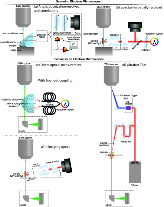
Apart from optical-CL instruments in which an electron beam is generated under moderate vacuum via discharge in a chamber (small enough to be mounted on a standard optical microscope) most modern CL instruments are based on a scanning electron microscope (SEM) or a transmission electron microscope (TEM). Fig. 5 illustrates representative types of SEM- and TEM-based CL instruments. In all instruments, the light generated by the interaction of a focused electron beam with a sample is out-coupled (with free space collection optics or an optical fiber). Collection optics that have been used for CL measurement and characterization include parabolic mirrors (for angular resolution) and objective lenses (for spatial resolution). Depending on how the light is collected and what additional components are utilized, a wide range of measurements can be performed from obtaining spatially resolved spectral information to angular, polarization, and even time-sensitive detection. Alternatively, placing the nanostructures on the tip of a fiber enables to directly collect the radiation through the fiber, a technique shown to enhance the evanescent field of the free electrons interacting with a nanostructure. So, MacDonald, and Zheludev (2014) It has also been demonstrated that the evanescent field of free electrons can by amplified as electrons fly over a plasmonic surface. So et al. (2015) Having to pass through a window to exit a vacuum chamber can limit the detectable wavelength range. An extensive review of current CL measurement techniques was recently published.Coenen and Haegel (2017)
Relevant to this Review, we highlight selected papers detailing different experimental methods useful for nanophotonic applications of CL. The use of tightly focused electron beams allows for collecting spectral information directly from nanoscale samples with spatial resolution limited by the size of the focused electron beam. CL found applications in plasmonics,Bashevoy et al. (2006, 2007); Vesseur et al. (2007); Hofmann et al. (2007); Denisyuk et al. (2010); Schefold et al. (2019); Han et al. (2018); Bauman et al. (2017); Liu et al. (2020); Ron, Zielinski, and Salomon (2020); Myroshnychenko et al. (2012); Krehl et al. (2018) photonics,Brenny et al. (2016); Talebi et al. (2015) semiconductors,Yan et al. (2008); Prabaswara et al. (2017); Vu et al. (2022) electron-beam lithography,Edwards et al. (2021) and tomographic reconstruction.Atre et al. (2015) Additionally, the combination of spatial and spectral resolution can be combined to measure the dispersion of CL effects such as SPR.Kaminer et al. (2017b); Massuda et al. (2017a); Roques-Carmes et al. (2019); Yang et al. (2018a) Methods were also proposed to disentangle several types of emission from farfield measurements.Fiedler et al. (2022) Inspired by the early days of radio, it has been shown that a nanoscopic dipole Herzian antenna acts as an efficient emitter of visible light when an electron beam is injected in the dipole gap. Denisyuk et al. (2010)
Thermal measurements can also be performed with CL, such as nanoscale thermometry and thermal transport measurements both in low current conditions (where sample heating is avoided) and higher current conditionsMauser et al. (2021) (where electron beam induced heating is present). Other electron-beam induced effects such as phase transitions in gallium nanoparticles at picojoule excitation energies have also been observed. Transformations between coexisting structural phases are accompanied by continuous changes in the nanoparticle film’s reflectivity Pochon et al. (2004) and luminescence, Denisyuk et al. (2008) which may be used for modulating light and optical data storage. Promising new techniques for resolving and hyperspectrally mapping picometric movements by detecting secondary electron emission from the edge of the nanostructure in an electron microscope Liu et al. (2021a) might also be realized via cathodoluminescence microscopy in the future.
Next, phase-sensitive imaging measurements were performed utilizing both angle-resolved CL and hyperspectral angle-resolved cathodoluminescence to characterize the far-field phase signal from scattering off of plasmonic nanostructures allowing for the reconstruction of the angle-dependent phase distributionsSchilder et al. (2020) and the coupling between nanoparticles and SPPs.Sannomiya et al. (2020) Similar techniques were used in the Fourier domain to determine the emission polarization properties of sub-wavelength structures like optical nanoantennas.Coenen et al. (2012) CL measurements have also been used to directly image plasmonic modes in annular nanoresonators, ultrathin plasmonic strip antennas, and metallic thin films, Hofmann et al. (2007); Barnard et al. (2011); Bashevoy et al. (2007) and to measure the statistics of the emitted light.Meuret et al. (2015, 2017, 2019, 2018); Bourrellier et al. (2016) Finally, CL measurements can be employed to characterize photonic band structures and measure the local density of states in nanostructured metallic, semiconductor and dielectric materials.Kuttge et al. (2009); Peng et al. (2019); García De Abajo and Kociak (2008); Mignuzzi et al. (2018)
III.1.2 Probing electron-light interactions in ultrafast electron microscopes
Several TEM-based CL instruments are similar to their SEM analogues, as shown in Fig. 5(c). For instance, collecting mirrors can be used to couple light out of the TEM vacuum chambers. The presence of two collecting mirrors can even allow the measurement of backward and forward radiation independently in some commercial systems.Silver et al. (2015); Stöger-Pollach et al. (2017, 2019, 2021) A TEM analogue of the spectrally/spatially-resolved SEM CL setup was also built,Remez et al. (2017, 2019) with a low-numerical-aperture objective outside the vacuum chamber and an additional rotation degree of freedom to measure radiation at various angles. An interesting advantage of TEM-based solutions is the availability of EELS which allows the measurement of electron energy loss and gain after interacting with a sample. EELS provides a method to probe near-field electron-light interactions and is complementary to CL measurement techniques. Polman, Kociak, and García de Abajo (2019) Since EELS is directly related to the photonic local density of states, García De Abajo and Kociak (2008) tomographic techniques have been demonstrated to reconstruct the full three-dimensional local density of states in nanoparticles. Horl et al. (2017)
Several techniques have been developed to add time-resolved measurements to the field of SEM- and TEM-based CL. This field has also been inspired by the techniques developed for ultrafast TEM in the Zewail group.Barwick and Zewail (2015); Yang, Mohammed, and Zewail (2010) Currently, time-resolved CL involves modifications to the electron beam emitter to generate electron pulses by the use of ultrashort laser pulses or by using fast electrostatic beam blanking to modulate a continuous electron beam. An example of a time-resolved TEM instrument is shown in Fig. 5(d), wherein an ultrafast laser is used to generate short time duration electron pulses from the electron source. The ultrafast laser can also be used to excite or probe the sample as a function of the of arrival of the electron pulse. Alternatively, instruments with beam blanking devices located after the electron source can provide time-resolved measurements, albeit at a lower time resolution than the laser-driven emitters. One example of a beam blanking measurement is found in Ref.Moerland et al. (2016), where a modified standard SEM with beam blanking electronics was used to produce electron pulses in the 80 to 90 ps duration range. This provided sufficient time resolution to characterize the spontaneous emission decay rate in a cerium-doped yttrium aluminium garnet sample. Moerland et al. (2016)
III.1.3 Free-electron analogues
Physical analogues of free electrons are convenient platforms to observe some of the above-mentioned physical phenomena. There are many free-electron analogues in the context of CR, since it is a general wave phenomenon, with analogues in classical and quantum electromagnetics, superfluid hydrodynamics, and classical hydrodynamics.Carusotto and Rousseaux (2013) In electromagnetics, CR can also be observed with superluminal polarization waves in SPP,Genevet et al. (2015) quantum cascade lasers,Belkin and Capasso (2015), solitons in optical fibers, Yuan et al. (2011); Chang, Chen, and Kärtner (2010); Skryabin et al. (2003); Zhang et al. (2013) and superluminal domain perturbations in rapidly time-modulated systems.Yablonovitch (1989); Sloan et al. (2021); Oue, Ding, and Pendry (2021); Dikopoltsev et al. (2022) Synchrotron-like radiation has also been observed by nonlinear polarization induced in a metasurface.Henstridge et al. (2018)
Most of the physical effects discussed in this Review are typically observed from electron beams generated in electron microscopes, with kinetic energies from few keV to hundreds of keV. Analogues of those effects have been predicted with hot electrons in graphene for CRKaminer et al. (2016b) and two-dimensional electrons in a driving field for SPR in the THz regime.Mikhailov (2013)
Some of those analogue systems have been utilized as a test bed for novel physical phenomena. In particular, metamaterial-loaded and slot waveguides have been used to directly emulate the propagation of an electron beam.Chen and Chen (2011); Xi et al. (2009); Jing et al. (2021); Antipov et al. (2007, 2008) The slot waveguide platform was first used to demonstrate backward Cherenkov radiation,Xi et al. (2009) an intriguing effect which has otherwise not been demonstrated in free-electron experiments. They have also been used to demonstrate polarization control in SPRJing et al. (2021) and, in combination with helical metastructures, SPR vortex beams.Zhu et al. (2020); Zhang et al. (2020)
III.2 Controlling free-electron radiation in nanophotonic structures
III.2.1 Angular and spectral control
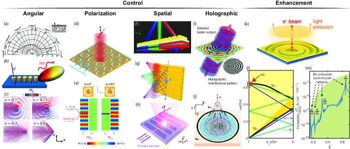
The spectral-angular distribution of coherent CL effects is, to first order, embedded into their dispersion relation (e.g., Eq. (9) for SPR). When free electrons emit in a nanophotonic medium, certain spectral and angular components of the radiative fields can be selectively enhanced. The possibility of selective enhancement is evident in periodic structures, where the emitted energy is proportional to the overlap between the electron trajectory and Fourier components of the photonic modes (as in Eqs. (8, 10)). The shaping of the spectral-angular distribution via photonic engineering has been the focus of many recent works in coherent CL. Certain notable results are shown in Fig. 6.
It was first proposed by Van den Berg that even perfect reflectors with sinusoidal corrugations could significantly alter the angular distribution of SPR.Van den Berg (1973) Controlling the emission direction can also be achieved by exciting plasmonic resonances in periodic metallic gratings with a free electron.Chuang and Kong (1984) SPR spectral-angular shaping has also been proposed and demonstrated with engineered grating profiles, Remez et al. (2017) resulting in multi-peaked spectra, and aperiodic gratings.Saavedra, Castells-Graells, and García De Abajo (2016)
III.2.2 Polarization control
Coherent CL effects are strongly linearly polarized, with limited tunability of the polarization angle. For instance, the polarization angle in SPR is set by the propagation direction of the electron beam. The control of the linear polarization angle and the generation of spin angular momentum in CL has therefore received recent interest, with some notable works shown in Fig. 6(d-e).
In CR, small components of circular polarization can be acquired when considering spin-polarized electron beams.Kaminer et al. (2016a); Peshkov (2020) With unpolarized electron beams, metasurfaces on waveguides have been proposed as platforms to generate circularly polarized CR.Li et al. (2020) Because of its potential applications in spectroscopy, the generation of circularly-polarized TR light in the mm-wave regime was realized by interfering its forward and backward componentsShibata et al. (2001)
In particular, SPR is highly polarized along the direction of the electron beam (a feature which has been known since its original discoverySmith and Purcell (1953)). Recent work using cross-coupled electric and magnetic dipoles in THz Babinet metasurfaces suggested a path to steer the angle of linear polarization.Wang et al. (2016); Yang et al. (2018b); Liu et al. (2017b) In principle, graphene metasurfaces can also be utilized to generate circular polarization states in the THz regime.Su et al. (2019) More recently, an experimental demonstration was provided by exciting cross-polarized resonances in a PhC.Yang et al. (2021) The generation and control of SPR with orbital angular momentum (vortex beams) could also be achieved with holographic gratings.Wang et al. (2020b) Full control of the optical angular momentum (spin and angular) in SPR could also find applications in on-chip spectroscopy, but its realization has remained elusive thus far.
III.2.3 Spatial control
As coherent CL relies on the excitation of photonic eigenmodes, phase relationships between different wave vector components are constrained, which prevents the control of the radiation far-field profile. With the promise of on-chip electron-driven light sources, there has been a growing interest in manipulating the far-field spatial distribution of emitted radiation in CL. If successful, this effect could be leveraged to realize integrated sources and collimators into a single component, with some notable works shown in Fig. 6(f-h).
The design of a CR lens was first proposed by adjusting the boundary of the Cherenkov target based on ray optics considerations to concentrate light into a focal spot.Galyamin and Tyukhtin (2014); Galyamin, Tyukhtin, and Vorobev (2018) Given the well-defined spectral-angular relation in SPR, a natural design for a SPR concentrator is a chirped grating (with decreasing pitch along the beam trajectory). That way, the emission angle is tuned along the direction of propagation or, equivalently, a thin-lens-like phase modulation is imparted to light generated via SPR. This concept was first proposed theoretically and demonstrated numerically for concentrators working at single wavelengths,Remez et al. (2017); Lai et al. (2017) and exhibiting strong chromatic aberrations. Alternatively, graphene metasurfaces can also generate converging SPR in the THz regime.Su et al. (2019) Recently, signatures of SPR lensing have been reportedKarnieli et al. (2022) using a chirped grating design.
The most general type of far-field wavefront engineering, holography, has also been proposed using tailored nanophotonic structures (see Fig. 6(i, j)). Li et al. (2016) This method relies on the controlled interference of transition radiation, generated by a focus electron beam, with an interference holographic mask. This enables the generation of light with prescribed wavelength, direction, divergence and topological charge via point-excitation of CL holography in plasmonic Li et al. (2016); Clarke, MacDonald, and Zheludev (2018); Schilder et al. (2020) and dielectric Clarke et al. (2018) metasurfaces. Free-electron holographic light sources offer a universal approach to generate light with prescribed wavelength, direction, divergence and topological charge via point-excitation of holographic metasurfaces with an electron beam. Lastly, inspired by transformation optics, several nanophotonic structure designs have been demonstrated to realize broadband focusing of transition radiation, Talebi et al. (2019) vortex light beams, Van Nielen et al. (2020) and more generally structured light from free electrons. Christopher et al. (2020) Such structures have been proposed as platforms to measure time-energy correlation functions in electron microscopes, paving the way towards attosecond electron-based spectroscopy techniques. Talebi (2016a).
III.3 Enhancing free-electron-light interactions in nanophotonics
III.3.1 Coherent cathodoluminescence
The existence of fundamental bounds on CL, as presented in section II.3, begs the following question: could one enhance coherent CL with nanophotonics to achieve emission efficiencies approaching such bounds?
The possibility of resonant enhancement in electron-light interaction is highlighted in the phase-matching relation Eq. (1). Emission into phase-matched photonic modes is selectively enhanced by adjusting the electron velocity. This concept has been proposed to enhance SPR by coupling electrons to photonicYamaguti et al. (2002) and plasmonic resonances,Chuang and Kong (1984) and CR in PhCs.Luo et al. (2003) Resonant enhancement of the electron-light interaction is also observed in an increase in PINEM signal when exciting photonic resonances.Wang et al. (2020a); Kfir et al. (2020)
Specifically, one can design a resonance mode of interest in metamaterials and excite it with a beam of free electrons. In particular, localized free-electron-beam excitation can create a low-divergence spatially coherent free-space light beam that bears similarity with laser light through coherent collective oscillation of an ensemble of coupled metamolecules.Adamo et al. (2012)
An interesting feature of the general bound from Eq. (11) is its apparent divergence in the limit of small losses. Recently, the use of BICs Hsu et al. (2016) was theoretically proposed as a new mechanism for enhanced SPR: coupling of electrons with BICs.Yang et al. (2018a) Such photonic modes have the extreme quality factors of guided modes but are, crucially, embedded in the radiation continuum, with no resulting SPR into the far field. Fig. 6(j) shows that by tuning the electron velocity (here, a sheet electron beam translationally invariant along the direction), one can achieve strong emission enhancements (such as in CL into a guided mode), while keeping the radiative coupling into a continuum resonance. This enhancement technique also requires a large modal overlap between the BIC and the electron near field (see Fig. 6(i)). This enhancement mechanism is in line with the upper limits from Eq. (11), since the enhancement is limited by the material’s non-zero losses at the emission wavelengths. Nevertheless, it has been theoretically shown that BIC-enhanced coupling enables the radiation intensity to closely approach this upper limit at several resonant velocities. In the presence of an absorptive channel, the maximum enhancement occurs at a small offset from the BIC where the -matching condition is satisfied.
Photonics can also provide CL enhancement via band structure engineering in periodic structures. Specifically, the perspective of enhancing SPR and CR was first proposed in two-dimensional periodic PhCs, where the intersection of the electron plane with photonic band structures can be manipulated.Kremers, Chigrin, and Kroha (2009) In particular, it was predicted that bands with vanishing transverse components of the group velocity would display strong emission enhancement; this has been suggested recently as a platform to realize full phase-matching of point electrons with photonic modes (with a continuum of phase-matched transverse modes). Recent experimental demonstration of resonant enhancement from flatbands also shows their potential in enhancing electron-light interactions.Yang et al. (2021)
III.3.2 Incoherent cathodoluminescence and scintillation
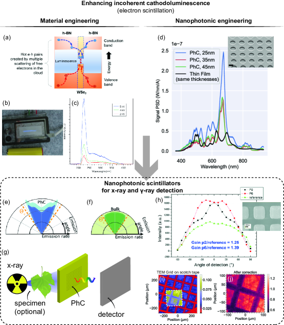
The process of light emission from fluctuating current sources in samples pumped by high-energy particles is called ICL (for free-electron pumps) or scintillation (for x- and -ray pumps). From the multi-physics picture illustrated in Fig. 1(e), there appears to be at least two ways to enhance it: (1) control of the available emitting states in the electronic band structure (material engineering) and; (2) control of the nanophotonic environment (nanophotonic engineering).
Material engineering.
ICL can be enhanced by engineering the electronic band structure of emitting materials, and specifically available defect states or band gap to make bright emitters. Such techniques are also ubiquitous in the context of brighter and faster x-ray scintillator development, which rely on the development of new materialsGektin and Korzhik (2017) (typically, with large atomic numbers, high density, and doped with rare earth elements to emit strongly in the UV-NIR).
In the context of ICL, two-dimensional materials and van der Waals heterostructures have received particular attention, given their bright emission properties and exceptional nanophotonic properties.Basov, Fogler, and García De Abajo (2016); Caldwell et al. (2019) Specifically, bright UV emission from hexagonal boron nitride has been demonstrated, with emission patterns reminiscent of a lasing behavior.Watanabe, Taniguchi, and Kanda (2004) Such substrates were then integrated into handheld devices to demonstrate their applicability to sanitizationWatanabe et al. (2009) (see Fig. 7(c, d)). Those achievements have been enabled by the control of hexagonal boron nitride’s excitonic properties, by growing crystals under high pressure and temperature. Recent work utilized ICL as a probe of the material’s properties down to the few-layer regime.Elias et al. (2019)
In semiconductors, the emission process is similar to LEDs, and the electron-hole pair recombination probability limits the emission efficiency. In van der Waals heterostructures, engineering of the excitonic properties of an ICL emitter can be realized, for instance by "sandwiching" a transition metal dichalcogenide monolayers between two layers of hexagonal boron nitride, resulting in several orders of magnitude ICL enhancement.Zheng et al. (2017)
Another common method for enhancing ICL and scintillation is by fabricating semiconductor quantum dots whose small dimensions cause quantum confinement of the excited charge carriers, resulting in larger dipoles and enhanced emission rates. Chen et al. (2018)
Nanophotonic engineering.
The other avenue to enhance ICL and scintillation is to engineer the nanophotonic environment in which the fluctuating current sources from Eq. (4) radiate. The possibility of nanophotonic enhancement can be explained by Eqs. (12, 13).
Scintillation and ICL being in essence spontaneous emission, they can be enhanced with nanophotonics in two distinct ways: (1) via direct enhancement of the rate of spontaneous emission by shaping the density of optical states Kurman et al. (2020); Purcell (1946) (as evident from the eigenmode formulation in Eq. (12)); or (2) through improved light extraction from bulk scintillators (as evident from the far-field formulation in Eq. (13)). The prospect of enhancing scintillation through the local density of states, as well as the prospect of large scintillation enhancements, by either mechanism, had remained unrealized until recently.Roques-Carmes et al. (2022a)
A recent demonstration of enhanced ICL from self-trapped hole defects in silica is shown in Fig. 7(e), where the emitted ICL from a defect transition emitting around nm is enhanced and spectrally-shaped. This is enabled by the presence of several high- photonic resonances due to the PhC patterning at the surface of the scintillator. In the same work, ICL is used as a spectroscopic probe of the nonlinear microscopic dynamics accounting for competing emission processes. Given the generality of Eqs. (12, 13), we note that similar techniques can be used to enhance emission from a stochastic ensemble of emitters, which could find applications beyond ICL and scintillation: in thermal radiation,Greffet et al. (2002, 2018); Overvig, Mann, and Alù (2021); Iyer et al. (2020) LEDs,Liu et al. (2018a) and photoluminescence.Liu et al. (2018a)
III.3.3 Nanophotonic scintillators for x- and -ray detection
Our approach highlights the common physical origin of ICL (from free electrons) and scintillation from x- and -rays. In a broader context scintillators are used for applications across medical imaging, non-destructive testing, and night vision technologies.Gektin and Korzhik (2017) This analogy holds especially for low-energy x-rays whose penetration depths are comparable to electrons in SEM and TEM.
The possibility of nanophotonic scintillation enhancement for x- and -ray application was recently revived by the availability of large-scale deposition and patterning techniques compatible with state-of-the-art scintillators in x-ray and nuclear imaging. Early work demonstrated enhanced light extraction provided by a PhC coating atop a bulk scintillator Liu et al. (2018b); Knapitsch et al. (2012); Knapitsch and Lecoq (2015); Zhu et al. (2015); Pignalosa et al. (2012); Liu et al. (2021b); Ouyang et al. (2020) (see Fig. 7(i)). It was then realized that x-ray scintillation could be enhanced via local photonic density of state enhancement in multi-layered structure, a convenient platform to also shape the scintillation angular emission profile.Kurman et al. (2020) Taking a step forward, a general framework for scintillation in nanophotonics was proposed, Roques-Carmes et al. (2022a) with experimental demonstrations of both enhancement mechanisms in two distinct platforms (ICL and x-ray scintillation). The perspective of using such nanophotonic scintillators for x-ray imaging was demonstrated in the same work (see Fig. 7(j,k)).
IV Future perspectives
In this section, we outline several research directions, which we believe will significantly flourish in the next few years. In particular, we highlight the use of nanophotonics in enabling these novel applications of (quantum) electron-light interactions.
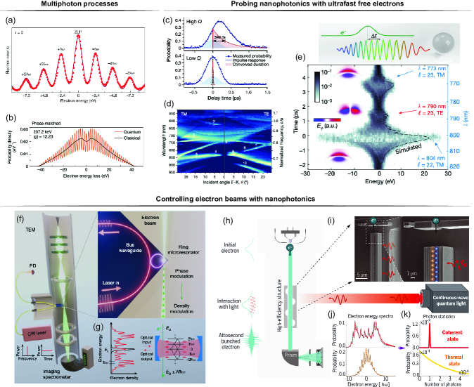
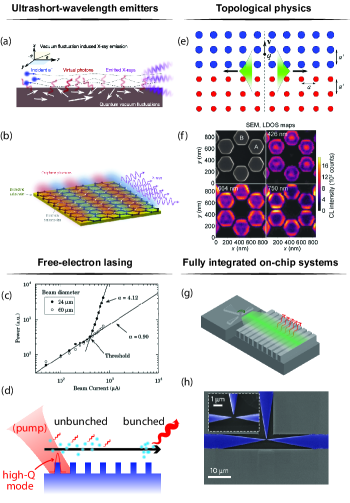
IV.1 Quantum effects
Much of the content of this Review can be understood in purely classical terms: in terms of solutions to the classical Maxwell’s wave equation in a dielectric medium, with a source (a moving electron). Even in the case of ICL (scintillation), the radiation may be understood in terms of radiation from classically non-deterministic current sources, although the correlations of the current sources may be dictated by the quantum statistics of the radiating currents (e.g., Bose-Einstein or Fermi-Dirac statistics). Nowadays, there is a great deal of effort in theoretically predicting, as well as experimentally isolating, genuinely quantum mechanical effects in free-electron radiation, which may not be described in terms of the classical wave equation with a source. In this section, we give a brief outline of the different types of quantum effects that can emerge in free-electron radiation. For more discussion of such effects (with different emphasis), we refer the reader to Ref. Rivera and Kaminer (2020); García De Abajo and Di Giulio (2021).
IV.1.1 Recoil effects
As discussed in section II, much of the spectral and angular properties of coherent cathodoluminescence may be understood from the phase-matching relation Eq. (1). This phase-matching, while naturally following from the classical Maxwell’s equations, can also be understood in quantum mechanical terms. Taking the emitted photon to have a four-momentum and the electron to have its own (initial / final ) four-momentum , with the electron energy and the electron momentum - one finds that the phase-matching condition is nothing other than an approximate solution to the energy-momentum conservation condition : under the approximation that the electron energy and momentum change only weakly due to the emission. For almost all cases in electron-photon interactions in nanophotonics, this approximation is very accurate, since the typical energy of the photon (1 eV), is quite small compared to that of the electron (in excess of 1 keV). The main regimes in which the recoil approximation may break down are: low electron energies,Tsesses, Bartal, and Kaminer (2017); Talebi (2016b); Huang et al. (2022a) high photon energies,Wong et al. (2015); Rivera et al. (2019); Rosolen et al. (2018) and near velocity thresholdsKaminer et al. (2016a) (between which the emission is kinematically forbidden or allowed). All three of these regimes have been subject of theoretical investigation: as of this writing, clear quantum recoil effects in coherent CL have not been observed (for SPR, the region where such quantum effects should arise is shown in Fig. 4).
IV.1.2 Quantum interference and waveshaping effects
Another way by which quantum effects can alter light emission by electrons is through electron waveshaping. One might imagine that controlling the wavefunction of the emitting electron will control the emitted radiation in much the same way that controlling the shape of the current density in Maxwell’s equations controls the radiation. The situation in quantum mechanics is more subtle, as the wavefunction of the electron does not represent a smeared out charge density. If it did, one could imagine that simply expanding or compressing the electron wavefunction transverse to its motion would change its radiation (e.g., by Cherenkov or Smith-Purcell emission): tunable transverse coherent size has shown that the intensity does not depend on the electron wavefunction. Remez et al. (2019) In fact, the transverse size had no influence on the emitted radiation, as first predicted for CR. Kaminer et al. (2016a) Despite this, the shape of the electron wavefunction can alter the radiation, under specific circumstances: namely, if the emitted photon has enough symmetries broken. For example, in a recent experiment involving EELS, it was found that the electron energy-loss spectrum could be modified by shaping the electron wavefunction to have its symmetry be compatible with different localized (plasmonic) modes of a structure.Vanacore et al. (2017) Namely, the electron could lose energy primarily through a dipolar mode of the structure (of one energy) when the electron wavefunction had a certain symmetry, and lose energy primarily through a quadrupolar mode (of a different energy) when the electron had a different symmetry. This shows the influence of the electron wavefunction on plasmon emission.
The key constraint that enables the dependence on the wavefunction is that the measurement system performs post-selection on the electron (by conditioning the measurement on the electron state). Without electron post-selection, the radiation intensity is independent on the electron wavefunction. Kaminer et al. (2016a); García De Abajo and Di Giulio (2021); Di Giulio et al. (2021) Unlike with the radiation intensity, other properties of the radiation such as coherence can be modified by shaping the electron wavefunction. This was first predicted in Ref. Karnieli et al. (2021), showing that the pulse duration of CR depends on the electron wavefunction. Building on this concept, electron wavefunction shaping was proposed as a method to control the quantum states of free-electron radiation. Ben Hayun et al. (2021); Wong et al. (2022)
Recent theoretical work has also shown how the emission of high-energy photons such as x-rays can be controlled through quantum interference effects.Wong et al. (2021) In that work, it was shown how quantum interference of amplitudes could lead to strong control over phenomena such as bremsstrahlung radiation, where an electron emits radiation after scattering off of an external (Coulomb) potential. In particular, two different initial electron states can scatter into the same final electron and emitted photon states, leading to quantum interference. Such interference can only occur when the external potential breaks sufficient symmetries to enable both transitions to conserve energy and momentum simultaneously. Beyond this, the effect of the electron wavefunction on the entanglement structure of the emitted radiation has been explored theoretically in other examples of coherent CL (e.g., CR).Kaminer et al. (2016a) Ref. Kfir et al. (2021) also showed the existence of phase correlations between the emitted CL field and the electron-modulating laser in the quantum-optical regime.
IV.1.3 High-order effects in quantum electrodynamics
A third class of important effects, which are now a rich and vibrant field on their own, is related to strong-field interactions, in which an electron can interact with a strong external field, leading to absorption or stimulated versions of coherent CL (see Fig. 8). For the types of fields that are now typical in such experiments, an electron can emit and absorb multiple (even hundreds) of quanta from the external field, leading to a large number of quantized peaks in the electron energy loss/gain spectrum of the post-interaction electron.Dahan et al. (2020) Since the first observations of this effect in 2009,Barwick, Flannigan, and Zewail (2009) which forms the basis for PINEM, this effect has been observed and extended in many guises and has been exploited for imaging electromagnetic fields in materials with high spatial (nanometer) resolution and high temporal (sub-picosecond) resolution.Park, Lin, and Zewail (2010); Piazza et al. (2015); Feist et al. (2015); Priebe et al. (2017); Morimoto and Baum (2018); Vanacore et al. (2018, 2017, 2019); Wang et al. (2020a); Dahan et al. (2020) Of particular note is that the electron energy spectrum after the interaction is sensitive to the quantum statistics of the field, Gorlach et al. (2020) as recently demonstrated in Ref. Dahan et al. (2021) to distinguish between thermal and coherent light. Such effects form the basis for the extension of quantum optics to free electrons. High-order processes have also been proposed in CR from heavy ions, resulting in modified emission angles and suppression of radiation pathways close to the threshold. Roques-Carmes et al. (2018) More recently, quantum regimes of interactions between free electrons and photons were observed at much lower energies in a SEM. Shiloh, Chlouba, and Hommelhoff (2022)
IV.2 Ultrashort-wavelength emitters
Free electrons are ideal platforms to generate high-energy radiation, since they carry large kinetic energies (in the keV to MeV range in most electron microscopes) that can be converted into electromagnetic radiation. Among the effects we have discussed in this Review, SPR displays the most evident wavelength tunability, via the incident electron velocity and the structure periodicity. In Fig. 4, we highlighted recent work towards SPR from low-energy (sub-keV) electrons and short-wavelength photons. Specifically, there has been excitement around achieving tunable SPR in the UV.García de Abajo (1999) There has been renewed interest in using SPR to generate UV light, with recent experimental demonstrations down to a wavelength of 230 nm. The perspective of generating UV light with SPR is especially promising for sanitization applications.Garciá De Abajo et al. (2020)
Generation of much shorter wavelength radiation has been reported in crystals, whose structure is naturally "modulated" at ångström scales. Recently, such effects found renewed interest in van der Waals heterostructures pumped by free electrons emitting soft x-ray light,Shentcis et al. (2020) with enhanced functionality demonstrated in Ref. Huang et al. (2022b). The ability to control coherent CL with engineered nanostructures is especially exciting for short-wavelength radiation generation, given the unavailability of efficient focusing optics in this wavelength range. Therefore, the ability to concentrate coherent CL has received specific attention recently, with proposals in the near-UVRemez et al. (2017) and x-ray regime.Shi et al. (2022); Balanov, Gorlach, and Kaminer (2021)
Pumping of wide-bandgap materials with free electrons is another way to generate bright UV light. In particular, hexagonal boron nitride was proposed as a bright UV emitter, exhibiting lasing behavior,Watanabe, Taniguchi, and Kanda (2004) and the possibility of being integrated in compact devices.Watanabe et al. (2009) The existence of a large catalogue of wide-bandgap semiconductors,Takahashi et al. (2007) in addition to the availability of nanophotonic enhancement and shaping,Roques-Carmes et al. (2022a) and the theoretical possibility of large conversion efficiencies,Klein (1968) makes UV ICL a particularly promising research direction.
IV.3 Nanophotonic dielectric laser accelerators
The time-reversal process of free-electron emission generation is particle acceleration. Similarly, nanophotonic development leads to new designs, device miniaturization, and performance improvement in particle accelerators. Plettner and Byer (2008); England et al. (2014); Breuer and Hommelhoff (2013); Peralta et al. (2013); Shiloh et al. (2021)
A channel for electron propagation usually has to be defined for accelerators; this limits the current applied to the device. To address this issue, a multi-channel accelerator structure was proposed, Zhao et al. (2020) which naturally transforms the seminal dual-grating accelerating structure into PhCs, thus allowing device designs via 2D and 3D band structure engineering. The use of photonic flatband proposed for enhancing SPR Yang et al. (2021) can be used in reverse to aid in optical localization and enhancement of electron acceleration without the need for electron channels, thus allowing more electrons to interact with accelerator devices.
Experimentally, an inverse-designed (see section below), waveguide-integrated, microscale dielectric laser accelerator was fabricated and tested, showing acceleration gradients around 30 MeV/m in a recent demonstration. Sapra et al. (2020) Meanwhile, electron phase space control was realized via introducing phase discontinuity (similar to a lattice dislocation) in a high-aspect-ratio, silicon-based, integrated structure, Shiloh et al. (2021) indicating the possibility for particle acceleration of confined beams to the MeV range with minimal particle loss over extended distances. Quantized peaks in electron energy spectra have also been observed in dielectric laser accelerators, revealing the quantum nature of such devices.Adiv et al. (2021)
IV.4 Inverse-designed free-electron radiators and accelerators
Computational inverse design has now penetrated every single field of nanophotonics. Molesky et al. (2018); Jensen and Sigmund (2011) Free-electron radiation will be no exception. In general, inverse design enables the automatic discovery of nanophotonics structures that are locally optimal for a pre-defined objective function. Among the various inverse-design techniques available, topology optimization is one of the most popular, given the many degrees of freedom it can investigate, while remaining computationally tractable, thanks to efficient gradient calculation methods (such as the adjoint methodMolesky et al. (2018) and automatic differentiation techniquesJin et al. (2020)). Topology optimization enables the inverse design "pixel by pixel" of various nanophotonic components, such as metasurfaces,Lin et al. (2018b) multiplexers,Piggott et al. (2015) and nonlinear frequency converters,Lin et al. (2016) while taking into account fabrication constraints.Hammond et al. (2021)
Topology optimization has recently been utilized to discover optimal nanophotonic structures as free-electron radiators, nanophotonic scintillators,Roques-Carmes et al. (2022a) radiators and accelerators.Haeusler et al. (2022); Sapra et al. (2020) Several features of free-electron radiation make it amenable to inverse design. First, electron-light interaction lends itself to closed-form expressions describing radiation and acceleration processes (such as the total emitted power in a range of angles, frequencies, and polarizations; or the particle acceleration gradient). Those expressions are enabled by the modeling of electron-light interactions mediated by the current sources from Eqs. (2, 4). Second, numerical methods are available in the time- and frequency- domains to calculate those expressions and their respective gradients efficiently, either via the adjoint method, or via automatic differentiation algorithms. This enables the implementation of efficient gradient-based discovery of complex nanophotonic structures interacting efficiently with free electrons. We also expect that recent efforts in inverse design for free-electron physics will bolster interest in fully-differentiable multi-physics pipelines, integrating electron trajectories and energy loss, microscopic dynamics, and nanophotonics.Roques-Carmes et al. (2022a)
IV.5 Free-electron lasing in nanophotonic structures and other exotic effects
As discussed earlier in this Review, the various coherent mechanisms by which a free electron can emit light (e.g., CR, SPR, TR) can be thought of as elementary processes of spontaneous emission in which an electron transitions to a state of lower kinetic energy while emitting a quantized mode of Maxwell’s equations (a photonic quasiparticle Rivera and Kaminer (2020)), whose properties (e.g., energy, momentum, polarization, and other quantum numbers) strongly control the observed properties of the emission. From this “quantum” perspective of free-electron radiation, the general principles of quantum mechanics determine that an electron should be able to absorb photonic quasiparticles (leading to inverse processes, such as inverse CRFontana and Pantell (1998)) and also stimulatedly emit photonic quasiparticles - leading to amplification and lasing phenomena.Schächter and Ron (1989)
Although we have used quantum arguments to infer the possibility of stimulated emission and free-electron lasing, much of the known phenomena can be described classically. In fact, the "stimulated emission" of light by a beam of classical electrons simply corresponds to net deceleration of a beam of electrons in an applied electromagnetic field: by energy conservation, the lost energy of the electrons goes into the field (into photons). Meanwhile, "absorption" of photons by the electrons corresponds to net acceleration.
Net stimulated emission by a beam of electrons forms the basis for a number of developed and developing technologies. In the microwave domain, these effects form the basis for established high-power microwave sources such as traveling wave tubes and klystrons used in space communication systems.Pierce (1950); Xu and Xu (2020) In traveling wave tubes and klystrons, such “lasing” is largely based on SPR. In the x-ray domain, amplification has also been achieved based on the collective interactions of a dense beam of electrons with a spatially periodic magnetic field. In such systems, the electrons can amplify their own spontaneous emission, when the density of the electron beam is sufficiently high.
As of this writing, free-electron lasing based on coherent CL in nanophotonic systems is yet to be observed, despite reported signatures of superradiance at THz frequencies,Urata et al. (1998) shown in Fig. 9(c). Such observations represent not only a logical step in the development of compact and integrated free electron radiation sources, but may also grant some practical advantages. Compared to CL, the coherence of the lasing field should be quite high (especially in architectures based on an optical feedback cavity,Andrews and Brau (2004); Pellegrini, Marinelli, and Reiche (2016) such as a PhC resonance,Rivera et al. (2020); Kumar and Kim (2006) as shown in Fig. 9(d)).
Lastly, other unconventional effects in free-electron radiation, such as backward CR, remain unobserved as of this writing. Originally predicted in left-handed materials, Veselago (1968); Chen and Chen (2011); Luo et al. (2003); Wu et al. (2007); Matloob and Ghaffari (2004) and then in PhCs, Luo et al. (2003) this effect has not been observed yet with free electrons (despite observations using free-electron analogues Xi et al. (2009)). The observation of this effect in nanophotonics with free electrons could open a way to novel high-energy particle detector designs.
IV.6 Topological effects in free-electron radiation
Topological photonics Lu, Joannopoulos, and Soljačić (2014); Ozawa et al. (2019) has undergone rapid development over the past decade. So far, optical excitations have been mostly employed for these studies. In previous sections, we have shown that photonic topological features, such as BICs Yang et al. (2018a); Song et al. (2018) and flatbands, Yang et al. (2021) can be used for boosting radiation generation from free electrons. In the following, we outline other opportunities at the intersection between free-electron nanophotonics and topological photonics.
Several recent advances show that free electrons can serve as probes for photonic topology. In Ref. Peng et al. (2019), the local density of states and the band structure of a topological PhC were mapped out via CL techniques, as shown in Fig. 9(f). In Ref.Yu et al. (2019), highly relativistic electron beams were used to excite the domain-wall edge states of a quantum-spin-Hall-like metamaterial, where localization to the edge was identified (see Fig. 9(e)). In Ref.Yu et al. (2020), free electrons were proposed for resolving the optically-driven phase transition of a graphene-dielectric metamaterial. Besides, it is worth mentioning that photonic topological properties can also be potentially adapted, in turn, for diagnosing the location, energy, and duration of charged particle beams.
Furthermore, free electrons can serve as pumps for photonic topology. It has been shown that free electron beams can drive nonreciprocity in metallic waveguides, therefore giving rise to unidirectionally-propagating modes. Fallah et al. (2021) Because the free electron velocity can be substantially higher than that in solids, the bandwidth of the resulting unidirectionality could be dramatically increased.
IV.7 Non-equilibrium emission processes with free electrons
The recent development of a general framework to predict non-equilibrium radiation phenomena such as scintillation (or ICL) reveals the sensitive dependence of the scintillation spectrum on the non-equilibrium distribution of the excited electrons generated by the high-energy electron beam. Roques-Carmes et al. (2022a) In particular, the radiated photons can inherit the non-equilibrium statistical properties of the excited electrons and holes generated in the scintillation process. Consider an interesting example which has yet to be observed in scintillation: when electrons and holes in semiconductors are produced by a high-energy pump, the electrons and holes quickly relax into a non-equilibrium quasi-steady state which can be described by quasi-Fermi levels for the electrons and holes which are different. In such a case, the radiated photons must (by detailed balance) experience an effective chemical potential dictated by the difference between the electron and hole chemical potentials.Wurfel (1982); Greffet et al. (2018) This is of course in complete contrast to thermal radiation, in which the photons have no chemical potential. More broadly, by controlling the energy of the high-energy electron pump, different non-equilibrium distributions of electrons and holes can be conceivably created, shaping the quantum statistical properties of the radiated photons. The ability to achieve this would represent a new way to control the quantum optical properties of photons. Such effects could be measured through second-order intensity correlation () measurements, as applied to ICL,Meuret et al. (2015); Bourrellier et al. (2016) where it was shown that for low enough electron-currents, the radiation could show signatures of antibunching, indicating single-photon emission, or signatures of bunching, indicating super-Poissonian statistics, depending on the material.
IV.8 Fully integrated on-chip systems
The use of compact nanoscale structures to convert energy from free electrons into radiation has spurred interest into realizing fully on-chip systems. This perspective is enabled by the realization of on-chip electron emitters, such as field emitter arrays. Guerrera and Akinwande (2016a, b); Temple (1999); Spindt et al. (1991). Such devices can realize focused electron beams, in continuous wave or pulsed regimes, with relatively low kinetic energies (from few tens of keV to few hundreds of eV).
It was first realized that Smith-Purcell and Cherenkov systems could be fully integrated on chip Tang et al. (1996); Ishizuka et al. (2001, 2000); Neo et al. (2006), thereby enabling integrated, tunable light emitters. One of the additional advantages of fully integrated systems is the ability to precisely align the electron emitter with nanophotonics structures, thereby optimizing free-electron emission. One such device is shown in Fig. 9(g), with a recent design of electron emitter shown in Fig. 9(h). Recent work focused on the realization of all-silicon Smith-Purcell emitters Roques-Carmes et al. (2019) and on-chip systems emitting CR from hyperbolic metamaterials. Liu et al. (2017a) The realization of fully integrated electron accelerator devices has also been the focus of much research recently. Sapra et al. (2020); Shiloh et al. (2021); Niedermayer et al. (2021); Niedermayer, Boine-Frankenheim, and Egenolf (2017); Black et al. (2019)
V Conclusion
In conclusion, we have provided an extensive review of the fundamental physics and applications of free-electron interactions with nanophotonic structures. We first proposed a general classical framework to model cathodoluminescence as the excitation of photonic eigenmodes by the free-electron current density. This excitation can be coherent, as is the case for all coherent CL effects, or partially incoherent, as is the case for ICL and scintillation. This framework enabled us to re-derive fundamental formula in free-electron physics, and to provide a unified method to shape and enhance CL. It also enabled us to derive fundamental bounds on electromagnetic radiation from free electrons in arbitrary nanophotonic environments.
Those methods have been confirmed and observed thanks to the development of experimental methods to observe and characterize light emission from free electrons in nanophotonic structures. Specifically, we reviewed the development of CL and EELS measurement techniques in SEM and (ultrafast) TEM. The several experimental methods we reviewed have enabled the measurement of all optical properties of interest: spectral, angular, time, spatial, polarization distributions, and quantum correlations. They can also be complemented by measurement of the electron properties with EELS.
Finally, we shared our enthusiasm for a few avenues in free-electron radiation enabled by nanophotonics, namely the emerging fields of ultrafast, topological, and quantum effects in electron microscopy, the design of ultrashort-wavelength emitters pumped by free electrons, miniaturized particle accelerators, novel inverse-design techniques applied to free-electron emitters, and the aspiration to reach stimulated emission and lasing by free electrons in nanophotonics. While the realization of some of those effects might still be a few years ahead of us, we believe that the strong interest from our community has already demonstrated the potential of nanophotonics in controlling and enhancing CL. Future developments in the field might unlock some of the most promising prospects in free-electron physics, such as the realization of widely tunable integrated light sources from x-rays to mm-wave, particle accelerators, and high-energy particle detectors. They should find direct applications across particle detection and acceleration, electron microscopy, dosimetry, water sanitization, medical imaging and therapy.
VI Authors’ contribution
C. R.-C. wrote the manuscript with inputs from S. E. K., Y. Y., N. R., and P. D. K. All authors read and reviewed the final manuscript.
VII Data availability
The data that support the findings of this study are available within the article and upon request for Fig. 4.
VIII Acknowledgments
The authors would like to thank Nikolay I. Zheludev, Kevin MacDonald, and Liang Jie Wong for their helpful comments on the Review. This material is based on work supported in part by the US Army Research Laboratory and the US Army Research Office through the Institute for Soldier Nanotechnologies under contract W911NF-18-2-0048. This material is also in part based on work supported by the Air Force Office of Scientific Research under awards FA9550-20-1-0115 and FA9550-21-1-0299.
Appendix A Appendix: Unified theory of coherent cathodoluminescence
In this Appendix, we provide more details on the derivation of emitted power densities for coherent CL. We then re-derive emblematic formulas of the field for CR, TR, and excitation of SPPs.
We derive our theory in the context of classical electrodynamics, with the analogy of a moving free electron as a time-dependent current source. We consider the case of a moving electron of charge propagating along the linear trajectory defined by the speed vector , which is described by the current distribution of Eqs. (2, 3).
This expression of the current in Fourier domain already highlights the existence of preferred field wavevectors to which the electron beam will couple to, which is the essence of the phase-matching condition shown in Eq. (1). The nanophotonic environment is described by the Green’s function expansion shown in Eq. (5).
The total energy radiated by a dipole can be calculated as an integral in frequency domain:Kremers, Chigrin, and Kroha (2009)
| (14) |
We then get the master equation shown in Eq. (6), which describes all coherent CL effects considered in this Review. In the next sections, we provide more details on its application the cases of CR, TR, and excitation of SPPs.
A.1 Cherenkov radiation
CR in its simplest embodiment occurs in a homogeneous dielectric environment and consists in the spontaneous emission of plane waves by a charged particle (see Fig. 3(a)). Its description entails eigenmodes as plane waves of the form , in a uniform medium of refractive index with polarization vector orthogonal to , and . The dispersion relation is . We also make the following adjustment . Injecting this expression into Eq. (6), we get the Frank-Tamm formulaFrank and Tamm (1937) for the spectral density per unit propagation length:
| (15) |
with the Heaviside function ( if , otherwise ). Here, the Cherenkov angle is defined such as . The Heaviside function defines the well-known Cherenkov threshold , which is equivalent to satisfying the phase-matching condition from Eq. (1) in a bulk medium.
A.2 Transition radiation
To describe transition radiation within the same framework, we consider the simplified case of a charge impinging at normal incidence on a perfect conductor (see Fig. 3(c)). We resort to the introduction of an image charge with opposite charge and velocity . The current distribution is modified accordingly and its emission in free space is considered. The current distribution is given by:
| (16) | ||||
| (17) |
and its Fourier transform
| (18) |
with the sign function. Utilizing the free space mode expansion as in the case of Cherenkov radiation, we get the following spectral distribution, first derived by Ginzburg and Tamm:Ginzburg (1940)
| (19) |
A.3 Coherent excitation of SPPs by free electrons
Excitation of SPPs is often considered in tandem with TR, since it typically occurs at the interface between two media supporting SPP. This phenomenon is observed even in the absence of corrugation at the surface because the electron point nature enables coupling directly to the SPP modes. Our formalism captures this effect by considering emission into the SPP modes rather than free space modes as before.
For simplicity, we consider an electron flying parallel to a planar interface at (reduced to a 2D problem). We consider the simplified case of a lossless interface between a dielectric (relative permittivity ) and a metal (relative permittivity ). The -polarized modes take the form (in the basis):
| (20) | ||||
| (21) |
And the following dispersion relation:
| (22) |
We obtained the following expression for the energy emitted by the free electron per unit length:
| (23) |
with the phase-matching condition:
| (24) |
which can be solved graphically by finding the intersection of the SPP’s dispersion and the electron line . Such an intersection always exists for the lower branch (corresponding to the excitation of a SPP).
References
References
- Crooke (1879) W. Crooke, “XVI. Contributions to molecular physics in high vacua. Magnetic deflection of molecular trajectory. - Laws of magnetic rotation in high and low vacua. - Phosphorogenic properties of molecular discharge,” Philosophical Transactions of the Royal Society of London 170, 641–662 (1879).
- Cherenkov (1934) P. A. Cherenkov, “Visible light from clear liquids under the action of gamma radiation,” Dokl. Akad. Nauk SSSR 2 (1934).
- Ginzburg and Frank (1945) V. Ginzburg and I. Frank, “Radiation of a uniformly moving electron due to its transition from one medium into another,” Zh.Eksp.Teor.Fiz. 9, 353–362 (1945).
- Smith and Purcell (1953) S. J. Smith and E. M. Purcell, “Visible light from localized surface charges moving across a grating,” Physical Review 92, 1069 (1953).
- Ritchie (1957) R. H. Ritchie, “Plasma Losses by Fast Electrons in Thin Films,” Physical Review 106, 874 (1957).
- Luo et al. (2003) C. Luo, M. Ibanescu, S. G. Johnson, and J. D. Joannopoulos, “Cerenkov radiation in photonic crystals,” Science 299, 368–371 (2003).
- Bashevoy et al. (2006) M. V. Bashevoy, F. Jonsson, A. V. Krasavin, N. I. Zheludev, Y. Chen, and M. I. Stockman, “Generation of traveling surface plasmon waves by free-electron impact,” Nano Letters 6, 1113–1115 (2006).
- Van Wijngaarden et al. (2006) J. T. Van Wijngaarden, E. Verhagen, A. Polman, C. E. Ross, H. J. Lezec, and H. A. Atwater, “Direct imaging of propagation and damping of near-resonance surface plasmon polaritons using cathodoluminescence spectroscopy,” Applied Physics Letters 88, 221111 (2006).
- Bashevoy et al. (2007) M. V. Bashevoy, F. Jonsson, K. F. MacDonald, Y. Chen, and N. I. Zheludev, “Hyperspectral imaging of plasmonic nanostructures with nanoscale resolution,” Optics Express, Vol. 15, Issue 18, pp. 11313-11320 15, 11313–11320 (2007).
- Vesseur et al. (2007) E. J. R. Vesseur, R. De Waele, M. Kuttge, and A. Polman, “Direct Observation of Plasmonic Modes in Au Nanowires Using High-Resolution Cathodoluminescence Spectroscopy,” Nano Letters 7, 2843–2846 (2007).
- Hofmann et al. (2007) C. E. Hofmann, E. J. R. Vesseur, L. A. Sweatlock, H. J. Lezec, F. J. G. De Abajo, A. Polman, and H. A. Atwater, “Plasmonic Modes of Annular Nanoresonators Imaged by Spectrally Resolved Cathodoluminescence,” Nano Letters 7, 3612–3617 (2007).
- Adamo et al. (2009) G. Adamo, K. F. MacDonald, Y. H. Fu, C. M. Wang, D. P. Tsai, F. J. García De Abajo, and N. I. Zheludev, “Light well: A tunable free-electron light source on a chip,” Physical Review Letters 103 (2009).
- Adamo et al. (2010) G. Adamo, K. F. MacDonald, Y. H. Fu, D. P. Tsai, F. J. García De Abajo, and N. I. Zheludev, “Tuneable electron-beam-driven nanoscale light source,” Journal of Optics A: Pure and Applied Optics 12 (2010).
- Barnard et al. (2011) E. S. Barnard, T. Coenen, E. J. R. Vesseur, A. Polman, and M. L. Brongersma, “Imaging the Hidden Modes of Ultrathin Plasmonic Strip Antennas by Cathodoluminescence,” Nano Letters 11, 4265–4269 (2011).
- Adamo et al. (2012) G. Adamo, J. Y. Ou, J. A. So, S. D. Jenkins, F. Deaangelis, K. F. MacDonald, E. Diafabrizio, J. Ruostekoski, and N. I. Zheludev, “Electron-beam-driven collective-mode metamaterial light source,” Physical Review Letters 109, 1–5 (2012).
- Kaminer et al. (2017a) I. Kaminer, S. Kooi, R. Shiloh, B. Zhen, Y. Shen, J. López, R. Remez, S. A. Skirlo, Y. Yang, J. D. Joannopoulos, A. Arie, and M. Soljačić, “Spectrally and Spatially Resolved Smith-Purcell Radiation in Plasmonic Crystals with Short-Range Disorder,” Physical Review X 7, 011003 (2017a).
- Liu et al. (2017a) F. Liu, L. Xiao, Y. Ye, M. Wang, K. Cui, X. Feng, W. Zhang, and Y. Huang, “Integrated Cherenkov radiation emitter eliminating the electron velocity threshold,” Nature Photonics 2017 11:5 11, 289–292 (2017a).
- Massuda et al. (2017a) A. Massuda, C. Roques-Carmes, Y. Yang, S. E. Kooi, Y. Yang, C. Murdia, K. K. Berggren, I. Kaminer, and M. Soljačić, “Smith-Purcell radiation from low-energy electrons,” ACS Photonics 5 (2017a).
- Yang et al. (2018a) Y. Yang, A. Massuda, C. Roques-Carmes, S. E. Kooi, T. Christensen, S. G. Johnson, J. D. Joannopoulos, O. D. Miller, I. Kaminer, and M. Soljačić, “Maximal spontaneous photon emission and energy loss from free electrons,” Nature Physics 14, 894–899 (2018a).
- Sapra et al. (2020) N. V. Sapra, K. Y. Yang, D. Vercruysse, K. J. Leedle, D. S. Black, R. Joel England, L. Su, R. Trivedi, Y. Miao, O. Solgaard, R. L. Byer, and J. Vučković, “On-chip integrated laser-driven particle accelerator,” Science 367, 79–83 (2020).
- Shentcis et al. (2020) M. Shentcis, A. K. Budniak, X. Shi, R. Dahan, Y. Kurman, M. Kalina, H. Herzig Sheinfux, M. Blei, M. K. Svendsen, Y. Amouyal, S. Tongay, K. S. Thygesen, F. H. Koppens, E. Lifshitz, F. J. García de Abajo, L. J. Wong, and I. Kaminer, “Tunable free-electron X-ray radiation from van der Waals materials,” Nature Photonics 2020 14:11 14, 686–692 (2020).
- Kfir et al. (2020) O. Kfir, H. Lourenço-Martins, G. Storeck, M. Sivis, T. R. Harvey, T. J. Kippenberg, A. Feist, and C. Ropers, “Controlling free electrons with optical whispering-gallery modes,” Nature 2020 582:7810 582, 46–49 (2020).
- Wang et al. (2020a) K. Wang, R. Dahan, M. Shentcis, Y. Kauffmann, A. Ben Hayun, O. Reinhardt, S. Tsesses, and I. Kaminer, “Coherent interaction between free electrons and a photonic cavity,” Nature 582, 50–54 (2020a).
- Nussupbekov et al. (2021) A. Nussupbekov, G. Adamo, J. K. So, L. Wu, Y. D. Chong, and L. J. Wong, “Enhanced photon emission from free electron excitation of a nanowell,” APL Photonics 6, 096101 (2021).
- Roques-Carmes et al. (2022a) C. Roques-Carmes, N. Rivera, A. Ghorashi, S. E. Kooi, Y. Yang, Z. Lin, J. Beroz, A. Massuda, J. Sloan, N. Romeo, Y. Yu, J. D. Joannopoulos, I. Kaminer, S. G. Johnson, and M. Soljačić, “A framework for scintillation in nanophotonics,” Science 375 (2022a).
- García De Abajo (2010) F. J. García De Abajo, “Optical excitations in electron microscopy,” Reviews of Modern Physics 82, 209–275 (2010).
- Polman, Kociak, and García de Abajo (2019) A. Polman, M. Kociak, and F. J. García de Abajo, “Electron-beam spectroscopy for nanophotonics,” Nature Materials 18, 1158–1171 (2019).
- Talebi (2017) N. Talebi, “Interaction of electron beams with optical nanostructures and metamaterials: from coherent photon sources towards shaping the wave function,” Journal of Optics 19, 103001 (2017).
- Coenen, Hoedt, and Polman (2016) T. Coenen, S. V. d. Hoedt, and A. Polman, “A New Cathodoluminescence System for Nanoscale Optics, Materials Science, and Geology,” Microscopy Today 24, 12–19 (2016).
- Christopher et al. (2020) J. Christopher, M. Taleb, A. Maity, M. Hentschel, H. Giessen, and N. Talebi, “Electron-driven photon sources for correlative electron-photon spectroscopy with electron microscopes,” Nanophotonics 9, 4381–4406 (2020).
- Marie Curie (1904) Marie Curie, Radio-active substances (Faculte des Sciences de Paris. Reprinted by Chemical News Office., 1904).
- Ginzburg (1996) V. L. Ginzburg, “Radiation by uniformly moving sources (Vavilov–Cherenkov effect, transition radiation, and other phenomena),” Physics-Uspekhi 39, 973–982 (1996).
- Akopov et al. (2002) N. Akopov, E. C. Aschenauer, K. Bailey, S. Bernreuther, N. Bianchi, G. P. Capitani, P. Carter, E. Cisbani, R. De Leo, E. De Sanctis, D. De Schepper, V. Djordjadze, B. W. Filippone, S. Frullani, F. Garibaldi, J. O. Hansen, B. Hommez, M. Iodice, H. E. Jackson, P. Jung, R. Kaiser, J. Kanesaka, R. Kowalczyk, L. Lagamba, A. Maas, V. Muccifora, E. Nappi, K. Negodaeva, W. D. Nowak, T. O’Connor, T. G. O’Neill, D. H. Potterveld, D. Ryckbosch, Y. Sakemi, F. Sato, A. Schwind, T. A. Shibata, K. Suetsugu, E. Thomas, M. Tytgat, G. M. Urciuoli, K. Van de Kerckhove, R. Van de Vyver, S. Yoneyama, H. Zohrabian, and L. F. Zhang, “The HERMES dual-radiator ring imaging Cherenkov detector,” Nuclear Instruments and Methods in Physics Research, Section A: Accelerators, Spectrometers, Detectors and Associated Equipment 479, 511–530 (2002).
- Adams et al. (2001) G. Adams, V. Burkert, R. Carl, T. Carstens, V. Frolov, L. Houghtlin, G. Jacobs, M. Kossov, M. Klusman, B. Kross, M. Onuk, J. Napolitano, J. W. Price, C. Riggs, Y. Sharabian, A. Stavinsky, L. C. Smith, W. A. Stephens, P. Stoler, W. Tuzel, K. Ullrich, A. Vlassov, A. Weisenberger, M. Witkowski, B. Wojtekhowski, P. F. Yergin, and C. Zorn, “The CLAS Cherenkov detector,” Nuclear Instruments and Methods in Physics Research, Section A: Accelerators, Spectrometers, Detectors and Associated Equipment 465, 414–427 (2001).
- Nakamura (2003) K. Nakamura, “Hyper-Kamiokande - A next generation water Cherenkov detector,” International Journal of Modern Physics A 18, 4053–4063 (2003).
- Kleinknecht (1982) K. Kleinknecht, “Particle detectors,” Physics Reports 84, 85–161 (1982).
- Papanestis (2014) A. Papanestis, “The LHCb RICH system; detector description and operation,” Nuclear Instruments and Methods in Physics Research Section A: Accelerators, Spectrometers, Detectors and Associated Equipment 766, 14–18 (2014).
- Carusotto and Rousseaux (2013) I. Carusotto and G. Rousseaux, “The Cherenkov Effect Revisited: From Swimming Ducks to Zero Modes in Gravitational Analogues,” in Analogue Gravity Phenomenology (Springer, 2013) pp. 109–144.
- Pellegrini, Marinelli, and Reiche (2016) C. Pellegrini, A. Marinelli, and S. Reiche, “The physics of x-ray free-electron lasers,” Reviews of Modern Physics 88, 015006 (2016).
- Joannopoulos et al. (2011) J. D. Joannopoulos, S. G. Johnson, J. N. Winn, and R. D. Meade, Photonic Crystals: Molding the Flow of Light (Second Edition) (Princeton University Press, 2011).
- Novotny and Hecht (2009) L. Novotny and B. Hecht, Principles of Nano-Optics, Vol. 9781107005 (Cambridge University Press, 2009) pp. 1–564.
- Yu and Capasso (2014) N. Yu and F. Capasso, “Flat optics with designer metasurfaces,” Nature Materials 13, 139–150 (2014).
- Yablonovitch (1994) E. Yablonovitch, “Photonic crystals,” Journal of Modern Optics 41, 173–194 (1994).
- Vahala (2003) K. J. Vahala, “Optical microcavities,” Nature 424, 839–846 (2003).
- Armani et al. (2003) D. K. Armani, T. J. Kippenberg, S. M. Spillane, and K. J. Vahala, “Ultra-high-Q toroid microcavity on a chip,” Nature 2003 421:6926 421, 925–928 (2003).
- Gektin and Korzhik (2017) A. Gektin and M. Korzhik, Inorganic scintillators for detector systems (Springer, 2017).
- Shaffer, Pratt, and Grimm (2017) T. M. Shaffer, E. C. Pratt, and J. Grimm, “Utilizing the power of Cerenkov light with nanotechnology,” Nature Nanotechnology 12, 106–117 (2017).
- Lin et al. (2021) X. Lin, H. Hu, S. Easo, Y. Yang, Y. Shen, K. Yin, M. P. Blago, I. Kaminer, B. Zhang, H. Chen, J. Joannopoulos, M. Soljačić, and Y. Luo, “A Brewster route to Cherenkov detectors,” Nature Communications 2021 12:1 12, 1–7 (2021).
- Lin et al. (2018a) X. Lin, S. Easo, Y. Shen, H. Chen, B. Zhang, J. D. Joannopoulos, M. Soljačić, and I. Kaminer, “Controlling Cherenkov angles with resonance transition radiation,” Nature Physics 2018 14:8 14, 816–821 (2018a).
- Niedermayer, Boine-Frankenheim, and Egenolf (2017) U. Niedermayer, O. Boine-Frankenheim, and T. Egenolf, “Designing a Dielectric Laser Accelerator on a Chip,” Journal of Physics: Conference Series 874 (2017).
- Black et al. (2019) D. S. Black, K. J. Leedle, Y. Miao, U. Niedermayer, R. L. Byer, and O. Solgaard, “Laser-Driven Electron Lensing in Silicon Microstructures,” Physical Review Letters 122, 104801 (2019).
- Karnieli et al. (2021) A. Karnieli, N. Rivera, A. Arie, and I. Kaminer, “The coherence of light is fundamentally tied to the quantum coherence of the emitting particle,” Science Advances 7 (2021).
- Ben Hayun et al. (2021) A. Ben Hayun, O. Reinhardt, J. Nemirovsky, A. Karnieli, N. Rivera, and I. Kaminer, “Shaping quantum photonic states using free electrons,” Science Advances 7, 4270–4280 (2021).
- Wong et al. (2021) L. J. Wong, N. Rivera, C. Murdia, T. Christensen, J. D. Joannopoulos, M. Soljačić, and I. Kaminer, “Control of quantum electrodynamical processes by shaping electron wavepackets,” Nature Communications 2021 12:1 12, 1–10 (2021).
- García De Abajo and Di Giulio (2021) F. J. García De Abajo and V. Di Giulio, “Optical Excitations with Electron Beams: Challenges and Opportunities,” ACS Photonics 8, 945–974 (2021).
- Di Giulio et al. (2021) V. Di Giulio, O. Kfir, C. Ropers, and F. J. García De Abajo, “Modulation of Cathodoluminescence Emission by Interference with External Light,” ACS Nano 15, 7290–7304 (2021).
- Kaminer et al. (2016a) I. Kaminer, M. Mutzafi, A. Levy, G. Harari, H. H. Sheinfux, S. Skirlo, J. Nemirovsky, J. D. Joannopoulos, M. Segev, and M. Soljacic, “Quantum Čerenkov radiation: Spectral cutoffs and the role of spin and orbital angular momentum,” Physical Review X 6, 011006 (2016a).
- Kfir et al. (2021) O. Kfir, V. D. Giulio, F. Javier García de Abajo, and C. Ropers, “Optical coherence transfer mediated by free electrons,” Science Advances 7 (2021).
- Rivera and Kaminer (2020) N. Rivera and I. Kaminer, “Light–matter interactions with photonic quasiparticles,” Nature Reviews Physics 2020 2:10 2, 538–561 (2020).
- Adiv et al. (2021) Y. Adiv, K. Wang, R. Dahan, P. Broaddus, Y. Miao, D. Black, K. Leedle, R. L. Byer, O. Solgaard, R. J. England, and I. Kaminer, “Quantum Nature of Dielectric Laser Accelerators,” Physical Review X 11, 041042 (2021).
- Schächter (1997) L. Schächter, “Periodic Structures,” in Beam-Wave Interaction in Periodic and Quasi-Periodic Structures (Springer Berlin Heidelberg, Berlin, Heidelberg, 1997) pp. 195–234.
- Kurman et al. (2020) Y. Kurman, A. Shultzman, O. Segal, A. Pick, and I. Kaminer, “Photonic-Crystal Scintillators: Molding the Flow of Light to Enhance X-Ray and -Ray Detection,” Physical Review Letters 125, 040801 (2020).
- Ginzburg (1940) V. L. Ginzburg, “Quantum Theory of Radiation of Electron Uniformly Moving in Medium,” Zh. Eksp. Teor. Fiz. 10 (1940).
- Cox (1944) R. T. Cox, “Momentum and Energy of Photon and Electron in the Čerenkov Radiation,” Physical Review 66, 106 (1944).
- Hsu et al. (2013) C. W. Hsu, B. Zhen, J. Lee, S. L. Chua, S. G. Johnson, J. D. Joannopoulos, and M. Soljačić, “Observation of trapped light within the radiation continuum,” Nature 499, 188–191 (2013).
- Müller et al. (2021) N. Müller, V. Hock, H. Koch, N. Bach, C. Rathje, and S. Schäfer, “Broadband coupling of fast electrons to high-Q whispering-gallery mode resonators,” ACS Photonics 8, 1569–1575 (2021).
- Hall and Giaccia (2012) E. J. Hall and A. J. Giaccia, Radiobiology for the Radiologist: Seventh Edition (Lippincottt Williams & Wilkins; 7th edition, 2012).
- Birks (1967) J. B. Birks, “The Theory and Practice of Scintillation Counting: International Series of Monographs on Electronics and Instrumentation,” Pergamon Press (1967).
- Klein (1968) C. A. Klein, “Bandgap dependence and related features of radiation ionization energies in semiconductors,” Journal of Applied Physics 39 (1968).
- Wurfel (1982) P. Wurfel, “The chemical potential of radiation,” Journal of Physics C: Solid State Physics 15, 3967–3985 (1982).
- Greffet et al. (2018) J. J. Greffet, P. Bouchon, G. Brucoli, and F. Marquier, “Light Emission by Nonequilibrium Bodies: Local Kirchhoff Law,” Physical Review X 8, 021008 (2018).
- Brenny, Coenen, and Polman (2014) B. J. Brenny, T. Coenen, and A. Polman, “Quantifying coherent and incoherent cathodoluminescence in semiconductors and metals,” Journal of Applied Physics 115, 244307 (2014).
- Potylitsyn et al. (2010) A. P. Potylitsyn, M. I. Ryazanov, M. N. Strikhanov, and A. A. Tishchenko, “Diffraction Radiation from Relativistic Particles,” Springer Tracts in Modern Physics 239 (2010).
- Yuan et al. (2011) J. Yuan, X. Sang, C. Yu, Y. Han, G. Zhou, S. Li, and L. Hou, “Highly efficient and broadband cherenkov radiation at the visible wavelength in the fundamental mode of photonic crystal fiber,” IEEE Photonics Technology Letters 23, 786–788 (2011).
- Chang, Chen, and Kärtner (2010) G. Chang, L.-J. Chen, and F. X. Kärtner, “Highly efficient Cherenkov radiation in photonic crystal fibers for broadband visible wavelength generation,” Optics Letters, Vol. 35, Issue 14, pp. 2361-2363 35, 2361–2363 (2010).
- Skryabin et al. (2003) D. V. Skryabin, F. Luan, J. C. Knight, and P. S. J. Russell, “Soliton self-frequency shift cancellation in photonic crystal fibers,” Science 301, 1705–1708 (2003).
- Zhang et al. (2013) X. B. Zhang, X. Zhu, L. Chen, F. G. Jiang, X. B. Yang, J. G. Peng, and J. Y. Li, “Enhanced violet Cherenkov radiation generation in GeO2-doped photonic crystal fiber,” Applied Physics B: Lasers and Optics 111, 273–277 (2013).
- Belkin and Capasso (2015) M. A. Belkin and F. Capasso, “New frontiers in quantum cascade lasers: high performance room temperature terahertz sources,” Physica Scripta 90, 118002 (2015).
- Vavilov (1934) S. Vavilov, “On the possible causes of blue -glow of liquids,” CR Dokl Akad Nauk SSSR (1934).
- Frank and Tamm (1937) I. Frank and I. Tamm, “Coherent Visible Radiation from Fast Electrons Passing Through Matter,” C.R. Acad. Sci. USSR (1937).
- Cherenkov (1937) P. Cherenkov, “The Visible Light Emitted by Electrons Moving with Velocities Exceeding that of Light in a Medium,” Cr Acad Sci USSR 14 (1937).
- Cherenkov (1938) P. Cherenkov, “Spatial distribution of visible radiation produced by fast electrons,” CR (Dokl.) Acad. Sci. URSS 21 (1938).
- IE Tamm (1959) IE Tamm, “General characteristics of radiation Emitted by systems moving with superlight Velocities with some applications to plasma Physics,” Nobel Lectures (1959).
- Ginzburg and Frank (1946) V. Ginzburg and I. Frank, “To the theory of transition radiation,” Sov. Phys. JETP 16, 15 (1946).
- Garibyan (1960) G. Garibyan, “Transition Radiation Effects in Particle Energy Losses,” Soviet Physics J 37, 372–376 (1960).
- Frank (1942) I. Frank, “Doppler effect in a refractive medium,” Izv. Akad. Nauk USSR, Ser. Fiz. 6, 3 (1942).
- Ginzburg and Frank (1947) V. Ginzburg and I. Frank, “On the Doppler effect at the superluminal velocity,” Dokl. Akad. Nauk SSSR 56, 583–586 (1947).
- Urbain (1909) G. Urbain, La phosphorescence cathodique des terres rares (Gauthier-Villars, 1909).
- Röntgen (1896) W. C. Röntgen, “On a new kind of rays,” Science 3, 227–231 (1896).
- Lecoq (2016) P. Lecoq, “Development of new scintillators for medical applications,” Nuclear Instruments and Methods in Physics Research Section A: Accelerators, Spectrometers, Detectors and Associated Equipment 809, 130–139 (2016).
- Remond, Phillips, and Roques-Carmes (2000) G. Remond, M. R. Phillips, and C. Roques-Carmes, “Importance of Instrumental and Experimental Factors on the Interpretation of Cathodoluminescence Data from Wide Band Gap Materials,” in Cathodoluminescence in Geosciences (Springer Berlin Heidelberg, 2000) pp. 59–126.
- Yacobi and Holt (1986) B. G. Yacobi and D. B. Holt, “Cathodoluminescence scanning electron microscopy of semiconductors,” Journal of Applied Physics 59, 1 (1986).
- Garlick (1950) G. F. Garlick, “Cathodoluminescence,” Advances in Electronics and Electron Physics 2, 151–184 (1950).
- Tsimring (2006) S. E. Tsimring, Electron Beams and Microwave Vacuum Electronics (Wiley, 2006) pp. 1–573.
- Lampel (1997) M. C. Lampel, “Coherent Smith-Purcell radiation as a pulse length diagnostic,” Nuclear Instruments and Methods in Physics Research, Section A: Accelerators, Spectrometers, Detectors and Associated Equipment 385, 19–25 (1997).
- Note (1) This Review focuses on spontaneous emission from free electrons, which is why we do not display major achievements like the first operation of a free-electron laser,Deacon et al. (1977) or work on dielectric laser acceleratorsEngland et al. (2014) in the timeline (though we discuss their connection with nanophotonics in section IV).
- Ishizuka et al. (2001) H. Ishizuka, Y. Kawamura, K. Yokoo, H. Shimawaki, and A. Hosono, “Smith–Purcell experiment utilizing a field-emitter array cathode: measurements of radiation,” Nuclear Instruments and Methods in Physics Research A 475, 593–598 (2001).
- Roques-Carmes et al. (2019) C. Roques-Carmes, S. Kooi, Y. Yang, A. Massuda, P. D. Keathley, A. Zaidi, Y. Yang, J. D. Joannopoulos, K. K. Berggren, I. E. Kaminer, and M. Soljačić, “Towards integrated tunable all-silicon free-electron light sources,” Nature Communications 10 (2019).
- Vick et al. (2018) D. Vick, H. Sahasrabudhe, M. Malac, P. Shekhar, R. Rahman, S. Pendharker, and Z. Jacob, “Extreme ultraviolet plasmonics and Cherenkov radiation in silicon,” Optica, Vol. 5, Issue 12, pp. 1590-1596 5, 1590–1596 (2018).
- Zhang et al. (2021) X. Zhang, M. Hu, Z. Zhang, Y. Wang, T. Zhang, X. Xu, T. Zhao, Z. Wu, R. Zhong, D. Liu, Y. Wei, Y. Gong, and S. Liu, “High-efficiency threshold-less Cherenkov radiation generation by a graphene hyperbolic grating in the terahertz band,” Carbon 183, 225–231 (2021).
- Guerrera and Akinwande (2016a) S. A. Guerrera and A. I. Akinwande, “Silicon Field Emitter Arrays With Current Densities Exceeding 100 A/cm2 at Gate Voltages Below 75 V,” IEEE Electron Device Letters 37, 96–99 (2016a).
- Guerrera and Akinwande (2016b) S. A. Guerrera and A. I. Akinwande, “Nanofabrication of arrays of silicon field emitters with vertical silicon nanowire current limiters and self-aligned gates,” Nanotechnology 27 (2016b).
- Temple (1999) D. Temple, “Recent progress in field emitter array development for high performance applications,” Materials Science and Engineering: R: Reports 24, 185–239 (1999).
- Yang et al. (2021) Y. Yang, C. Roques-Carmes, S. E. Kooi, H. Tang, J. Beroz, E. Mazur, I. Kaminer, J. D. Joannopoulos, and M. Soljačić, “Observation of enhanced free-electron radiation from photonic flatband resonances,” Preprint available at arXiv:2110.03550 (2021).
- Remez et al. (2017) R. Remez, N. Shapira, C. Roques-Carmes, R. Tirole, Y. Yang, Y. Lereah, M. Soljačić, I. Kaminer, and A. Arie, “Spectral and spatial shaping of Smith-Purcell radiation,” Physical Review A 96 (2017).
- Chuang and Kong (1984) S. L. Chuang and J. A. Kong, “Enhancement of Smith-Purcell radiation from a grating with surface-plasmon excitation,” Journal of the Optical Society of America A 1, 672 (1984).
- Coenen, Vesseur, and Polman (2011) T. Coenen, E. J. R. Vesseur, and A. Polman, “Angle-resolved cathodoluminescence spectroscopy,” Applied Physics Letters 99, 143103 (2011).
- Yang et al. (2018b) Y. Yang, C. Roques-Carmes, I. Kaminer, A. Zaidi, A. Massuda, Y. Yang, S. E. Kooi, K. K. Berggren, and M. Soljacic, “Manipulating Smith-Purcell radiation polarization with metasurfaces,” in 2018 Conference on Lasers and Electro-Optics, CLEO 2018 - Proceedings (2018).
- Veselago (1968) V. Veselago, “Electrodynamics of substances with simultaneously negative electrical and magnetic permeabilities,” Soviet Physics Uspekhi 10, 509–514 (1968).
- Ye et al. (2019) Y. U. Ye, F. Liu, M. Wang, L. Tai, K. Cui, X. Feng, W. Zhang, and Y. Huang, “Deep-ultraviolet Smith–Purcell radiation,” Optica 6, 592–597 (2019).
- Watanabe et al. (2009) K. Watanabe, T. Taniguchi, T. Niiyama, K. Miya, and M. Taniguchi, “Far-ultraviolet plane-emission handheld device based on hexagonal boron nitride,” Nature Photonics 3, 591–594 (2009).
- Watanabe and Taniguchi (2011) K. Watanabe and T. Taniguchi, “Hexagonal boron nitride as a new ultraviolet luminescent material and its application,” International Journal of Applied Ceramic Technology 8, 977–989 (2011).
- Watanabe, Taniguchi, and Kanda (2004) K. Watanabe, T. Taniguchi, and H. Kanda, “Direct-bandgap properties and evidence for ultraviolet lasing of hexagonal boron nitride single crystal,” Nature Materials 3, 404–409 (2004).
- Urata et al. (1998) J. Urata, M. Goldstein, M. F. Kimmitt, A. Naumov, C. Platt, and J. E. Walsh, “Superradiant Smith-Purcell emission,” Physical Review Letters 80, 516–519 (1998).
- Goldstein et al. (1998) M. Goldstein, J. E. Walsh, M. F. Kimmitt, J. Urata, and C. L. Platt, “Demonstration of a micro far-infrared Smith–Purcell emitter,” Applied Physics Letters 71, 452 (1998).
- Coenen and Haegel (2017) T. Coenen and N. M. Haegel, “Cathodoluminescence for the 21st century: Learning more from light,” Applied Physics Reviews 4, 031103 (2017).
- Dahan et al. (2020) R. Dahan, S. Nehemia, M. Shentcis, O. Reinhardt, Y. Adiv, X. Shi, O. Be’er, M. H. Lynch, Y. Kurman, K. Wang, and I. Kaminer, “Resonant phase-matching between a light wave and a free-electron wavefunction,” Nature Physics 16, 1123–1131 (2020).
- Morimoto and Baum (2018) Y. Morimoto and P. Baum, “Diffraction and microscopy with attosecond electron pulse trains,” Nature Physics 14, 252–256 (2018).
- Priebe et al. (2017) K. E. Priebe, C. Rathje, S. V. Yalunin, T. Hohage, A. Feist, S. Schäfer, and C. Ropers, “Attosecond electron pulse trains and quantum state reconstruction in ultrafast transmission electron microscopy,” Nature Photonics 11, 793–797 (2017).
- Vanacore et al. (2018) G. M. Vanacore, I. Madan, G. Berruto, K. Wang, E. Pomarico, R. J. Lamb, D. McGrouther, I. Kaminer, B. Barwick, F. J. García De Abajo, and F. Carbone, “Attosecond coherent control of free-electron wave functions using semi-infinite light fields,” Nature Communications 2018 9:1 9, 1–11 (2018).
- Kozák, Schönenberger, and Hommelhoff (2018) M. Kozák, N. Schönenberger, and P. Hommelhoff, “Ponderomotive Generation and Detection of Attosecond Free-Electron Pulse Trains,” Physical Review Letters 120, 103203 (2018).
- Feist et al. (2015) A. Feist, K. E. Echternkamp, J. Schauss, S. V. Yalunin, S. Schäfer, and C. Ropers, “Quantum coherent optical phase modulation in an ultrafast transmission electron microscope,” Nature 521, 200–203 (2015).
- Di Giulio, Kociak, and de Abajo (2019) V. Di Giulio, M. Kociak, and F. J. G. de Abajo, “Probing quantum optical excitations with fast electrons,” Optica 6, 1524 (2019).
- Lim et al. (2021) J. Lim, Y. S. Ang, L. K. Ang, and L. J. Wong, “Control of atom-photon interactions with shaped quantum electron wavepackets,” Preprint available at arXiv:2111.13317 (2021).
- Barwick, Flannigan, and Zewail (2009) B. Barwick, D. J. Flannigan, and A. H. Zewail, “Photon-induced near-field electron microscopy,” Nature 462, 902–906 (2009).
- Park, Lin, and Zewail (2010) S. T. Park, M. Lin, and A. H. Zewail, “Photon-induced near-field electron microscopy (PINEM): Theoretical and experimental,” New Journal of Physics 12, 123028 (2010).
- Garcia De Abajo, Asenjo-Garcia, and Kociak (2010) F. J. Garcia De Abajo, A. Asenjo-Garcia, and M. Kociak, “Multiphoton absorption and emission by interaction of swift electrons with evanescent light fields,” Nano Letters 10, 1859–1863 (2010).
- Vanacore et al. (2019) G. M. Vanacore, G. Berruto, I. Madan, E. Pomarico, P. Biagioni, R. J. Lamb, D. McGrouther, O. Reinhardt, I. Kaminer, B. Barwick, H. Larocque, V. Grillo, E. Karimi, F. J. García de Abajo, and F. Carbone, “Ultrafast generation and control of an electron vortex beam via chiral plasmonic near fields,” Nature Materials 18, 573–579 (2019).
- Dahan et al. (2021) R. Dahan, A. Gorlach, U. Haeusler, A. Karnieli, O. Eyal, P. Yousefi, M. Segev, A. Arie, G. Eisenstein, P. Hommelhoff, and I. Kaminer, “Imprinting the quantum statistics of photons on free electrons,” Science 373 (2021).
- Henke et al. (2021) J. W. Henke, A. S. Raja, A. Feist, G. Huang, G. Arend, Y. Yang, F. J. Kappert, R. N. Wang, M. Möller, J. Pan, J. Liu, O. Kfir, C. Ropers, and T. J. Kippenberg, “Integrated photonics enables continuous-beam electron phase modulation,” Nature 600, 653–658 (2021).
- Feist et al. (2022) A. Feist, G. Huang, G. Arend, Y. Yang, J.-W. Henke, A. Sajid Raja, F. J. Kappert, R. N. Wang, H. Lourenço-Martins, Z. Qiu, J. Liu, O. Kfir, T. J. Kippenberg, and C. Ropers, “Cavity-mediated electron-photon pairs,” Preprint available at arXiv:2202.12821 (2022).
- Varkentina et al. (2022) N. Varkentina, Y. Auad, S. Y. Woo, A. Zobelli, L. Bocher, J.-D. Blazit, X. Li, M. Tencé, K. Watanabe, T. Taniguchi, O. Stéphan, M. Kociak, and T. Luiz, “Cathodoluminescence excitation spectroscopy: nanoscale imaging of excitation pathways,” Preprint available at arXiv:2202.12520 (2022).
- Adiv et al. (2022) Y. Adiv, H. Hu, S. Tsesses, R. Dahan, K. Wang, Y. Kurman, A. Gorlach, H. Chen, X. Lin, G. Bartal, and I. Kaminer, “Observation of 2D Cherenkov radiation,” Preprint available at arXiv:2203.01698 (2022).
- John David Jackson (1999) John David Jackson, Classical Electrodynamics (John Wiley, 1999).
- Szczepkowicz, Schächter, and England (2020) A. Szczepkowicz, L. Schächter, and R. J. England, “Frequency-domain calculation of Smith–Purcell radiation for metallic and dielectric gratings,” Applied Optics 59, 11146 (2020).
- Chan, Soljačić, and Joannopoulos (2006) D. L. C. Chan, M. Soljačić, and J. D. Joannopoulos, “Direct calculation of thermal emission for three-dimensionally periodic photonic crystal slabs,” Physical Review E 74, 036615 (2006).
- Kremers, Chigrin, and Kroha (2009) C. Kremers, D. N. Chigrin, and J. Kroha, “Theory of Cherenkov radiation in periodic dielectric media: Emission spectrum,” Physical Review A 79 (2009).
- Roques-Carmes et al. (2018) C. Roques-Carmes, N. Rivera, J. D. Joannopoulos, M. Soljačić, and I. Kaminer, “Nonperturbative Quantum Electrodynamics in the Cherenkov Effect,” Physical Review X 8, 041013 (2018).
- Kaminer et al. (2016b) I. Kaminer, Y. T. Katan, H. Buljan, Y. Shen, O. Ilic, J. J. Lopez, L. J. Wong, J. D. Joannopoulos, and M. Soljacic, “Efficient plasmonic emission by the quantum Čerenkov effect from hot carriers in graphene,” Nature Communications 7, 1–9 (2016b).
- Salisbury (1970) W. W. Salisbury, “Generation of Light from Free Electrons,” JOSA, Vol. 60, Issue 10, pp. 1279-1284 60, 1279–1284 (1970).
- Yamamoto, Javier García De Abajo, and Myroshnychenko (2015) N. Yamamoto, F. Javier García De Abajo, and V. Myroshnychenko, “Interference of surface plasmons and Smith-Purcell emission probed by angle-resolved cathodoluminescence spectroscopy,” Physical Review B - Condensed Matter and Materials Physics 91 (2015).
- Note (2) We note that SPR was also observed with relativistic electrons.Doucas et al. (1992).
- Tsesses, Bartal, and Kaminer (2017) S. Tsesses, G. Bartal, and I. Kaminer, “Light generation via quantum interaction of electrons with periodic nanostructures,” Physical Review A 95, 013832 (2017).
- Baryshevsky et al. (2006) V. G. Baryshevsky, K. G. Batrakov, I. D. Feranchuk, A. O. Grubich, A. A. Gurinovich, A. S. Lobko, A. A. Rouba, P. F. Safronov, V. I. Stolyarsky, B. A. Tarnopolsky, and A. P. Ulyanenkov, “Coherent bremsstrahlung and parametric X-ray radiation from nonrelativistic electrons in a crystal,” Technical Physics Letters 32, 392–395 (2006).
- Baryshevsky and Feranchuk (1983) V. G. Baryshevsky and I. D. Feranchuk, “Parametric X-rays from ultrarelativistic electrons in a crystal: theory and possibilities of practical utilization,” Journal de physique Paris 44, 913–922 (1983).
- Überall (1956) H. Überall, “High-energy interference effect of Bremsstrahlung and pair production in crystals,” Physical Review 103, 1055–1067 (1956).
- Korobochko, Kosmach, and VI Mineev (1965) Y. Korobochko, V. Kosmach, and VI Mineev, “On coherent electron bremsstrahlung,” Sov. Phys. JETP 21, 1248–1256 (1965).
- Wong and Kaminer (2021) L. J. Wong and I. Kaminer, “Prospects in x-ray science emerging from quantum optics and nanomaterials,” Applied Physics Letters 119, 130502 (2021).
- Tan et al. (2021) Y. J. Tan, P. Pitchappa, N. Wang, R. Singh, and L. J. Wong, “Space-Time Wave Packets from Smith-Purcell Radiation,” Advanced Science 8, 2100925 (2021).
- Balanov, Gorlach, and Kaminer (2021) A. Balanov, A. Gorlach, and I. Kaminer, “Temporal and spatial design of x-ray pulses based on free-electron–crystal interaction,” APL Photonics 6, 070803 (2021).
- Bachheimer (1972) J. P. Bachheimer, “Experimental investigation of the interaction radiation of a moving electron with a metallic grating: The Smith-Purcell effect,” Physical Review B 6, 2985–2994 (1972).
- Kaminer et al. (2017b) I. Kaminer, S. E. Kooi, R. Shiloh, B. Zhen, Y. Shen, J. J. López, R. Remez, S. A. Skirlo, Y. Yang, J. D. Joannopoulos, A. Arie, and M. Soljačić, “Spectrally and spatially resolved Smith-Purcell radiation in plasmonic crystals with short-range disorder,” APS 7 (2017b).
- Huang et al. (2022a) S. Huang, R. Duan, N. Pramanik, J. S. Herrin, C. Boothroyd, Z. Liu, and L. J. Wong, “Quantum Recoil in Free Electron-Driven Spontaneous Emission from Van Der Waals Crystals,” Tech. Rep. (2022).
- VL Ginzburg and VN Tsytovich (1984) VL Ginzburg and VN Tsytovich, “Transition radiation and transition scattering-some questions regarding the theory,” Moscow Izdatel Nauka (1984).
- KA Barsukov (1960) KA Barsukov, “Transition radiation in a waveguide,” Sov. Phys. JETP 37, 1106–1109 (1960).
- Yamada, Hosokawa, and Takenaka (1999) K. Yamada, T. Hosokawa, and H. Takenaka, “Observation of soft x rays of single-mode resonant transition radiation from a multilayer target with a submicrometer period,” Physical Review A 59, 3673 (1999).
- Pardo and André (2000) B. Pardo and J. M. André, “Classical theory of resonant transition radiation in multilayer structures,” Physical Review E 63, 016613 (2000).
- Keller (1994) O. Keller, “Electrodynamic surface dressing of a moving electron: Cherenkov-Landau surface shock waves,” Physics Letters A 188, 272–280 (1994).
- Liu et al. (2012) S. Liu, P. Zhang, W. Liu, S. Gong, R. Zhong, Y. Zhang, and M. Hu, “Surface polariton Cherenkov light radiation source,” Physical Review Letters 109, 153902 (2012).
- Garciía De Abajo (2013) F. J. Garciía De Abajo, “Multiple excitation of confined graphene plasmons by single free electrons,” ACS Nano 7, 11409–11419 (2013).
- Gong et al. (2014) S. Gong, M. Hu, R. Zhong, X. Chen, P. Zhang, T. Zhao, and S. Liu, “Electron beam excitation of surface plasmon polaritons,” Optics Express 22, 19252 (2014).
- Lin et al. (2017) X. Lin, I. Kaminer, X. Shi, F. Gao, Z. Yang, Z. Gao, H. Buljan, J. D. Joannopoulos, M. Soljačić, H. Chen, and B. Zhang, “Splashing transients of 2D plasmons launched by swift electrons,” Science Advances 3 (2017).
- Hu et al. (2022) H. Hu, X. Lin, L. J. Wong, Q. Yang, D. Liu, B. Zhang, and Y. Luo, “Surface Dyakonov–Cherenkov radiation,” eLight 2 (2022).
- Chao et al. (2021) P. Chao, B. Strekha, R. K. Defo, S. Molesky, and A. W. Rodriguez, “Physical limits on electromagnetic response,” Preprint available at arXiv:2109.05667 (2021).
- Miller et al. (2016) O. D. Miller, A. G. Polimeridis, M. T. Homer Reid, C. W. Hsu, B. G. DeLacy, J. D. Joannopoulos, M. Soljačić, and S. G. Johnson, “Fundamental limits to optical response in absorptive systems,” Optics Express 24, 3329 (2016).
- Shim, Chung, and Miller (2020) H. Shim, H. Chung, and O. D. Miller, “Maximal Free-Space Concentration of Electromagnetic Waves,” Phys. Rev. Appl. 10, 14007 (2020).
- Michon et al. (2019) J. Michon, M. Benzaouia, M. Benzaouia, W. Yao, O. D. Miller, and S. G. Johnson, “Limits to surface-enhanced Raman scattering near arbitrary-shape scatterers,” Optics Express 27, 35189–35202 (2019).
- Shim et al. (2019) H. Shim, L. Fan, S. G. Johnson, and O. D. Miller, “Fundamental Limits to Near-Field Optical Response over Any Bandwidth,” Physical Review X 9 (2019).
- Talebi (2019) N. Talebi, Near-Field-Mediated Photon–Electron Interactions, springer ed., Springer Series in Optical Sciences, Vol. 228 (Springer International Publishing, Cham, 2019).
- Talebi (2020) N. Talebi, “Strong Interaction of Slow Electrons with Near-Field Light Visited from First Principles,” Physical Review Letters 125, 080401 (2020).
- Liebtrau et al. (2021) M. Liebtrau, M. Sivis, A. Feist, H. Lourenço-Martins, N. Pazos-Pérez, R. A. Alvarez-Puebla, F. J. G. de Abajo, A. Polman, and C. Ropers, “Spontaneous and stimulated electron–photon interactions in nanoscale plasmonic near fields,” Light: Science & Applications 10, 1–14 (2021).
- Roques-Carmes et al. (2022b) C. Roques-Carmes, Z. Lin, R. E. Christiansen, Y. Salamin, S. E. Kooi, J. D. Joannopoulos, S. G. Johnson, and M. Soljačić, “Toward 3D-Printed Inverse-Designed Metaoptics,” ACS Photonics 9, 43–51 (2022b).
- Osorio et al. (2015) C. I. Osorio, T. Coenen, B. J. M. Brenny, A. Polman, and A. F. Koenderink, “Angle-Resolved Cathodoluminescence Imaging Polarimetry,” ACS Photonics 3, 147–154 (2015).
- Meuret et al. (2015) S. Meuret, L. H. Tizei, T. Cazimajou, R. Bourrellier, H. C. Chang, F. Treussart, and M. Kociak, “Photon bunching in cathodoluminescence,” Physical Review Letters 114, 197401 (2015).
- Solà-Garcia et al. (2021) M. Solà-Garcia, K. W. Mauser, N. van Nielen, T. Coenen, S. Meuret, and A. Polman, “Pump-probe cathodoluminescence microscopy,” Preprint available at arXiv:2112.03034 (2021).
- Meuret et al. (2019) S. Meuret, M. Solà Garcia, T. Coenen, E. Kieft, H. Zeijlemaker, M. Lätzel, S. Christiansen, S. Y. Woo, Y. H. Ra, Z. Mi, and A. Polman, “Complementary cathodoluminescence lifetime imaging configurations in a scanning electron microscope,” Ultramicroscopy 197, 28–38 (2019).
- Massuda et al. (2017b) A. Massuda, C. Roques-Carmes, A. Solanki, Y. Yang, S. E. Kooi, F. Habbal, I. Kaminer, M. Soljačić, F. Habbal, I. Kaminer, and M. Soljačić, “High-order Smith-Purcell radiation in Silicon Nanowires,” in Conference on Lasers and Electro-Optics (OSA, Washington, D.C., 2017) p. JTh5B.8.
- Silver et al. (2015) J. Silver, X. Yan, G. R. Fern, and N. Wilkinson, “Cathodoluminescent images and spectra of single crystals of Y2O2S:Tb3+ and Gd2O2S:Tb3+ nanometer sized phosphor crystals excited in a field emission scanning transmission electron microscope,” Journal of Physics: Conference Series 619, 012049 (2015).
- Stöger-Pollach et al. (2017) M. Stöger-Pollach, L. Kachtík, B. Miesenberger, and P. Retzl, “Transition radiation in EELS and cathodoluminescence,” Ultramicroscopy 173, 31–35 (2017).
- Stöger-Pollach et al. (2019) M. Stöger-Pollach, K. Bukvišová, S. Schwarz, M. Kvapil, T. Šamořil, and M. Horák, “Fundamentals of cathodoluminescence in a STEM: The impact of sample geometry and electron beam energy on light emission of semiconductors,” Ultramicroscopy 200, 111–124 (2019).
- Stöger-Pollach et al. (2021) M. Stöger-Pollach, C. F. Pichler, T. Dan, G. A. Zickler, K. Bukvišová, O. Eibl, and F. Brandstätter, “Coherent light emission in cathodoluminescence when using GaAs in a scanning (transmission) electron microscope,” Ultramicroscopy 224, 113260 (2021).
- Remez et al. (2019) R. Remez, A. Karnieli, S. Trajtenberg-Mills, N. Shapira, I. Kaminer, Y. Lereah, and A. Arie, “Observing the Quantum Wave Nature of Free Electrons through Spontaneous Emission,” Physical Review Letters 123, 060401 (2019).
- Piazza et al. (2015) L. Piazza, T. T. Lummen, E. Quiñonez, Y. Murooka, B. W. Reed, B. Barwick, and F. Carbone, “Simultaneous observation of the quantization and the interference pattern of a plasmonic near-field,” Nature Communications 6, 1–7 (2015).
- Vanacore et al. (2017) G. M. Vanacore, I. Madan, G. Berruto, K. Wang, E. Pomarico, R. J. Lamb, D. McGrouther, I. Kaminer, B. Barwick, F. J. G. de Abajo, and F. Carbone, “From attosecond to zeptosecond coherent control of free-electron wave functions using semi-infinite light fields,” Preprint available at arXiv:1712.08441v1 (2017).
- So, MacDonald, and Zheludev (2014) J. K. So, K. F. MacDonald, and N. I. Zheludev, “Fiber optic probe of free electron evanescent fields in the optical frequency range,” Applied Physics Letters 104, 201101 (2014).
- So et al. (2015) J.-K. So, F. J. García de Abajo, K. F. MacDonald, and N. I. Zheludev, “Amplification of the Evanescent Field of Free Electrons,” ACS Photonics 2, 1236–1240 (2015).
- Denisyuk et al. (2010) A. I. Denisyuk, G. Adamo, K. F. MacDonald, J. Edgar, M. D. Arnold, V. Myroshnychenko, M. J. Ford, F. J. García De Abajo, and N. I. Zheludev, “Transmitting hertzian optical nanoantenna with free-electron feed,” Nano Letters 10 (2010).
- Schefold et al. (2019) J. Schefold, S. Meuret, N. Schilder, T. Coenen, H. Agrawal, E. C. Garnett, and A. Polman, “Spatial Resolution of Coherent Cathodoluminescence Super-Resolution Microscopy,” ACS Photonics 6, 1067–1072 (2019).
- Han et al. (2018) T. Han, S. Zu, Z. Li, M. Jiang, X. Zhu, and Z. Fang, “Reveal and Control of Chiral Cathodoluminescence at Subnanoscale,” Nano Letters 18, 567–572 (2018).
- Bauman et al. (2017) S. J. Bauman, Q. Yan, M. Benamara, and J. B. Herzog, “Plasmonic nanogap structures studied via cathodoluminescence imaging,” in Proceedings Volume 10346, Plasmonics: Design, Materials, Fabrication, Characterization, and Applications XV; SPIE Nanoscience + Engineering (SPIE-Intl Soc Optical Eng, 2017) p. 6.
- Liu et al. (2020) A. C. Liu, J. Lloyd, T. Coenen, and D. E. Gómez, “Mapping Local Surface Plasmon Modes in a Nanoplasmonic Trimer Using Cathodoluminescence in the Scanning Electron Microscope,” Microscopy and Microanalysis 26, 808–813 (2020).
- Ron, Zielinski, and Salomon (2020) R. Ron, M. S. Zielinski, and A. Salomon, “Cathodoluminescence Nanoscopy of 3D Plasmonic Networks,” Nano Letters 20, 8205–8211 (2020).
- Myroshnychenko et al. (2012) V. Myroshnychenko, J. Nelayah, G. Adamo, N. Geuquet, J. Rodríguez-Fernández, I. Pastoriza-Santos, K. F. MacDonald, L. Henrard, L. M. Liz-Marzán, N. I. Zheludev, M. Kociak, and F. J. García De Abajo, “Plasmon spectroscopy and imaging of individual gold nanodecahedra: A combined optical microscopy, cathodoluminescence, and electron energy-loss spectroscopy study,” Nano Letters 12 (2012).
- Krehl et al. (2018) J. Krehl, G. Guzzinati, J. Schultz, P. Potapov, D. Pohl, J. Martin, J. Verbeeck, A. Fery, B. Büchner, and A. Lubk, “Spectral field mapping in plasmonic nanostructures with nanometer resolution,” Nature Communications 9, 1–6 (2018).
- Brenny et al. (2016) B. J. Brenny, D. M. Beggs, R. E. Van Der Wel, L. Kuipers, and A. Polman, “Near-Infrared Spectroscopic Cathodoluminescence Imaging Polarimetry on Silicon Photonic Crystal Waveguides,” ACS Photonics 3, 2112–2121 (2016).
- Talebi et al. (2015) N. Talebi, W. Sigle, R. Vogelgesang, M. Esmann, S. F. Becker, C. Lienau, and P. A. Van Aken, “Excitation of Mesoscopic Plasmonic Tapers by Relativistic Electrons: Phase Matching versus Eigenmode Resonances,” ACS Nano 9, 7641–7648 (2015).
- Yan et al. (2008) J. Yan, X. Fang, L. Zhang, Y. Bando, U. K. Gautam, B. Dierre, T. Sekiguchi, and D. Golberg, “Structure and cathodoluminescence of individual ZnS/ZnO biaxial nanobelt heterostructures,” Nano Letters 8, 2794–2799 (2008).
- Prabaswara et al. (2017) A. Prabaswara, D. J. Stowe, B. Janjua, T. K. Ng, D. H. Anjum, P. Longo, C. Zhao, R. T. Elafandy, X. Li, A. Y. Alyamani, M. M. El-Desouki, and B. S. Ooi, “Spatially resolved investigation of competing nanocluster emission in quantum-disks-in-nanowires structure characterized by nanoscale cathodoluminescence,” Journal of Nanophotonics 11, 026015 (2017).
- Vu et al. (2022) D. T. Vu, N. Matthaiakakis, H. Saito, and T. Sannomiya, “Exciton-dielectric mode coupling in MoS2nanoflakes visualized by cathodoluminescence,” Nanophotonics 11, 2129–2137 (2022).
- Edwards et al. (2021) P. R. Edwards, R. W. Martin, B. J. Kowalski, A. Pieniek, A. Reszka, S. Rodt, and S. Reitzenstein, “High-performance deterministic in situ electron-beam lithography enabled by cathodoluminescence spectroscopy,” Nano Express 2, 014007 (2021).
- Atre et al. (2015) A. C. Atre, B. J. Brenny, T. Coenen, A. García-Etxarri, A. Polman, and J. A. Dionne, “Nanoscale optical tomography with cathodoluminescence spectroscopy,” Nature Nanotechnology 10, 429–436 (2015).
- Fiedler et al. (2022) S. Fiedler, P. E. Stamatopoulou, A. Assadillayev, C. Wolff, H. Sugimoto, M. Fujii, N. A. Mortensen, S. Raza, and C. Tserkezis, “Disentangling Cathodoluminescence Spectra in Nanophotonics: Particle Eigenmodes vs Transition Radiation,” Nano Letters 22, 2320–2327 (2022).
- Mauser et al. (2021) K. W. Mauser, M. Solà-Garcia, M. Liebtrau, B. Damilano, P. M. Coulon, S. Vézian, P. A. Shields, S. Meuret, and A. Polman, “Employing Cathodoluminescence for Nanothermometry and Thermal Transport Measurements in Semiconductor Nanowires,” ACS Nano 15, 11385–11395 (2021).
- Pochon et al. (2004) S. Pochon, K. F. MacDonald, R. J. Knize, and N. I. Zheludev, “Phase coexistence in gallium nanoparticles controlled by electron excitation,” Physical Review Letters 92 (2004).
- Denisyuk et al. (2008) A. I. Denisyuk, F. Jonsson, K. F. MacDonald, N. I. Zheludev, and F. J. García De Abajo, “Luminescence readout of nanoparticle phase state,” Applied Physics Letters 92 (2008).
- Liu et al. (2021a) T. Liu, J. Y. Ou, E. Plum, K. F. MacDonald, and N. I. Zheludev, “Visualization of Subatomic Movements in Nanostructures,” Nano Letters 21, 7746–7752 (2021a).
- Schilder et al. (2020) N. J. Schilder, H. Agrawal, E. C. Garnett, and A. Polman, “Phase-Resolved Surface Plasmon Scattering Probed by Cathodoluminescence Holography,” ACS Photonics 7, 1476–1482 (2020).
- Sannomiya et al. (2020) T. Sannomiya, A. Konečná, T. Matsukata, Z. Thollar, T. Okamoto, F. J. García De Abajo, and N. Yamamoto, “Cathodoluminescence Phase Extraction of the Coupling between Nanoparticles and Surface Plasmon Polaritons,” Nano Letters 20, 592–598 (2020).
- Coenen et al. (2012) T. Coenen, A. Polman, K. J. Klein Koerkamp, S. Enoch, F. B. Segerink, N. F. van Hulst, and L. Kuipers, “Polarization-sensitive cathodoluminescence Fourier microscopy,” Optics Express 20, 18679–18691 (2012).
- Meuret et al. (2017) S. Meuret, T. Coenen, H. Zeijlemaker, M. Latzel, S. Christiansen, S. Conesa-Boj, and A. Polman, “Photon bunching reveals single-electron cathodoluminescence excitation efficiency in InGaN quantum wells,” Physical Review B 96, 035308 (2017).
- Meuret et al. (2018) S. Meuret, T. Coenen, S. Y. Woo, Y. H. Ra, Z. Mi, and A. Polman, “Nanoscale Relative Emission Efficiency Mapping Using Cathodoluminescence g(2) Imaging,” Nano Letters 18, 2288–2293 (2018).
- Bourrellier et al. (2016) R. Bourrellier, S. Meuret, A. Tararan, O. Stéphan, M. Kociak, L. H. Tizei, and A. Zobelli, “Bright UV single photon emission at point defects in h-BN,” Nano Letters 16, 4317–4321 (2016).
- Kuttge et al. (2009) M. Kuttge, E. J. Vesseur, A. F. Koenderink, H. J. Lezec, H. A. Atwater, F. J. De Abajo, and A. Polman, “Local density of states, spectrum, and far-field interference of surface plasmon polaritons probed by cathodoluminescence,” Physical Review B - Condensed Matter and Materials Physics 79, 113405 (2009).
- Peng et al. (2019) S. Peng, N. J. Schilder, X. Ni, J. Van De Groep, M. L. Brongersma, A. Alù, A. B. Khanikaev, H. A. Atwater, and A. Polman, “Probing the Band Structure of Topological Silicon Photonic Lattices in the Visible Spectrum,” Physical Review Letters 122, 117401 (2019).
- García De Abajo and Kociak (2008) F. J. García De Abajo and M. Kociak, “Probing the photonic local density of states with electron energy loss spectroscopy,” Physical Review Letters 100, 106804 (2008).
- Mignuzzi et al. (2018) S. Mignuzzi, M. Mota, T. Coenen, Y. Li, A. P. Mihai, P. K. Petrov, R. F. M. Oulton, S. A. Maier, and R. Sapienza, “Energy–Momentum Cathodoluminescence Spectroscopy of Dielectric Nanostructures,” ACS Photonics 5, 1381–1387 (2018).
- Horl et al. (2017) A. Horl, G. Haberfehlner, A. Trugler, F. P. Schmidt, U. Hohenester, and G. Kothleitner, “Tomographic imaging of the photonic environment of plasmonic nanoparticles,” Nature Communications 8, 1–7 (2017).
- Barwick and Zewail (2015) B. Barwick and A. H. Zewail, “Photonics and Plasmonics in 4D Ultrafast Electron Microscopy,” ACS Photonics 2, 1391–1402 (2015).
- Yang, Mohammed, and Zewail (2010) D. S. Yang, O. F. Mohammed, and A. H. Zewail, “Scanning ultrafast electron microscopy,” Proceedings of the National Academy of Sciences of the United States of America 107, 14993–14998 (2010).
- Moerland et al. (2016) R. J. Moerland, I. Gerward, C. Weppelman, M. W. H. Garming, P. Kruit, and J. P. Hoogenboom, “Time-resolved cathodoluminescence microscopy with sub-nanosecond beam blanking for direct evaluation of the local density of states,” Optics Express 24, 24760–24772 (2016).
- Genevet et al. (2015) P. Genevet, D. Wintz, A. Ambrosio, A. She, R. Blanchard, and F. Capasso, “Controlled steering of Cherenkov surface plasmon wakes with a one-dimensional metamaterial,” Nature Nanotechnology 10, 804–809 (2015).
- Yablonovitch (1989) E. Yablonovitch, “Accelerating reference frame for electromagnetic waves in a rapidly growing plasma: Unruh-Davies-Fulling-DeWitt radiation and the nonadiabatic Casimir effect,” Physical Review Letters 62, 1742 (1989).
- Sloan et al. (2021) J. Sloan, N. Rivera, J. D. Joannopoulos, and M. Soljačić, “Controlling two-photon emission from superluminal and accelerating index perturbations,” Nature Physics 18, 67–74 (2021).
- Oue, Ding, and Pendry (2021) D. Oue, K. Ding, and J. B. Pendry, “Cerenkov radiation in vacuum from a superluminal grating,” Physical Review Research 4, 013064 (2021).
- Dikopoltsev et al. (2022) A. Dikopoltsev, Y. Sharabi, M. Lyubarov, Y. Lumer, S. Tsesses, E. Lustig, I. Kaminer, and M. Segev, “Light emission by free electrons in photonic time-crystals,” Proceedings of the National Academy of Sciences of the United States of America 119 (2022).
- Henstridge et al. (2018) M. Henstridge, C. Pfeiffer, D. Wang, A. Boltasseva, V. M. Shalaev, A. Grbic, and R. Merlin, “Synchrotron radiation from an accelerating light pulse,” Science 362, 439–442 (2018).
- Mikhailov (2013) S. A. Mikhailov, “Graphene-based voltage-tunable coherent terahertz emitter,” Physical Review B 87, 115405 (2013).
- Chen and Chen (2011) H. Chen and M. Chen, “Flipping photons backward: reversed Cherenkov radiation,” Materials Today 14, 34–41 (2011).
- Xi et al. (2009) S. Xi, H. Chen, T. Jiang, L. Ran, J. Huangfu, B. I. Wu, J. A. Kong, and M. Chen, “Experimental verification of reversed Cherenkov radiation in Left-Handed metamaterial,” Physical Review Letters 103, 194801 (2009).
- Jing et al. (2021) L. Jing, X. Lin, Z. Wang, I. Kaminer, H. Hu, E. Li, Y. Liu, M. Chen, B. Zhang, and H. Chen, “Polarization Shaping of Free-Electron Radiation by Gradient Bianisotropic Metasurfaces,” Laser & Photonics Reviews 15, 2000426 (2021).
- Antipov et al. (2007) S. Antipov, L. Spentzouris, W. Liu, W. Gai, and J. G. Power, “Wakefield generation in metamaterial-loaded waveguides,” Journal of Applied Physics 102, 034906 (2007).
- Antipov et al. (2008) S. Antipov, L. Spentzouris, W. Gai, M. Conde, F. Franchini, R. Konecny, W. Liu, J. G. Power, Z. Yusof, and C. Jing, “Observation of wakefield generation in left-handed band of metamaterial-loaded waveguide,” Journal of Applied Physics 104, 014901 (2008).
- Zhu et al. (2020) J. F. Zhu, C. H. Du, Z. W. Zhang, F. H. Li, P. K. Liu, A. W. Cross, and L. Zhang, “Free-Electron-Driven Orbital Angular Momentum Emitter,” 2020 13th UK-Europe-China Workshop on Millimetre-Waves and Terahertz Technologies, UCMMT 2020 - Proceedings (2020).
- Zhang et al. (2020) Z. W. Zhang, C. H. Du, J. F. Zhu, F. H. Li, P. K. Liu, A. W. Cross, and L. Zhang, “Orbital Angular Momentum Generator Based on Super Smith-Purcell Radiation,” 2020 13th UK-Europe-China Workshop on Millimetre-Waves and Terahertz Technologies, UCMMT 2020 - Proceedings (2020).
- Wang et al. (2016) Z. Wang, K. Yao, M. Chen, H. Chen, and Y. Liu, “Manipulating Smith-Purcell Emission with Babinet Metasurfaces,” Physical Review Letters 117, 1–6 (2016).
- Lai et al. (2017) Y. C. Lai, T. C. Kuang, B. H. Cheng, Y. C. Lan, and D. P. Tsai, “Generation of convergent light beams by using surface plasmon locked Smith-Purcell radiation,” Scientific Reports 2017 7:1 7, 1–8 (2017).
- Karnieli et al. (2022) A. Karnieli, D. Roitman, M. Liebtrau, S. Tsesses, N. Van Nielen, I. Kaminer, A. Arie, and A. Polman, “Cylindrical Metalens for Generation and Focusing of Free-Electron Radiation,” Nano Letters 22, 5641–5650 (2022).
- Su et al. (2019) Z. Su, F. Cheng, L. Li, and Y. Liu, “Complete Control of Smith-Purcell Radiation by Graphene Metasurfaces,” ACS Photonics 6, 1947–1954 (2019).
- Li et al. (2016) G. Li, B. P. Clarke, J.-K. So, K. F. MacDonald, and N. I. Zheludev, “Holographic free-electron light source.” Nature communications 7, 13705 (2016).
- Talebi et al. (2019) N. Talebi, S. Meuret, S. Guo, M. Hentschel, A. Polman, H. Giessen, and P. A. van Aken, “Merging transformation optics with electron-driven photon sources,” Nature Communications 10, 1–8 (2019).
- Van den Berg (1973) P. M. Van den Berg, “Smith–Purcell radiation from a point charge moving parallel to a reflection grating,” Journal of the Optical Society of America 63, 1588 (1973).
- Saavedra, Castells-Graells, and García De Abajo (2016) J. R. Saavedra, D. Castells-Graells, and F. J. García De Abajo, “Smith-Purcell radiation emission in aperiodic arrays,” Physical Review B 94, 035418 (2016).
- Coenen et al. (2014) T. Coenen, F. Bernal Arango, A. Femius Koenderink, and A. Polman, “Directional emission from a single plasmonic scatterer,” Nature Communications 5, 1–8 (2014).
- Coenen and Polman (2019) T. Coenen and A. Polman, “Energy-Momentum Cathodoluminescence Imaging of Anisotropic Directionality in Elliptical Aluminum Plasmonic Bullseye Antennas,” ACS Photonics 6, 573–580 (2019).
- Peshkov (2020) A. A. Peshkov, “Spin-polarization effects in cherenkov radiation from electrons,” Canadian Journal of Physics 98, 660–663 (2020).
- Li et al. (2020) W. Li, J. Liu, Y. Gao, K. Zhou, and S. Liu, “Circular polarization of Cherenkov radiation assisted by a metasurface on waveguides,” Optics Letters 45, 315–318 (2020).
- Shibata et al. (2001) Y. Shibata, S. Sasaki, K. Ishi, T. Takahashi, T. Matsuyama, and K. Kobayashi, “Generation of circularly polarized light by superposition of coherent transition radiation in the millimeter wavelength region,” Review of Scientific Instruments 72, 3221 (2001).
- Liu et al. (2017b) L. Liu, H. Chang, C. Zhang, Y. Song, and X. Hu, “Terahertz and infrared Smith-Purcell radiation from Babinet metasurfaces: Loss and efficiency,” Physical Review B 96, 165435 (2017b).
- Wang et al. (2020b) M. Wang, M. Wang, M. Wang, F. Liu, F. Liu, F. Liu, Y. Lin, Y. Lin, Y. Lin, K. Cui, K. Cui, K. Cui, X. Feng, X. Feng, X. Feng, W. Zhang, W. Zhang, W. Zhang, Y. Huang, Y. Huang, and Y. Huang, “Vortex Smith-Purcell radiation generation with holographic grating,” Photonics Research 8, 1309–1315 (2020b).
- Galyamin and Tyukhtin (2014) S. N. Galyamin and A. V. Tyukhtin, “Dielectric concentrator for Cherenkov radiation,” Physical Review Letters 113, 064802 (2014).
- Galyamin, Tyukhtin, and Vorobev (2018) S. N. Galyamin, A. V. Tyukhtin, and V. V. Vorobev, “Focusing the Cherenkov radiation using dielectric concentrator: simulations and comparison with theory,” Journal of Instrumentation 13, C02029 (2018).
- Clarke, MacDonald, and Zheludev (2018) B. P. Clarke, K. F. MacDonald, and N. I. Zheludev, “Direction-division multiplexed holographic free-electron-driven light sources,” Applied Physics Letters 112 (2018).
- Clarke et al. (2018) B. P. Clarke, B. Gholipour, K. F. Macdonald, and N. I. Zheludev, “All-dielectric free-electron-driven holographic light sources,” Applied Physics Letters 113, 241902 (2018).
- Van Nielen et al. (2020) N. Van Nielen, M. Hentschel, N. Schilder, H. Giessen, A. Polman, and N. Talebi, “Electrons Generate Self-Complementary Broadband Vortex Light Beams Using Chiral Photon Sieves,” Nano Letters 20, 5975–5981 (2020).
- Talebi (2016a) N. Talebi, “Spectral Interferometry with Electron Microscopes,” Scientific Reports 6, 1–10 (2016a).
- Yamaguti et al. (2002) S. Yamaguti, J. i. Inoue, O. Haeberlé, and K. Ohtaka, “Photonic crystals versus diffraction gratings in Smith-Purcell radiation,” Physical Review B 66, 195202 (2002).
- Hsu et al. (2016) C. W. Hsu, B. Zhen, A. D. Stone, J. D. Joannopoulos, and M. Soljačić, “Bound states in the continuum,” Nature Reviews Materials 1, 16048 (2016).
- Zheng et al. (2017) S. Zheng, J. K. So, F. Liu, Z. Liu, N. Zheludev, and H. J. Fan, “Giant Enhancement of Cathodoluminescence of Monolayer Transitional Metal Dichalcogenides Semiconductors,” Nano Letters 17, 6475–6480 (2017).
- Knapitsch and Lecoq (2015) A. Knapitsch and P. Lecoq, “Review on photonic crystal coatings for scintillators,” International Journal of Modern Physics A 29, 1430070 (2015).
- Basov, Fogler, and García De Abajo (2016) D. N. Basov, M. M. Fogler, and F. J. García De Abajo, “Polaritons in van der Waals materials,” Science 354 (2016).
- Caldwell et al. (2019) J. D. Caldwell, I. Aharonovich, G. Cassabois, J. H. Edgar, B. Gil, and D. N. Basov, “Photonics with hexagonal boron nitride,” Nature Reviews Materials 4, 552–567 (2019).
- Elias et al. (2019) C. Elias, P. Valvin, T. Pelini, A. Summerfield, C. J. Mellor, T. S. Cheng, L. Eaves, C. T. Foxon, P. H. Beton, S. V. Novikov, B. Gil, and G. Cassabois, “Direct band-gap crossover in epitaxial monolayer boron nitride,” Nature Communications 10, 1–7 (2019).
- Chen et al. (2018) Q. Chen, J. Wu, X. Ou, B. Huang, J. Almutlaq, A. A. Zhumekenov, X. Guan, S. Han, L. Liang, Z. Yi, J. Li, X. Xie, Y. Wang, Y. Li, D. Fan, D. B. Teh, A. H. All, O. F. Mohammed, O. M. Bakr, T. Wu, M. Bettinelli, H. Yang, W. Huang, and X. Liu, “All-inorganic perovskite nanocrystal scintillators,” Nature 561, 88–93 (2018).
- Purcell (1946) E. M. Purcell, “Spontaneous Emission Probabliities at Radio Frquencies,” Physical Review 69 (1946).
- Greffet et al. (2002) J.-J. Greffet, R. Carminati, K. Joulain, J.-P. Mulet, S. Mainguy, and Y. Chen, “Coherent emission of light by thermal sources,” Nature 416, 61–64 (2002).
- Overvig, Mann, and Alù (2021) A. C. Overvig, S. A. Mann, and A. Alù, “Thermal Metasurfaces: Complete Emission Control by Combining Local and Nonlocal Light-Matter Interactions,” Physical Review X 11, 021050 (2021).
- Iyer et al. (2020) P. P. Iyer, R. A. DeCrescent, Y. Mohtashami, G. Lheureux, N. A. Butakov, A. Alhassan, C. Weisbuch, S. Nakamura, S. P. DenBaars, and J. A. Schuller, “Unidirectional luminescence from InGaN/GaN quantum-well metasurfaces,” Nature Photonics 14, 543–548 (2020).
- Liu et al. (2018a) S. Liu, A. Vaskin, S. Addamane, B. Leung, M. C. Tsai, Y. Yang, P. P. Vabishchevich, G. A. Keeler, G. Wang, X. He, Y. Kim, N. F. Hartmann, H. Htoon, S. K. Doorn, M. Zilk, T. Pertsch, G. Balakrishnan, M. B. Sinclair, I. Staude, and I. Brener, “Light-Emitting Metasurfaces: Simultaneous Control of Spontaneous Emission and Far-Field Radiation,” Nano Letters 18, 6906–6914 (2018a).
- Liu et al. (2018b) B. Liu, H. Chen, J. Zhu, J. Liu, L. Chen, M. Gu, Q. Wu, X. Ouyang, X. Chen, and Z. Zhu, “Enhanced light extraction of plastic scintillator using large-area photonic crystal structures fabricated by hot embossing,” Optics Express 26, 11438–11446 (2018b).
- Knapitsch et al. (2012) A. Knapitsch, E. Auffray, C. W. Fabjan, J. L. Leclercq, X. Letartre, R. Mazurczyk, and P. Lecoq, “Results of photonic crystal enhanced light extraction on heavy inorganic scintillators,” IEEE Transactions on Nuclear Science 59, 2334–2339 (2012).
- Zhu et al. (2015) Z. Zhu, S. Wu, C. Xue, J. Zhao, L. Wang, Y. Wu, B. Liu, C. Cheng, M. Gu, H. Chen, and R. Tai, “Enhanced light extraction of scintillator using large-area photonic crystal structures fabricated by soft-X-ray interference lithography,” Applied Physics Letters 106, 241901 (2015).
- Pignalosa et al. (2012) P. Pignalosa, B. Liu, H. Chen, H. Smith, and Y. Yi, “Giant light extraction enhancement of medical imaging scintillation materials using biologically inspired integrated nanostructures,” Optics Letters 37, 2808 (2012).
- Liu et al. (2021b) B. Liu, C. Zhang, F. Zhang, H. Tang, J. Liu, J. Xu, L. Chen, M. Gu, X. Ouyang, Z. Zhu, L. Chen, J. Liu, and X. Ouyang, “Improved light output from thick beta-Ga2O3 scintillation crystals via graded-refractive-index photonic crystals,” Optics Express 29, 18646–18653 (2021b).
- Ouyang et al. (2020) X. Ouyang, B. Liu, X. Xiang, L. Chen, M. Xu, X. Song, J. Ruan, J. Liu, C. Chen, Z. Zhu, and Y. Li, “Enhanced light output of CsI(Na) scintillators by photonic crystals,” Nuclear Instruments and Methods in Physics Research Section A: Accelerators, Spectrometers, Detectors and Associated Equipment 969, 164007 (2020).
- Rivera et al. (2019) N. Rivera, L. J. Wong, J. D. Joannopoulos, M. Soljačić, and I. Kaminer, “Light emission based on nanophotonic vacuum forces,” Nature Physics 15, 1284–1289 (2019).
- Wong et al. (2015) L. J. Wong, I. Kaminer, O. Ilic, J. D. Joannopoulos, and M. Soljačić, “Towards graphene plasmon-based free-electron infrared to X-ray sources,” Nature Photonics 10, 46–52 (2015).
- Yu et al. (2019) Y. Yu, K. Lai, J. Shao, J. Power, M. Conde, W. Liu, S. Doran, C. Jing, E. Wisniewski, and G. Shvets, “Transition Radiation in Photonic Topological Crystals: Quasiresonant Excitation of Robust Edge States by a Moving Charge,” Physical Review Letters 123, 057402 (2019).
- Talebi (2016b) N. Talebi, “Schrödinger electrons interacting with optical gratings: quantum mechanical study of the inverse Smith–Purcell effect,” New Journal of Physics 18, 123006 (2016b).
- Rosolen et al. (2018) G. Rosolen, L. J. Wong, N. Rivera, B. Maes, M. Soljačić, and I. Kaminer, “Metasurface-based multi-harmonic free-electron light source,” Light: Science & Applications 7, 64 (2018).
- Wong et al. (2022) L. Wong, X. Shi, A. Karnieli, I. Kaminer, and L. Wong, “Tailoring Free Electron Spontaneous Emission from Graphene using Shaped Electron Wavepackets,” CLEO, San Jose, CA, USA, JTh5P.3 (2022).
- Gorlach et al. (2020) A. Gorlach, A. Karnieli, R. Dahan, E. Cohen, A. Pe’er, and I. Kaminer, “Ultrafast non-destructive measurement of the quantum state of light using free electrons,” Preprint available at arXiv:2012.12069 (2020).
- Shiloh, Chlouba, and Hommelhoff (2022) R. Shiloh, T. Chlouba, and P. Hommelhoff, “Quantum-Coherent Light-Electron Interaction in a Scanning Electron Microscope,” Physical Review Letters 128, 235301 (2022).
- García de Abajo (1999) F. J. García de Abajo, “Interaction of Radiation and Fast Electrons with Clusters of Dielectrics: A Multiple Scattering Approach,” Physical Review Letters 82, 2776–2779 (1999).
- Garciá De Abajo et al. (2020) F. J. Garciá De Abajo, R. J. Hernández, I. Kaminer, A. Meyerhans, J. Rosell-Llompart, and T. Sanchez-Elsner, “Back to Normal: An Old Physics Route to Reduce SARS-CoV-2 Transmission in Indoor Spaces,” ACS Nano 14, 7704–7713 (2020).
- Huang et al. (2022b) S. Huang, R. Duan, N. Pramanik, C. Boothroyd, Z. Liu, and L. J. Wong, “Enhanced Versatility of Table-Top X-Rays from Van der Waals Structures,” Advanced Science 9 , 2105401 (2022b).
- Shi et al. (2022) X. Shi, Y. Kurman, M. Shentcis, L. J. Wong, F. J. G. de Abajo, and I. Kaminer, “Free-electron-driven X-ray caustics from strained van der Waals materials,” in CLEO: QELS Fundamental Science (Optica Publishing Group, 2022) pp. FTh4A–5.
- Takahashi et al. (2007) K. Takahashi, A. Yoshikawa, A. Sandhu, Y. Ishitani, and Y. Kawakami, “Wide bandgap semiconductors: Fundamental properties and modern photonic and electronic devices,” Wide Bandgap Semiconductors: Fundamental Properties and Modern Photonic and Electronic Devices , 1–460 (2007).
- Pizzi et al. (2020) A. Pizzi, G. Rosolen, L. J. Wong, R. Ischebeck, M. Soljačić, T. Feurer, and I. Kaminer, “Graphene Metamaterials for Intense, Tunable, and Compact Extreme Ultraviolet and X-Ray Sources,” Advanced Science 7, 1901609 (2020).
- Plettner and Byer (2008) T. Plettner and R. L. Byer, “Proposed dielectric-based microstructure laser-driven undulator,” Physical Review Special Topics - Accelerators and Beams 11, 030704 (2008).
- England et al. (2014) R. J. England, R. J. Noble, K. Bane, D. H. Dowell, C.-K. Ng, J. E. Spencer, S. Tantawi, Z. Wu, R. L. Byer, E. Peralta, K. Soong, C.-M. Chang, B. Montazeri, S. J. Wolf, B. Cowan, J. Dawson, W. Gai, P. Hommelhoff, Y.-C. Huang, C. Jing, C. McGuinness, R. B. Palmer, B. Naranjo, J. Rosenzweig, G. Travish, A. Mizrahi, L. Schachter, C. Sears, G. R. Werner, and R. B. Yoder, “Dielectric laser accelerators,” Reviews of Modern Physics 86, 1337–1389 (2014).
- Breuer and Hommelhoff (2013) J. Breuer and P. Hommelhoff, “Laser-Based Acceleration of Nonrelativistic Electrons at a Dielectric Structure,” Physical Review Letters 111, 134803 (2013).
- Peralta et al. (2013) E. A. Peralta, K. Soong, R. J. England, E. R. Colby, Z. Wu, B. Montazeri, C. Mcguinness, J. Mcneur, K. J. Leedle, D. Walz, E. B. Sozer, B. Cowan, B. Schwartz, G. Travish, and R. L. Byer, “Demonstration of electron acceleration in a laser-driven dielectric microstructure,” Nature 503 (2013).
- Shiloh et al. (2021) R. Shiloh, J. Illmer, T. Chlouba, P. Yousefi, N. Schönenberger, U. Niedermayer, A. Mittelbach, and P. Hommelhoff, “Electron phase-space control in photonic chip-based particle acceleration,” Nature 597, 498–502 (2021).
- Zhao et al. (2020) Z. Zhao, D. S. Black, R. J. England, T. W. Hughes, Y. Miao, O. Solgaard, R. L. Byer, and S. Fan, “Design of a multichannel photonic crystal dielectric laser accelerator,” Photonics Research, Vol. 8, Issue 10, pp. 1586-1598 8, 1586–1598 (2020).
- Molesky et al. (2018) S. Molesky, Z. Lin, A. Y. Piggott, W. Jin, J. Vucković, and A. W. Rodriguez, “Inverse design in nanophotonics,” Nature Photonics 12, 659–670 (2018).
- Jensen and Sigmund (2011) J. Jensen and O. Sigmund, “Topology optimization for nano-photonics,” Laser Photonics Rev. 5, 308–321 (2011).
- Jin et al. (2020) W. Jin, W. Li, M. Orenstein, and S. Fan, “Inverse Design of Lightweight Broadband Reflector for Relativistic Lightsail Propulsion,” ACS Photonics 7 (2020).
- Lin et al. (2018b) Z. Lin, B. Groever, F. Capasso, A. W. Rodriguez, and M. Lončar, “Topology-Optimized Multilayered Metaoptics,” Phys. Rev. Appl. 9, 044030 (2018b).
- Piggott et al. (2015) A. Y. Piggott, J. Lu, K. G. Lagoudakis, J. Petykiewicz, T. M. Babinec, and J. Vucković, “Inverse design and demonstration of a compact and broadband on-chip wavelength demultiplexer,” Nature Photonics 9, 374–377 (2015).
- Lin et al. (2016) Z. Lin, X. Liang, M. Lončar, S. G. Johnson, A. W. Rodriguez, and J. A. Paulson, “Cavity-enhanced second-harmonic generation via nonlinear-overlap optimization,” Optica 3, 233–238 (2016).
- Hammond et al. (2021) A. M. Hammond, A. Oskooi, A. Oskooi, S. G. Johnson, and S. E. Ralph, “Photonic topology optimization with semiconductor-foundry design-rule constraints,” Optics Express 29, 23916–23938 (2021).
- Haeusler et al. (2022) U. Haeusler, M. Seidling, P. Yousefi, and P. Hommelhoff, “Boosting the Efficiency of Smith–Purcell Radiators Using Nanophotonic Inverse Design,” ACS Photonics 9, 664–671 (2022).
- Fontana and Pantell (1998) J. R. Fontana and R. H. Pantell, “A high-energy, laser accelerator for electrons using the inverse Cherenkov effect,” Journal of Applied Physics 54, 4285 (1998).
- Schächter and Ron (1989) L. Schächter and A. Ron, “Smith-Purcell free-electron laser,” Physical Review A 40, 876 (1989).
- Pierce (1950) J. R. Pierce, “Traveling-Wave Tubes,” Bell System Technical Journal 29, 189–250 (1950).
- Xu and Xu (2020) W. Xu and H. Xu, “Review of the high-power vacuum tube microwave sources based on Cherenkov radiation,” Preprint available at arXiv:2003.04288 (2020).
- Andrews and Brau (2004) H. L. Andrews and C. A. Brau, “Gain of a Smith-Purcell free-electron laser,” Physical Review Special Topics - Accelerators and Beams 7, 22–28 (2004).
- Rivera et al. (2020) N. Rivera, C. Roques-Carmes, I. Kaminer, and M. Soljačić, “Toward Nanophotonic Free-Electron Lasers,” in Conference on Lasers and Electro-Optics (Optical Society of America, 2020) p. FM2Q.3.
- Kumar and Kim (2006) V. Kumar and K. J. Kim, “Analysis of Smith-Purcell free-electron lasers,” Physical Review E - Statistical, Nonlinear, and Soft Matter Physics 73, 026501 (2006).
- Wu et al. (2007) B. I. Wu, J. Lu, J. A. Kong, and M. Chen, “Left-handed metamaterial design for Čerenkov radiation,” Journal of Applied Physics 102, 114907 (2007).
- Matloob and Ghaffari (2004) R. Matloob and A. Ghaffari, “Čerenkov radiation in a causal permeable medium,” Physical Review A 70, 052116 (2004).
- Lu, Joannopoulos, and Soljačić (2014) L. Lu, J. D. Joannopoulos, and M. Soljačić, “Topological photonics,” Nature Photonics 8, 821–829 (2014).
- Ozawa et al. (2019) T. Ozawa, H. M. Price, A. Amo, N. Goldman, M. Hafezi, L. Lu, M. C. Rechtsman, D. Schuster, J. Simon, O. Zilberberg, and I. Carusotto, “Topological photonics,” Reviews of Modern Physics 91, 015006 (2019).
- Song et al. (2018) Y. Song, N. Jiang, L. Liu, X. Hu, and J. Zi, “Cherenkov Radiation from Photonic Bound States in the Continuum: Towards Compact Free-Electron Lasers,” Physical Review Applied 10, 064026 (2018).
- Yu et al. (2020) R. Yu, R. Alaee, R. W. Boyd, and F. J. G. De Abajo, “Ultrafast Topological Engineering in Metamaterials,” Physical Review Letters 125, 037403 (2020).
- Fallah et al. (2021) A. Fallah, Y. Kiasat, M. G. Silveirinha, and N. Engheta, “Nonreciprocal guided waves in the presence of swift electron beams,” Physical Review B 103, 214303 (2021).
- Wang et al. (2009) Z. Wang, Y. Chong, J. D. Joannopoulos, and M. Soljačić, “Observation of unidirectional backscattering-immune topological electromagnetic states,” Nature 461, 772–775 (2009).
- Lu et al. (2015) L. Lu, Z. Wang, D. Ye, L. Ran, L. Fu, J. D. Joannopoulos, and M. Soljačić, “Experimental observation of Weyl points,” Science 349, 622–624 (2015).
- Spindt et al. (1991) C. A. Spindt, C. E. Holland, A. Rosengreen, and I. Brodie, “Field-Emitter Arrays for Vacuum Microelectronics,” IEEE Transactions on Electron Devices 38, 2355–2363 (1991).
- Tang et al. (1996) C.-M. Tang, T. A. Swyden, K. A. Thomason, L. N. Yadon, D. Temple, C. A. Ball, W. D. Palmer, J. E. Mancusi, D. Vellenga, and G. E. McGuire, “Emission measurements and simulation of silicon field-emitter arrays with linear planar lenses,” Journal of Vacuum Science & Technology B: Microelectronics and Nanometer Structures 14, 3455 (1996).
- Ishizuka et al. (2000) H. Ishizuka, Y. Kawamura, K. Yokoo, H. Shimawaki, and A. Hosono, “Smith–Purcell radiation experiment using a field-emission array cathode,” Nuclear Instruments and Methods in Physics Research Section A: Accelerators, Spectrometers, Detectors and Associated Equipment 445, 276–280 (2000).
- Neo et al. (2006) Y. Neo, H. Shimawaki, T. Matsumoto, and H. Mimura, “Smith-Purcell radiation from ultraviolet to infrared using a Si field emitter,” Journal of Vacuum Science & Technology B: Microelectronics and Nanometer Structures Processing, Measurement, and Phenomena 24, 924 (2006).
- Niedermayer et al. (2021) U. Niedermayer, J. Lautenschläger, T. Egenolf, and O. Boine-Frankenheim, “Design of a Scalable Integrated Nanophotonic Electron Accelerator on a Chip,” Physical Review Applied 16, 024022 (2021).
- Lemoine (2009) D. Lemoine, “Vacuum Packaging at the Wafer Level for the Monolithic Integration of MEMS and CMOS, Thesis, McGill University,” (2009).
- Behnam et al. (2008) M. Behnam, G. V. Kaigala, M. Khorasani, P. Marshall, C. J. Backhouse, and D. G. Elliott, “An integrated CMOS high voltage supply for lab-on-a-chip systems,” Lab on a Chip 8, 1524–1529 (2008).
- Deacon et al. (1977) D. A. Deacon, L. R. Elias, J. M. Madey, G. J. Ramian, H. A. Schwettman, and T. I. Smith, “First operation of a free-electron laser,” Physical Review Letters 38, 892–894 (1977).
- Doucas et al. (1992) G. Doucas, J. H. Mulvey, M. Omori, J. Walsh, and M. F. Kimmitt, “First observation of Smith-Purcell radiation from relativistic electrons,” Physical Review Letters 69, 1761 (1992).