CathSim: An Open-source Simulator for Endovascular Intervention
Abstract
Autonomous robots in endovascular operations have the potential to navigate circulatory systems safely and reliably while decreasing the susceptibility to human errors. However, there are numerous challenges involved with the process of training such robots, such as long training duration and safety issues arising from the interaction between the catheter and the aorta. Recently, endovascular simulators have been employed for medical training but generally do not conform to autonomous catheterization. Furthermore, most current simulators are closed-source, which hinders the collaborative development of safe and reliable autonomous systems. In this work, we introduce CathSim, an open-source simulation environment that accelerates the development of machine learning algorithms for autonomous endovascular navigation. We first simulate the high-fidelity catheter and aorta with a state-of-the-art endovascular robot. We then provide the capability of real-time force sensing between the catheter and the aorta in simulation. Furthermore, we validate our simulator by conducting two different catheterization tasks using two popular reinforcement learning algorithms. The experimental results show that our open-source simulator can mimic the behaviour of real-world endovascular robots and facilitate the development of different autonomous catheterization tasks. Our simulator is publicly available at https://github.com/robotvisionlabs/cathsim.
I INTRODUCTION
Endovascular intervention has been continuously evolving since the traditional approach of direct, open-cut surgery. It involves the use of a small incision that allows surgical equipment (such as catheters and guidewires) to be manoeuvred within the vasculature. This type of minimally invasive surgery (MIS) provides numerous advantages where the patient benefits from reduced blood loss, shorter recovery time, lower postoperative pain, and diminished inflammatory response compared to the traditional approaches [1]. In typical clinical conditions, the catheter and guidewire are navigated to the diagnosis zone through the use of fluoroscopy, a medical visualization procedure that obtains real-time X-Ray images from the operating theatre. Despite the relative advantages, endovascular intervention still presents some drawbacks such as lack of sensory feedback, surgeon exposure to radiation, and the need for highly dexterous manipulation [2].
To reduce the continuous risk of radiation imposed on the surgeon throughout the fluoroscopic procedure, many robotic systems with leader-follower teleoperation architecture have been proposed [3, 4, 5]. The surgeon actuates the leader device, from which, the information is mapped to the follower robot that executes the related action. The use of leader-follower robots allows the surgeon to perform the procedure remotely from a safe, radiation-free zone. Recent work has further focused on creating Magnetic Resonance (MR) safe robotic platforms[3], which eliminates the ionizing radiation exposure whilst allowing the soft tissue, such as the vasculature, to be visualized [6, 7]. Furthermore, in academic settings, the robotic system is developed to provide additional information to the surgeon through assistive features such as force/torque information [8], haptic feedback [9], and real-time segmentation and tracking [10]. Nonetheless, the procedure continues to be manually conducted by the surgeon due to the inherent technical, regulatory, ethical, and legal challenges [11].

| Simulator | Physics Engine | Catheter | Guidewire | Aorta |
| Molinero et al. [9] | Unity Physics [12] | Discretized | ✗ | 3D artery |
| Karstensen et al. [13] | SOFA [14] | Timoshenko Beam theory [15] | ✗ | 2D vessel |
| Behr et al. [16] | SOFA [14] | Timoshenko Beam theory [15] | ✗ | 2D vessel |
| Omisore et al. [17] | CopelliaSim [18] | ✗ | Discretized | 3D vessel |
| Schegg et al. [19] | SOFA [14] | ✗ | Timoshenko Beam theory [15] | 3D artery |
| CathSim (ours) | MuJoCo [20] | Discretized | ✗ | 3D artery |
Whilst recent robotic platforms for endovascular intervention demonstrate their assistive potential in the successful completion of the procedure, they share two problems: i) the lack of autonomy [21] and ii) increased duration of robotic procedure compared to its non-robotic counterpart [22]. Firstly, the surgeon operates within the tridimensional space of the vasculature whilst relying on the information provided by two-dimensional fluoroscopic images and haptic feedback. Additionally, the surgeon has to avoid inflicting extensive damage to the vasculature system, thus operating under mentally strenuous conditions which can be diminished through the automation of the procedure. A more autonomous surgery would ideally inflict little damage whilst operating in a timely manner. However, this is not a trivial task in practice as it requires a complex vision, learning, and control system that can guarantee the safety of the procedure [21].
Recent developments in machine learning promise a greater degree of autonomy in many robotic systems. Such systems leverage deep learning architectures such as convolutional neural networks [23, 24], long-short-term memory [25, 26, 27], and generative adversarial imitation learning [10] to facilitate force estimation and catheter segmentation. Whilst those systems confer a lower level autonomy through the use of robotic assistive features, the higher level autonomy is left unaddressed. However, the navigation task has been addressed through many works [13, 17, 22]. The environment employed by those works makes tradeoffs between the use of physical and virtual environments, and they generally rely on closed-source environments which cannot be replicated by fellow researchers. Furthermore, albeit the use of simulated environments, they generally do not adhere to the Reinforcement Learning (RL) paradigm.
In this work, our goal is to provide a new minimally invasive surgery environment for endovascular procedures. We aim at facilitating the development of endovascular autonomy through the provision of a standardized environment that confers familiarity to the machine learning community. As such, we propose CathSim, a real-time simulation environment for autonomous cannulation based on MuJoCo [20]. We choose MuJoCo as the base simulator as it is a real-time and accurate physics engine that facilitates optimal control applications, hence well-suited for our task. Fig. 1 shows the overview of our simulator. We summarize our contributions and the potential usage of our simulator as follows:
-
1.
We propose CathSim, a new open-source simulation environment for endovascular procedures.
-
2.
We implement the baseline and provide the benchmark for autonomous cannulation tasks in our simulator using two popular RL algorithms.
-
3.
With real-time force sensing and high-fidelity visualization, our open-source simulator can be used in various tasks such as medical training or Augmented Reality (AR) and Virtual Reality (VR) applications.
II Related Work
Simulation Environments. Research within the simulation of environments for minimally invasive surgery divides the simulation level into four distinct categories: synthetic, animal, virtual reality and human cadaver[28] where each possesses its own limitations and advantages [29, 30, 31]. They mostly concentrate on the skill development of the trainee [32, 28], or the development of assistive features such as haptic feedback [9], or rely on physical materials which do not offer an RL-compliant environment. Recent research has been undertaken using synthetic simulators such as a high fidelity phantom [22] by means of imitation learning, whereas other research used the SOFA [14] simulation environment [33] and tested on a bi-dimensional synthetic phantom. Whilst the literature is advancing, the physical or closed-source nature of such simulators hinders the ability for collaborative improvement.
Autonomous Catheterization. Machine learning has facilitated the shift from assistive features to semi-autonomous navigation in autonomous catheterization [34]. Our research focuses on deep reinforcement learning (RL) for its role in complex decision-making, as proven in fields like autonomous driving. Many studies utilize RL, particularly using images from fluoroscopy [13, 35, 9, 16, 17]. While alternative methods, like the Dijkstra algorithm or breadth-first search exist [36, 19], model-free RL is suitable for managing the uncertainty and complexity of achieving higher autonomy. However, most research is still in the early stages of autonomy [34], making comprehensive autonomous navigation of the vascular system a challenging yet prospective target for reinforcement learning.
In Table I, we show a detailed comparison of current learning-based works that make use of environments based on a variety of physics engines. A limitation of such environments is that they are not publicly available, which hinders the reproducibility. CathSim, in contrast to other simulators, provides an open-source environment that is well-suited for training autonomous agents using different machine-learning approaches. Based on MuJoCo’s [20] framework, our simulator offers an advanced simulation environment for real-time applications. Furthermore, our simulator provides real-time force sensing capability and high-fidelity realistic visualization of the aorta, catheter, and endovascular robots. In practice, CathSim can be used to train RL agents or serve as a practice platform for healthcare professionals.
III The CathSim Simulator
Our CathSim environment has three components: i) the follower robotic model for endovascular procedures [7], ii) the aortic arch phantoms, and iii) the catheter. Our simulator enables real-time simulation and supports the training of state-of-the-art learning algorithms.
III-A Robot Simulation
In this work, we aim at transferring CathBot [3] to simulation with the purpose of autonomous agents training. We choose CathBot as it is a state-of-the-art robot and is not bounded by a commercial licence. The design of CathBot follows the popular leader-follower architecture with the leader robot uses haptic feedback generated by the catheter’s interaction with the environment through the navigation system [9] and maintains an intuitive control that replicates human motion patterns such as insertion, retraction, and rotation. The follower robot mimics the leader motion, and it is made up of two pneumatic linear motors for translation, one pneumatic rotating stepper motor, and two pneumatic J-clamps for clamping the instrument while performing translational motions.
Given the linear mapping between the leader and the follower robot in CathBot’s design [3], we focus only on simulating the follower robot for simplicity. We simulate the follower robot by constructing four modular platforms that are attached to the main rail. On two of those platforms, a pair of clamps is set to secure the guidewire in place during the translational movements, whilst on the other two, rotary catheter and guidewire platforms are attached for performing the angular motions. The parts that account for the translational movements on the main rail as well as the clamps are joined using prismatic joints. Furthermore, revolute joints are used to bind the wheels, thus providing the catheter with rotational movements. The rotational aspect of the catheter implies frictional movement. That is, when the rotational movement is actuated, the clams lock the catheter in place and rotate it. A similar procedure is carried out when the catheter is linearly displaced. However, this friction-reliant rotation is difficult to undertake in simulation, and therefore we assume a perfect motion throughout the system and actuate the joints directly.
We simulate the follower of the CathBot robot together with the aortic arch phantoms and the catheter. We chose to model these elements using MuJoCo’s [20] physics engine, given its stability and computational speed while enabling real-time interactions.
III-B Aorta Simulation
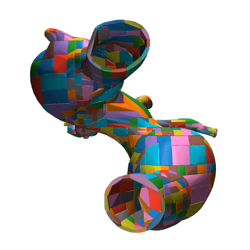
To simulate the aorta, we first scan the silicone-based, transparent, anthropomorphic phantom, of the Type-I and Type-II aortic arch model (Elastrat Sarl, Switzerland) to create high-fidelity 3D mesh models. The concave mesh is then decomposed into a set of nearly convex surfaces using the volumetric hierarchical approximate decomposition [37] resulting in convex hulls for Type-I aortic arch and convex hulls for Type-II aortic arch. The difference in the number of convex hulls is given by the difference in the measure of concavity of the two meshes [38]. The convex hull is used to model the collision, as it aids the computational process and allows the use of soft contacts by the physics engine [20]. We show the aorta simulation in Fig. 2.
III-C Catheter Simulation
One form of modelling the catheter represents the discretization of the continuous shape into a series of rigid bodies, interconnected by revolute or spherical joints [5]. This approach has been further categorized into discrete and serpentine methods, where the latter differs by having shorter rigid segments and, implicitly, a higher number of links [39]. The discretization approach has been proven to confer reasonable accuracy for shape prediction of the continuum robot [40]. In contrast to the discretization approach, the continuous curve estimation methods [5] assume that the described object is composed of an elastic backbone. The constant curvature model describes the continuum robot geometry with a finite number of mutually tangent curved segments, each with a constant curvature along its length or a variable curvature approach that takes into account the different curvatures across the backbone of the robot [5].
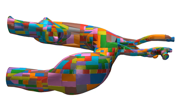
In this work, to enable real-time performance, we model the catheter based on a discretization approach by creating a serpentine-like model in which its continuous shape and deformation are approximated by a series of rigid bodies and revolute joints. A prismatic joint was added for mimicking the direct linear movement of the catheter. Similarly to [41], we limit the actuation to the tip of the catheter, where the revolute joints are intrinsically actuated by motors. This approach allows the use of standardized modelling based on Newton-Euler equation [42] which is a good approximation of the inherent continuous shape that benefits from the computational efficiency of the method. The catheter model is shown in Fig. 3.
III-D Contact Simulation
As our CathSim is based on MuJoCo [20], the contact between the aorta and the catheter are simulated using point contacts. In practice, point contact is defined geometrically as a point between two geometric objects and a spatial frame centered at that point in a global coordinate frame. The first axis of this frame is the contact normal direction, while the two other axes define the tangent plane. The contact distance is then used to determine if penetration happens (i.e., the contact distance is positive if two geometric objects are separated, zero when they are in contact, and negative when they penetrate) [20]. While this approach does present some limitations, such as a simplified representation of real-life contact, it is a reasonable compromise that balances computational efficiency and a sufficient degree of realism [43].
III-E How will CathSim be useful to the community?
We discuss some interesting research directions that can be benefited from our simulator:
- •
- •
Besides these tasks, we believe our simulator can be used in other scenarios such as developing surgical planning [44] and medical training [28]. The research community is free to explore other applications of our simulator.
IV Reinforcement Learning for Autonomous Cannulation
We consider the task of autonomous cannulation, where our system represents an episodic Partially Observable Markov Decision Process (POMDP). Our agent, represented by the catheter, interacts with an environment represented by an aortic arch type. At each time step , the agent, receives an observation , chooses an action , receives a reward and arrives in a new state . The episode terminates when the agent reaches the goal position within the aorta .
IV-A Observations
We consider three types of observations for the experiments, namely Internal, Image, and Sequential. In the Internal setup, we fed the system extensive data such as position, velocity, center of mass inertia and velocity, actuator-generated force, and external forces on the body. For the Image observation, we utilized a virtual RGB camera positioned above the aortic phantom, producing resolution grayscale images akin to X-ray images used in clinical procedures. Despite the possibility of higher resolution, our experiments indicated no performance improvement, only increased computational demand and memory requirements.
Moreover, due to the difficulty of inferring the catheter actions given one image as in the “Image” observation setup, we consider the “Sequential” observation by inserting the temporal dimension through the concatenation of three subsequent images . This observation would take into account the temporal domain of the catheter action and potentially provide more information to the RL agent.
IV-B Actions and Rewards
The actions are represented by a vector , where the first elements are associated with motors that actuate the revolute joints inside the tip of the catheter plus the prismatic joint responsible for the translational movement. Furthermore, the actions are normalized within an interval, giving a space of .
Given the sparsity of the reward function, we chose to convey more spatial information through reward shaping. Considering the navigation task, we provide the agent with an informational reward regarding the distance towards the target by computing the negative Euclidean distance between the head of the catheter and the goal , such as . If the agent is within a distance of the goal, the episode terminates and the agent receives a further reward of . The distance of mm was selected as within this distance, the catheter tip is fully inserted within the artery.
| (1) |
Whilst this reward function assists in agent convergence, it is also prone to local minima. An example would be the erroneous insertion of the catheter in another artery. In this case, the catheter would have to increase the distance from the target to achieve the goal objective.
| Aorta | Observation | Reward | Mean Force | Max Force | Success % | ||||
| BCA | LCCA | BCA | LCCA | BCA | LCCA | BCA | LCCA | ||
| Type-I | PPO-Internal | ||||||||
| SAC-Internal | |||||||||
| PPO-Image | 37 | ||||||||
| SAC-Image | |||||||||
| PPO-Sequential | 97 | ||||||||
| SAC-Sequential | |||||||||
| Type-II | PPO-Internal | ||||||||
| SAC-Internal | 100 | ||||||||
| PPO-Image | 97 | ||||||||
| SAC-Image | |||||||||
| PPO-Sequential | |||||||||
| SAC-Sequential | |||||||||
IV-C Network Architectures
Considering the continuous action representation, we employ two state-of-the-art RL algorithms, namely PPO [45] and SAC [46] with the parameters utilized in [47]. A Multi-Layered Perceptron (MLP) based policy is used for the Internal observation and a Convolutional Neural Network (CNN) is used for the Image and Sequential observation. Note that, for simplicity, we follow [48] and use CNN for both Image and Sequential observations as they only have one and four input channels respectively where each channel represents a greyscale image. In our implementation, the MLP has two hidden layers of sizes with a tanh activation function. The CNN has three convolutional layers with a ReLU activation function. The ADAM optimizer is used to train all networks with a learning rate of .
V Experiments
In this section, we perform intensive experiments to validate our CathSim. We start with the experiment to verify whether our simulator can mimic the behavior of the real-robot (CathBot). We then demonstrate how CathSim can be used for autonomous cannulation tasks using RL algorithms.
V-A Simulator Validation

V-A1 Setup
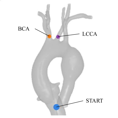
We assess the validity of our simulator, considering the distribution of forces generated throughout cannulation of the brachiocephalic artery (BCA). Ideally, we want to compare the force measured by our simulator and the force measured in the real-world experiment setup [3]. Note that, in the experiments conducted in [3], a load cell (Mini40, ATI Industrial Automation, Apex, NC, USA) was used to capture the force generated by the interaction of the instruments with the silicon phantom.
To extract the force from our simulator, we manually perform the cannulation using the keyboard, then in each simulation time step, we extract the collision points between the catheter and the aorta along with the tridimensional force expressed as the normal force and frictional forces and . We further compute the magnitude of the force given the previous, such as given a time instance , the magnitude of the force is given by
| (2) |
We proceed by offering a comparison between the observed empirical distribution and a normal distribution derived from the real experiments conducted in [3]. We sample the same number of samples generated in our simulation and the real experiment in [3] for a fair comparison. We use the distributions to generate a cumulative distribution function , which can be visualized in Fig. 5. We further assess the normality of the sampled data, followed by a comparison of the two given distributions.
V-A2 Results
We begin our comparison with the Shapiro-Wilk test of normality on the data extracted from the simulator and, given a p-value of and a statistic of , we conclude that the sampled data does not represent a normal distribution . This can be visualized in Fig. 5, where the sampled data is plotted against a normal distribution. Furthermore, we assess the homoscedasticity of the sample distribution and normal distribution by using Levene’s tests, which results in a statistic of and a p-value of , therefore, concluding that . Given the previous statistics (i.e., non-normal distribution and unequal variances), we select the non-parametric Mann-Whitney test to compare the given distributions. The resulting statistic given the test is , with a p-value of . Given that the p-value is higher than the threshold of , we can conclude that the differences in the distributions are merely given to chance and therefore the distributions can be considered as being part of the same population and thus convene that the force distribution of our simulator closely represents the distribution of forces encountered in the real-life system. Therefore, we can see that our CathSim successfully mimics the behavior of the real-world system.
V-B Reinforcement Learning Results
V-B1 Setups
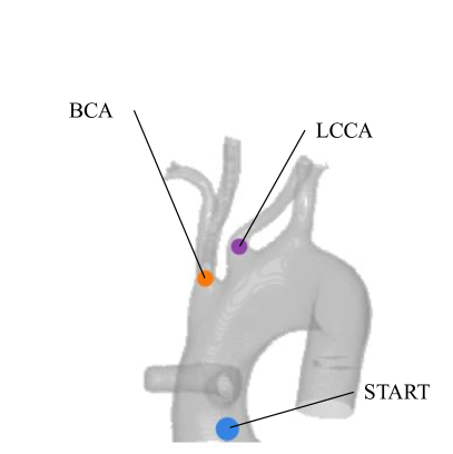
We consider the autonomous catheterization of two principal arteries into the aortic arch, namely the brachiocephalic artery (BCA) and the left common carotid artery (LCCA). Within both setups, we position the catheter tip at the starting locations within the ascending aorta and terminate the training when the catheter is fully inserted within the artery. We follow the same procedure for both the Type-I and the Type-II aortic arches. Please see Fig. 4 for our experimental setup.
We trained the model for time steps. Each episode started with a random catheter displacement of mm, followed by navigation through the ascending aorta to reach the goal. If not achieved within steps, the episode ended. Data on the aorta’s contact points and exerted force, as per Eq. 2, was gathered at each step to form a force heatmap overlaid on the RGB virtual image. The model’s performance was evaluated over episodes. The maximum and mean forces for samples were computed as:
| (3) |
The training time ranged from hour for PPO to hours for SAC, making PPO approximately five times faster. Regardless, our simulator showed a performance of FPS for the image-based environment and FPS for the internal-based environment.

V-B2 Quantitative Results
Table II shows the force, reward, and success rate of all methods. Type-I Aortic Arch experiments show that PPO relying on a sequential observation space achieved the greatest reward when cannulating the BCA target, although it shows the least performance when the target is LCCA. A more coherent reward has been achieved while using a singular image observation, where the cannulation of LCCA presents the greatest success (). In contrast, the cannulation of BCA presents close results to the sequential observation (). The performance gap between the cannulation of BCA and LCCA is mainly because of the start configuration and the position of the BCA and LCCA in the aortic arch. Naturally, from Fig. 4 we can see that it would be easier for both humans and RL agents to reach BCA rather than LCCA since LCCA’s position is further away from the navigation direction and is surrounded by other vascular branches.
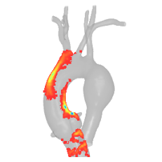
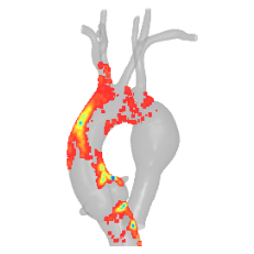
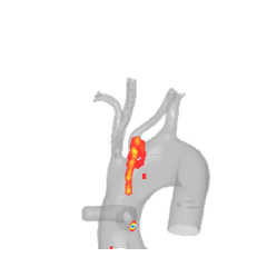
Given the success rates of the cannulation, the catheterization of the Type-II aortic arch appears to be a more straightforward procedure. The task implies that the dependability of the vessel wall for maneuvering is reduced, leading to a more straightforward catheterization procedure. This phenomenon can be also observed in Fig. 6, where the catheter exerted a greater amount of force on the aortic walls in order to reach the designated target. Overall, from our experiments, PPO shows better suitability for the task, especially when the observation is image-based.

V-B3 Qualitative Result
Force frames from each time step help us calculate the mean of in-contact regions, which are then superimposed on the phantom model to generate force heatmaps (see Fig. 6). These maps show increased catheter force at bends required for target access. For example, the catheter often overshoots the LCCA target in Fig. 6 (c) whilst exerting greater force on the ascending aortic walls within Type-I Aortic Arch. Within the Type-II Aortic Arch, more force exerted on the wall between BCA and LCCA. Regardless, the Type-I Aortic Arch, poses more difficulties.
We further display the results of the force interaction in Fig. 7. Analyzing the figure, it can be seen that the force distribution is quite similar along both the Type-I and Type-II Aortic Arch, where most of the force is concentrated within the to range. Despite this, there are outliers which exert a force greater than . Specifically, the BCA within the Type-I Aortic Arch has a notably wider interquartile range than the LCCA, suggesting that the BCA generally exerts more force than the LCCA.

At each time step, we compute the mean reward of the last steps and display the results in Fig. 8. The algorithms show continuous learning with slight convergence. In our experiments, PPO with an image observation obtained the highest or close to the highest reward. Furthermore, it shows that an image-based observation that correlates to the medical procedure undertaken in real clinical scenarios is capable of reaching the most reward. While using the internal observation space, SAC performed reasonably well, managing to obtain the highest reward within the Type-I aortic arch when cannulating the LCCA target. Within all cases and coherent with the evaluation undertaken in the previous section, the sequential observation space yielded the least reward.
VI Discussion
While our current framework represents promising progress, it remains challenged by the complexity of fully mimicking real-world dynamics within a simulated environment. There exist critical gaps in simulating intricate biological structures like a deformable aorta, implementing realistic guidewire simulation, and factoring in the nuanced interplay between surgical instruments and fluid mechanics. Our systematic experimentation, however, has shed light on crucial directions for further advancement. These include: i) the development of comprehensive and sophisticated features that aim to lessen the divergence between real-world conditions and simulated environments, ii) the integration of our simulation system into Augmented Reality/Virtual Reality (AR/VR) platforms to foster a more immersive and interactive experience, and iii) the execution of robust comparative studies that pit the simulated environment against its physical counterparts to validate the simulation’s accuracy and reliability. Despite the inherent challenges in achieving a perfect simulation of real-world medical procedures, our research has illuminated promising pathways for subsequent development. We aim to refine our system into a more robust, comprehensive, and precise research tool. Furthermore, to promote collaborative scientific growth, we intend to disseminate our source code to stimulate additional research and innovation in this rapidly evolving field.
VII Conclusions
We present CathSim, an open-source simulation environment designed as a comprehensive benchmarking platform for autonomous endovascular navigation. Our proposed simulator allows for the development and testing of a diverse range of algorithms for autonomous cannulation, eliminating the need for physical robotic systems and thereby reducing the associated risks and costs of experimental hardware. It serves a dual purpose as a development and benchmarking platform for computer scientists and roboticists, as well as a training platform for healthcare professionals. Furthermore, with the rapid advancements in medical robotics, CathSim provides a risk-free, controlled environment where innovative procedures and techniques can be tested and refined before their implementation in real-world clinical scenarios. By maintaining an open-source model, our simulator also encourages collaborative advancements and knowledge sharing within the scientific community.
References
- [1] I. Wamala, E. T. Roche, et al., “The use of soft robotics in cardiovascular therapy,” Expert Review of Cardiovascular Therapy, 2017.
- [2] O. M. Omisore, S. Han, J. Xiong, H. Li, Z. Li, and L. Wang, “A review on flexible robotic systems for minimally invasive surgery,” SMC, 2020.
- [3] D. Kundrat, G. Dagnino, et al., “An MR-Safe endovascular robotic platform: Design, control, and ex-vivo evaluation,” TBME, 2021.
- [4] V. M. Pereira, N. M. Cancelliere, et al., “First-in-human, robotic-assisted neuroendovascular intervention,” Journal of Neurointerventional Surgery, 2020.
- [5] J. Burgner-Kahrs, D. C. Rucker, and H. Choset, “Continuum robots for medical applications: A survey,” T-RO, 2015.
- [6] T. Heidt, S. Reiss, et al., “Real-time magnetic resonance imaging–guided coronary intervention in a porcine model,” Scientific Reports, 2019.
- [7] M. E. M. K. Abdelaziz, D. Kundrat, et al., “Toward a Versatile Robotic Platform for Fluoroscopy and MRI-Guided Endovascular Interventions: A Pre-Clinical Study,” in IROS, 2019.
- [8] J. Konstantinova, A. Jiang, K. Althoefer, P. Dasgupta, and T. Nanayakkara, “Implementation of tactile sensing for palpation in robot-assisted minimally invasive surgery: A review,” IEEE Sensors Journal, 2014.
- [9] M. B. Molinero, G. Dagnino, J. Liu, W. Chi, M. E. Abdelaziz, T. M. Kwok, C. Riga, and G.-Z. Yang, “Haptic guidance for robot-assisted endovascular procedures: implementation and evaluation on surgical simulator,” in IROS, 2019.
- [10] A. Nguyen, D. Kundrat, et al., “End-to-end real-time catheter segmentation with optical flow-guided warping during endovascular intervention,” in ICRA, 2020.
- [11] P. E. Dupont, B. J. Nelson, et al., “A decade retrospective of medical robotics research from 2010 to 2020,” Science Robotics, 2021.
- [12] A. Juliani, V.-P. Berges, E. Teng, A. Cohen, J. Harper, C. Elion, C. Goy, Y. Gao, H. Henry, M. Mattar, et al., “Unity: A general platform for intelligent agents,” arXiv preprint arXiv:1809.02627, 2018.
- [13] L. Karstensen, T. Behr, T. P. Pusch, F. Mathis-Ullrich, and J. Stallkamp, “Autonomous guidewire navigation in a two dimensional vascular phantom,” CDBME, 2020.
- [14] F. Faure, C. Duriez, H. Delingette, J. Allard, B. Gilles, S. Marchesseau, H. Talbot, H. Courtecuisse, G. Bousquet, I. Peterlik, et al., “SOFA: A Multi-Model Framework for Interactive Physical Simulation,” in Soft Tissue Biomechanical Modeling for Computer Assisted Surgery, 2012.
- [15] R. Davis, R. Henshell, and G. Warburton, “A Timoshenko beam element,” JSV, 1972.
- [16] T. Behr, T. P. Pusch, M. Siegfarth, D. Hüsener, T. Mörschel, and L. Karstensen, “Deep Reinforcement Learning for the Navigation of Neurovascular Catheters,” Current Directions in Biomedical Engineering, 2019.
- [17] O. M. Omisore, T. Akinyemi, W. Duan, W. Du, and L. Wang, “A Novel Sample-efficient Deep Reinforcement Learning with Episodic Policy Transfer for PID-Based Control in Cardiac Catheterization Robots,” arXiv preprint arXiv:2110.14941, 2021.
- [18] E. Rohmer, S. P. Singh, and M. Freese, “V-REP: A versatile and scalable robot simulation framework,” in IROS, 2013.
- [19] P. Schegg, J. Dequidt, E. Coevoet, E. Leurent, R. Sabatier, P. Preux, and C. Duriez, “Automated Planning for Robotic Guidewire Navigation in the Coronary Arteries,” in RoboSoft, 2022.
- [20] E. Todorov, T. Erez, and Y. Tassa, “Mujoco: A physics engine for model-based control,” in IROS, 2012.
- [21] A. Attanasio, B. Scaglioni, E. De Momi, P. Fiorini, and P. Valdastri, “Autonomy in surgical robotics,” Control, Robotics, and Autonomous Systems, 2021.
- [22] W. Chi, G. Dagnino, T. M. Kwok, A. Nguyen, D. Kundrat, M. E. Abdelaziz, C. Riga, C. Bicknell, and G.-Z. Yang, “Collaborative robot-assisted endovascular catheterization with generative adversarial imitation learning,” in ICRA, 2020.
- [23] A. Marban, V. Srinivasan, W. Samek, J. Fernández, and A. Casals, “A recurrent convolutional neural network approach for sensorless force estimation in robotic surgery,” Biomed. Signal Process. Control, 2019.
- [24] C. Gao, X. Liu, M. Peven, M. Unberath, and A. Reiter, “Learning to see forces: Surgical force prediction with rgb-point cloud temporal convolutional networks,” in MICCAI Workshop, 2018.
- [25] A. I. Aviles, S. M. Alsaleh, J. K. Hahn, and A. Casals, “Towards retrieving force feedback in robotic-assisted surgery: A supervised neuro-recurrent-vision approach,” ToH, 2016.
- [26] A. I. Aviles, S. M. Alsaleh, E. Montseny, P. Sobrevilla, and A. Casals, “A deep-neuro-fuzzy approach for estimating the interaction forces in robotic surgery,” in FUZZ-IEEE, 2016.
- [27] A. Marban, V. Srinivasan, W. Samek, J. Fernández, and A. Casals, “Estimation of interaction forces in robotic surgery using a semi-supervised deep neural network model,” in IROS, 2018.
- [28] C. I. Nesbitt, N. Birdi, S. Mafeld, and G. Stansby, “The Role of Simulation in the Development of Endovascular Surgical Skills,” Perspect Med Educ, 2016.
- [29] Y. Wei, S. Cotin, J. Dequidt, C. Duriez, J. Allard, E. Kerrien, et al., “A (near) real-time simulation method of aneurysm coil embolization,” Aneurysm, 2012.
- [30] J. Dequidt, C. Duriez, S. Cotin, and E. Kerrien, “Towards interactive planning of coil embolization in brain aneurysms,” in MICCAI, 2009.
- [31] H. Talbot, F. Spadoni, C. Duriez, M. Sermesant, S. Cotin, and H. Delingette, “Interactive training system for interventional electrocardiology procedures,” in ISBMS, 2014.
- [32] S. Sinceri, M. Carbone, M. Marconi, A. Moglia, M. Ferrari, and V. Ferrari, “Basic Endovascular Skills Trainer: A surgical simulator for the training of novice practitioners of endovascular procedures,” in EMBC, 2015.
- [33] T. P. Lillicrap, J. J. Hunt, A. Pritzel, N. Heess, T. Erez, Y. Tassa, D. Silver, and D. Wierstra, “Continuous control with deep reinforcement learning,” arXiv preprint arXiv:1509.02971, 2015.
- [34] G.-Z. Yang, J. Cambias, K. Cleary, E. Daimler, J. Drake, P. E. Dupont, N. Hata, P. Kazanzides, S. Martel, R. V. Patel, et al., “Medical robotics–Regulatory, ethical, and legal considerations for increasing levels of autonomy,” Science Robotics, 2017.
- [35] J. Kweon, K. Kim, C. Lee, H. Kwon, J. Park, K. Song, Y. I. Kim, J. Park, I. Back, J.-H. Roh, et al., “Deep reinforcement learning for guidewire navigation in coronary artery phantom,” IEEE Access, 2021.
- [36] Y. Cho, J.-H. Park, J. Choi, and D. E. Chang, “Image Processing Based Autonomous Guidewire Navigation in Percutaneous Coronary Intervention,” in ICCE-Asia, 2021.
- [37] M. Silcowitz, S. Niebe, and K. Erleben, “Interactive rigid body dynamics using a projected gauss–seidel subspace minimization method,” in Computer Vision, Imaging and Computer Graphics. Theory and Applications: International Joint Conference, VISIGRAPP 2010, Angers, France, May 17-21, 2010. Revised Selected Papers, 2010.
- [38] K. Mamou and F. Ghorbel, “A simple and efficient approach for 3D mesh approximate convex decomposition,” in ICIP, 2009.
- [39] G. Robinson and J. B. C. Davies, “Continuum robots-a state of the art,” in ICRA, 1999.
- [40] C. Shi, X. Luo, P. Qi, T. Li, S. Song, Z. Najdovski, T. Fukuda, and H. Ren, “Shape sensing techniques for continuum robots in minimally invasive surgery: A survey,” TBME, 2016.
- [41] K.-W. Kwok, K. H. Tsoi, V. Vitiello, J. Clark, G. C. Chow, W. Luk, and G.-Z. Yang, “Dimensionality reduction in controlling articulated snake robot for endoscopy under dynamic active constraints,” T-RO, 2012.
- [42] R. Featherstone, Rigid body dynamics algorithms, 2014.
- [43] E. Todorov, “A convex, smooth and invertible contact model for trajectory optimization,” in 2011 IEEE International Conference on Robotics and Automation. IEEE, 2011, pp. 1071–1076.
- [44] Z. Li, J. Dankelman, and E. De Momi, “Path planning for endovascular catheterization under curvature constraints via two-phase searching approach,” International Journal of Computer Assisted Radiology and Surgery, vol. 16, pp. 619–627, 2021.
- [45] J. Schulman, F. Wolski, P. Dhariwal, A. Radford, and O. Klimov, “Proximal policy optimization algorithms,” arXiv preprint arXiv:1707.06347, 2017.
- [46] T. Haarnoja, A. Zhou, P. Abbeel, and S. Levine, “Soft actor-critic: Off-policy maximum entropy deep reinforcement learning with a stochastic actor,” in ICML, 2018.
- [47] G. Dalal, K. Dvijotham, M. Vecerik, T. Hester, C. Paduraru, and Y. Tassa, “Safe exploration in continuous action spaces,” arXiv preprint arXiv:1801.08757, 2018.
- [48] V. Mnih, K. Kavukcuoglu, D. Silver, A. Graves, I. Antonoglou, D. Wierstra, and M. Riedmiller, “Playing atari with deep reinforcement learning,” arXiv preprint arXiv:1312.5602, 2013.