Actomyosin-driven division of a synthetic cell
Abstract
One of the major challenges of bottom-up synthetic biology is rebuilding a minimal division machinery. The animal cell division apparatus is mechanically the simplest, in which an actin-based ring constricts the membrane, as compared to microbes and plant cells where a cell wall is involved. Furthermore, reconstitution of the actin division machinery helps to understand the physical and molecular mechanisms of cytokinesis in animal cells and thus our own cells. In this review, we describe the state-of-the-art research on reconstitution of minimal actin-mediated cytokinetic machineries. Based on the conceptual requirements that we obtained from the physics of the shape changes involved in cell division, we propose two major routes for building a minimal actin apparatus capable of division. Importantly, we acknowledge both the passive and active roles that the confining lipid membrane can play in synthetic cytokinesis. We conclude this chapter by identifying the most pressing challenges for future reconstitution work, thereby laying out a roadmap for building a synthetic cell equipped with a minimal actin division machinery.
keywords:
bottom-up reconstitution, synthetic cell, cell division, actin, myosinEqual contributions \altaffiliationEqual contributions \altaffiliationCorresponding author \abbreviations
1 Introduction
Bottom-up synthetic biology is an emerging field at the interface of cell biology, (bio)chemistry and (bio)physics. Several national and international initiatives have been founded recently, which are aimed at reconstituting synthetic cells that can autonomously grow and divide 1, 2. As a chassis, usually giant unilamellar vesicles (GUVs) are used, which are cell-sized (5-50 µm) containers enveloped in a lipid bilayer 3, 4, 5, 6. One of the key functions that a synthetic cell must be able to perform in order to be considered life-like is cytokinesis 7, a process in which a cell physically splits into two daughter cells. To reconstitute cytokinesis, various strategies are being pursued, inspired by biological strategies employed by prokaryotic, archaeal or eukaryotic cells 8, 7. These biological systems have in common that cell division is accomplished by a cytoskeletal protein machinery, often ring-shaped, that assembles at the cell equator. In microbial cells (bacteria and yeast), this protein machinery collaborates with a complex cell wall synthesis machinery 9, 10. By contrast, animal cells lack a cell wall and cytokinesis is entirely driven by the actin cytoskeleton. Actin-based cell division could thus be an ideal basis for engineering synthetic cell division.
Bottom-up reconstitution of actin-based cell division is interesting not only from an engineering perspective, but also as a means to understand how cytokinesis works at the molecular level in animal cells. Although cytokinesis is a well-studied cellular process, surprisingly many fundamental questions about its working principles remain unanswered 11: how are mechanical forces generated and sustained? How much molecular complexity is needed to ensure that the actin cortex retains its structural integrity during cytokinesis? What are the requirements for cortex-membrane interactions to promote furrow ingression? These questions are difficult to address in cell-based studies because of the enormous molecular complexity of cells combined with substantial variation between cytokinetic mechanisms employed by different species and different cell types 12, 13, 10.
In this review, we propose a roadmap towards the bottom-up reconstitution of actin-driven cytokinesis in minimal cells. For brevity, we consider only the process of furrow ingression, neglecting other aspects such as membrane abscission and chromosome and cytoplasmic segregation, which are reviewed elsewhere 14, 15, 16, 17. Based on theoretical models of cytokinesis in animal cells, we first identify four central biophysical requirements for actin-driven furrow ingression. Next we review experimental insights obtained from recent efforts to reconstitute minimal actin systems. We also emphasize the importance of controlling the surface area of the synthetic plasma membrane to enable cell division. Finally we propose a roadmap towards building a molecular machinery that can successfully deform a minimal cell-like container.
2 Biophysical requirements for making a cell divide
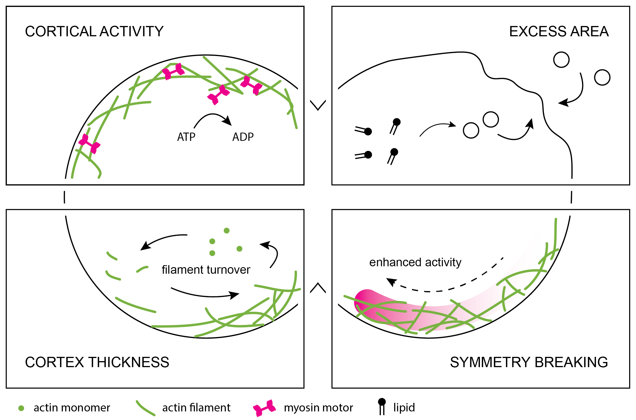
Cytokinesis in animal cells is a complicated process that involves many different molecular components (lipids and proteins) whose interactions and localization are tightly regulated. At a coarse-grained level, however, it is possible to formulate general biophysical requirements for cell division based on a consideration of the mechanical forces at play. Pioneering experimental work from the 1950s onward has demonstrated that cytokinesis is accompanied by membrane furrowing 18, cortical stiffening 19, 20 and the appearance of ordered filamentous structures in the cytokinetic ring 21, 22. These observations have served as input for coarse-grained theoretical and computational models that describe cytokinesis as the shape evolution of a thin, viscoelastic and active shell around a (nearly) constant volume of cytoplasm. From the models, we can infer several key requirements that a cell, living or synthetic, must fulfil in order to successfully divide (fig. 1):
1. Cortical activity. The actin cortex driving cytokinesis in animal cells must be active. This means that it should include elements that hydrolyse adenosine triphosphate (ATP), an energy-carrying nucleotide, to generate contractile forces that produce cellular shape changes. The viscoelastic and active nature of the cortex can be described using the framework of active gel theory as proposed by Kruse et al. 23. This formalism is usually applied in the viscous limit 24, 25, 26, 27, as cytokinesis is slow (minutes) compared to the fluidization time scale (10 s) of the actin cortex 24. The molecular origins of active force production are complex and depend on molecular detail, as discussed below.
2. Cortical thickness. Cortex activity, at least when mediated by myosin motors, is roughly proportional to cortical thickness 25, 24, 26. To maintain cortical activity, the cortex must consequently be of a controlled thickness. Cortical thickness is regulated by a balance of actin polymerization and depolymerization, or turnover, and cortical flows: cortical flows accumulate material in the cytokinetic furrow, whereas turnover redistributes actin throughout the cell. This suggests two requirements for synthetic cell division. Firstly, components of the cortex must be laterally mobile to be effectively redistributed by cortical flows 24, 26, 28. Secondly, actin turnover rates must be low enough to allow local actin accumulation and therefore increased contractility in the furrow region. If actin is removed too rapidly, furrow constriction slows down significantly and may be halted altogether 26. On the other hand, complete lack of filament turnover in a 2D actomyosin cortex is theoretically predicted to lead to irreversible clustering of actin, inhibiting effective stress generation 29. This prediction has yet to be reconciled with experimental evidence from yeast cells, which suggests that the persistent presence of filamentous actin, rather than turnover, is key for successful contraction of the cytokinetic ring 30.
3. Cortical symmetry breaking. From the 1930s onwards, various models have been proposed to explain the mechanical basis of cytokinesis. The early models range from active expansion of the cell poles 31, through active pushing by the mitotic spindle32, to spindle-mediated relaxation of the cell poles 33, 28 and finally active constriction of the cytokinetic furrow 34, 35, 26, 22. While details vary widely between these models, they share a key characteristic: they all posit that there must be a difference in activity between the polar and equatorial regions to drive furrow ingression. After decades of research it is now widely accepted (reviewed e.g. in 36) that the main driving factor of animal cell cytokinesis is actin-based constriction at the cleavage furrow. However, in vitro reconstitution may be the ideal tool to understand actin’s role in molecular detail, and to assess to which extent other mechanisms 37, 38 also contribute.
4. Cell surface area and volume regulation. Consistent with observations in cells, models have generally assumed that the cytoplasm is very weakly, if at all, compressible 28, 26. The apparent cell surface-to-volume ratio, however, changes dramatically during cytokinesis 39. It follows that the cell’s (visible) surface area must be changing. In theoretical works this change in surface area is generally assumed to be energetically ‘free’, as living cells can regulate the available membrane area through a variety of processes like blebbing 40, or caveolae disassembly and membrane trafficking 41, 42. This supply of membrane on demand is probably one of the most challenging aspects to recapitulate in a reconstituted system.
3 Roadmap towards actin-driven synthetic cell division
Cytokinesis of animal cells is a highly complex and tightly regulated process. Yet, fairly minimal computational models are able to recapitulate cytokinesis, suggesting that the underlying mechanisms may be recreated with simplified molecular mechanisms. Here, we propose a roadmap towards reconstituting actin-driven cell division by considering lessons from recent cell and in vitro (i.e. cell-free reconstitution) studies. Basically, there are two routes for reconstitution of actin-driven cytokinesis (see fig. 2). First, cell division can be recreated via reconstitution of an actin cortex that, upon symmetry breaking, is more contractile at the cell equator as compared to the poles. This route is most close to cytokinesis in mammalian cells, and we therefore name it the naturalistic route. The second route is by construction of a cytokinetic ring that anchors and contracts at the cell equator, coined the engineering route. We will first discuss the design of an actin-based machinery fit for driving cytokinesis in both scenarios, and in the next section consider the design of the lipid membrane envelope.
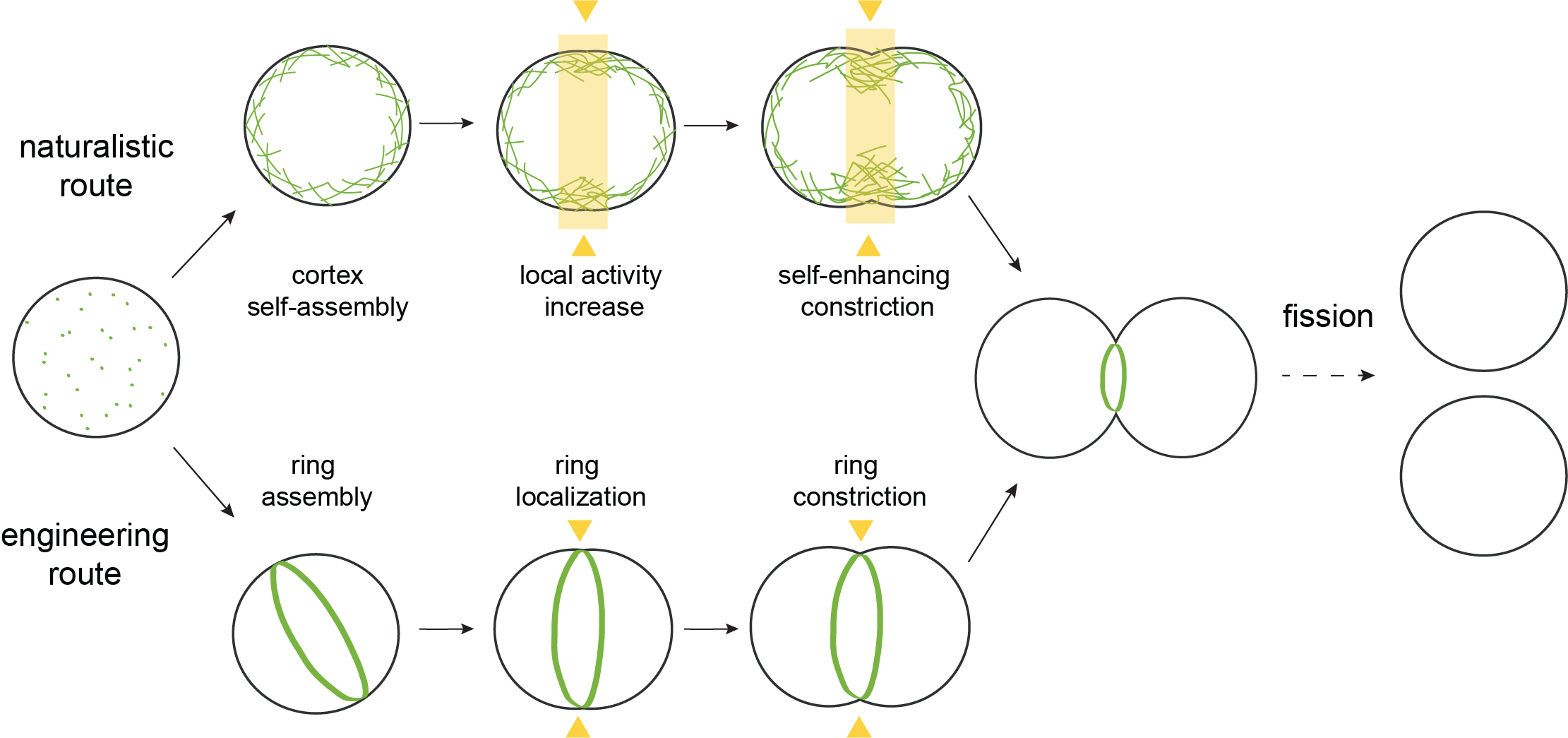
3.1 Naturalistic route: building a self-assembling cytokinetic ring
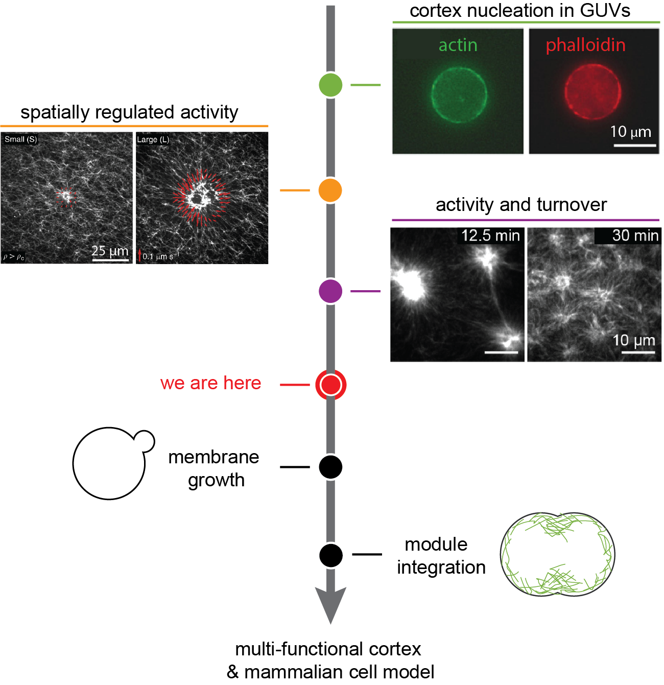
During interphase, mammalian cells have a continuous actin cortex that lines the plasma membrane 43. When cells enter mitosis, the cortex is remodelled and self-assembles into a contractile ring at the cell equator. Symmetry breaking and midplane localization of the cytokinetic furrow is initiated by biochemical signalling, which includes Rho-dependent myosin phosphorylation in the furrow region 26, 44. The locally enhanced activation of myosin is thought to lead to cortical flows from the poles to the equator 28, 45, which further accumulate and organize contractile elements in the furrow 46 that drive furrow ingression 26. Such a complex self-assembling system has not been built to date, but steps have been taken along the road (fig. 3).
3.1.1 Reconstitution of active actin networks
Both cell-free experiments and theoretical models of cortex-like disordered actin networks have been used to elucidate why disordered actomyosin networks are contractile in the first place. The detailed mechanisms are reviewed elsewhere 47, 48, 49, but they broadly comprise two scenarios. Actin filaments are semiflexible polymers with a thermal persistence length of 10-15 µm, of the same order as their contour length 50. The first contraction scenario, relevant for well-connected networks of long filaments, is that the anisotropic mechanical force-extension response of actin filaments causes them to buckle and break under motor-induced compressive stress 51, 52. The second scenario, relevant for networks with short actin filaments, is that the structural polarity of actin filaments in combination with the tendency of myosin II motors to dwell at the filament plus end before detachment causes contraction via polarity sorting 53, 25, 54. In the actin cortex of mammalian cells there may be a combination of both mechanisms, since distinct populations of short and long filaments are present there 55.
Notably, the combined effect of contractile motor activity and actin turnover remains poorly explored. Theoretical models generally assume that the cytokinetic cortex does undergo actin turnover 26, 24, 56, and have even indicated that turnover is key for sustained stress generation during furrow ingression 29. Experimentally, besides one study with a cell extract 57, only one minimal in vitro study has so far combined actin turnover and myosin activity 58. This work showed that myosin activity alone can be sufficient to induce turnover in minimal actin networks (see fig. 3, purple). Myosin-driven compaction and fragmentation of Arp2/3-nucleated actin led to the removal of actin from the network, and subsequent redistribution and re-incorporation of network components, creating a cortex in dynamic steady state. Strikingly, actin turnover rates were observed to be much slower here than typical rates in cells, with actin turning over within tens of minutes rather than tens of seconds, respectively 59, 60. This discrepancy is likely due to the absence of dedicated actin severing proteins in the minimal system. More rapid turnover has been observed in vitro in volume-spanning entangled actin networks where filaments were severed by cofilin and polymerization was driven by formin 61. Combining more rapid turnover with motor activity in vitro may open a rich field of network behaviours, with complex implications for both the regulation of cortical thickness and of stress propagation and relaxation 62.
To build and control a system that allows actin to turn over, we can turn to the growing body of work studying the functions of various actin regulators on the single molecule or filament level. Research into the two key nucleators of cortical actin, Arp2/3 and formins 65, 55, 66 has uncovered new complexities in recent years. Both the processivity and the actin filament elongation rate of different formins have been shown to be regulated by the physicochemical environment, the presence of profilin, and mechanical stress 67, 68, 69. Even more complex co-regulation of formin with other barbed-end binding proteins is emerging 70. Regulation of Arp2/3 by profilin 71, 72 as well as by actin filament curvature 73 has been known for a number of years, but the true diversity and complexity of the various isoforms of Arp2/3 is only just emerging 74. In addition, there are promoting factors that control cortex architecture by controlling both formin and Arp2/3 activity 66. Besides formin and Arp2/3, other actin nucleators such as the recently identified spire 75 have barely been used in reconstitution experiments and may offer yet other routes towards reconstituting a minimal dynamic cortex. Actin depolymerization can equally be controlled by various factors. Disassembly of filamentous actin in vitro is usually mediated by proteins of the ADF/cofilin family 76. The activity of ADF/cofilin proteins has been shown to depend on cooperation with other proteins 77, 78 and on actin crosslinking 79. ADF/cofilin also facilitates debranching in actin networks nucleated by Arp2/3 76, which is furthermore sensitive to force and actin filament age 80.
3.1.2 Reconstitution of actin cortices inside GUVs
Controlled actin encapsulation in GUVs has proven to be a challenge. Over the years, many different methods have been explored for protein encapsulation, either based on lipid swelling 81 or on emulsion transfer 63, 82, 83, 84 (reviewed in 3). Of these, methods based on emulsion transfer are currently most successful, although the encapsulation efficiency and the ability to upscale the number of encapsulates remain to be characterized 83. Most prior GUV studies focused on the effect of crosslink proteins and myosin motors on bulk-nucleated actin. By contrast, membrane-nucleated actin networks with turnover in GUVs remain poorly explored. Early works from the Sykes lab 63, 85 demonstrated that Arp2/3 nucleated cortices can be reconstituted at the inner leaflet of GUVs (fig. 3, green), and that such cortex-bearing vesicles reproduce aspects of the mechanics of living cells. More recently, Dürre et al. demonstrated that Arp2/3 nucleated cortices can induce local deformations of the GUV membrane by either polymerization forces alone or in combination with contractility induced by non-muscle myosin-II 86. New work from the Liu lab shows that membrane-bound Arp2/3 in combination fascin and -actinin is sufficient to yield ring-like membrane-bound actin networks 87. Myosin-initiated contraction of these networks resulted in membrane constriction, thus getting one step closer to cell division.
More extensive work, especially with myosin-driven cortices, has been performed with stable actin filaments anchored to the membrane by streptavidin or actin-binding membrane proteins. In such systems, cortical tension was shown to depend on the ratio of active versus passive crosslinkers 88 and excessive cortical tension was shown to cause full or partial detachment of the cortex from the membrane 88, 89. Recently, Litschel et al. demonstrated the formation of actomyosin rings in GUVs 82. However, these structures were unable to deform the GUV membrane on large length scales because they slipped on the membrane. Based on our understanding of cell division, this is likely due to (at least) three missing factors: cortex turnover, symmetry breaking between the poles and equator of the synthetic cell, and a severely limited supply of extra membrane area. Symmetry breaking is likely necessary for productive and sustained membrane deformations. There are several artificial means by which symmetry breaking could be triggered in synthetic cells. Myosin activity could, for instance, be locally light-activated by targeting either the light-sensitive myosin inhibitor blebbistatin 90, 64, 91 (see (fig. 3, orange) or myosin-II directly 92. Similar approaches could be used to locally modulate the crosslink density of the actin cortex or the interaction strength of the cortex with the synthetic cell membrane. Finally, it would likely help to make GUVs shape-anisometric, for instance by using microfluidic channels 93.
Conceptually, building a dynamic actin cortex and pushing it towards self-assembly of a cytokinetic furrow is very attractive. Such a system would mimic many core attributes of the cortex of living animal cells. Further, the continuous nature of such a cortex would allow it take on a dual function, both as a mechanoprotective module for the synthetic cell and as a division apparatus, which sets it apart from other cytoskeletal systems such as FtsZ 94. A life-like actin cortex offers the opportunity to test existing theoretical models of cell division and to tease out the essential functions needed for cytokinesis in living cells. On the other hand, a dynamic actin cortex will necessarily comprise more proteins and hence a higher level of complexity than one composed of stable actin filaments. From an experimental perspective, reconstituting sustained actin turnover in combination with motor activity will in particular be challenging, as it requires fine control over both stoichiometry and activity of cytoskeletal components.
3.2 Engineering route: building an isolated contractile ring
A more engineering-type approach to synthetic cell division may also be interesting: instead of building a cortex that self-organizes into a ring, one could build an isolated ring directly (fig. 2, bottom). This would inherently fulfil the requirement for different activities in polar and equatorial contractility, as by definition the poles are not contractile in such a case. If a sufficient supply of long actin filaments throughout furrow ingression can be ensured, the need for controlled turnover may be diminished and the complexities of such regulated filament assembly and disassembly may be avoidable. This approach will need to address three key challenges: 1) to build an actin ring, 2) to make it contractile, and 3) to control its position and also place it mid-center such that membrane invagination rather than ring slippage occurs.
3.2.1 How to build an isolated ring?
Actin filaments can be bundled and bent into ringlike structures in various ways (fig. 4, green). Most simply, ring formation can be induced by entropic effects through macromolecular crowding 95 or by cross-linking with multivalent ions 96. Alternatively, proteins can be used to bend actin into rings. Septins spontaneously bend actin into ringlike structures 97 and are recruited to the cytokinetic ring, where they cooperate with anillin in actin-membrane binding 98, 99, 100, 101, 102. Anillin itself also promotes the formation of actin rings 103. Further, the IQGAP fragment ‘curly’ has recently been shown to bend actin into rings on model membranes 104. The fact that all three of these proteins are enriched in the cytokinetic furrow 105 suggests that these ring-forming capabilities may provide a cellular mechanism to promote successful cytokinesis.
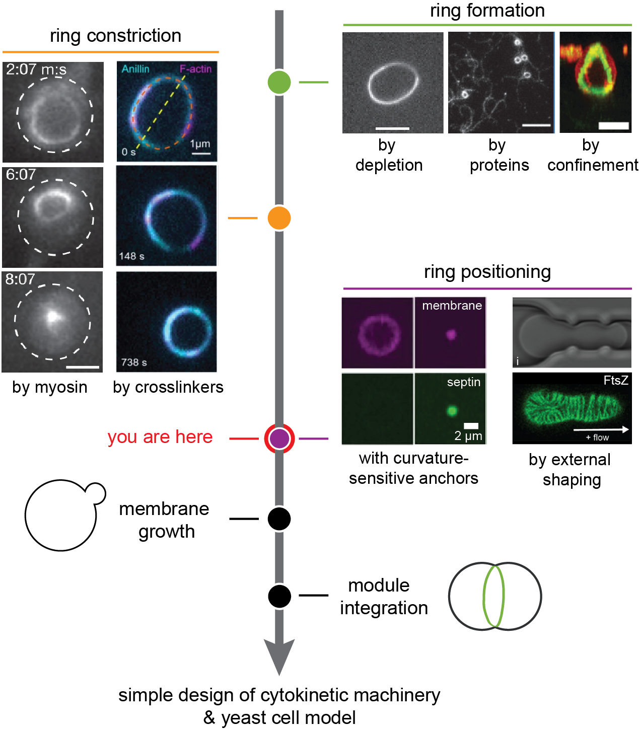
Confinement of actin filaments inside spherical droplets or vesicles tends to promote the formation of actin rings because the confinement forces the semiflexible filaments to minimize the filament bending energy 109. Entangled or crosslinked actin networks inside emulsion droplets and inside lipid vesicles form peripheral cortex-like networks 110, 111, 112, 113, 87, 114, while bundled actin forms one or more closed rings 81, 106, 112, 113, 87. Single rings form when the container size is smaller than the persistence length of the actin filament or bundle 113, 87. Recent theoretical 115 and experimental work 82 has shown that ring formation can be further enhanced by introducing actin-membrane adhesion. It should be noted, however, that ring formation requires a subtle balance of filament-filament and filament-wall adhesion, as well as size and stiffness of the confinement, and is not trivial to precisely control experimentally.
3.2.2 How to contract an isolated ring?
Contracting a once-formed actin ring can again proceed in different ways (fig. 4, orange). The classical purse-string model posits a well-organized cytokinetic ring that closes by myosin-mediated translocation of actin filaments 22, 116. Although this model does not appear to hold in all cell types 117, 118, 119, recent superresolution and electron microscopy showed convincing evidence that it does apply at least in some cell types 120, 121. Contracting actin-myosin rings have been succesfully reconstituted on supported lipid bilayers 104 and inside water-in-oil droplets 106 and GUVs 82. The efficiency of ring closure is likely determined by the orientation and arrangement of the actin filaments in the ring, which can be tuned by crosslinker composition and concentration 113, 122, 52, 123, 124.
Alternatively, ring contraction may be driven by mechanisms that do not require molecular motors. For instance, anillin was recently shown to drive actin bundle contraction even though it is a passive crosslinker 103. Contraction was attributed to an energetically driven process whereby actin filaments increase their overlap as long as energy can be gained by accumulating diffusive crosslinkers in the overlap region 103. This mechanism was enhanced when anillin was combined with actin depolymerization. Since contraction driven by passive crosslinkers does not consume energy from an external energy source such as ATP, it can only bring the system into a configuration of minimal free energy, at which point rearrangement will stop 125. Intriguingly, recent theoretical modelling 126 suggests that a crosslinker that consumes ATP to unbind from actin filaments, but does not actively translocate them like myosin, could in principle induce contraction indefinitely. In this case, the consumption of an energy carrier breaks detailed balance in the system, and in combination with the asymmetric mechanical properties of actin, overall contractile forces can arise.
3.2.3 How to keep an isolated ring in place?
Although contractile actin rings have been successfully reconstituted inside GUVs, so far none of these efforts have yielded anything close to furrow-like membrane invaginations. The rings either detached or slipped along the membrane upon myosin activation 88, 106, 82, 89, at best producing rare instances of slight membrane deformation 82. In cells, positioning of the cytokinetic ring is ensured by a complex and poorly understood interplay between the actin and microtubule cytoskeleton, local changes in lipid composition, and soluble signalling molecules 127, 128. Reconstituting this interplay in GUVs seems too technically challenging to be expected in the coming years. We therefore expect that simpler, if less biological, solutions may be more promising. To the best of our knowledge, no such efforts have been reported so far. However a few options present themselves (fig. 4, purple): curvature-sensing or –inducing scaffolding proteins such as septins 107 or I-BAR-domain proteins 129, 130 may help in templating a furrow and inhibiting slippage of contractile actin rings. These proteins may have to be combined with more engineering-type solutions designed to deform GUVs from the outside, either by confinement in traps 93, 108 or by membrane-binding complexes 131, 132, 133.
Building an isolated contractile actin ring in principle offers an elegant way to drive synthetic cytokinesis. Formation of such a ring requires only few components and tuning ring contractility is certainly subtle, but most likely achievable. The biggest technical challenge in this approach is to localize the ring at the equator and keep it in place during contraction so as to foster productive membrane deformation. On a more conceptual level, reconstituting isolated contractile rings likely will not bring us much insight into the mechanisms of cytokinesis in animal cells. It may however be a valid strategy for understanding mechanisms in yeast cytokinesis, in tandem with top-down work on yeast cell ghosts 134.
4 Involving the membrane
So far, we have largely ignored an important assumption in the key requirements that we set out earlier, which is that the GUV membrane and actin cortex are intrinsically coupled. However, it is far from trivial that actomyosin contraction is followed by deformation of the cellular membrane. While actomyosin networks and membranes have separately been thoroughly investigated by biophysicists, their interplay has received much less attention and presents a crucial challenge to address in the coming years.
4.1 Membrane-cortex anchoring
In vivo, a multitude of cytoplasmic proteins is known to be involved in actin-membrane adhesion, many of which have binding sites for both actin and membrane lipids. These proteins include ERM (ezrin, radixin, moesin)-proteins, myosin1b, anillin and septins 138, 139, 140, 141. How these proteins cooperate in adhesion and how they are spatially organized at the membrane remains elusive. Electron microscopy and superresolution microscopy have revealed that the distance between the filamentous actin and the plasma membrane is surprisingly large, ranging from 10 to 20 nm in the cell cortex of animal cells 142 and from 60 to 160 nm in the cytokinetic ring of fission yeast 143, 144. It is unclear how this large gap, which is often wider than the distance that known linker proteins span, arises. There is evidence that the actin cortex itself is stratified, with myosin filaments being restricted towards the cytoplasmic side of the cortex due to steric exclusion from the dense cortex 145. Interestingly, a recent in vitro reconstitution study showed that actin-myosin networks on supported lipid bilayers spontaneously self-organize into radial actin structures (asters) with myosin at the core and layered atop to relieve steric constraints 146.
Mechanical measurements on cells indicate that the cortex adheres to the membrane via a high density of weak links. With optical tweezers, one can pull membrane tubes from cells with membrane-bound beads. These tubes can easily be moved over the cell surface 147, indicating that the membrane easily zips off the cortex and quickly rebinds. Various tube pulling experiments have shown that the force required for tube extrusion is dependent on the levels of ezrin 148 and phosphatidylinositol-4,5-bisphosphate (PIP2) lipids 149. PIP2 lipids specifically interact with many actin-binding proteins including ezrin (reviewed in 150). In S. Pombe cells, PIP2 depletion causes sliding of the cytokinetic ring, indicating that PIP2-dependent actin-membrane adhesion is essential for anchoring of the ring 151. Although PIP2-protein interactions are individually weak, their high density collectively causes a tight yet dynamic seam between bilayer and cytoskeleton.
In stark contrast to the reversible actin-membrane binding observed in vivo, in vitro reconstitution efforts have mostly relied on anchoring interactions with unphysiologically high binding affinity (fig. 5, left). Many studies used either direct coupling of biotinylated actin filaments to biotinylated lipids via streptavidin 88, 152, 82 or indirect coupling using His-tagged actin-binding proteins coupled to Ni-NTA lipids 153, 89. These bonds are virtually permanent and unbreakable 154, 155, 156. As described above, actin-myosin cortices anchored in this manner typically detach from the membrane upon myosin activation 89, 88. In two studies with high anchor density, the acto-myosin cortex did remain attached to the membrane upon contraction, but it slid towards one side so the membrane was only minimally deformed 88, 82. Cortex slippage is likely due to the fluid nature of the lipid bilayer membrane. Actin and microtubule gliding assays with motors proteins anchored onto supported lipid bilayers have shown that motor activity is accompanied by lipid slippage 157, 158. The interplay between the dynamics of the actin cortex and the dynamics of the lipids is complicated. Adhesion to the actin cortex slows down lipid diffusion 159, 160, while myosin-driven actin cortex contraction can actively cluster lipids into microdomains 161, 162, 163, 164, 165, 166. Altogether, it remains poorly understood what conditions are necessary for the actin cortex to remain stably anchored and cause sustained membrane deformation.
Dynamic actin-membrane linkage has so far been reconstituted only on supported lipid bilayers. Using ezrin recruited to the bilayer via PIP2 lipids, indeed a dynamic actin network was created that could be remodelled by passive filament cross-linkers 167. Bead tracking microrheology showed that ezrin serves as a dynamic cross-linker for the membrane-attached actin layer with the network stiffness being controlled by the pinning point density 168. Ezrin-anchored actin filaments could diffuse over the membrane but longer filaments were immobilized, being pinned by a larger number of actin-membrane links 169. This indicates that collective binding with transient links can fix cytoskeletal structures in place on top of a fluid membrane. Other promising candidates for in vitro transient actin binding are septins and anillin. Septins themselves can bind to membranes and self-assemble into filamentous scaffolds 170. Membrane binding is curvature-sensitive 137, 171, which renders septins interesting candidates for spatially controlling actin organization in synthetic cells. In solution, septins can bind and crosslink actin filaments into curved bundles 97. This could explain the role of septins in the formation and stabilization of contractile acto-myosin rings observed in vivo 97. However, the simultaneous interplay of septins with lipid membranes and actin has yet to be reconstituted in vitro. Like septins, also anillin possesses both actin-binding and membrane-binding domains. Anillin has been shown by reconstitution to be able to anchor actin filaments to lipid membranes in a RhoA-dependent manner 172. In combination with anillin’s ability to bundle and constrict actin rings via condensation forces 103, it would be interesting to explore anillin’s ability to promote synthetic cell division. Besides protein-based binding, actin filaments can also be bound to lipid membranes by electrostatic interactions that can be tuned by the choice of ions, offering an alternative route for studying and modulating transient actin-membrane binding 173.
Besides actin-membrane linkers, also membrane-localized actin nucleation contributes to cortex-membrane adhesion. The main nucleators of cortical actin filaments in vivo are Arp2/3 and formin 65. Arp2/3 in combination with membrane-bound nucleation promoting factors such as WASP are responsible for the formation of branched actin filament arrays, whereas formins nucleate linear filaments. Actin nucleation has been successfully reconstituted in vitro both with formins, often for simplicity with constitutively active mutants 174, and with Arp2/3, often activated by WASP fragments such as VCA 175, 63, 176. Actin turnover can be introduced by addition of severing proteins such as ADF/cofilin 177.
It is unknown how filament nucleation in conjunction with actin-membrane anchoring by dynamic linker proteins such as ezrin will influence the ensemble mechanics of the actin-membrane composite. Tailoring actin-based division machineries towards synthetic cell division will require careful tuning of the cortex itself, the anchoring strategy, and also the membrane physico-chemical properties.
4.2 Membrane engineering
The membrane should not be considered just a passive player in cytokinesis. In contrast, membrane properties can be exploited to aid cytokinesis, for example by shaping the contractile network (fig. 5, middle). In vivo, the plasma membrane in the cleavage furrow has a distinct lipid composition that is thought to contribute to cytokinesis by biochemical signalling and perhaps also by induction of spontaneous curvature 178. Elevated PIP2 levels at the cleavage furrow probably contribute to furrow ingression by recruiting anillin, septins and ERM-proteins 179. Furthermore, PIP2-mediated signalling promotes the formation and maintenance of a stable actin cortex by promoting actin nucleation and slowing down actin filament severing via actin regulatory proteins 180. Other membrane compontents such as gangliosides and cholesterol also accumulate in the cleavage furrow, where they regulate and bind the cortex 179. In addition, the distribution of phosphatidylethanolamine (PE) lipids over the two bilayer leaflets changes significantly during cell division: while PE lipids reside in the inner leaflet during interphase, they are exposed in the outer leaflet of the cleavage furrow during cytokinesis 181. This asymmetric distribution of PE lipids has been shown to be important for disassembly of the contractile ring after cytokinesis 181. It is possible that the specialized lipid composition of the cleavage furrow also directly affects cytokinesis by changing the mechanical properties of the membrane, but this remains to be shown.
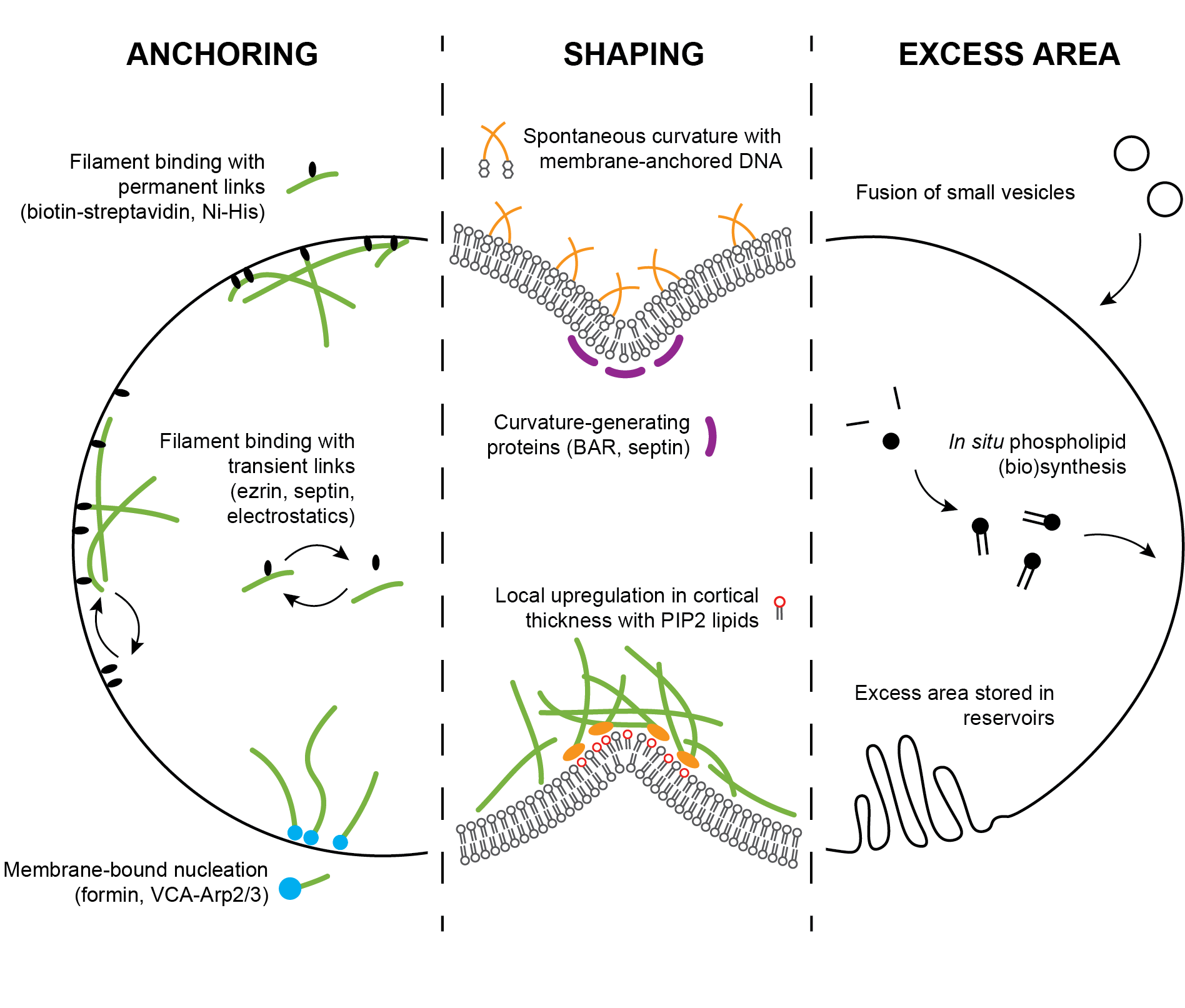
For engineering artificial cell division, it could be useful to exploit known mechanical effects of lipids. An important characteristic of lipid bilayers is that asymmetries between the two membrane leaflets give rise to membrane spontaneous curvature. Asymmetries can be generated in many different ways (reviewed in 182), such as by different lipid compositions or different numbers of lipids in the two leaflets 183, proteins binding to one leaflet133, membrane-anchored DNA oligos inserting into one leaflet135, or different solutes on both sides of the membrane 184. In the context of actomyosin-based synthetic cell division, spontaneous curvature effects could be exploited for spatial control and symmetry breaking. Binding of proteins to the outer leaflet of vesicles can be used to make vesicles dumbbell-shaped and to constrict and even split the neck 133. Generation of negative membrane curvature could be used to locally recruit septins, which selectively bind to membrane areas with micrometric curvature 107, 137. In addition, membrane-binding proteins that not only sense, but also generate curvature could be used, such as BAR-domain proteins. I-BAR proteins were shown to directly bind to actin in fission yeast 130 and are therefore interesting candidates for promoting actomyosin-driven membrane invagination. Interestingly, I-BAR domain proteins promote ezrin enrichment in negatively curved membrane protrusions 129, providing further prospects for boosting membrane invagination in vitro.
4.3 Addition of new membrane area
To create two daughter cells from a single mother cell, assuming spherical geometry, the cell surface area has to increase by 28% 39. In vivo, this extra membrane area is delivered to the cleavage furrow by targeted endosomal transport 185. This mechanism does not only lead to a local area increase, but also allows fast and localized delivery of specific lipids and regulatory proteins (reviewed in 186). For reconstitution of cell division, various strategies can be followed to increase the membrane area (fig. 5, right). First, GUV membranes can be grown by external addition of small unilamellar vesicles (SUVs) that can be forced to fuse with the GUV using fusogenic peptides, DNA, or charge-based interactions 187, 188, 189, 190. Second, lipid membranes can be grown by in situ synthesis of lipids from their precursors. Examples are non-enzymatic reactions from synthetic reactive precursors 191 or enzyme-catalysed biosynthesis using either purified proteins 192 or in vitro transcription-translation 193. Although there is evidence that mammalian cells do not use area reservoirs, such as microvilli, to supply extra membrane area for division 194, 195, this mechanism could be exploited for engineering division in a synthetic cells. Asymmetries between the two leaflets of the bilayer generated by different means (see preceding section) can be used to store excess area in membrane tubes and buds 133, 184, 196, 197. Low forces suffice to access these reservoirs 184, 197. To achieve synthetic cell division, it will be important to match timing of membrane areal growth with the timing of actin-driven constriction. To achieve multiple cycles of division, it will moreover be important to build in a mechanism to maintain lipid homeostasis.
5 Challenges ahead
In the past decades, our knowledge of cell division and its molecular actors has increased tremendously. To understand the physical mechanisms governing actomyosin-driven cell division, focus is put increasingly on bottom-up reconstitution experiments. Bulk and SLB experiments have helped us to understand mechanics of active actomyosin networks in 3D and 2D. However, translating these insights to the process of cell division is not trivial. To summarise, we list here the critical challenges that need to be overcome before we can reconstitute a minimal version of actin-driven cell division.
First, we need to understand how actin network contraction is sustained to drive division all the way. This will require myosin activity working in concert with actin turnover. While activity and turnover have been studied to great extent individually, we still have minimal understanding how they together govern actin network mechanics and contractility. Not only is this a challenging system to understand from a physical and biological perspective, also from an experimental perspective it is difficult to recapitulate, as it involves a large number of components whose concentration and activity need to be tightly controlled. More in vitro work in this direction, both in 3D and 2D, will be essential to explore the parameter space.
Second, it remains elusive how the actomyosin network should be anchored onto the membrane in order to achieve membrane deformations. A multitude of anchoring strategies has been developed and investigated, but only minimally in combination with a deformable membrane. Combined with our limited understanding of cortex-membrane molecular organization in vivo, this might prove one of the most important challenges. Future studies need to focus on understanding the influence of linker density and strength, as well as membrane composition and organization. In addition, the respective roles of filament-membrane linkers and membrane-bound nucleators need to be investigated.
Third, attention must be paid to the supplying of extra membrane area during constriction. Additional area can be present in membrane reservoirs, be synthesized, or be added by fusion of small vesicles. However, none of these approaches have to our knowledge been co-reconstituted with actin-driven contraction and resulting membrane deformation.
Fourth, there is to date only a minimal body of work on contractile actomyosin networks in GUVs. Confining the system in GUVs requires that all components are encapsulated in the right concentration and stoichiometric ratio, while preserving functionality. Although there are numerous GUV formation techniques, they have been minimally characterized for their potential to encapsulate complex mixtures of biochemically active components. More work in this direction is crucial to perform controlled reconstitution in GUVs, but also to be able to extrapolate findings from bulk and SLB experiments to vesicle systems.
Fifth, spatial and temporal control of the components and their activity is crucial. On the short term, some of the involved challenges may be by-passed by taking a semi-autonomous approach to synthetic cell division. For example, optogenetics, external mechanical or chemical cues, or fusion-based delivery of components with small vesicles provide handles to control the system even after encapsulation of the components inside GUVs. However, if the goal is to create a synthetic cell that divides fully autonomously, reconstitution will be more complicated, requiring for example feedback loops, signalling molecules and internal clocks.
As a concluding remark, we note that the most pressing challenges to achieve in vitro actin-driven cell division require integration of modules. Only when actomyosin studies meet membrane biophysics, when myosin motor activity is combined with actin turnover, and when protein biochemistry becomes integrated in GUV formation, we can start thinking about reconstituting cell division. In the coming years, perspectives from experimental work, theoretical studies and simulations need to be combined to guide future work with the ultimate goal to develop a full understanding of actin-driven synthetic cell division.
6 Acknowledgements
We would like to thank Ilina Bareja, Gerard Castro-Linares and Fred MacKintosh for useful discussions about actin cross-linkers. We acknowledge financial support from The Netherlands Organization of Scientific Research (NWO/OCW) Gravitation program Building A Synthetic Cell (BaSyC) (024.003.019).
References
- Staufer et al. 2021 Staufer, O. et al. Building a community to engineer synthetic cells and organelles from the bottom-up. eLife 2021, 10
- Frischmon et al. 2021 Frischmon, C.; Sorenson, C.; Winikoff, M.; Adamala, K. P. Build-a-Cell: Engineering a Synthetic Cell Community. Life 2021, 11
- Mulla et al. 2018 Mulla, Y.; Aufderhorst-Roberts, A.; Koenderink, G. H. Shaping up synthetic cells. Physical Biology 2018, 15, 041001
- Spoelstra et al. 2018 Spoelstra, W. K.; Deshpande, S.; Dekker, C. Tailoring the appearance: what will synthetic cells look like? Current Opinion in Biotechnology 2018, 51, 47–56
- Gaut and Adamala 2021 Gaut, N. J.; Adamala, K. P. Reconstituting Natural Cell Elements in Synthetic Cells. Advanced Biology 2021, 5
- Litschel and Schwille 2021 Litschel, T.; Schwille, P. Protein Reconstitution Inside Giant Unilamellar Vesicles. Annual Review of Biophysics 2021, 50, 525–548
- Olivi et al. 2021 Olivi, L.; Berger, M.; Creyghton, R. N.; De Franceschi, N.; Dekker, C.; Mulder, B. M.; Claassens, N. J.; ten Wolde, P. R.; van der Oost, J. Towards a synthetic cell cycle. Nature communications 2021, 12
- Kretschmer et al. 2019 Kretschmer, S.; Ganzinger, K. A.; Franquelim, H. G.; Schwille, P. Synthetic cell division via membrane-transforming molecular assemblies. BMC Biology 2019, 17
- Mahone and Goley 2020 Mahone, C. R.; Goley, E. D. Bacterial cell division at a glance. Journal of cell science 2020, 133
- Wang et al. 2020 Wang, K.; Okada, H.; Bi, E. Comparative Analysis of the Roles of Non-muscle Myosin-IIs in Cytokinesis in Budding Yeast, Fission Yeast, and Mammalian Cells. Frontiers in Cell and Developmental Biology 2020, 8, 1397
- Pollard 2017 Pollard, T. D. Nine unanswered questions about cytokinesis. Journal of Cell Biology 2017, 216, 3007–3016
- Cortes et al. 2018 Cortes, D. B.; Dawes, A.; Liu, J.; Nickaeen, M.; Strychalski, W.; Maddox, A. S. Unite to divide – How models and biological experimentation have come together to reveal mechanisms of cytokinesis. Journal of Cell Science 2018, 131
- Leite et al. 2019 Leite, J.; Osorio, D. S.; Sobral, A. F.; Silva, A. M.; Carvalho, A. X. Network Contractility During Cytokinesis-from Molecular to Global Views. Biomolecules 2019, 9
- Jongsma et al. 2015 Jongsma, M. L.; Berlin, I.; Neefjes, J. On the move: organelle dynamics during mitosis. Trends in Cell Biology 2015, 25, 112–124
- Addi et al. 2018 Addi, C.; Bai, J.; Echard, A. Actin, microtubule, septin and ESCRT filament remodeling during late steps of cytokinesis. Current opinion in cell biology 2018, 50, 27–34
- Horváth and Müller-Reichert 2020 Horváth, P.; Müller-Reichert, T. A Structural View on ESCRT-Mediated Abscission. Frontiers in Cell and Developmental Biology 2020, 8
- Anjur-Dietrich et al. 2021 Anjur-Dietrich, M. I.; Kelleher, C. P.; Needleman, D. J. Mechanical Mechanisms of Chromosome Segregation. Cells 2021, 10, 1–26
- Roberts 1961 Roberts, H. S. Mechanisms of Cytokinesis: A Critical Review. The Quarterly Review of Biology 1961, 36, 155–177
- Mitchison and Swann 1955 Mitchison, J. M.; Swann, M. M. The mechanical properties of the cell surface: III. The sea-urchin egg from fertilization to cleavage. Journal of Experimental Biology 1955, 32, 734–750
- Wolpert 1966 Wolpert, L. The mechanical properties of the membrane of the sea urchin egg during cleavage; 1966; Vol. 41; p 396
- Arnold 1969 Arnold, J. M. Cleavage furrow formation in a teoloecithal egg (Loligo Pealii) I. Filaments in Early Furrow Formation. Journal of Cell Biology 1969, 41, 894–904
- Schroeder 1968 Schroeder, T. E. Preliminary notes: Cytokinesis: filaments in the cleavage furrow. Experimental Cell Research 1968, 53, 272–316
- Kruse et al. 2005 Kruse, K.; Joanny, J. F.; Jülicher, F.; Prost, J.; Sekimoto, K. Generic theory of active polar gels: A paradigm for cytoskeletal dynamics. European Physical Journal E 2005, 16, 5–16
- Salbreux et al. 2009 Salbreux, G.; Prost, J.; Joanny, J. F. Hydrodynamics of cellular cortical flows and the formation of contractile rings. Physical Review Letters 2009, 103
- Zumdieck et al. 2005 Zumdieck, A.; Lagomarsino, M. C.; Tanase, C.; Kruse, K.; Mulder, B.; Dogterom, M.; Julicher, F. Continuum description of the cytoskeleton: Ring formation in the cell cortex. Physical Review Letters 2005, 95
- Turlier et al. 2014 Turlier, H.; Audoly, B.; Prost, J.; Joanny, J.-F. F. Furrow constriction in animal cell cytokinesis. Biophysical Journal 2014, 106, 114–123
- Reymann et al. 2016 Reymann, A.-C.; Staniscia, F.; Erzberger, A.; Salbreux, G.; Grill, S. W. Cortical flow aligns actin filaments to form a furrow. eLife 2016,
- White and Borisy 1983 White, J. G.; Borisy, G. G. On the Mechanisms of Cytokinesis in Animal Cells; 1983; Vol. 101; pp 289–316
- Hiraiwa and Salbreux 2016 Hiraiwa, T.; Salbreux, G. Role of Turnover in Active Stress Generation in a Filament Network. Physical Review Letters 2016, 116
- Chew et al. 2017 Chew, T. G.; Huang, J.; Palani, S.; Sommese, R.; Kamnev, A.; Hatano, T.; Gu, Y.; Oliferenko, S.; Sivaramakrishnan, S.; Balasubramanian, M. K. Actin turnover maintains actin filament homeostasis during cytokinetic ring contraction. Journal of Cell Biology 2017, 216, 2657–2667
- Swann and Mitchison 1958 Swann, M. M.; Mitchison, J. M. The mechanism of cleavage in animal cells. Biological Reviews 1958, 33, 103–135
- Dan 1948 Dan, J. C. On the Mechanism of Astral Cleavage. Physical Zoology 1948, 21, 191–218
- Wolpert 1960 Wolpert, L. The Mechanics and Mechanism of Cleavage. International Review of Cytology 1960, 10
- Marsland 1950 Marsland, D. The mechanisms of cell division, temperature-pressure experiments on the cleaving eggs of arbacia punctulata. Journal of Cellular and Comparative Physiology 1950, 36, 205–227
- Yoneda and Dan 1972 Yoneda, M.; Dan, K. Tension at the surface of the dividing sea-urchin egg; 1972; Vol. 57; pp 575–587
- Green et al. 2012 Green, R. A.; Paluch, E.; Oegema, K. Cytokinesis in Animal Cells. Annual Review of Cell and Developmental Biology 2012, 28, 29–58
- Gudejko et al. 2012 Gudejko, H. F. M.; Alford, L. M.; Burgess, D. R. Polar expansion during cytokinesis. Cytoskeleton 2012, 69, 1000–1009
- Wang 2001 Wang, Y.-L. The Mechanism of Cytokinesis: Reconsideration and Reconciliation. Cell structure and function 2001, 26, 633–638
- Frey and Idema 2021 Frey, F.; Idema, T. More than just a barrier: using physical models to couple membrane shape to cell function. Soft matter 2021, 17, 3533–3549
- Sedzinski et al. 2011 Sedzinski, J.; Biro, M.; Oswald, A.; Tinevez, J. Y.; Salbreux, G.; Paluch, E. Polar actomyosin contractility destabilizes the position of the cytokinetic furrow. Nature 2011, 476, 462–468
- Sinha et al. 2011 Sinha, B. et al. Cells respond to mechanical stress by rapid disassembly of caveolae. Cell 2011, 144, 402–413
- Albertson et al. 2005 Albertson, R.; Riggs, B.; Sullivan, W. Membrane traffic: A driving force in cytokinesis. Trends in Cell Biology 2005, 15, 92–101
- Chugh and Paluch 2018 Chugh, P.; Paluch, E. K. The actin cortex at a glance. Journal of Cell Science 2018, 131
- Asano et al. 2009 Asano, S.; Hamao, K.; Hosoya, H. Direct evidence for roles of phosphorylated regulatory light chain of myosin II in furrow ingression during cytokinesis in HeLa cells. Genes to Cells 2009, 14, 555–568
- Bray and White 1988 Bray, D.; White, J. G. Cortical flow in animal cells. Science 1988, 239, 883–888
- Najafabadi et al. 2022 Najafabadi, F. R.; Leaver, M.; Grill, S. W. Orchestrating Non-muscle myosin II filament assembly at the onset of cytokinesis. Molecular biology of the cell 2022,
- Koenderink and Paluch 2018 Koenderink, G. H.; Paluch, E. K. Architecture shapes contractility in actomyosin networks. Current Opinion in Cell Biology 2018, 50, 79–85
- Mendes Pinto et al. 2013 Mendes Pinto, I.; Rubinstein, B. Y.; Li, R. Force to divide: Structural and mechanical requirements for actomyosin ring contraction. Biophysical Journal 2013, 105, 547–554
- Murrell et al. 2015 Murrell, M.; Oakes, P. W.; Lenz, M.; Gardel, M. L. Forcing cells into shape: the mechanics of actomyosin contractility. Nature Reviews Molecular Cell Biology 2015 16:8 2015, 16, 486–498
- Kang et al. 2012 Kang, H.; Bradley, M. J.; McCullough, B. R.; Pierre, A.; Grintsevich, E. E.; Reisler, E.; De La Cruz, E. M. Identification of cation-binding sites on actin that drive polymerization and modulate bending stiffness. Proceedings of the National Academy of Sciences of the United States of America 2012, 109, 16923–16927
- Murrell and Gardel 2012 Murrell, M. P.; Gardel, M. L. F-actin buckling coordinates contractility and severing in a biomimetic actomyosin cortex. Proceedings of the National Academy of Sciences of the United States of America 2012, 109, 20820–20825
- Lenz et al. 2012 Lenz, M.; Thoresen, T.; Gardel, M. L.; Dinner, A. R. Contractile units in disordered actomyosin bundles arise from f-actin buckling. Physical Review Letters 2012, 108
- Kruse and Jülicher 2000 Kruse, K.; Jülicher, F. Actively Contracting Bundles of Polar Filaments. Physical Review Letters 2000, 85, 1778–1781
- Wollrab et al. 2019 Wollrab, V.; Belmonte, J. M.; Baldauf, L.; Leptin, M.; Nédeléc, F.; Koenderink, G. H. Polarity sorting drives remodeling of actin-myosin networks. Journal of Cell Science 2019, 132
- Fritzsche et al. 2016 Fritzsche, M.; Erlenkämper, C.; Moeendarbary, E.; Charras, G.; Kruse, K. Actin kinetics shapes cortical network structure and mechanics. Science Advances 2016, 2
- Berthoumieux et al. 2014 Berthoumieux, H.; Maître, J. L.; Heisenberg, C. P.; Paluch, E. K.; Jülicher, F.; Salbreux, G. Active elastic thin shell theory for cellular deformations. New Journal of Physics 2014, 16
- Malik-Garbi et al. 2019 Malik-Garbi, M.; Ierushalmi, N.; Jansen, S.; Abu-Shah, E.; Goode, B. L.; Mogilner, A.; Keren, K. Scaling behaviour in steady-state contracting actomyosin networks. Nature Physics 2019, 15, 509–516
- Sonal et al. 2019 Sonal,; Ganzinger, K. A.; Vogel, S. K.; Mücksch, J.; Blumhardt, P.; Schwille, P. Myosin-II activity generates a dynamic steady state with continuous actin turnover in a minimal actin cortex. Journal of Cell Science 2019, 132
- Fritzsche et al. 2013 Fritzsche, M.; Lewalle, A.; Duke, T.; Kruse, K.; Charras, G. Analysis of turnover dynamics of the submembranous actin cortex. Molecular Biology of the Cell 2013, 24, 757–767
- Robin et al. 2014 Robin, F. B.; McFadden, W. M.; Yao, B.; Munro, E. M. Single-molecule analysis of cell surface dynamics in Caenorhabditis elegans embryos. Nature Methods 2014, 11, 677–682
- McCall et al. 2019 McCall, P. M.; MacKintosh, F. C.; Kovar, D. R.; Gardel, M. L. Cofilin drives rapid turnover and fluidization of entangled F-actin. Proceedings of the National Academy of Sciences of the United States of America 2019, 116, 12629–12637
- Arzash et al. 2019 Arzash, S.; McCall, P. M.; Feng, J.; Gardel, M. L.; MacKintosh, F. C. Stress relaxation in F-actin solutions by severing. Soft Matter 2019, 15, 6300–6307
- Pontani et al. 2009 Pontani, L. L.; Van Der Gucht, J.; Salbreux, G.; Heuvingh, J.; Joanny, J. F.; Sykes, C. Reconstitution of an actin cortex inside a liposome. Biophysical Journal 2009, 96, 192–198
- Linsmeier et al. 2016 Linsmeier, I.; Banerjee, S.; Oakes, P. W.; Jung, W.; Kim, T.; Murrell, M. P. Disordered actomyosin networks are sufficient to produce cooperative and telescopic contractility. Nature Communications 2016, 7, 1–9
- Bovellan et al. 2014 Bovellan, M. et al. Cellular control of cortical actin nucleation. Current Biology 2014, 24, 1628–1635
- Cao et al. 2020 Cao, L. et al. SPIN90 associates with mDia1 and the Arp2/3 complex to regulate cortical actin organization. Nature cell biology 2020, 22, 803–814
- Zimmermann et al. 2017 Zimmermann, D.; Homa, K. E.; Hocky, G. M.; Pollard, L. W.; De La Cruz, E. M.; Voth, G. A.; Trybus, K. M.; Kovar, D. R. Mechanoregulated inhibition of formin facilitates contractile actomyosin ring assembly. Nature Communications 2017, 8, 1–12
- Cao et al. 2018 Cao, L.; Kerleau, M.; Suzuki, E. L.; Wioland, H.; Jouet, S.; Guichard, B.; Lenz, M.; Romet-Lemonne, G.; Jegou, A. Modulation of formin processivity by profilin and mechanical tension. eLife 2018, 7, 1–24
- Suarez et al. 2015 Suarez, C.; Carroll, R. T.; Burke, T. A.; Christensen, J. R.; Bestul, A. J.; Sees, J. A.; James, M. L.; Sirotkin, V.; Kovar, D. R. Profilin regulates F-Actin network homeostasis by favoring formin over Arp2/3 complex. Developmental Cell 2015, 32, 43–53
- Shekhar et al. 2015 Shekhar, S.; Kerleau, M.; Kühn, S.; Pernier, J.; Romet-Lemonne, G.; Jégou, A.; Carlier, M. F. Formin and capping protein together embrace the actin filament in a ménage à trois. Nature Communications 2015, 6
- Mullins et al. 1998 Mullins, R. D.; Kelleher, J. F.; Xu, J.; Pollard, T. D. Arp2/3 complex from Acanthamoeba binds profilin and cross-links actin filaments. Molecular Biology of the Cell 1998, 9, 841–852
- Blanchoin et al. 2000 Blanchoin, L.; Pollard, T. D.; Mullins, R. D. Interactions of ADF/cofilin, Arp2/3 complex, capping protein and profilin in remodeling of branched actin filament networks. Current Biology 2000, 10, 1273–1282
- Risca et al. 2012 Risca, V. I.; Wang, E. B.; Chaudhuri, O.; Chia, J. J.; Geissler, P. L.; Fletcher, D. A. Actin filament curvature biases branching direction. Proceedings of the National Academy of Sciences of the United States of America 2012, 109, 2913–2918
- Abella et al. 2016 Abella, J. V. G.; Galloni, C.; Pernier, J.; Barry, D. J.; Kjær, S.; Carlier, M. F.; Way, M. Isoform diversity in the Arp2/3 complex determines actin filament dynamics. Nature Cell Biology 2016, 18, 76–86
- Quinlan et al. 2005 Quinlan, M. E.; Heuser, J. E.; Kerkhoff, E.; Mullins, R. D. Drosophila Spire is an actin nucleation factor. Nature 2005, 433, 382–388
- Chan et al. 2009 Chan, C.; Beltzner, C. C.; Pollard, T. D. Cofilin Dissociates Arp2/3 Complex and Branches from Actin Filaments. Current Biology 2009, 19, 537–545
- Kotila et al. 2019 Kotila, T.; Wioland, H.; Enkavi, G.; Kogan, K.; Vattulainen, I.; Jégou, A.; Romet-Lemonne, G.; Lappalainen, P. Mechanism of synergistic actin filament pointed end depolymerization by cyclase-associated protein and cofilin. Nature Communications 2019, 10, 1–14
- Jansen et al. 2014 Jansen, S.; Collins, A.; Golden, L.; Sokolova, O.; Goode, B. L. Structure and mechanism of mouse cyclase-associated protein (CAP1) in regulating actin dynamics. Journal of Biological Chemistry 2014, 289, 30732–30742
- Wioland et al. 2019 Wioland, H.; Jegou, A.; Romet-Lemonne, G. Torsional stress generated by ADF/cofilin on cross-linked actin filaments boosts their severing. Proceedings of the National Academy of Sciences of the United States of America 2019, 116, 2595–2602
- Pandit et al. 2020 Pandit, N. G.; Cao, W.; Bibeau, J.; Johnson-Chavarria, E. M.; Taylor, E. W.; Pollard, T. D.; De La Cruz, E. M. Force and phosphate release from Arp2/3 complex promote dissociation of actin filament branches. Proceedings of the National Academy of Sciences of the United States of America 2020, 117, 13519–13528
- Tsai and Koenderink 2015 Tsai, F.-C.; Koenderink, G. Shape control of lipid bilayer membranes by confined actin bundles. Soft Matter 2015, 11, 8834–8847
- Litschel et al. 2021 Litschel, T.; Kelley, C. F.; Holz, D.; Adeli Koudehi, M.; Vogel, S. K.; Burbaum, L.; Mizuno, N.; Vavylonis, D.; Schwille, P. Reconstitution of contractile actomyosin rings in vesicles. Nature Communications 2021, 12, 1–10
- van de Cauter et al. 2021 van de Cauter, L.; Fanalista, F.; van Buren, L.; Franceschi, N. D.; Godino, E.; Bouw, S.; Danelon, C.; Dekker, C.; Koenderink, G. H.; Ganzinger, K. A. Optimized cDICE for Efficient Reconstitution of Biological Systems in Giant Unilamellar Vesicles. ACS Synthetic Biology 2021, 10, 1690
- Bashirzadeh et al. 2021 Bashirzadeh, Y.; Wubshet, N.; Litschel, T.; Schwille, P.; Liu, A. P. Rapid Encapsulation of Reconstituted Cytoskeleton inside Giant Unilamellar Vesicles. Journal of Visualized Experiments : Jove 2021,
- Murrell et al. 2011 Murrell, M.; Pontani, L. L.; Guevorkian, K.; Cuvelier, D.; Nassoy, P.; Sykes, C. Spreading dynamics of biomimetic actin cortices. Biophysical Journal 2011, 100, 1400–1409
- Dürre et al. 2018 Dürre, K.; Keber, F. C.; Bleicher, P.; Brauns, F.; Cyron, C. J.; Faix, J.; Bausch, A. R. Capping protein-controlled actin polymerization shapes lipid membranes. Nature Communications 2018, 9, 1–11
- Bashirzadeh et al. 2022 Bashirzadeh, Y.; Moghimianavval, H.; Liu, A. P. Encapsulated actomyosin patterns drive cell-like membrane shape changes. iScience 2022, 25, 104236
- Carvalho et al. 2013 Carvalho, K. K. K.; Tsai, F.-C. C.; Lees, E.; Voituriez, R.; Koenderink, G. H.; Sykes, C. Cell-sized liposomes reveal how actomyosin cortical tension drives shape change. Proceedings of the National Academy of Sciences 2013, 110, 16456–16461
- Loiseau et al. 2016 Loiseau, E.; Schneider, J. A. M.; Keber, F. C.; Pelzl, C.; Massiera, G.; Salbreux, G.; Bausch, A. R. Shape remodeling and blebbing of active cytoskeletal vesicles. Science Advances 2016, 2
- Sakamoto et al. 2005 Sakamoto, T.; Limouze, J.; Combs, C. A.; Straight, A. F.; Sellers, J. R. Blebbistatin, a myosin II inhibitor, is photoinactivated by blue light. Biochemistry 2005, 44, 584–588
- Schuppler et al. 2016 Schuppler, M.; Keber, F. C.; Krüger, M.; Bausch, A. R. Boundaries steer the contraction of active gels. Nature Communications 2016, 7, 1–10
- Yamamoto et al. 2021 Yamamoto, K.; Miura, H.; Ishida, M.; Sawai, S.; Kondo, Y.; Aoki, K. Optogenetic relaxation of actomyosin contractility uncovers mechanistic roles of cortical tension during cytokinesis. bioRxiv 2021, 2021.04.19.440549
- Fanalista et al. 2019 Fanalista, F.; Birnie, A.; Maan, R.; Burla, F.; Charles, K.; Pawlik, G.; Deshpande, S.; Koenderink, G. H.; Dogterom, M.; Dekker, C. Shape and Size Control of Artificial Cells for Bottom-Up Biology. ACS Nano 2019, 13, 5439–5450
- Barrows and Goley 2021 Barrows, J. M.; Goley, E. D. FtsZ dynamics in bacterial division: What, how, and why? Current opinion in cell biology 2021, 68, 163–172
- Lau et al. 2009 Lau, A. W.; Prasad, A.; Dogic, Z. Condensation of isolated Semi-flexible filaments driven by depletion interactions. Europhysics Letters 2009, 87
- Tang et al. 2001 Tang, J. X.; Käs, J. A.; Shah, J. V.; Janmey, P. A. Counterion-induced actin ring formation. European Biophysics Journal 2001, 30, 477–484
- Mavrakis et al. 2014 Mavrakis, M.; Azou-Gros, Y.; Tsai, F.-C. C.; Alvarado, J.; Bertin, A.; Iv, F.; Kress, A.; Brasselet, S.; Koenderink, G. H.; Lecuit, T. Septins promote F-actin ring formation by crosslinking actin filaments into curved bundles. Nature Cell Biology 2014, 16, 322–334
- Kinoshita et al. 2002 Kinoshita, M.; Field, C. M.; Coughlin, M. L.; Straight, A. F.; Mitchison, T. J. Self- and actin-templated assembly of mammalian septins. Developmental Cell 2002, 3, 791–802
- Field et al. 2005 Field, C. M.; Coughlin, M.; Doberstein, S.; Marty, T.; Sullivan, W. Characterization of anillin mutants reveals essential roles in septin localization and plasma membrane integrity. Development 2005, 132, 2849–2860
- Piekny and Glotzer 2008 Piekny, A. J.; Glotzer, M. Anillin Is a Scaffold Protein That Links RhoA, Actin, and Myosin during Cytokinesis. Current Biology 2008, 18, 30–36
- Piekny and Maddox 2010 Piekny, A. J.; Maddox, A. S. The myriad roles of Anillin during cytokinesis. Seminars in Cell and Developmental Biology 2010, 21, 881–891
- Garno et al. 2021 Garno, C.; Irons, Z. H.; Gamache, C. M.; McKim, Q.; Reyes, G.; Wu, X.; Shuster, C. B.; Henson, J. H. Building the cytokinetic contractile ring in an early embryo: Initiation as clusters of myosin II, anillin and septin, and visualization of a septin filament network. PLoS ONE 2021, 16
- Kučera et al. 2021 Kučera, O.; Janda, D.; Siahaan, V.; Dijkstra, S. H.; Pilátová, E.; Zatecka, E.; Diez, S.; Braun, M.; Lansky, Z. Anillin propels myosin-independent constriction of actin rings. Nature Communications 2021, 12, 1–12
- Palani et al. 2021 Palani, S.; Ghosh, S.; Ivorra-Molla, E.; Clarke, S.; Suchenko, A.; Balasubramanian, M. K.; Köster, D. V. Calponin-homology domain mediated bending of membrane-associated actin filaments. eLife 2021, 10, 1–19
- Biro et al. 2013 Biro, M.; Romeo, Y.; Kroschwald, S.; Bovellan, M.; Boden, A.; Tcherkezian, J.; Roux, P. P.; Charras, G.; Paluch, E. K. Cell cortex composition and homeostasis resolved by integrating proteomics and quantitative imaging. Cytoskeleton 2013, 70, 741–754
- Miyazaki et al. 2015 Miyazaki, M.; Chiba, M.; Eguchi, H.; Ohki, T.; Ishiwata, S. Cell-sized spherical confinement induces the spontaneous formation of contractile actomyosin rings in vitro. Nature Cell Biology 2015, 17, 480–489
- Bridges et al. 2016 Bridges, A. A.; Jentzsch, M. S.; Oakes, P. W.; Occhipinti, P.; Gladfelter, A. S. Micron-scale plasma membrane curvature is recognized by the septin cytoskeleton. Journal of Cell Biology 2016, 213, 23–32
- Ganzinger et al. 2020 Ganzinger, K. A.; Merino-Salomón, A.; García-Soriano, D. A.; Butterfield, A. N.; Litschel, T.; Siedler, F.; Schwille, P. FtsZ Reorganization Facilitates Deformation of Giant Vesicles in Microfluidic Traps**. Angewandte Chemie 2020, 132, 21556–21560
- Morrison and Thirumalai 2009 Morrison, G.; Thirumalai, D. Semiflexible chains in confined spaces. Physical Review E - Statistical, Nonlinear, and Soft Matter Physics 2009, 79, 1–14
- Soares E Silva et al. 2011 Soares E Silva, M.; Alvarado, J.; Nguyen, J.; Georgoulia, N.; Mulder, B. M.; Koenderink, G. H. Self-organized patterns of actin filaments in cell-sized confinement. Soft Matter 2011, 7, 10631–10641
- Claessens et al. 2006 Claessens, M. M. A. E.; Tharmann, R.; Kroy, K.; Bausch, A. R. Microstructure and viscoelasticity of confined semiflexible polymer networks. Nature Physics 2006, 2, 186–189
- Limozin and Sackmann 2002 Limozin, L.; Sackmann, E. Polymorphism of Cross-Linked Actin Networks in Giant Vesicles. Physical Review Letters 2002, 89, 14–17
- Bashirzadeh et al. 2021 Bashirzadeh, Y.; Redford, S. A.; Lorpaiboon, C.; Groaz, A.; Moghimianavval, H.; Litschel, T.; Schwille, P.; Hocky, G. M.; Dinner, A. R.; Liu, A. P. Actin crosslinker competition and sorting drive emergent GUV size-dependent actin network architecture. Communications Biology 2021, 4
- Schäfer et al. 2015 Schäfer, E.; Vache, M.; Kliesch, T. T.; Janshoff, A. Mechanical response of adherent giant liposomes to indentation with a conical AFM-tip. Soft Matter 2015, 11, 4487–4495
- Adeli Koudehi et al. 2019 Adeli Koudehi, M.; Rutkowski, D. M.; Vavylonis, D. Organization of associating or crosslinked actin filaments in confinement. Cytoskeleton 2019, 76, 532–548
- Schroeder 1972 Schroeder, T. E. The contractile ring II. Determining its brief existence, volumetric changes, and vital role in cleaving arbacia eggs. Journal of Cell Biology 1972, 53, 419–434
- Ma et al. 2012 Ma, X.; Kovaćs, M.; Conti, M. A.; Wang, A.; Zhang, Y.; Sellers, J. R.; Adelstein, R. S. Nonmuscle myosin II exerts tension but does not translocate actin in vertebrate cytokinesis. Proceedings of the National Academy of Sciences of the United States of America 2012, 109, 4509–4514
- Reichl et al. 2008 Reichl, E. M.; Ren, Y.; Morphew, M. K.; Delannoy, M.; Effler, J. C.; Girard, K. D.; Divi, S.; Iglesias, P. A.; Kuo, S. C.; Robinson, D. N. Interactions between Myosin and Actin Crosslinkers Control Cytokinesis Contractility Dynamics and Mechanics. Current Biology 2008, 18, 471–480
- Fishkind and Wang 1993 Fishkind, D. J.; Wang, Y. L. Orientation and three-dimensional organization of actin filaments in dividing cultured cells. Journal of Cell Biology 1993, 123, 837–848
- Fenix et al. 2016 Fenix, A. M.; Taneja, N.; Buttler, C. A.; Lewis, J.; Van Engelenburg, S. B.; Ohi, R.; Burnette, D. T. Expansion and concatenation of nonmuscle myosin IIA filaments drive cellular contractile system formation during interphase and mitosis. Molecular Biology of the Cell 2016, 27, 1465–1478
- Henson et al. 2017 Henson, J. H.; Ditzler, C. E.; Germain, A.; Irwin, P. M.; Vogt, E. T.; Yang, S.; Wu, X.; Shuster, C. B. The ultrastructural organization of actin and myosin II filaments in the contractile ring: New support for an old model of cytokinesis. Molecular Biology of the Cell 2017, 28, 613–623
- Lenz 2014 Lenz, M. Geometrical origins of contractility in disordered actomyosin networks. Physical Review X 2014, 4, 041002
- Ennomani et al. 2016 Ennomani, H.; Letort, G.; Guérin, C.; Martiel, J. L.; Cao, W.; Nédélec, F.; De La Cruz, E. M.; Théry, M.; Blancholn, L. Architecture and Connectivity Govern Actin Network Contractility. Current Biology 2016, 26, 616–626
- Schwayer et al. 2016 Schwayer, C.; Sikora, M.; Slováková, J.; Kardos, R.; Heisenberg, C. P. Actin Rings of Power. Developmental Cell 2016, 37, 493–506
- Odde 2015 Odde, D. J. Mitosis, diffusible crosslinkers, and the ideal gas law. Cell 2015, 160, 1041–1043
- Chen et al. 2020 Chen, S.; Markovich, T.; MacKintosh, F. C. Motor-free Contractility in Active Gels. arXiv preprint 2020,
- Rizzelli et al. 2020 Rizzelli, F.; Malabarba, M. G.; Sigismund, S.; Mapelli, M. The crosstalk between microtubules, actin and membranes shapes cell division. Open biology 2020, 10
- Lancaster and Baum 2014 Lancaster, O. M.; Baum, B. Shaping up to divide: coordinating actin and microtubule cytoskeletal remodelling during mitosis. Seminars in cell & developmental biology 2014, 34, 109–115
- Tsai et al. 2018 Tsai, F. C.; Bertin, A.; Bousquet, H.; Manzi, J.; Senju, Y.; Tsai, M. C.; Picas, L.; Miserey-Lenkei, S.; Lappalainen, P.; Lemichez, E.; Coudrier, E.; Bassereau, P. Ezrin enrichment on curved membranes requires a specific conformation or interaction with a curvature-sensitive partner. eLife 2018, 7, 1–27
- Quinones et al. 2010 Quinones, G. A.; Jin, J.; Oro, A. E. I-BAR protein antagonism of endocytosis mediates directional sensing during guided cell migration. Journal of Cell Biology 2010, 189, 353–367
- Bae et al. 2019 Bae, W.; Kocabey, S.; Liedl, T. DNA nanostructures in vitro, in vivo and on membranes. Nano Today 2019, 26, 98–107
- Czogalla et al. 2015 Czogalla, A.; Kauert, D. J.; Franquelim, H. G.; Uzunova, V.; Zhang, Y.; Seidel, R.; Schwille, P. Amphipathic DNA Origami Nanoparticles to Scaffold and Deform Lipid Membrane Vesicles. Angewandte Chemie International Edition 2015, 54, 6501–6505
- Steinkühler et al. 2020 Steinkühler, J.; Knorr, R. L.; Zhao, Z.; Bhatia, T.; Bartelt, S. M.; Wegner, S.; Dimova, R.; Lipowsky, R. Controlled division of cell-sized vesicles by low densities of membrane-bound proteins. Nature Communications 2020, 11, 1–11
- Mishra et al. 2013 Mishra, M.; Kashiwazaki, J.; Takagi, T.; Srinivasan, R.; Huang, Y.; Balasubramanian, M. K.; Mabuchi, I. In vitro contraction of cytokinetic ring depends on myosin II but not on actin dynamics. Nature Cell Biology 2013, 15, 853–859
- Franceschi et al. 2021 Franceschi, N. D.; Pezeshkian, W.; Fragasso, A.; Bruininks, B. M. H.; Tsai, S.; Marrink, S. J.; Dekker, C. A synthetic membrane shaper for controlled liposome deformation. bioRxiv 2021, 2021.12.22.473854
- McMahon and Boucrot 2015 McMahon, H. T.; Boucrot, E. Membrane curvature at a glance. Journal of cell science 2015, 128, 1065–1070
- Beber et al. 2019 Beber, A.; Taveneau, C.; Nania, M.; Tsai, F. C.; Di Cicco, A.; Bassereau, P.; Lévy, D.; Cabral, J. T.; Isambert, H.; Mangenot, S.; Bertin, A. Membrane reshaping by micrometric curvature sensitive septin filaments. Nature Communications 2019, 10, 1–12
- Taubenberger et al. 2020 Taubenberger, A. V.; Baum, B.; Matthews, H. K. The Mechanics of Mitotic Cell Rounding. Frontiers in cell and developmental biology 2020, 8
- Prospéri et al. 2015 Prospéri, M. T.; Lépine, P.; Dingli, F.; Paul-Gilloteaux, P.; Martin, R.; Loew, D.; Knölker, H. J.; Coudrier, E. Myosin 1b functions as an effector of EphB signaling to control cell repulsion. The Journal of cell biology 2015, 210, 347–361
- Russo and Krauss 2021 Russo, G.; Krauss, M. Septin Remodeling During Mammalian Cytokinesis. Frontiers in cell and developmental biology 2021, 9
- Michie et al. 2019 Michie, K. A.; Bermeister, A.; Robertson, N. O.; Goodchild, S. C.; Curmi, P. M. G. Two Sides of the Coin: Ezrin/Radixin/Moesin and Merlin Control Membrane Structure and Contact Inhibition. International journal of molecular sciences 2019, 20
- Clausen et al. 2017 Clausen, M. P.; Colin-York, H.; Schneider, F.; Eggeling, C.; Fritzsche, M. Dissecting the actin cortex density and membrane-cortex distance in living cells by super-resolution microscopy. Journal of Physics D: Applied Physics 2017, 50, 64002
- Swulius et al. 2018 Swulius, M. T.; Nguyen, L. T.; Ladinsky, M. S.; Ortega, D. R.; Aich, S.; Mishra, M.; Jensen, G. J. Structure of the fission yeast actomyosin ring during constriction. Proceedings of the National Academy of Sciences of the United States of America 2018, 115, E1455–E1464
- McDonald et al. 2017 McDonald, N. A.; Lind, A. L.; Smith, S. E.; Li, R.; Gould, K. L. Nanoscale architecture of the Schizosaccharomyces pombe contractile ring. eLife 2017, 6
- Truong Quang et al. 2021 Truong Quang, B. A.; Peters, R.; Cassani, D. A. D.; Chugh, P.; Clark, A. G.; Agnew, M.; Charras, G.; Paluch, E. K. Extent of myosin penetration within the actin cortex regulates cell surface mechanics. Nature Communications 2021, 12, 1–12
- Das et al. 2020 Das, A.; Bhat, A.; Sknepnek, R.; Köster, D.; Mayor, S.; Rao, M. Stratification relieves constraints from steric hindrance in the generation of compact actomyosin asters at the membrane cortex. Science Advances 2020, 6, 6093–6104
- Sheetz 2001 Sheetz, M. P. Cell control by membrane-cytoskeleton adhesion. Nature Reviews Molecular Cell Biology 2001, 2, 392–396
- Paraschiv et al. 2021 Paraschiv, A.; Lagny, T. J.; Campos, C. V.; Coudrier, E.; Bassereau, P.; Šarić, A. Influence of membrane-cortex linkers on the extrusion of membrane tubes. Biophysical Journal 2021, 120, 598–606
- Raucher et al. 2000 Raucher, D.; Stauffer, T.; Chen, W.; Shen, K.; Guo, S.; York, J. D.; Sheetz, M. P.; Meyer, T. Phosphatidylinositol 4,5-bisphosphate functions as a second messenger that regulates cytoskeleton-plasma membrane adhesion. Cell 2000, 100, 221–228
- Janmey et al. 1999 Janmey, P. A.; Xian, W.; Flanagan, L. A. Controlling cytoskeleton structure by phosphoinositide-protein interactions: Phosphoinositide binding protein domains and effects of lipid packing. Chemistry and Physics of Lipids 1999, 101, 93–107
- Snider et al. 2017 Snider, C. E.; Willet, A. H.; Chen, J. S.; Arpag, G.; Zanic, M.; Gould, K. L. Phosphoinositide-mediated ring anchoring resists perpendicular forces to promote medial cytokinesis. Journal of Cell Biology 2017, 216, 3041–3050
- Simon et al. 2018 Simon, C.; Caorsi, V.; Campillo, C.; Sykes, C. Interplay between membrane tension and the actin cytoskeleton determines shape changes. Physical Biology 2018, 15
- Shrivastava et al. 2015 Shrivastava, R.; Köster, D.; Kalme, S.; Mayor, S.; Neerathilingam, M. Tailor-made ezrin actin binding domain to probe its interaction with actin in-vitro. PLoS ONE 2015, 10, e0123428
- Rico et al. 2019 Rico, F.; Russek, A.; González, L.; Grubmüller, H.; Scheuring, S. Heterogeneous and rate-dependent streptavidin–biotin unbinding revealed by high-speed force spectroscopy and atomistic simulations. Proceedings of the National Academy of Sciences of the United States of America 2019, 116, 6594–6601
- Nye and Groves 2008 Nye, J. A.; Groves, J. T. Kinetic control of histidine-tagged protein surface density on supported lipid bilayers. Langmuir 2008, 24, 4145–4149
- Raghunath and Dyer 2019 Raghunath, G.; Dyer, R. B. Kinetics of Histidine-Tagged Protein Association to Nickel-Decorated Liposome Surfaces. Langmuir 2019, 35, 12550–12561
- Grover et al. 2016 Grover, R.; Fischer, J.; Schwarz, F. W.; Walter, W. J.; Schwille, P.; Diez, S. Transport efficiency of membrane-anchored kinesin-1 motors depends on motor density and diffusivity. Proceedings of the National Academy of Sciences of the United States of America 2016, 113, E7185–E7193
- Pernier et al. 2019 Pernier, J.; Kusters, R.; Bousquet, H.; Lagny, T.; Morchain, A.; Joanny, J. F.; Bassereau, P.; Coudrier, E. Myosin 1b is an actin depolymerase. Nature Communications 2019, 10, 1–7
- Schneider et al. 2017 Schneider, F.; Waithe, D.; Clausen, M. P.; Galiani, S.; Koller, T.; Ozhan, G.; Eggeling, C.; Sezgin, E. Diffusion of lipids and GPI-anchored proteins in actin-free plasma membrane vesicles measured by STED-FCS. Molecular Biology of the Cell 2017, 28, 1507–1518
- Heinemann et al. 2013 Heinemann, F.; Vogel, S. K.; Schwille, P. Lateral Membrane Diffusion Modulated by a Minimal Actin Cortex. Biophysical Journal 2013, 104, 1465–1475
- Vogel et al. 2017 Vogel, S. K.; Greiss, F.; Khmelinskaia, A.; Schwille, P. Control of lipid domain organization by a biomimetic contractile actomyosin cortex. eLife 2017, 6
- Köster et al. 2016 Köster, D. V.; Husain, K.; Iljazi, E.; Bhat, A.; Bieling, P.; Mullins, R. D.; Rao, M.; Mayor, S. Actomyosin dynamics drive local membrane component organization in an in vitro active composite layer. Proceedings of the National Academy of Sciences of the United States of America 2016, 113, E1645–E1654
- Honigmann et al. 2014 Honigmann, A.; Sadeghi, S.; Keller, J.; Hell, S. W.; Eggeling, C.; Vink, R. A lipid bound actin meshwork organizes liquid phase separation in model membranes. eLife 2014, 2014
- Rao and Mayor 2014 Rao, M.; Mayor, S. Active organization of membrane constituents in living cells. Current Opinion in Cell Biology 2014, 29, 126–132
- Gowrishankar et al. 2012 Gowrishankar, K.; Ghosh, S.; Saha, S.; Rumamol, C.; Mayor, S.; Rao, M. Active remodeling of cortical actin regulates spatiotemporal organization of cell surface molecules. Cell 2012, 149, 1353–1367
- Liu and Fletcher 2006 Liu, A. P.; Fletcher, D. A. Actin polymerization serves as a membrane domain switch in model lipid bilayers. Biophysical Journal 2006, 91, 4064–4070
- Schön et al. 2019 Schön, M.; Mey, I.; Steinem, C. Influence of cross-linkers on ezrin-bound minimal actin cortices. Progress in Biophysics and Molecular Biology 2019, 144, 91–101
- Nöding et al. 2018 Nöding, H.; Schön, M.; Reinermann, C.; Dörrer, N.; Kürschner, A.; Geil, B.; Mey, I.; Heussinger, C.; Janshoff, A.; Steinem, C. Rheology of Membrane-Attached Minimal Actin Cortices. Journal of Physical Chemistry B 2018, 122, 4537–4545
- Mosby et al. 2020 Mosby, L. S.; Hundt, N.; Young, G.; Fineberg, A.; Polin, M.; Mayor, S.; Kukura, P.; Köster, D. V. Myosin II Filament Dynamics in Actin Networks Revealed with Interferometric Scattering Microscopy. Biophysical Journal 2020, 118, 1946–1957
- Szuba et al. 2021 Szuba, A.; Bano, F.; Castro Linares, G.; Iv, F.; Mavrakis, M.; Richter, R. P.; Bertin, A.; Koenderink, G. H. Membrane binding controls ordered self-assembly of animal septins. eLife 2021, 10
- Cannon et al. 2019 Cannon, K. S.; Woods, B. L.; Crutchley, J. M.; Gladfelter, A. S. An amphipathic helix enables septins to sense micrometer-scale membrane curvature. Journal of Cell Biology 2019, 218, 1128–1137
- Sun et al. 2015 Sun, L.; Guan, R.; Lee, I. J.; Liu, Y.; Chen, M.; Wang, J.; Wu, J. Q.; Chen, Z. Mechanistic Insights into the Anchorage of the Contractile Ring by Anillin and Mid1. Developmental Cell 2015, 33, 413–426
- Schroer et al. 2020 Schroer, C. F.; Baldauf, L.; van Buren, L.; Wassenaar, T. A.; Melo, M. N.; Koenderink, G. H.; Marrink, S. J. Charge-dependent interactions of monomeric and filamentous actin with lipid bilayers. Proceedings of the National Academy of Sciences of the United States of America 2020, 117, 5861–5872
- Zigmond 2004 Zigmond, S. H. Formin-induced nucleation of actin filaments. Current Opinion in Cell Biology 2004, 16, 99–105
- Romero et al. 2004 Romero, S.; Le Clainche, C.; Didry, D.; Egile, C.; Pantaloni, D.; Carlier, M. F. Formin is a processive motor that requires profilin to accelerate actin assembly and associated ATP hydrolysis. Cell 2004, 119, 419–429
- Guevorkian et al. 2015 Guevorkian, K.; Manzi, J.; Pontani, L. L.; Brochard-Wyart, F.; Sykes, C. Mechanics of Biomimetic Liposomes Encapsulating an Actin Shell. Biophysical Journal 2015, 109, 2471–2479
- Bleicher et al. 2020 Bleicher, P.; Sciortino, A.; Bausch, A. R. The dynamics of actin network turnover is self-organized by a growth-depletion feedback. Scientific Reports 2020, 10, 1–11
- Ng et al. 2005 Ng, M. M.; Chang, F.; Burgess, D. R. Movement of membrane domains and requirement of membrane signaling molecules for cytokinesis. Developmental Cell 2005, 9, 781–790
- Cauvin and Echard 2015 Cauvin, C.; Echard, A. Phosphoinositides: Lipids with informative heads and mastermind functions in cell division. Biochimica et Biophysica Acta - Molecular and Cell Biology of Lipids 2015, 1851, 832–843
- Logan and Mandato 2006 Logan, M. R.; Mandato, C. A. Regulation of the actin cytoskeleton by PIP2 in cytokinesis. Biology of the Cell 2006, 98, 377–388
- Emoto and Umeda 2001 Emoto, K.; Umeda, M. Membrane Lipid Control of Cytokinesis. Cell Structure and Function 2001, 26, 659–665
- Bassereau et al. 2018 Bassereau, P. et al. The 2018 biomembrane curvature and remodeling roadmap. Journal of Physics D: Applied Physics 2018, 51, 343001
- Dasgupta et al. 2018 Dasgupta, R.; Miettinen, M. S.; Fricke, N.; Lipowsky, R.; Dimova, R. The glycolipid GM1 reshapes asymmetric biomembranes and giant vesicles by curvature generation. Proceedings of the National Academy of Sciences of the United States of America 2018, 115, 5756–5761
- Karimi et al. 2018 Karimi, M.; Steinkühler, J.; Roy, D.; Dasgupta, R.; Lipowsky, R.; Dimova, R. Asymmetric Ionic Conditions Generate Large Membrane Curvatures. Nano Letters 2018, 18, 7816–7821
- Shuster and Burgess 2002 Shuster, C. B.; Burgess, D. R. Targeted new membrane addition in the cleavage furrow is a late, separate event in cytokinesis. Proceedings of the National Academy of Sciences 2002, 99, 3633–3638
- Schiel and Prekeris 2013 Schiel, J. A.; Prekeris, R. Membrane dynamics during cytokinesis. Current Opinion in Cell Biology 2013, 25, 92–98
- Marsden et al. 2009 Marsden, H. R.; Elbers, N. A.; Bomans, P. H. H.; Sommerdijk, N. A. J. M.; Kros, A. A Reduced SNARE Model for Membrane Fusion. Angewandte Chemie 2009, 121, 2366–2369
- Lira et al. 2019 Lira, R. B.; Robinson, T.; Dimova, R.; Riske, K. A. Highly Efficient Protein-free Membrane Fusion: A Giant Vesicle Study. Biophysical Journal 2019, 116, 79–91
- Deshpande et al. 2019 Deshpande, S.; Wunnava, S.; Hueting, D.; Dekker, C. Membrane Tension–Mediated Growth of Liposomes. Small 2019, 15, 1902898
- Dreher et al. 2021 Dreher, Y.; Jahnke, K.; Bobkova, E.; Spatz, J. P.; Göpfrich, K. Division and Regrowth of Phase-Separated Giant Unilamellar Vesicles. Angewandte Chemie 2021, 133, 10756–10764
- Kurihara et al. 2011 Kurihara, K.; Tamura, M.; Shohda, K. I.; Toyota, T.; Suzuki, K.; Sugawara, T. Self-reproduction of supramolecular giant vesicles combined with the amplification of encapsulated DNA. Nature Chemistry 2011, 3, 775–781
- Exterkate et al. 2018 Exterkate, M.; Caforio, A.; Stuart, M. C. A.; Driessen, A. J. M. Growing Membranes in Vitro by Continuous Phospholipid Biosynthesis from Free Fatty Acids. ACS Synthetic Biology 2018, 7, 153–165
- Blanken et al. 2020 Blanken, D.; Foschepoth, D.; Serrão, A. C.; Danelon, C. Genetically controlled membrane synthesis in liposomes. Nature Communications 2020, 11, 1–13
- Schroeder 1981 Schroeder, T. E. The origin of cleavage forces in dividing eggs: A mechanism in two steps. Experimental Cell Research 1981, 134, 231–240
- Wong et al. 1997 Wong, G. K.; Allen, P. G.; Begg, D. A. Dynamics of filamentous actin organization in the sea urchin egg cortex during early cleavage divisions: Implications for the mechanism of cytokinesis. Cytoskeleton 1997, 36, 30–42
- Bhatia et al. 2018 Bhatia, T.; Agudo-Canalejo, J.; Dimova, R.; Lipowsky, R. Membrane Nanotubes Increase the Robustness of Giant Vesicles. ACS Nano 2018, 12, 4478–4485
- Bhatia et al. 2020 Bhatia, T.; Christ, S.; Steinkühler, J.; Dimova, R.; Lipowsky, R. Simple sugars shape giant vesicles into multispheres with many membrane necks. Soft Matter 2020, 16, 1246–1258