Control of magnetoelastic coupling in Ni/Fe multilayers using He+ ion irradiation
Abstract
This study reports the effects of post-growth He+ irradiation on the magneto-elastic properties of a / multi-layered stack. The progressive intermixing caused by He+ irradiation at the interfaces of the multilayer allows us to tune the saturation magnetostriction value with increasing He+ fluences, and even to induce a reversal of the sign of the magnetostrictive effect. Additionally, the critical fluence at which the absolute value of the magnetostriction is dramatically reduced is identified. Therefore insensitivity to strain of the magnetic stack is nearly reached, as required for many applications. All the above mentioned effects are attributed to the combination of the negative saturation magnetostriction of sputtered Ni, Fe layers and the positive magnetostriction of the NixFe1-x alloy at the intermixed interfaces, whose contribution is gradually increased with irradiation. Importantly the irradiation does not alter the layers polycrystalline structure, confirming that post-growth He+ ion irradiation is an excellent tool to tune the magneto-elastic properties of magnetic samples. A new class of spintronic devices can be envisioned with a material treatment able to arbitrarily change the magnetostriction with ion-induced "magnetic patterning".
The magnetoelastic properties of thin films are of major interest for technological use as well as for scientific investigations. The requirements for the magnetoelastic coefficient () strongly depend on the application. Magnetic sensors often need, for example, strain immunityOta, Ando, and Chiba (2018), i.e. zero magnetostriction, to reduce strain cross-sensitivity, while actuators require giant strain effects, achieved in materials such as (terfenol)García-Miquel et al. (2016). One way to obtain the optimal value of the magnetostriction for a specific application, is to use the combination of two or more materials with different magnetic and magnetoelastic properties. Multilayer systems have been widely investigated exploiting the combination of different parameters to achieve a target valueNagai, Senda, and Toshima (1988); Senda and Nagai (1989); Rengarajan et al. (1997); Jen and Lin (2005). In these studies, atomic intermixing at the multilayer interfaces has been identified to severely influence the total magnetostriction and this interface magnetostriction has been exploited to engineer the total magnetoelastic coupling of the multilayer Nagai, Senda, and Toshima (1988); Senda and Nagai (1989). In ion-sputtered films, where interface mixing naturally occurs, Nagai et al.Nagai, Senda, and Toshima (1988) were able to change the sign of the magnetostriction of a multilayer magnetic stack by changing the relative thickness of the layers. However, a clear limit to this approach is the lack of control of the inter-layer roughness and degree of intermixing. The latter is indeed fixed by the deposition conditions. This imposes limitations to the usability of this method, as the magnetostriction cannot be arbitrarily changed.
An established technique to induce mixing at interfaces is ion irradiationZhao et al. (2019); Fassbender, Ravelosona, and Samson (2004). Specifically, the use of light ions such as He+ at energies in the range of induces short range atomic displacements without generation of surface defects in the material, which instead is more prevailing for heavy atomsTerris et al. (1999) such as Ar+ or Ga+ . If compared to alternative techniques to promote atomic diffusion, e.g. annealing, the use of ion irradiation confines the intermixing to the magnetic layer boundaries and avoids mixing with the nonmagnetic seed layers (for details see S1 of the supplementary materials). Additionally, annealing is a uniform process while the local nature of irradiation interaction can be applied to the magnetic patterning of multilayer film system. For these reasons, ion irradiation is an excellent candidate to obtain a desired value of the magnetostriction in a multilayer, by controlling the vertical extension of the intermixed part. Previous work Juraszek et al. (2006) reported intermixing induced magnetostriction changes using heavy ions and high energies ( ). However, the use of these type of atoms can be harmful for thin magnetic layersCureton, Tracy, and Lang (2021), whose magnetic properties such as saturation magnetization or perpendicular magnetic anisotropy can be easily degraded. Moreover, the presence of cascade collisions in the material and long-range atomic displacements Devolder et al. (2013) makes the precise control of magnetic properties a difficult task.
In this work, we study the effect of progressive intermixing at the interfaces of a multilayer caused by light-ion irradiation at different fluences. We report that He+ ion irradiation can be used to tune locally the magnetoelastic properties of in-plane magnetized multilayers, changing the saturation magnetostriction of the magnetic stack from negative to positive. Importantly, we confirm that the above mentioned method not only preserves the layers polycrystalline structure, but also improves the magnetic softness of the material, reducing the coercive field up to and the anisotropy. The key advantages of the proposed method are the high repeatability of the process and the surface uniformity of the magnetic properties. Moreover this technology allows for ion-induced "magnetic patterning", performing the irradiation through a mask in a similar fashion to semiconductor dopingFassbender, Ravelosona, and Samson (2004); Devolder et al. (1999).
The samples have been prepared by magnetron sputtering using a Singulus Rotaris system on a substrate. A multilayer of is sputtered in the presence of a rotating magnetic field of on a seed layer and capped with of . After that, optical lithography and ion etching have been used to pattern arrays of circles ( of diameter and of spacing) on the samples in order to probe the local film properties. Multiple copies of the samples have been irradiated at an energy of with different fluences of He+ ions from to . As reported elsewhere for similar irradiation conditions Fassbender, Ravelosona, and Samson (2004), collision cascades are absent and the structural modifications are confined to the vicinity of the ion path in the metal.
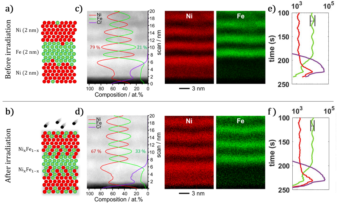
At low fluences, it has been shownDevolder et al. (2001) that room temperature irradiation releases strain, whereas, at high fluences, one major structural effect of irradiation is intermixing, as schematically presented in figure 1 a) and b) and confirmed by Montecarlo (TRIM) simulations (figure S3 of the supplementary material). X-ray diffraction and scanning transmission electron microscopy (STEM) studies indicate in our sample a polycrystalline structure of (110)-textured layers of Fe and (111)-textured layers of Ni which is not significantly altered by the process of irradiation. In-depth investigation on the structural changes induced by He+ irradiation and annealing can be found in section S1 of the supplementary material, where atomic diffusion activated by thermal energy is compared with ion irradiation. According to TRIM simulationsZiegler, Ziegler, and Biersack (2010), the majority () of the ions reaches the substrate therefore a uniform intermixing in the vertical direction of the sample is expected, moreover, the effect of ion implantation in the multilayer is negligible.
To have a more quantitative estimation of the formation of the alloy for increasing ion fluences, a series of experiments to probe structural and chemical modifications occurring at the layer interfaces caused by ion irradiation were performed and are summarized in figure 1. Cross sections of Fe/Ni/NiFeCr on SiO2/Si were prepared using the focused-ion-beam method (FIB). High-angle annular darkfield scanning transmission electron microscopy (HAADF-STEM, 80-200 mrad) images were acquired and nanoscale chemical analysis via energy dispersive X-ray spectroscopy (EDX) was performed in STEM mode. A vertical EDX profile across the bottom layers of the multilayer stack is shown in figure 1 c) and d) together with corresponding EDX maps of the elemental distribution recorded on multilayers before and after He+ irradiation with fluence, respectively. After sputtering (figure 1 c) ) the interfaces between the magnetic layers are well defined. The EDX profile of the relative atomic composition indicates 21(2) of Fe in a Ni layer before the irradiation. After irradiation (figure 1 d) ) the ratio of Fe atoms in a Ni layer increases to 33(4). This measured stoichiometric change in the layer composition is reflected in the displayed EDX elemental maps by the increased diffuse scattering of signal intensity across the layer interfaces after irradiation. This suggests the formation of an alloy of at the Ni/Fe interfaces when the different atoms are displaced under the effect of incoming He+ ions.
Figure 1 e) and f) display the atomic depth distribution measured by Time-of-Flight Secondary Ion Mass Spectrometry (ToF-SIMS) Benninghoven, Rudenauer, and Werner (1987); Sodhi (2004); Vickerman and Briggs (2013); Lamperti et al. (2013); Conte et al. (2015). The presence of Fe, Ni and Cr atoms in the multilayer is reported for samples as-deposited and irradiated with fluence, respectively. Observing figure 1 e) the position of the periodic oscillations of Ni and Fe appear well defined and have the same periodicity. The peak position, minima of Ni at maxima of Fe, reflects the layer distribution. The atomic distribution after irradiation is shown in figure 1 f). In this case, the amplitude of Ni and Fe oscillations is significantly attenuated with respect to the as-deposited case. This is again attributed to the intermixing of the atoms in the neighbouring magnetic layers, leading to the formation of alloy. More details about simulations and measurements to probe structural modifications can be found in section S1 of the supplementary materials.
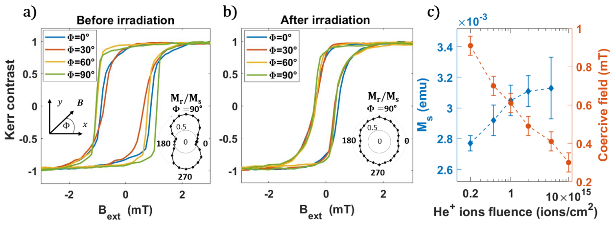
The thin film magnetic properties have been measured with Kerr microscopy and Vibrating Sample Magnetometry (VSM). Figures 2 a) and b) show in-plane hysteresis loops before and after the ion irradiation, respectively. In figure 2 a), the as-deposited sample presents different magnetization curves for different angular directions of the magnetic field, indicating the presence of uniaxial crystalline anisotropy as can be observed in the inset. The coercivity, measured along the easy axis of magnetization is . The same magnetic measurements are reported in figure 2 b) for the sample after He+ irradiation of . The magnetic in-plane anisotropy is now negligible, as the the different hysteresis loops overlap. The coercivity is reduced to . The reduction in coercivity and anisotropy might be related to a possible increase in the concentration of nucleation sites after irradiation, which allow domain formation and switching of the magnetization at lower fields. As the and the magnetic anisotropy are reduced, the magnetic softness of our multilayer is improved by this material treatment. Figure 2 c) reports systematic measurements of the magnetic properties of our multilayer as a function of the He+ fluence during irradiation. With increasing He+ fluence the magnetic moment of the sample increases by about , from 2.8(1) to 3.1(2) . As reported elsewhereSrivastava et al. (2006), this is an indication of increased level of intermixing of our magnetic layers (Ni and Fe).
In order to evaluate the potential of ion irradiation to finely tune the magnetoelastic properties of a magnetic multilayer, the effective magnetic anisotropy in our sample has been measured under the application of mechanical strain by three-point bending method as previously reported Masciocchi et al. (2021). Here the substrate is bent to exert a uniaxial strain on the sample. Since the magnetization is coupled to the external strain via the expression of the anisotropy energyBur et al. (2011) one way to probe the effect of the strain is to observe changes in the hysteretic behavior before and after mechanical deformation. More details can be found in section S2 of the supplementary material. The expression of the magnetoelastic anisotropy depends on the saturation magnetostriction of the material according toFinizio et al. (2014)
| (1) |
where is the Young’s modulus and is the uniaxial tensile strain. If the directions of the crystalline and magnetoelastic uniaxial anisotropy are such that , the strain dependent effective anisotropy measured in the system can be written as sum of two terms according toMartin et al. (2009)
| (2) |
As the sign of can be negative or positive, depending on the value of , the total magnetic anisotropy can, respectively, increase or decrease in the presence of strain. To quantify hysteresis loops are measured using Kerr microscopy, where the magnetic field and the tensile strain are applied along the fixed direction .
The hysteresis loops measured along the direction of the applied strain are reported in figure 3 a) for samples irradiated with different fluences of ions. In response to the applied strain, the irradiated samples have a different magnetic anisotropy field. By comparison with the magnetization curve in the absence of strain (dashed line) two potential scenarios are identified. When a tensile strain increases the anisotropy field in the direction parallel to , and are negative. Our sample exhibits negative magnetoelastic coupling in the as-deposited state. On the other hand, if the strain direction becomes an easy-axis of magnetization (reduced anisotropy field), and are positive. This behavior is reported for larger fluences in the same magnetic stack.
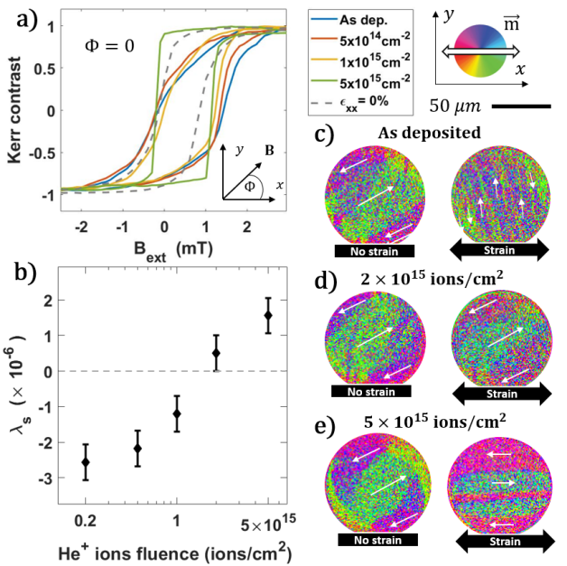
As the difference between magnetic loops before and after the application of strain is proportional to the magnetoelastic anisotropy, the saturation magnetostriction () of our magnetic multilayer can be estimatedChoe and Megdal (1999); Raghunathan, Snyder, and Jiles (2009); Hill et al. (2013) using eq. 1 and eq. 2. Figure 3 b) shows the saturation magnetostriction as a function of the fluence of He+ ions. In agreement with the behavior of the magnetic loops, the value of magnetostriction of the as deposited multilayer is . He+ fluences larger than gradually reduce the absolute value of magnetostriction that then increases through positive values. The change of sign of the magnetoelastic coupling occurs for fluences between and .
An additional confirmation of the magnetic behavior of the magnetic stack under strain is obtained by imaging domain formation using the magneto-optical Kerr (MOKE) effect. A vector image of the in-plane magnetization is obtained by the sum of horizontal and vertical components of the magnetic contrast. The MOKE images shown in figure 3 c) - e) present how the preferred direction of magnetization changes before and after the application of uniaxial strain in disk patterned samples. This particular shape has been chosen since it minimizes the in-plane shape anisotropy. The remanent magnetic domain pattern of the multilayer as-deposited is presented in figure 3 c). Before the application of strain (left) the magnetization aligns to the crystalline anisotropy. After the application of strain magnetic domains orient along the direction, perpendicular to the uniaxial strain . This is a clear experimental proof of the development of stress induced magnetic anisotropy that overcomes the initial anisotropy direction. is perpendicular to the tensile strain direction due to the negative sign of the magnetostriction. The domain structure of the sample irradiated with a He+ dose of is displayed in figure 3 d), where a value of magnetostriction close to zero is measured. In this case, the orientation of the magnetization is almost unchanged by the presence of strain, meaning that is negligible, compared to the crystalline anisotropy of the material . For higher values of fluences as reported in figure 3 e), the effects of strain on the remanent magnetization state become again significant. This time the dominant magnetic anisotropy contribution in the system is as the domains orient along the x direction, parallel to the applied strain . Thus, the magnetoelastic coupling of the stack has been altered using ion irradiation obtaining values of magnetostriction that range from negative to positive.
As previously reported Senda and Nagai (1989), the small value of magnetostriction in our (as-deposited) periodic system is caused by the balance among the negative magnetostriction of Ni () and Fe () and the strongly positive magnetostriction of alloy film () with a relative composition close to Bozorth (1993); Cullity and Graham (2011). As shown by STEM-EDX measurements, a more intermixed interface region of is formed at the boundary between Ni and Fe layers by He+ ion irradiation. Hence, the thickness of the positive magnetostrictive alloy increases proportionally to the fluence of the He+ ions during irradiation, as also confirmed by ToF-SIMS measurements. This gradually shifts the magnetostriction of the full stack to positive values. A common way to describe the effective magnetostriction in the presence of intermixing is Nagai, Senda, and Toshima (1988); Hollingworth, Gibbs, and Murdoch (2003); Favieres, Vergara, and Madurga (2007); Jen and Lin (2005); Senda and Nagai (1989); Rengarajan et al. (1997); Jen et al. (2004)
| (3) |
where is the period thickness, describes the thickness of the alloy originated by the intermixing and is the saturation magnetostriction of the intermixed alloy. After deposition in similarly sputtered Ni/Fe multilayersNagai, Senda, and Toshima (1988) has been estimated to be around , under the assumption . Using this value of eq. 3 returns , close to the measured value after deposition. Moreover, the amount of induced intermixing caused by He+ ions can be estimated using eq. 3. The calculated value is at the magnetostriction compensation value () and for the highest fluence, where the magnetostriction is positive due to the dominant effect of the alloy. This corresponds to increase in the alloy thickness induced by He+ between and , in agreement with the information extracted from STEM-EDX and ToF-SIMS measurements.
In conclusion, this manuscript presents an experimental investigation in to the magnetoelastic properties of sputtered multilayers after He+ ion irradiation. Using different experimental techniques for structural analysis, the presence of moderate roughness and alloying is observed after sputtering at the Ni/Fe interface. This can justify the small negative value of magnetostriction in the as-deposited state. In the same way, it was found that light ion irradiation promotes the intermixing of the sputtered layers at the interfaces proportional to the ion fluence. This process can explain the reported changes in the saturation magnetostriction of the magnetic stack. The increasing fluence of the irradiating ions progressively changes the saturation magnetostriction inducing a change in sign of the magnetoelastic coupling of the material, from negative to positive for high fluences. Remarkably, strain insensitivity on the magnetic properties of the proposed material can be obtained with ion fluences between and . Importantly, the polycrystalline structure of the layers is confirmed to be unchanged after the used irradiation conditions.
As a result, post growth He+ ion irradiation has been demonstrated to be an excellent tool that allows to fine-tune the magneto-elastic properties of multilayer magnetic samples. Accordingly, this technique can be expected to be the next generation of material treatment offering the possibility to have local patterning of magnetostriction with high control and flexibility, allowing the realization of highly demanding applications.
Acknowledgements.
The authors acknowledge Prof. J. McCord from Kiel University for fruitful discussions. This project has received funding from the European Union’s Horizon 2020 research and innovation program under the Marie Skłodowska-Curie grant agreement No 860060 “Magnetism and the effect of Electric Field” (MagnEFi), the Deutsche Forschungsgemeinschaft (DFG, German Research Foundation) - TRR 173 - 268565370 (project A01 and B02), the DFG funded collaborative research center (CRC)1261 / project A6 and the Austrian Research Promotion Agency (FFG). The authors acknowledge support by the chip production facilities of Sensitec GmbH (Mainz, DE), where part of this work was carried out and the Max-Planck Graduate Centre with Johannes Gutenberg University.Author Declarations
The following article has been submitted to Applied Physics Letters. After it is published, it will be found at publishing.aip.org .
Conflict of interest
The authors have no conflicts to disclose.
Data Sharing Policy
The data that support the findings of this study are available from the corresponding author upon reasonable request.
References
- Ota, Ando, and Chiba (2018) S. Ota, A. Ando, and D. Chiba, “A flexible giant magnetoresistive device for sensing strain direction,” Nature Electronics 1, 124–129 (2018).
- García-Miquel et al. (2016) H. García-Miquel, D. Barrera, R. Amat, G. Kurlyandskaya, and S. Sales, “Magnetic actuator based on giant magnetostrictive material Terfenol-D with strain and temperature monitoring using FBG optical sensor,” Measurement 80, 201–206 (2016).
- Nagai, Senda, and Toshima (1988) Y. Nagai, M. Senda, and T. Toshima, “Properties of ion-beam-sputtered Ni/Fe artificial lattice film,” Journal of Applied Physics 63, 1136–1140 (1988).
- Senda and Nagai (1989) M. Senda and Y. Nagai, “Magnetic properties of Fe/Co, Fe/CoFe, and (Fe/Co)/ multilayer films,” Journal of Applied Physics 65, 3151–3156 (1989).
- Rengarajan et al. (1997) S. Rengarajan, E. Yun, W. Kang, and R. Walser, “Effect of intermixing on the magnetic properties of / multilayers,” Journal of Applied Physics 81, 4761–4763 (1997).
- Jen and Lin (2005) S. Jen and C. Lin, “Magnetostriction and Young’s modulus of [/] multilayers,” Thin Solid Films 471, 218–223 (2005).
- Zhao et al. (2019) X. Zhao, B. Zhang, N. Vernier, X. Zhang, M. Sall, T. Xing, L. H. Diez, C. Hepburn, L. Wang, G. Durin, et al., “Enhancing domain wall velocity through interface intermixing in W-CoFeB-MgO films with perpendicular anisotropy,” Applied Physics Letters 115, 122404 (2019).
- Fassbender, Ravelosona, and Samson (2004) J. Fassbender, D. Ravelosona, and Y. Samson, “Tailoring magnetism by light-ion irradiation,” Journal of Physics D: Applied Physics 37, R179 (2004).
- Terris et al. (1999) B. Terris, L. Folks, D. Weller, J. Baglin, A. Kellock, H. Rothuizen, and P. Vettiger, “Ion-beam patterning of magnetic films using stencil masks,” Applied Physics Letters 75, 403–405 (1999).
- Juraszek et al. (2006) J. Juraszek, A. Grenier, J. Teillet, N. Tiercelin, F. Petit, J. B. Youssef, and M. Toulemonde, “Swift ion irradiation of magnetostrictive multilayers,” Nuclear Instruments and Methods in Physics Research Section B: Beam Interactions with Materials and Atoms 245, 157–160 (2006).
- Cureton, Tracy, and Lang (2021) W. F. Cureton, C. L. Tracy, and M. Lang, “Review of swift heavy ion irradiation effects in CeO2,” Quantum Beam Science 5, 19 (2021).
- Devolder et al. (2013) T. Devolder, I. Barisic, S. Eimer, K. Garcia, J.-P. Adam, B. Ockert, and D. Ravelosona, “Irradiation-induced tailoring of the magnetism of CoFeB/MgO ultrathin films,” Journal of Applied Physics 113, 203912 (2013).
- Devolder et al. (1999) T. Devolder, C. Chappert, Y. Chen, E. Cambril, H. Launois, H. Bernas, J. Ferre, and J. Jamet, “Patterning of planar magnetic nanostructures by ion irradiation,” Journal of Vacuum Science & Technology B: Microelectronics and Nanometer Structures Processing, Measurement, and Phenomena 17, 3177–3181 (1999).
- Devolder et al. (2001) T. Devolder, S. Pizzini, J. Vogel, H. Bernas, C. Chappert, V. Mathet, and M. Borowski, “X-ray absorption analysis of sputter-grown Co/Pt stackings before and after helium irradiation,” The European Physical Journal B-Condensed Matter and Complex Systems 22, 193–201 (2001).
- Ziegler, Ziegler, and Biersack (2010) J. F. Ziegler, M. D. Ziegler, and J. P. Biersack, “SRIM–the stopping and range of ions in matter (2010),” Nuclear Instruments and Methods in Physics Research Section B: Beam Interactions with Materials and Atoms 268, 1818–1823 (2010).
- Benninghoven, Rudenauer, and Werner (1987) A. Benninghoven, F. Rudenauer, and H. W. Werner, “Secondary ion mass spectrometry: basic concepts, instrumental aspects, applications and trends,” (1987).
- Sodhi (2004) R. N. Sodhi, “Time-of-Flight Secondary Ion Mass Spectrometry (TOF-SIMS):—versatility in chemical and imaging surface analysis,” Analyst 129, 483–487 (2004).
- Vickerman and Briggs (2013) J. C. Vickerman and D. Briggs, ToF-SIMS: materials analysis by mass spectrometry (IM publications, 2013).
- Lamperti et al. (2013) A. Lamperti, E. Cianci, O. Salicio, L. Lamagna, S. Spiga, and M. Fanciulli, “Thermal stability of high- oxides on or for charge-trapping nonvolatile memories,” Surface and interface analysis 45, 390–393 (2013).
- Conte et al. (2015) R. L. Conte, E. Martinez, A. Hrabec, A. Lamperti, T. Schulz, L. Nasi, L. Lazzarini, R. Mantovan, F. Maccherozzi, S. Dhesi, et al., “Role of B diffusion in the interfacial Dzyaloshinskii-Moriya interaction in nanowires,” Physical Review B 91, 014433 (2015).
- Srivastava et al. (2006) S. Srivastava, R. Kumar, A. Gupta, R. Patel, A. Majumdar, and D. Avasthi, “Swift heavy ion induced mixing in Fe/Ni multilayer,” Nuclear Instruments and Methods in Physics Research Section B: Beam Interactions with Materials and Atoms 243, 304–312 (2006).
- Masciocchi et al. (2021) G. Masciocchi, M. Fattouhi, A. Kehlberger, L. Lopez-Diaz, M.-A. Syskaki, and M. Kläui, “Strain-controlled domain wall injection into nanowires for sensor applications,” Journal of Applied Physics 130, 183903 (2021).
- Bur et al. (2011) A. Bur, T. Wu, J. Hockel, C.-J. Hsu, H. K. Kim, T.-K. Chung, K. Wong, K. L. Wang, and G. P. Carman, “Strain-induced magnetization change in patterned ferromagnetic nickel nanostructures,” Journal of Applied Physics 109, 123903 (2011).
- Finizio et al. (2014) S. Finizio, M. Foerster, M. Buzzi, B. Krüger, M. Jourdan, C. A. Vaz, J. Hockel, T. Miyawaki, A. Tkach, S. Valencia, et al., “Magnetic anisotropy engineering in thin film Ni nanostructures by magnetoelastic coupling,” Physical Review Applied 1, 021001 (2014).
- Martin et al. (2009) N. Martin, J. McCord, A. Gerber, T. Strache, T. Gemming, I. Mönch, N. Farag, R. Schäfer, J. Fassbender, E. Quandt, et al., “Local stress engineering of magnetic anisotropy in soft magnetic thin films,” Applied Physics Letters 94, 062506 (2009).
- Choe and Megdal (1999) G. Choe and B. Megdal, “High precision magnetostriction measurement employing the BH looper bending method,” IEEE Transactions on Magnetics 35, 3959–3961 (1999).
- Raghunathan, Snyder, and Jiles (2009) A. Raghunathan, J. E. Snyder, and D. Jiles, “Comparison of alternative techniques for characterizing magnetostriction and inverse magnetostriction in magnetic thin films,” IEEE Transactions on Magnetics 45, 3269–3273 (2009).
- Hill et al. (2013) C. Hill, W. Hendren, R. Bowman, P. McGeehin, M. Gubbins, and V. Venugopal, “Whole wafer magnetostriction metrology for magnetic films and multilayers,” Measurement Science and Technology 24, 045601 (2013).
- Bozorth (1993) R. M. Bozorth, Ferromagnetism (1993).
- Cullity and Graham (2011) B. D. Cullity and C. D. Graham, Introduction to magnetic materials (John Wiley & Sons, 2011) Chap. 8, pp. 243–257.
- Hollingworth, Gibbs, and Murdoch (2003) M. Hollingworth, M. Gibbs, and S. Murdoch, “Magnetostriction and surface roughness of ultrathin NiFe films deposited on SiO2,” Journal of Applied Physics 94, 7235–7239 (2003).
- Favieres, Vergara, and Madurga (2007) C. Favieres, J. Vergara, and V. Madurga, “Interface effects on magnetostriction in pulsed laser deposited Co/Fe/Co cylindrical soft magnetic multilayers,” Journal of Physics D: Applied Physics 40, 4101 (2007).
- Jen et al. (2004) S. Jen, T. Wu, C. Lin, and K. Chang, “Anisotropic magnetoresistance and magnetostriction of [Fe15Ni85/Fe25Ni75] and [Co35Ni65/Fe25Ni75] multilayers,” Solid State Communications 132, 259–262 (2004).