Ensemble CNN models for Covid-19 Recognition and Severity Perdition From 3D CT-scan
Institute of Applied Sciences and Intelligent Systems,
National Research Council of Italy (CNR),
73100 Lecce, Italy
faresbougourzi@gmail.com
\And
Institute of Applied Sciences and Intelligent
Systems, National Research Council of Italy,
73100 Lecce, Italy
Department of Innovation Engineering, University
of Salento, 73100 Lecce, Italy
cosimo.distante@cnr.it
\And
University of the Basque Country UPV/EHU,
San Sebastian, SPAIN; IKERBASQUE, Basque
Foundation for Science, Bilbao, SPAIN
fadi.dornaika@ehu.eus
\And
IEMN UMR CNRS 8520, Université
Polytechnique Hauts de France, UPHF
Abdelmalik.Taleb-Ahmed@uphf.fr
Abstract
Since the appearance of Covid-19 in late 2019, Covid-19 has become an active research topic for the artificial intelligence (AI) community. One of the most interesting AI topics is Covid-19 analysis of medical imaging. CT-scan imaging is the most informative tool about this disease.
This work is part of the 2nd COV19D competition, where two challenges are set: Covid-19 Detection and Covid-19 Severity Detection from the CT-scans. For Covid-19 detection from CT-scans, we proposed an ensemble of 2D Convolution blocks with Densenet-161 models. Here, each 2D convolutional block with Densenet-161 architecture is trained separately and in testing phase, the ensemble model is based on the average of their probabilities. On the other hand, we proposed an ensemble of Convolutional Layers with Inception models for Covid-19 severity detection. In addition to the Convolutional Layers, three Inception variants were used, namely Inception-v3, Inception-v4 and Inception-Resnet.
Our proposed approaches outperformed the baseline approach in the validation data of the 2nd COV19D competition by 11% and 16% for Covid-19 detection and Covid-19 severity detection, respectively.
Keywords Covid-19 Deep Leaning CNNs Recognition Severity
1 Introduction
Since the appearance of the Covid-19 pandemic in the late of 2019, reverse transcription-polymerase chain reaction (RT-PCR) has been considered as the golden standards for Covid-19 Detection. However, the RT-PCR test has many drawbacks including inadequate supply of RT-PCR kits, time-consuming consumption, and considerable false negative results [1, 2, 3]. To deal with this limitations, Medical Imaging modalities have been widly used as supporting tools. These imaging modalities include X-rays and CT-scans [4, 5]. Indeed, CT-scans are not only used to detect Covid-19 infected cases, but they could be used to follow up the state of the patient and predicting the disease severity [6, 7].
In the last decade, Deep Learning methods have become dominant in most of the computer vision tasks and they have achieved high performance compared to traditional methods [8, 9]. However, the main drawback of deep learning, especially the CNN architecture is the need of huge labelled data, which is hard to be obtained in medical domains [6]. On the other hand, most of the proposed CNN architectures adopted for Static images (single image input) [7].
In this work, we adopted pretrained CNN architectures to detect Covid-19 infection and Covid-19 Severity from 3D CT-scans as part of 2nd COV19D Competition. furthermore, we trained a CNN model to filter the slices that do not show lungs region. For the Covid-19 Severity detection, Att-Unet model were trained to segment the lungs regions to remove non-important features. In addition, two volume input branches were used to reduce the lost information due to the resizing transformation and the variation of the number of slices from one CT-scan to another. The main contributions of this paper can be summarized as follows:
-
•
For Covid-19 detection from CT scans, we proposed an ensemble of Convolution 2D blocks with Densenet-161 models. Each convolution 2D block with densenet-161 architecture was trained separately, and the ensemble model is based on the average of their probabilities.
-
•
For detecting the severity of Covid-19 from CT scans, we proposed an ensemble of Convolutional Layer with Inception models. Three Inception variants were used in addition to the Convolutional layers, namely Inception-v3, Inception-v4, and Inception-Resnet. The Inception variants were trained twice separately and the ensemble model is the average of the probabilities of the six models.
-
•
Our proposed approaches outperformed the baseline approach in the validation data of the 2nd COV19D competition by 11% and 16% for Covid-19 detection and Covid-19 severity detection, respectively.
-
•
The codes used and the pre-trained models are publicly available in https://github.com/faresbougourzi/2nd-COV19D-Competition. ( Last accessed on June, 28th 2022).
2 Our Approaches
In the following two sections, we will describe our proposed approaches for Covid-19 Recognition and Severity prediction, respectively.
2.1 Covid-19 Detection
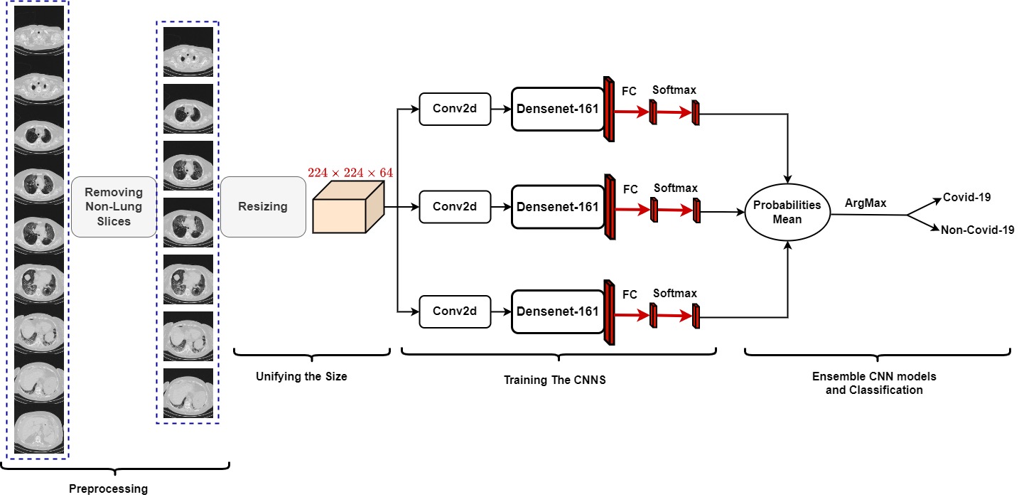
Our proposed approach to Covid-19 Detection for the 2nd COV19D competition is summarized in Figure 1. Because the 3D CT-scans have a different number of slices depending on the scanner and acquisition settings and contain multiple non-lung slices, we trained a model to remove the non-lung slices. For this purpose, we manually labelled 20 CT-scans from the training data as lung and non-lung. In addition, we used the following datasets: COVID-19 CT segmentation [10], Segmentation dataset nr.2 [10], and COVID-19-CT Seg dataset [11] to have more labelled data as lung and non-lung slices. After creating the lung and non-lung sets, we trained the ResneXt-50 model [12] to remove the slices that did not show lungs at all. Since the CT scans have different numbers of slices, we concatenated all lung slices and then reduced them to as shown in Figure 1. To distinguish between Covid-19 and Non-Covid-19 CT scans, we separately trained three Densenet-161 [13] models. Since the input size of Denesenet-161 model is an RGB image and we have a 3D volume of size , we added Convolution block with a 3 x 3 kernel, a stride of 1, and a padding of 1. The 3 x 3 convolution block takes 64 input channels and produces 3 output channels. It should be noted that each CNN model was trained separately. In the decision or test phase, we propose to form an ensemble of the three trained CNN architectures by averaging their probabilities.
2.2 Covid-19 Severity Detection
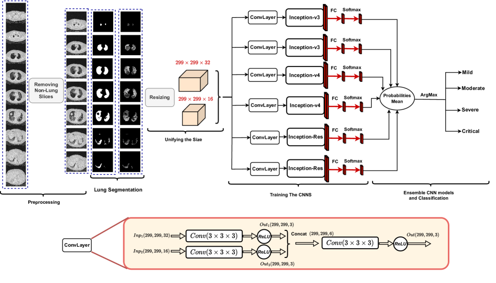
Our proposed approach for Covid-19 severity prediction for the 2nd COV19D competition is summarized in Figure 2. Similar to the Covid-19 detection approach in section 2.1, we used the trained ResneXt-50 model to remove non-lung slices. To segment the lungs, we trained the Att-Unet [14] architecture using the following datasets COVID-19 CT segmentation [10], Segmentation segmentation dataset nr.2 [10], and COVID-19-CT-Seg dataset [11]. After removing the non-lung slices and segmenting the lung regions to remove unnecessary features, we resized the obtained slices into two volumes and . The goal of the two volumes is to reduce the information lost due to resizing and to obtain two views of each CT scan. The two input volumes were fed into the convolution block. The convolution Layer consists of three 3 by 3 convolution blocks with stride 1 and badding 1. The first convolution block transforms the volume to . Similarly, the second convolution block transforms the volume into . The third convolution block transforms the concatenation of the outputs of the first and second convolution blocks () into . The output volume of the third convolution block is used as input to the architecture. In our experiments, we used Inception architectures as CNN backbones, namely inception-v3, inception-V4, and Inception-Resnet [15]. Our approach to predict Covid-19 severity consists of six ConvLayers + CNN branches, as shown in Figure 2, where each branch was trained separately. To create a more comprehensive ensemble model, we trained each CNN model twice. The final Covid-19 severity prediction was obtained by averaging the prediction probabilities of the six models.
3 Experiments and Results
3.1 The COV19-CT-DB Database
The COVID19-CT-Database (COV19-CT-DB) [16, 17, 18, 19, 20] consists of chest CT scans that are annotated for the existence of COVID-19.
| Set | Training | Validation |
|---|---|---|
| Covid-19 | 882 | 215 |
| Non-Covid-19 | 1110 | 289 |
| Severity Class | Training | Validation |
| 1 | 85 | 22 |
| 2 | 62 | 10 |
| 3 | 85 | 22 |
| 4 | 26 | 5 |
COVID19-CT-DB contains 3-D CT scans of the chest, which were analysed for the presence of COVID-19. Each of the 3-D scans contains a different number of slices, ranging from 50 to 700. The database was divided into a training set, a validation set, and a test set. The training set contains a total of 1992 3-D CT scans. The validation set consists of 494 3-D CT scans. The number of COVID-19 and non COVID-19 cases in each set is given in Table 1.
Further partitioning of the COVID-19 cases was based on the severity of COVID-19 , which was given by four medical experts in a range of 1 to 4, with 4 denoting critical status. The training set contains a total of 258 3-D CT scans. The validation set consists of 61 3-D CT scans. The number of scans in each severity class in these sets is shown in Table 2.
3.2 Experimental Setup
For Deep Learning training and testing, we used the Pytorch [21] Library with NVIDIA GPU Device GeForce TITAN RTX 24 GB. The batch size used consists of 16 CT-scan volumes for both tasks. We trained the networks for 40 epochs. The initial learning rate is 0.0001, which decreases by 0.1 after 15 epochs, followed by another 0.1 decrease after 30 epochs.
3.3 Covid-19 Recognition
Table 3 summarizes the Covid-19 detection results (F1 score) obtained on the validation data. As described in section 2.1, we trained three Convolutional Block + Densenet-161 architectures. For simplicity, we refer to Convolutional Block + Densenet-161 with Densenet-161 and the training iteration. Comparing with the baseline results, we find that all the trained Densenet models outperform the baseline approach by about 11%, 9% and 10% for model 1, 2 and 3, respectively. Moreover, the ensemble of these Densenet models improves the result compared to the best trained model (Model 1).
| Model | Architecture | Macro F1-Score |
|---|---|---|
| - | Baseline | 77 |
| 1 | Densenet-161 Model1 | 88.34 0.59 |
| 2 | Densenet-161 Model2 | 86.91 0.59 |
| 3 | Densenet-161 Model3 | 87.46 0.59 |
| 4 | Ensemble Models | 88.84 0.31 |
3.4 Covid-19 Severity Detection
Table 4 summarizes the results (F1 score) of detecting the severity of Covid-19 using the validation data. As described in section2.2, we trained six convolutional layers + CNN backbones. The CNN backbones used are Inception-v3, Inception-V4, and Inception-Resnet, and each model was trained twice to obtain a greater variety of predictors. For simplicity, we refer to ConvLayer + CNN backbone with the backbone architecture name. Compared to the baseline results, we find that our trained models achieve better performance of 11%, 11%, 12%, 10%, 10% and 9% for model 1-6, respectively. Moreover, the ensemble of trained models improves the result compared to all models and achieves 16% better result than the baseline approach.
| Model | Architecture | Macro F1-Score |
|---|---|---|
| - | Baseline | 63 |
| 1 | Inception-v3 Model1 | 74.86 0.5 |
| 2 | Inception-v3 Model2 | 74.75 0.5 |
| 3 | Inception-v4 Model1 | 75.60 1.1 |
| 4 | Inception-v4 Model2 | 73.48 1.1 |
| 5 | Inception-Res Model1 | 73.37 0.7 |
| 6 | Inception-Res Model2 | 72.01 0.7 |
| 7 | Ensemble Models | 79.10 0.6 |
4 Conclusion
In this work, we proposed two ensemble CNN-based approaches for the 2nd COV19D competition. For Covid-19 detection from CT scans, we proposed an ensemble of 2D convolution blocks with Densenet-161 models. Here, the individual convolution 2D blocks with Densenet-161 architecture are trained separately, and the ensemble model is based on the average of their probabilities at testing phase. On the other hand, we proposed an ensemble of Convolutional Layers with Inception models for Covid-19 severity detection. In addition to the Convolutional Layers, three Inception variants were used, namely Inception-v3, Inception-v4, and Inception-Resnet.
Our proposed approaches outperformed the baseline approach by a significant margin in the validation data of the 2nd COV19D competition with 11% and 16% in Covid-19 detection and Covid-19 severity detection, respectively. In future work, we will evaluate our approaches in other datasets and use the attention mechanism to select the representative slices for Covid-19 detection.
References
- [1] Ying-Hui Jin, Lin Cai, and Zhen-Shun et al. Cheng. A rapid advice guideline for the diagnosis and treatment of 2019 novel coronavirus (2019-nCoV) infected pneumonia (standard version). Military Medical Research, 7(1):4, February 2020.
- [2] Yu-Huan Wu, Shang-Hua Gao, Jie Mei, Jun Xu, Deng-Ping Fan, Rong-Guo Zhang, and Ming-Ming Cheng. JCS: An Explainable COVID-19 Diagnosis System by Joint Classification and Segmentation. IEEE Transactions on Image Processing, 30:3113–3126, 2021. Conference Name: IEEE Transactions on Image Processing.
- [3] Lauren M. Kucirka, Stephen A. Lauer, Oliver Laeyendecker, Denali Boon, and Justin Lessler. Variation in False-Negative rate of reverse transcriptase polymerase chain Reaction–Based SARS-CoV-2 Tests by time since exposure. Annals of Internal Medicine, 173(4):262–267, May 2020. Publisher: American College of Physicians.
- [4] Edoardo Vantaggiato, Emanuela Paladini, Fares Bougourzi, Cosimo Distante, Abdenour Hadid, and Abdelmalik Taleb-Ahmed. Covid-19 recognition using ensemble-cnns in two new chest x-ray databases. Sensors, 21(5):1742, 2021.
- [5] Fares Bougourzi, Riccardo Contino, Cosimo Distante, and Abdelmalik Taleb-Ahmed. Recognition of COVID-19 from CT Scans Using Two-Stage Deep-Learning-Based Approach: CNR-IEMN. Sensors, 21(17):5878, January 2021. Number: 17 Publisher: Multidisciplinary Digital Publishing Institute.
- [6] Fares Bougourzi, Cosimo Distante, Abdelkrim Ouafi, Fadi Dornaika, Abdenour Hadid, and Abdelmalik Taleb-Ahmed. Per-COVID-19: A Benchmark Dataset for COVID-19 Percentage Estimation from CT-Scans. Journal of Imaging, 7(9):189, September 2021. Number: 9 Publisher: Multidisciplinary Digital Publishing Institute.
- [7] Fares Bougourzi, Riccardo Contino, Cosimo Distante, and Abdelmalik Taleb-Ahmed. CNR-IEMN: A Deep Learning Based Approach To Recognize COVID-19 From CT-SCAN. In 2021 IEEE International Conference on Autonomous Systems (IEEE ICAS), 2021.
- [8] F. Bougourzi, F. Dornaika, K. Mokrani, A. Taleb-Ahmed, and Y. Ruichek. Fusing Transformed Deep and Shallow features (FTDS) for image-based facial expression recognition. Expert Systems with Applications, 156:113459, October 2020.
- [9] F. Bougourzi, F. Dornaika, and A. Taleb-Ahmed. Deep learning based face beauty prediction via dynamic robust losses and ensemble regression. Knowledge-Based Systems, 242:108246, April 2022.
- [10] RADIOLOGISTS. COVID-19 CT-scans segmentation datasets, available at: http://medicalsegmentation.com/covid19/, 2019. Last visited: 18-08-2021.
- [11] Jun Ma, Yixin Wang, Xingle An, Cheng Ge, Ziqi Yu, Jianan Chen, Qiongjie Zhu, Guoqiang Dong, Jian He, Zhiqiang He, Tianjia Cao, Yuntao Zhu, Ziwei Nie, and Xiaoping Yang. Toward data efficient learning: A benchmark for COVID 19 CT lung and infection segmentation. Medical Physics, 48:1197–1210, March 2021. ADS Bibcode: 2021MedPh..48.1197M.
- [12] Saining Xie, Ross Girshick, Piotr Dollár, Zhuowen Tu, and Kaiming He. Aggregated residual transformations for deep neural networks, 2017.
- [13] Gao Huang, Zhuang Liu, Laurens Van Der Maaten, and Kilian Q. Weinberger. Densely connected convolutional networks. In Proceedings of the IEEE conference on computer vision and pattern recognition, pages 4700–4708, 2017.
- [14] Ozan Oktay, Jo Schlemper, and Loic Le et al. Folgoc. Attention U-Net: Learning Where to Look for the Pancreas. arXiv:1804.03999 [cs], May 2018. arXiv: 1804.03999.
- [15] Christian Szegedy, Vincent Vanhoucke, Sergey Ioffe, Jonathon Shlens, and Zbigniew Wojna. Rethinking the inception architecture for computer vision, 2015.
- [16] Dimitrios Kollias, Anastasios Arsenos, and Stefanos Kollias. Ai-mia: Covid-19 detection & severity analysis through medical imaging. arXiv preprint arXiv:2206.04732, 2022.
- [17] Dimitrios Kollias, Anastasios Arsenos, Levon Soukissian, and Stefanos Kollias. Mia-cov19d: Covid-19 detection through 3-d chest ct image analysis. In Proceedings of the IEEE/CVF International Conference on Computer Vision, pages 537–544, 2021.
- [18] Dimitrios Kollias, N Bouas, Y Vlaxos, V Brillakis, M Seferis, Ilianna Kollia, Levon Sukissian, James Wingate, and S Kollias. Deep transparent prediction through latent representation analysis. arXiv preprint arXiv:2009.07044, 2020.
- [19] Dimitris Kollias, Y Vlaxos, M Seferis, Ilianna Kollia, Levon Sukissian, James Wingate, and Stefanos D Kollias. Transparent adaptation in deep medical image diagnosis. In TAILOR, page 251– 267, 2020.
- [20] Dimitrios Kollias, Athanasios Tagaris, Andreas Stafylopatis, Stefanos Kollias, and Georgios Tagaris. Deep neural architectures for prediction in healthcare. Complex & Intelligent Systems, 4(2):119–131, 2018.
- [21] Adam Paszke, Sam Gross, and Francisco et al. Massa. Pytorch: An imperative style, high-performance deep learning library. In Advances in neural information processing systems, pages 8026–8037, 2019.