Computer-aided diagnosis and prediction in brain disorders
Abstract
Computer-aided methods have shown added value for diagnosing and predicting brain disorders and can thus support decision making in clinical care and treatment planning. This chapter will provide insight into the type of methods, their working, their input data - such as cognitive tests, imaging and genetic data - and the types of output they provide. We will focus on specific use cases for diagnosis, i.e. estimating the current ‘condition’ of the patient, such as early detection and diagnosis of dementia, differential diagnosis of brain tumours, and decision making in stroke. Regarding prediction, i.e. estimation of the future ‘condition’ of the patient, we will zoom in on use cases such as predicting the disease course in multiple sclerosis and predicting patient outcomes after treatment in brain cancer. Furthermore, based on these use cases, we will assess the current state-of-the-art methodology and highlight current efforts on benchmarking of these methods and the importance of open science therein. Finally, we assess the current clinical impact of computer-aided methods and discuss the required next steps to increase clinical impact.
Keywords: Dementia, Stroke, Glioma, Cognitive impairment
1. Introduction
Computer-aided methods have major potential value for diagnosing and predicting outcomes in brain disorders such as dementia, brain cancer, and stroke. Diagnosis aims to determine the current ‘condition’ of the patient. Prediction, or prognosis, on the other hand, aims to forecast the future ‘condition’ of the patient. In this way, the patient’s current and future condition can be estimated in a more detailed and accurate way, which opens up possibilities for better patient care and personalised medicine, with interventions tailored to the individual patient. Moreover, diagnosis and prediction are not only crucial for decision making in clinical care and treatment planning, but also for managing the expectations of patients and their caregivers. This is particularly important in brain disorders as they may strongly affect life expectancy and quality of life, as symptoms of the disorder and side effects of the treatment can have a major impact on the patient’s cognitive skills, daily functioning, social interaction, and general well-being. In clinical practice, diagnosis and prediction are typically performed using multiple sources of information, such as symptomatology, medical history, cognitive tests, brain imaging, electroencephalography (EEG), magnetoencephalography (MEG), blood tests, cerebrospinal fluid (CSF) biomarkers, histopathological or molecular findings, and lifestyle and genetic risk factors. These various pieces of information are integrated by the treating clinician, often in consensus with other experts at a multidisciplinary team meeting, in order to reach a final diagnosis and/or treatment plan. The aim of computer-aided methods for diagnosis and prediction is to support this process, in order to achieve more accurate, objective, and efficient decision making.
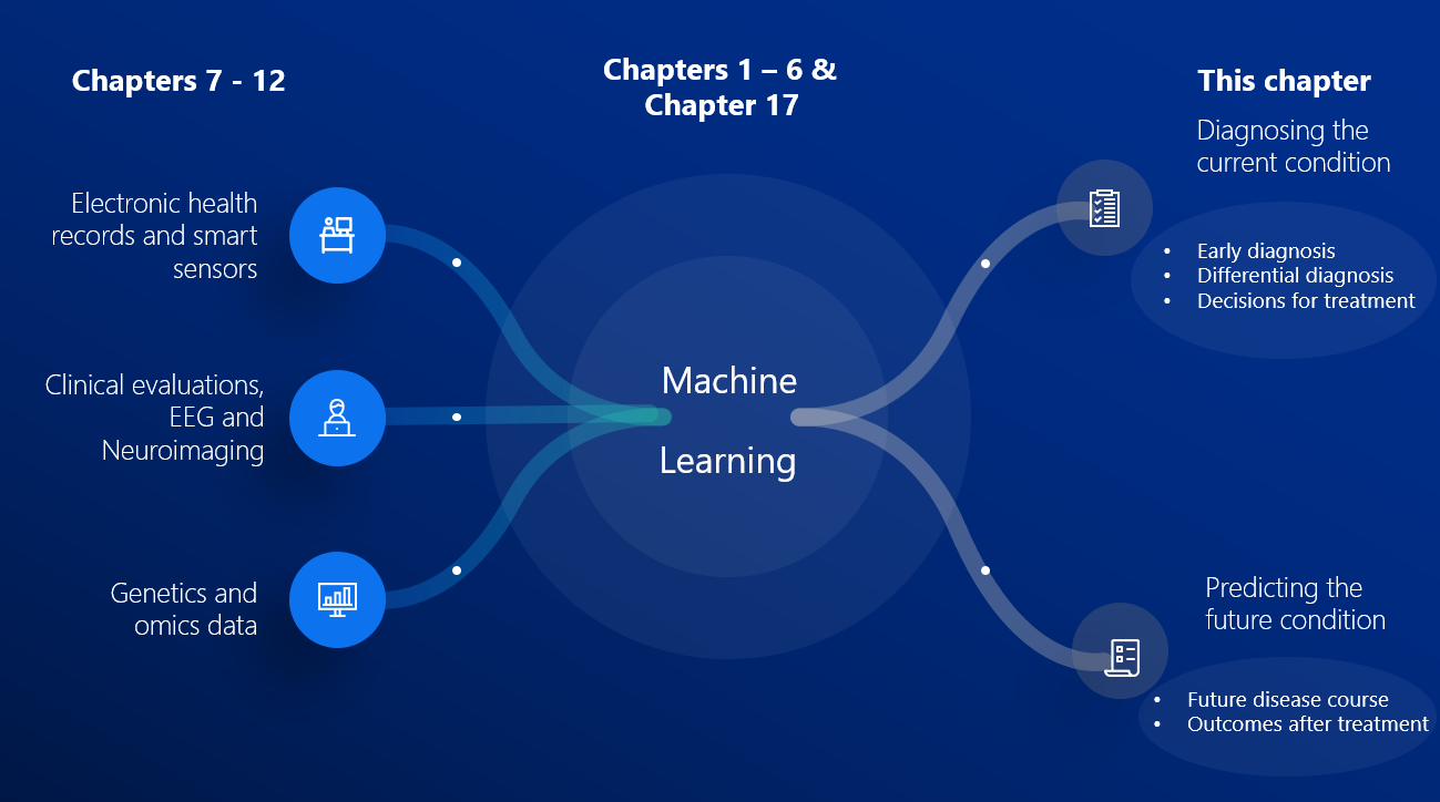
In the literature, numerous examples of computer-aided methods for diagnosis and prediction in brain disorders can be found. Most of the state-of-the-art methods use some form of machine learning to construct a model that maps (often high-dimensional) input data to the output variable of interest. There exists a large variation in machine learning technology, types of input data, and output variables. Chapters 1-6 introduced the main machine learning technologies used for computer-aided diagnosis and prediction. These include, on the one hand, classical methods such as linear models, support vector machines, and random forests, and on the other hand deep learning methods such as convolutional neural networks and recurrent neural networks. These methods can either be implemented as classification models (estimating discrete labels) or as regression models (estimating continuous quantities), possibly specialized for survival (or ‘time-to-event’) analysis. In addition, Chapter 17 highlights the category of disease progression modelling techniques, which could be considered as a specialized form of machine learning incorporating models of the disease evolution over time. Chapters 7-12 described the main types of input data used in machine learning for brain disorders: clinical evaluations, neuroimaging, EEG/MEG, genetics and omics data, electronic health records, smartphone and sensor data. The current chapter focuses on the choice of the output variable, i.e., the diagnosis or prediction of interest.
To illustrate the various ways in which machine learning could aid diagnosis and prediction, we focus on representative use cases organised according to the type of output. Section 1.1 presents diagnostic use cases, including early diagnosis, differential diagnosis, and decision making for treatment. Section 1.2 presents prediction use cases, including estimation of the natural disease course and prediction of patient outcomes after treatment. While the diagnostic use cases are the core of current clinical practice which could be aided by machine learning, the prediction use cases represent a potential future application. Currently, prediction is not so often made as clinicians are not yet able to make a reliable prediction in most cases. After these introductory sections, Section 2 provides a more comprehensive survey of the state-of-the-art methodology, and Section 3 analyses the clinical impact of such methodology and suggests a roadmap for further clinical translation. Finally, Section 5 concludes this chapter.
1.1 D iagnosis
Diagnosis aims to determine the current ‘condition’ of the patient to inform patient care and treatment decisions. Here, we introduce three categories of diagnostic tasks that occur in clinical practice, and describe why and how computer-aided models have or could have added value.
Early diagnosis is highly challenging in neurodegenerative diseases such as dementia and multiple sclerosis (MS). Dementia is a clinical syndrome which can be caused by several underlying diseases, Alzheimer’s disease (AD) being the most prevalent, and is estimated to affect 50 million people worldwide [1]. The mean age at dementia diagnosis is approximately 83 years [109]. MS is estimated to affect about 2 million people worldwide, and it primarily affects younger adults with the mean age of onset for incident MS being approximately 30 years [77]. Both for dementia and MS, establishing the diagnosis usually takes a substantial period of time after the first clinical symptoms arise [140, 59]. Early detection and accurate diagnosis is crucial for timely decision making regarding care and management of dementia symptoms, and as such can reduce healthcare costs and improve quality of life as it gives patients access to supportive therapies that help to delay institutionalisation [110]. Early diagnosis of MS is important, because patients who begin treatment earlier do reap more benefit than those who start late [93]. In addition, advancing the diagnosis in time is essential to support the development of new disease modifying treatments, since late treatment is expected to be a major factor in failure of clinical trials [91]. The clinical diagnosis of dementia is currently based on objective assessment of cognitive impairment, assessment of biomarkers [29] and evaluation of its interference with daily living [90, 4, 43, 115]. The clinical diagnosis of MS is based on frequency of relapsing inflammatory attacks, associated symptoms and distribution of lesions on MRI [135]. For a subset of MS patients with demyelinating lesions highly suggestive of MS, termed as radiologically isolated syndrome (RIS), a separate diagnostic criteria was formed by Okuda et al.[101] to improve the diagnostic accuracy. However, objective assessment of biomarkers of the underlying processes can advance diagnosis, since symptoms are known to arise relatively late in the disease process. This holds for example for cognitive impairment due to dementia and physical disability or cognitive impairment due to MS [53, 41, 26]. By combining neuroimaging and other biomarkers with machine learning based on large-datasets, computer-aided diagnosis algorithms aim to facilitate medical decision support by providing a potentially more objective diagnosis than that obtained by conventional clinical criteria [65, 116]. In addition to biomarkers, machine learning based on data from remote monitoring technology, such as wearables and smart watches, is an emerging field of research aimed at detecting cognitive, behavioural and physical symptoms in an objective way at the earliest stage possible [97, 129].
Beyond an early diagnosis, accurate identification of the underlying disease, i.e. differential diagnosis, is crucial for planning care and treatment decisions. For example, in dementia, the most common underlying diseases are AD, vascular cognitive impairment (VCI), dementia with Lewy bodies (DLB), and fronto-temporal lobar degeneration (FTLD). Although clinical symptomatology differs between the diseases, symptoms in the early stage may be unclear and can overlap [90, 43, 115]. The current clinical criteria for AD and FTLD for example, which entail qualitative inspection of neuroimaging, fail to accurately differentiate the two diseases [48]. Additionally, a young patient ( 65 years old) with behavioural problems could have a differential diagnosis of dementia (i.e., behavioural phenotypes of FTLD or AD) or primary psychiatric disorder, as symptomatology overlaps substantially [70]. An accurate diagnosis of primary psychiatric disorder can be informative in such patients by suggesting that progressive decline in the condition is not necessarily expected [30]. For some specific diseases, measurements of proteins causing the underlying pathology have in the last decade shown high accuracy for diagnosis of the pathology. AD is a good example with blood-based biomarkers measuring phosphorylated-tau (P-tau), CSF biomarkers measuring Amyloid , P-Tau and Tau, and PET imaging measuring Amyloid- and Tau. However, while highly promising, measurement of these proteins is not yet widely performed in clinical practice as blood-based biomarkers of AD are not widely available yet, CSF biomarkers require an invasive lumbar puncture, and PET imaging is too expensive and not sufficiently widely accessible to be done in each patient. Moreover, such markers of the underlying pathology are currently unavailable for other types of dementia. As an alternative, quantitative neuroimaging and other biomarkers, especially in combination with machine learning and large data sets, have shown to be beneficial in difficult cases of differential diagnosis [14, 113].
Another disorder where differential diagnosis is crucial is brain cancer. Diagnosis of brain tumours typically starts with the analysis of MRI brain data. A first diagnostic task is to differentiate between primary and secondary lesions. Primary lesions are tumours that originated from healthy brain cells, with glioma being the most common primary brain tumour type. Secondary lesions are metastases from tumours located elsewhere in the body, which may trigger very different care and treatment paths. Also the distinction between glioma and other less common malignant primary lesions such as lymphoma is relevant. Whereas neuroradiologists are trained to differentiate these different types of lesions, the large variation in appearance of tumours induces uncertainty in the differential diagnosis. Machine learning has been shown to be able to distinguish glioma from metastasis [21] and lymphoma [89] based on quantitative analysis of brain MRI, and may thus be used as a ‘second’ reader supporting the radiologists. Once a diagnosis of cancer is established, a second task in differential diagnosis is the further subtyping of the lesion. While glioma is one of the deadliest forms of cancer [100], there exist large differences in survival and treatment response between patients. These differences can be attributed to the glioma’s genetic and histological features, in particular the isocitrate dehydrogenase (IDH) mutation status, the 1p19q co-deletion status, MGMT promoter methylation status, and the tumour grade [28, 31, 39]. These insights have led to classification guidelines by the World Health Organization (WHO) [80]. In current clinical practice, these genetic and histological features are determined from tumour tissue after resection. However, there has been an increasing interest in complementary non-invasive alternatives that can provide the genetic and histological information before resection [152, 11]. Also here, neuroradiologists can be trained to visually distinguish the subtypes based on MRI [131, 27], but uncertainty often remains and the inherent subjectivity associated with visual inspection of subtle differences in appearance, by radiologists with varying levels of expertise, is undesirable. A large body of research has therefore focused on development of machine learning approaches to support MRI-based determination of genetic and histological features of glioma [125, 67, 42, 130].
The third diagnostic task we address is decision making for treatment. This is relevant when multiple therapeutic options are available, such as for patients with stroke. Multiple treatment options for stroke exist such as thrombolytic medication and endovascular clot retrieval (mechanical thrombectomy). Since depending on the situation different treatments or their combination may be optimal, and since the costs per patient are rising, there is a real and urgent need for computer-aided diagnosis techniques to aid in the streamlined care of patients and individualised treatment decisions [57]. To enable early treatment of acute stroke, early and reliable diagnosis is required, which heavily relies on imaging. The vast majority of strokes are of ischemic origin, caused by a blood clot occluding an artery resulting in oxygen deprivation of the brain tissue supplied by this artery. Typical causes are large vessel occlusion with or without thrombus dislodgement (e.g. carotid stenosis) or a cardiac cause resulting in embolies (e.g. atrial fibrillation). The less common subtype is hemorrhagic stroke, which has substantially different aetiology and is often caused by hypertension. Without early treatment of stroke, prognosis is poor. Each minute without treatment leads to loss of an estimated 1.8 million neurons [66]. Patients that enter the hospital with acute stroke symptoms often immediately undergo CT (or MR) scanning, even before detailed clinical evaluation of the patient [66]. Imaging here has three roles in decision making for treatment: 1) rule out hemorrhagic stroke; 2) establish the exact cause and the extent of ischemic stroke and 3) determine a patient’s suitability for (intra-arterial) treatment [33, 83]. Applications of machine learning for treatment decisions in stroke include identification of haemorrhage and early identification of imaging findings to determine the cause and extent of stroke and estimation of the time of onset. Time of onset is relevant since most current treatments aim for rapid reperfusion of ischemic tissue, either using intravenous thrombolytic medications or using endovascular techniques to mechanically remove the obstruction to blood flow, which should be performed within 4.5 hours of stroke onset [57].
1.2 P rediction
Prediction or prognosis aims to understand the future ‘condition’ of the patient, which can then be used for considering and planning therapeutic or lifestyle interventions proactively [23] that may slow the disease process or may reduce the risk for event recurrence. In addition, it can be used for effective patient management, for managing the expectations of patients and their caregivers [85], as well as for patient selection in clinical trials [36, 105]. We distinguish two main categories of prediction targets here: the natural disease course and patient outcomes after treatment.
Predicting the natural disease course, i.e. the future progression of the disease and its symptoms in a subject, is clinically relevant as it can aid care planning and managing the expectations of patients and caregivers about their future quality of life, physical health and dependency [84]. Additionally, in disorders where treatment options are limited, it would improve future clinical trials for new medication through identification of patients most likely to benefit from an effective treatment, i.e. those at early stages of disease who are likely to progress over the short-to-medium term (1-5 years) [86].
In dementia, prediction is challenging because of disease heterogeneity, i.e. differences in symptoms between patients along the disease process. For example, a patient can either have typical AD with memory problems, or atypical AD with either language problems [44] or behavioural problems [102]. Moreover, patients with comparable brain atrophy may decline differently as the disease progresses, reflecting cognitive resilience due to genetic or lifestyle factors that may help to compensate for the level of atrophy [147]. Lastly, a similar symptom in two patients could be resulting from different diseases altogether. For example, a patient with mild cognitive impairment (MCI) may have either early stage dementia or may have cognitive impairment due to a different cause such as older age, injury, or a virus such as SARS-CoV-2 [45]. The latter, i.e. cognitive impairment due to non-degenerative disorders, is almost twice as prevalent as cognitive impairment due to dementia [109]. Here it is of interest to predict how the symptoms will develop over time for an individual; while patients without dementia may remain stable over time or even improve, the symptoms of patients with dementia typically worsen with time. Hence, the applications of machine learning in predicting the future course of dementia include: i) predicting if a patient with cognitive impairment patient will develop dementia [137], ii) predicting when the patient will reach a clinical dementia stage (i.e. duration of the prodromal disease phase) [86], and iii) predicting the progression of biomarkers such as cognition and MRI measurements [68, 63].
In MS, especially in the early stages when patients experience clinical symptoms sporadically, prediction of the future disease course is highly relevant for care planning and expectation management. The early stage of MS, known as the relapsing-remitting phase, is characterised by sporadic inflammatory attacks on the neuronal protective coating called myelin. Over time, the recovery from these relapses becomes incomplete, resulting in permanent and progressive disability [144]. Because of this progressive nature and the variation between individuals, predicting the number of relapses and the time to permanent disability in a specific patient is highly important for care and treatment planning [19].
Next to prediction of the natural disease course, prediction of the future disease course after an intervention, i.e. outcome prediction after treatment, could be instrumental for planning of treatment and subsequent follow-up. This is of particular interest in MS where multiple treatment options are available. There are currently FDA-approved disease-modifying drugs available[2] that inhibit different aspects of pathological progression of MS mainly by immune modulation and sometimes through neuroprotection or remyelination. It is hence clinically highly relevant to choose the treatment option that an individual patient is expected to have most benefit from and to determine whether risks of second-line treatment are justified [134]. The same holds for stroke in the post-acute phase, where prediction of patient outcomes after treatment based on imaging may play a role for choosing between available treatments such as medication and rehabilitation therapy [83]. Here the focus is on the long term: reducing risk of recurrence and optimization of functioning. Computer-aided approaches can thus help in personalising the treatment for a patient.
Predicting the outcomes after treatment is also of major interest for patients with brain tumours, and specifically in case of glioma where treatment response varies greatly across patients. Treatment usually consists of surgical resection followed by radiotherapy and/or chemotherapy. Almost invariably tumour recurrence or regrowth occurs, however, the question is when. In case of high-grade glioma (i.e., glioblastoma), tumour regrowth typically happens within a few months. In low-grade glioma, progression after treatment is often slower, and it may take years before any significant regrowth is detected; at some point however, malignant transformation (to a high-grade glioma) may occur, leading to accelerated regrowth. As discussed in Section 1.1, computer-aided diagnosis methods can be used to identify the current tumour’s genetic and histological profile, which already provides important prognostic information. Beyond this example of computer-aided differential diagnosis, machine learning methods can contribute in different ways by directly predicting outcomes after treatment [67, 130]. First, machine learning methods have shown promise to aid the differentiation between tumour progression and treatment related abnormalities (pseudoprogression, radiation necrosis) [55, 67, 143, 130, 76]. Second, machine learning can be used to predict local relapse locations after radiotherapy, thus highlighting locations that should be targeted with a higher radiation dose, leading to personalised radiotherapy planning [117]. Third, a machine learning approach can predict local response to stereotactic radiosurgery of brain metastases, based on radiomics analysis of pretreatment MRI, where the outcome of interest (local tumour progression) was defined in terms of maximum axial diameter growth as measured on a follow-up scan [96]. Fourth, machine learning methods have been proposed for prediction of progression-free and overall survival, which aids care planning and managing the expectations of patients about their future [62, 125, 111, 130].
2. Method evaluation
2.1 S tate-of-the-art methodology for diagnosis and prediction
For early diagnosis in dementia, a large body of research has been published on classification of subjects into AD, mild cognitive impairment (MCI), and normal aging [145, 116, 37]. Overall, classification methods show high performance for classification of AD patients and cognitively normal controls with an area under the receiver-operating characteristic curve (AUC) of . Reported performances are somewhat lower for early diagnosis in patients with MCI, i.e. prediction of imminent conversion to AD (AUC: ). Dementia classification is usually based on clinical diagnosis as a reference standard for training and validation [90], but biological diagnosis based on assessment of amyloid pathology with PET imaging or CSF has been increasingly used over the last years [54, 132]. Structural T1-weighted (T1w) MRI to quantify neuronal loss is the most commonly used biomarker, whereas the support vector machine (SVM) is the most commonly used classifier. For T1w, both voxel-based maps (e.g. voxel-based morphometry maps quantifying local gray matter density [64]) and region-based features [81] have been frequently used. While using only region-based volumes may limit performance, combining those with regional shape and texture has been shown to perform competitively with using voxel-wise maps [25, 15, 16]. Using multimodal imaging such as FDG-PET or DTI in addition to structural MRI may have added value over structural MRI only, but limited data is available[150, 79]. Following the trends and successes in medical image analysis and machine learning, neural network classifiers - convolutional neural networks (CNN) in particular - have increasingly been used since a few years [145, 17], but have not been shown to significantly outperform conventional classifiers. In addition, data-driven disease progression models are being developed [104], which do not rely on a-priori defined labels but instead derive disease progression in a data-driven way.
Regarding differential diagnosis in dementia studies focus mostly on discriminating AD from other types of dementia. Differential diagnosis based on CSF and PET biomarkers of AD pathology has shown good performance for distinguishing AD from FTLD with sensitivities of 0.83 (p-tau/Amyloid- ratio from CSF) and 0.87 (amyloid PET) [114, 49, 120]. In addition, machine learning approaches have been published based on either structural or multimodal MRI as region-wise or voxel-wise imaging features and generally SVM as a classifier, similar to those used for early diagnosis in dementia. These methods focused mostly on differential diagnosis of AD and FTLD and reported performances in the range of AUC=0.75-0.85[113, 98, 14, 13]. A few studies addressed differential diagnosis of AD and vascular dementia (VaD) [151] or multiclass differential diagnosis (5+ classes including AD, FTLD, VaD, dementia with lewy bodies and subjective cognitive decline) [136, 95].
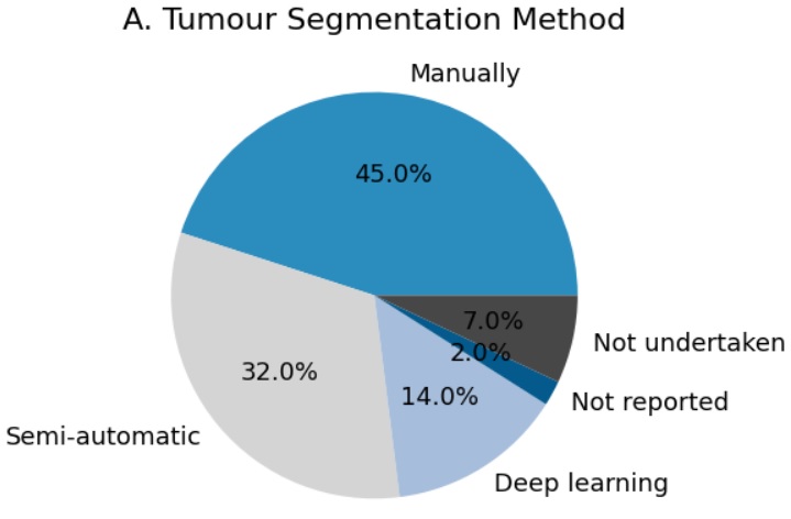
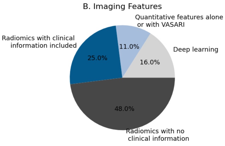
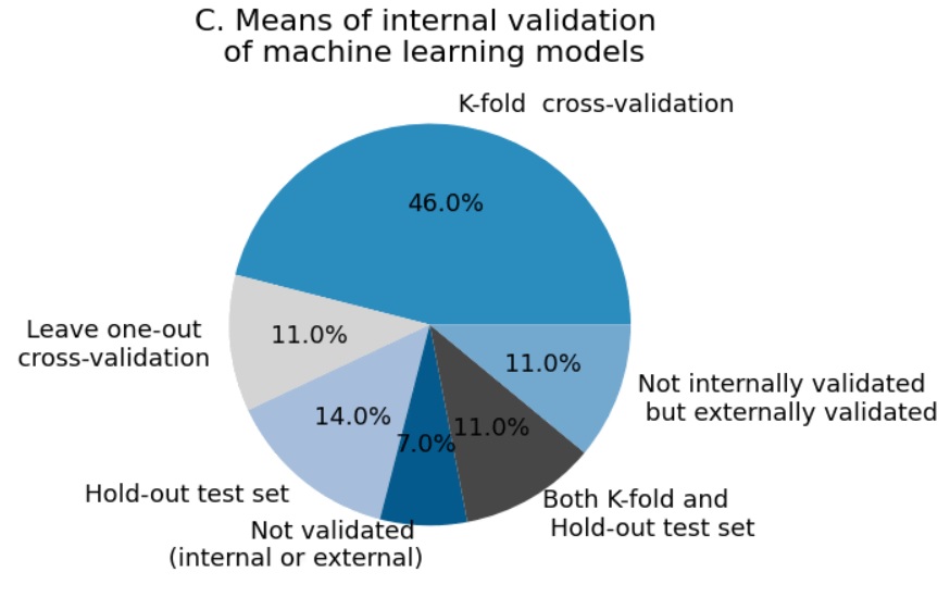
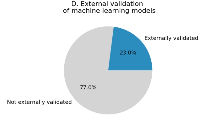
For differential diagnosis in brain cancer, numerous MRI-based machine learning approaches have been presented. These developments have partly been facilitated by the availability of several valuable public datasets, see for example the overviews in [141, 92]. Most literature is dedicated to glioma characterisation, which is therefore discussed in more detail here. Studies vary in the choice of input MRI sequences (T1w pre- and post-contrast, FLAIR, T2w, diffusion weighted imaging, perfusion weighted imaging, MR spectroscopy, APT CEST), the machine learning methodology (ranging from conventional radiomics approaches with hand-crafted features derived from manual tumour segmentations, to deep learning approaches that automatically segment the tumour), the classification target(s) (e.g. grade, IDH, 1p19q, and/or MGMT status), the selection of glioma subtypes on which the method is validated (e.g. only low-grade glioma, only high-grade glioma, or both), and the extent of validation performed (single train-test split, repeated cross-validation, internal versus external validation). A systematic review on the use of machine learning in neuro-oncology found four articles on glioma grading, and four articles on identifying genetic/molecular characteristics of glioma based on MRI [125]. Among those, only one study used convolutional neural networks as a machine learning tool - to predict 1p19q status in low-grade glioma [3]. A more recent systematic review identified 27 studies on glioma grading of which 6 used deep learning, and 48 studies on MRI-based estimation of genetic/molecular characteristics of which 8 used deep learning [20]. Another recent review dedicated to machine learning approaches for MRI-based glioma characterisation found 12 studies on glioma grading of which 2 used deep learning, and 43 studies on molecular characterisation out of which 10 used deep learning [42]. These numbers indicate a trend towards deep learning approaches as we see in the entire field, but with conventional machine learning approaches with pre-defined radiomics features still being used frequently. Regarding the performance, two recent systematic reviews performed a meta-analysis of studies on molecular characterisation of glioma. Jian et al. [56] found a pooled sensitivity/specificity/AUC in the validation set of 0.85/0.83/0.90 for IDH status prediction (12 studies), and 0.70/0.72/0.75 for 1p19q status prediction (5 studies). For MGMT, sensitivities and specificities ranging from 0.70 to 0.88 were found in 3 studies reporting validation performance, not allowing a meta-analysis. Van Kempen et al. [61] reported a pooled AUC of 0.91 for IDH status prediction (7 studies), 0.75 for 1p19q status prediction (3 studies), and 0.87 for MGMT promoter status prediction (3 studies). Thus, while the studies applied somewhat different criteria for inclusion in the meta-analysis and used different statistical analysis methods, they obtained similar performance estimates . Whereas both meta-analyses suggest promising accuracy for MRI-based MGMT promoter status prediction based on the results reported in literature, a comprehensive evaluation of deep learning approaches for MGMT promoter status prediction on the BraTS2021 dataset [7] yielded disappointing results, with AUCs ranging from 0.5 to 0.6 [123]. Also, the winning method of the BraTS2021 challenge achieved an AUC of 0.62 [9], suggesting that MGMT promoter status prediction from MRI is a very difficult task. Both systematic reviews [56, 61] also pointed out the low proportion of studies with external validation (10 out of 44 in [56] and 12 out of 60 in [61]). Figure 2, recreated based on [56], shows a number of other insightful statistics on the methodologies found in literature. Finally, both reviews also identified machine learning methods aimed at predicting other, less frequently considered molecular targets, including ATRX, TERT, EGFR, P53, and PTEN, indicating the broad range of possible future research directions in this area.
Beyond glioma characterisation, other differential diagnosis problems in brain cancer are differentiation between glioma and lymphoma, between glioblastoma and metastasis, between different types of meningioma, and between glioma, meningioma and pituitary tumours [125, 67, 20, 130, 149], with promising performances reported (AUC/accuracies around ). Of note, a recent study pointed out an important potential source of bias (the “Clever Hans effect”) in studies focused on differentiation between glioma, meningioma and pituitary tumours, due to implicit radiologist input in the selection of the 2D slices in a commonly used benchmark dataset [142].
For decision making in stroke, different targets for machine learning based on imaging data have been identified, mostly focused at determining the cause and extent of stroke and to a lesser extent, on informing treatment decisions [57]. Regarding cause and extent of acute stroke, automatic lesion detection and identification of tissue-at-risk include the most important elements. These remain challenging as there is a lot of variation in lesion shape and location depending on time-from-symptom onset, vessel occlusion site, and collateral status [72]. Machine learning methods for segmentation and detection are increasingly successful (see chapter 13). The step towards computer-aided diagnosis in stroke is also being taken using for example the CE-marked eASPECTS score [50], which is a machine learning-based assessment of the Alberta Stroke Program Early Computed Tomography Score (ASPECTS). This system for scoring acute ischemic damage to the brain has shown to be a simple, reliable and strong predictor of functional outcome after stroke. Regarding treatment decisions, machine learning is used in several studies to determine whether a patient qualifies for a specific stroke treatment. For thrombolytic treatment, this qualification depends on time elapsed after symptom onset and treatment should be performed within 4.5 hours. For this application, methods are developed that provide a binary estimation of stroke onset time (i.e., more or less than 4.5 hours) based on either DWI and FLAIR [73] or perfusion-weighted imaging (CT or MR) [51]. Both approaches used a radiomics-like approach of feature extraction (e.g. intensity/gradient/texture based or using an autoencoder) followed by a machine learning classifier (support vector machine, random forest, and logistic regression). These machine learning methods had greater sensitivity than human readers using the standard procedure of DWI-FLAIR mismatch and comparable specificity. In addition, thrombolysis may cause the rare complication of symptomatic intracranial haemorrhage. Several machine learning methods have been developed to predict the risk of this complication achieving promising predictive performance, for example using a support vector machine classifiers based on CT data (AUC=0.74) [10].
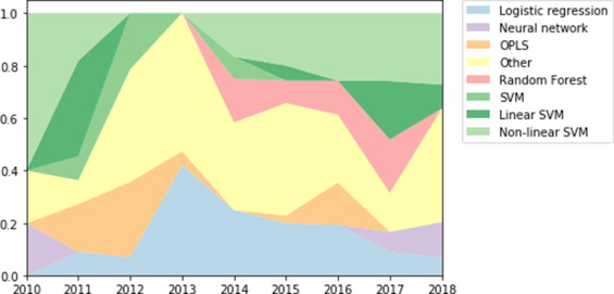
For prediction of the future course of subjects at-risk of developing dementia, there are three frequently used approaches for defining the prediction problem at hand. First, predicting whether the patient will develop dementia. In specific diseases, measurement of proteins causing underlying pathology has shown to be very promising to identify patients in a prodromal disease state. Here, prediction is performed either using univariate analysis or using logistic regression with few variables as input. Blood-based P-tau biomarker can predict incident AD within 4 years with an AUC of [106], CSF biomarkers and PET images of amyloid and Tau can predict clinical progression of subjects in their prodromal AD state with an AUC of [47]. Alternatively, in the absence of pathology-specific markers, MRI and cognitive markers of a patient together with machine learning approaches have been used to predict AD with an AUC of [139, 17, 78, 24]. For a systematic review of the different machine learning methods developed for the purpose of predicting AD, see [6]. Support vector machines (SVM) and logistic regressions are the most used algorithms in the last decade (Fig. 3). In FTD, where it is currently not possible to measure the pathological proteins in body fluids, prediction based on a combination of biomarkers that are non-specific to the underlying pathology is promising. This is demonstrated for example by [34], who predicted disease onset in familial FTD based on unspecific blood-based and CSF-based biomarkers using a disease progression model and identified presymptomatic subjects that developed dementia in the near future with an AUC of .
Second, predicting the time for conversion to dementia. While the previous problem predicts a dichotomous output variable, here it involves predicting a continuous variable of time to dementia. Bilgel et al. [12] predicted time to AD dementia with a mean error of years. In the TADPOLE challenge, machine learning approaches to predict time for conversion to AD dementia of 33 participating teams have been assessed quantitatively [86]. Ansart et al. [6] strongly favour predicting the exact time for conversion to dementia and argue against predicting converters within a given time interval (for example, within 3 years), because of the precision in the predictions. While this is indeed methodologically more elegant, the implications for clinical use and perception of patients regarding prediction precision and the inherent uncertainty remains to be established.
Third, prediction of disease markers could help to obtain insight into the clinical prognosis in an individual. Important disease markers are for example measures of global cognition (mini-mental state examination [MMSE] or Alzheimer’s disease assessment scale [ADAS] scores), or salient imaging markers (volume of the brain ventricles or longitudinal Tau protein accumulation). ADAS scores could not be reliably predicted by any participating team in the TADPOLE challenge [86], but a recent disease progression model called AD course map [68] could predict ADAS scores (which is scored from 0 to 150) after 3 years with a mean absolute error of 7.6 points. AD course map could also predict MMSE scores (which is scored from 0 to 30) after 3 years with a mean absolute error of 3.2 points. While these predictions used MRI as input, Tau PET was recently shown to be more predictive of future MMSE scores using linear mixed models [103]. However, a thorough validation of this Tau PET-based prediction is lacking. Predicting salient imaging markers such as volume of the ventricles [86], volume of the hippocampus [68] or longitudinal Tau accumulation [75] is a promising topic. Identifying the most clinically useful target to be predicted, the imaging modality that has the best cost-benefit ratio for prognosis of a patient, and the method that best predicts it are all important questions that still need answers in the future.
Most prediction methods in MS focus on predicting either physical disability, cognitive impairment, or treatment response in imaging data of an individual patient [58]. Physical disability as measured by expanded disability status scale (EDSS, range 0-10) has been the most commonly used predictor variable as recently used in [107, 121]. An ensemble of classifiers consisting of convolutional neural networks, random forests, and manifold learning was reported to predict EDSS with a mean square error of 3.0 [121]. Cognitive impairment has either been predicted as a global measure of cognition or as specific cognitive domains such as attention or working memory [32]. For predicting treatment response in MS, Signori et al. [127] used meta-analysis to identify subject characteristics that have higher treatment effects. In [35], the authors used an unsupervised disease progression model to identify subtypes of progression pathways in MS and found in post-hoc analysis that one of the subtypes predicted better treatment effects. Current challenges in this evolving field of predicting treatment response in MS and future directions have been summarized in [38].
For the prediction of patient outcomes after treatment of brain cancer, most machine learning studies have focused on MRI-based prediction of progression-free survival or overall survival, which will therefore be discussed in more detail here. A systematic review by Sarkiss et al. identified nine articles on survival prediction in glioma, and two on survival prediction for patients after stereotactic radiosurgery of brain metastases [125]. A more recent systematic review by Buchlak et al. identified 17 studies on survival prediction with performance estimates (AUC or accuracy) mostly in the range 0.7-0.8 [20]. Among those, only one study reported results of external validation, predicting overall survival of patients with low-grade glioma, and obtained an AUC of 0.71 with a model combining radiomics with non-imaging features including age, resection extent, grade, and IDH status [22]. Random (survival) forests and support vector machines were most often used methods. One study used a CNN as a pre-trained feature extractor [71]. Other recent approaches using CNNs to extract features that are subsequently combined with other factors into a final prognostic model include [99, 46, 52]. The 2017/2018 editions of the well-known BraTS challenges also included a task on overall survival prediction, with best teams obtaining accuracies around 0.6 in a three-class classification setting distinguishing short-, mid-, and long-survivors, [8]. Here, it was also pointed out that conventional machine learning methods outperformed deep learning methods, likely due to the limited size of available datasets for training.
Beyond MRI-based methods, methods using histopathology images and/or genomics data as input for the machine learning model are also considered in the literature on outcome prediction for glioma patients. In one of the pioneering studies on digital pathology images of glioma, better prognostication was obtained with deep learning when pathology images were combined with genetic markers (IDH, 1p19q) [94]. Preliminary work on so-called ‘radiopathomics’ in glioma is also available, supporting the notion that combining histology and radiology features improves prognostication (overall survival prediction) in glioma patients [119, 118].
2.2 B enchmarks and challenges
For 15 years, grand challenges have been organised in the biomedical image analysis research field. These are international benchmarks in competition form that have the goal of objectively comparing algorithms for a specific task on the same clinically representative data using the same evaluation protocol. In such challenges, the organisers supply reference data and evaluation measures on which researchers can evaluate their algorithms. Over the past years, the number and the impact of such grand challenges have increased [82]. Also in the field of computer-aided diagnosis and prediction, such grand challenges have been organised. For example, in the dementia field, four challenges have been organised focusing on early diagnosis [16, 5, 124] and predicting the natural disease course [5, 124, 86]. In general, algorithms winning the challenges performed rigorous data pre-processing and combined a wide range of input features [18]. In the field of brain cancer, the series of BraTS challenges has had a major impact [8, 7]. These benchmarks are instrumental to gaining insight into successful approaches and their potential for use in clinical practice and clinical trials.
2.3 O pen source software
Open source machine learning software such as Scikit Learn111https://scikit-learn.org/ and MONAI222https://monai.io/ have been fundamental to development of this field of research. More specifically for computer-aided diagnosis and prediction in brain diseases, dedicated platforms are available such as Clinica [122], NeuroPredict [112], and PRoNTo [126]. We also see a trend of researchers publishing their scripts and trained classifiers with their publications in order to promote reproducibility.
3. Clinical Impact
There are multiple ways in which computer-aided diagnosis and prediction models can make an impact on clinical practice. Key areas of impact are in decision making for treatment and care, replacing invasive diagnostic procedures and patient selection for clinical trials. Here we will discuss to what extent these clinical needs are addressed by current methods.
First, the most direct impact is on decision making for treatment and care. This not only affects clinical care and treatment planning in patients with for example dementia, stroke, MS or brain cancer, but is also important for managing the expectations of patients and their caregivers. Although high performances are achieved for some related tasks such as dementia classification, validation of those results on external datasets and clinical cohorts is still very limited as well as knowledge on the robustness of the methods. For other applications, there is still room for performance improvement, and key factors in achieving that would be the combination of multi-modal input and the availability of more well-maintained and large-scale datasets for training and evaluation. In general, there is room for improvement in how well real clinical questions are addressed by current methodology. Second, machine learning models can have an impact by replacing invasive diagnostic procedures. This is especially relevant in brain cancer, where machine learning techniques based on imaging data are developed to predict for example genetic mutation status or tumour grade, thereby avoiding or reducing the need for biopsies [152, 11]. As a motivating example, MRI-based prediction of MGMT methylation status could be beneficial to guide treatment decisions. This is supported by findings from a population-based study assessing survival in 131 patients with radiological diagnosis of glioblastoma who did not undergo surgery and thus lacked (histological or molecular) tissue-based verification of the diagnosis [146]. While patients without treatment had extremely poor prognosis with median survival of 3.6 months, those who received upfront temozolomide treatment did significantly better (with median survival of 6.8 months). Since the response to temozolomide is known to be highly dependent on the MGMT status, MRI-based prediction of MGMT status could give insight into which patients would benefit from treatment avoiding the need for biopsies in patients to frail for tumour biopsy. Third, patient selection for clinical trials is relevant in diseases where no to limited options for treatment exist, such as dementia, or diseases where existing treatments are suboptimal for some patients, such as MS. This can boost the power of trials by enrolling for example individuals who are more likely to progress based on prediction models. Several pilot studies demonstrated the added value of machine learning models to select a subgroup of participants to increase sensitivity to the treatment using phase III trial data (e.g. for Alzheimer’s disease treatment using donepezil or semagacestat) [36, 105]. This will ultimately reduce size, duration and cost of clinical trials.
The number of published methods is not evenly distributed over tasks. While many methods have been published on for example the classification of Alzheimer’s disease patients versus controls, much fewer publications exist on differential diagnosis in dementia. In addition, there seems to be a mismatch in some applications between published classification methods and clinical needs, e.g. the clinically relevant problem of early diagnosis does not directly translate to the frequently studied classification task of established Alzheimer’s disease versus healthy controls, but would instead require separation of early disease stage Alzheimer’s disease patients from those that have cognitive complaints but not dementia.
Several approved machine learning products to assist diagnosis and prediction are making their way into clinical practice, in particular in the imaging domain. Van Leeuwen et al. evaluated 100 commercially available products for AI in radiology, of which 38 are related to brain diseases [74]. These include mostly segmentation, quantification and normative comparison for neurodegenerative diseases and detection of lesions for stroke and oncology. Most methods generate a sample radiologist report which can be inspected and modified. In dementia, for example, 17 reporting tools that use automated brain MRI segmentation software and normative reference data for single-subject comparison are regulatory approved for use in the memory clinic [108].
One of these is Quantib ND (Quantib BV, Rotterdam, the Netherlands)333quantib.com/solutions/quantib-nd, which is an approved commercial software that performs automatic segmentation into 20 brain regions as well as normative volumetry reference curves based on data of 5000 subjects from a population based cohort. While Quantib ND and most other available tools use machine learning for brain segmentation, their output is not a diagnostic label produced by a machine learning algorithm. Another approved software, cDSI (Combinostics, Tampere, Finland)444combinostics.com/cdsi, does output diagnostic labels as confidence scores in addition to segmentation and normative volumetry based on MRI. It uses univariate machine learning to normalise individual biomarkers of different modalities based on reference values of patient and control groups, colour-codes these biomarkers to improve visualisation of large-data datasets and combines confidence scores based on individual biomarkers into one score [87, 88]. While cDSI is a machine learning tool for computer-aided diagnosis and prognosis, it does not exploit the power of machine learning to detect complex patterns in high dimensional data but rather focuses on visualisation and interpretability. Diagnosis and prediction algorithms that map high dimensional input, i.e. images and other clinical data, to an outcome measure using machine learning have not yet made their way into clinical practice.
4. Roadmap for clinical translation
There are numerous challenges for clinical translation of computer-aided diagnosis and prediction methods. Some key items that should be on the roadmap for translation relate to large and standardised datasets, to technical and clinical validation, to interpretability by clinicians and patients, and to practical issues related to implementation. In this section, we will discuss these requirements and related developments and initiatives.
The first requirement for translation are large and standardised datasets. For a few brain disorders, one or multiple large datasets (i.e. up to 2500 participants) are available to train machine learning algorithms for diagnosis and prediction tasks, facilitated by large multicenter initiatives such as the Alzheimer’s disease Neuroimaging initiative (ADNI) or the Parkinson’s progression markers initiative (PPMI). For validation in other cohorts and for development of algorithms in other diseases, there is only limited data available and a need for more (well-annotated) data exists. In particular there is a need for validation data that reflect the reality of clinical routine with no to limited data harmonization and large variation in imaging protocols and data quality. Setting up such large-scale datasets is complex due to various reasons including obstacles in inter-institutional data sharing and a lack of funding for collection, curation and labelling of data. To overcome these challenges, developments in research software and infrastructure may provide a solution by sharing easily reproducible algorithms rather than the data. Wrapping an algorithm in a container (e.g. Docker555www.docker.com, Singularity [69]) and applying the algorithms locally to the data (at one site or multiple sites in a federated approach) enables method validation on large sets of data within the confines of the local institute’s firewalls. Such an approach could be also used for enabling training on larger datasets (i.e. federated learning [128]). Standardisation of the data is important for eventual translation as it enables researchers to combine multiple datasets for development and validation of machine learning methods for diagnosis and prediction. Such standardisation entails both data collection (e.g. diagnostic criteria, protocols for image acquisition and clinical tests) and data organisation (e.g. through open source standards and platforms for data storage such as the Brain Imaging Data Structure (BIDS) and the Extensible Imaging Archive Toolkit (XNAT)).
Second, technical and clinical validation is a key focus area on the roadmap for translation. In the field of radiology, the quantitative neuroradiology initiative (QNI) framework has been developed as a model framework for translation defining the technical and clinical validation necessary to embed automated software into the clinical workflow [40]. Based on this framework, [108] reviewed the published evidence regarding commercial automated volumetric MRI tools for dementia diagnosis. For the 17 products identified, 11 companies have published some form of technical validation on their methods, but only 4 have published clinical validation in a dementia population. They concluded that there is a significant evidence gap in the literature regarding clinical validation and in-use evaluation. Whereas this review only addressed image volumetry in dementia, these findings likely extend to other brain diseases, applications and modalities. Hence, there is a need for both retrospective and prospective studies validating algorithms in a clinical setting. In addition, performance metrics used in validation studies should aim to capture real clinical applicability and address different aspects of the reliability of an algorithm, including accuracy, uncertainty estimation, reproducibility and generalizability to other data. Standards for validation and reporting are provided by guidelines such as STARD-AI [133] and TRIPOD-AI666osf.io/zyacb.
A third key item for clinical translation is interpretability by end-users such as clinicians and patients. As clinicians have responsibility for the decisions related to care and treatment, they should have trust in a computer-aided diagnosis or prediction system and understand its outputs to an extent that they can rely on them for decision making and explanation to a patient. Performance metrics should aim to capture real clinical applicability and be understandable to intended users [60]. High validation performance is important for building trust in methods, but not sufficient by itself, since performance may reduce in individual cases because of unaccounted inter-individual such as comorbidities or population differences such as MRI scan protocol. Therefore, apart from model accuracy, relevant questions for interpretation are for example: Is the model suitable for the data of this patient? What features contribute to the machine learning decision for this patient? How certain is the decision for this patient and can the algorithm know when it is uncertain about an individual’s decision? Such questions are important and methods should be designed and implemented in a way that facilitates answers to such questions. This could be obtained by using interpretability methods on top of ‘black box’ machine learning models or directly by using interpretable models. For the first category, many methods have been developed based on model weight visualisation, feature map visualisation, back-propagation methods or perturbation of inputs (see also Chapter 22). For interpretable models, an example in the field of computer-aided diagnosis and prognosis are disease progression models[148, 138]. These data-driven models are designed specifically for neurodegenerative diseases and explain their decisions based on their estimate of the natural progression of the disease in the cohort (see also Chapter 17).
As a final key item, we will discuss implementation feasibility. For machine learning models to be actually used in practice, it is essential that models and reporting are integrated into the clinical workflow and that the sending and processing of clinical data and receiving results is fully automated. Current commercial products for automatic volumetry in dementia all reported to have implemented an integration with radiology systems and the clinical workflow. While validation of the workflows is limited [108], this does support the feasibility for machine learning in clinical practice. While these products integrate with the radiological workflow, a key challenge for the clinical translation of algorithms that use non-imaging clinical data (such as cognitive scores) as input is to also integrate with the clinical workflow of multi-disciplinary diagnosis.
5. Final summary and conclusion
Computer-aided diagnosis and prediction of brain disorders is an important research area, with a wide variety of applications. While typically for these applications generic machine learning methods are used, domain knowledge of these brain disorders is crucial for selecting novel clinically relevant applications as well as for making domain specific methodological improvements. Regarding diagnosis, clinical challenges are in early diagnosis of dementia and MS, differential diagnosis of dementia and brain cancer, and decision making for treatment in stroke. Regarding prediction, challenges are in the prediction of the natural disease course in dementia and MS, and the prediction of patient outcomes after treatment in stroke, brain cancer, and MS. Even though the disorders on which we focused are important avenues for impact, computer aided diagnosis and prognosis would also be extremely useful in other disorders such as movement disorders for predicting response to treatment and side-effects, epilepsy for predicting response to epilepsy surgery, psychiatric disorders where diagnosis can be particularly difficult.
Key areas of impact are in 1) decision making for treatment and care in patients with dementia, stroke, MS or brain cancer, 2) replacing invasive diagnostic procedures in brain cancer, and 3) patient selection for clinical trials in dementia and MS. While the first AI methods are making their way to clinical practice, diagnosis and prediction algorithms that map high dimensional input, i.e. images and other clinical data, to an outcome measure using machine learning are not yet clinically available. To enable translation, major items on the roadmap relate to the availability of large and standardised datasets and technical and clinical validation of the developed machine learning methods. In addition, other important aspects are interpretability of the results by clinicians and patients, optimisation of the diagnostic or treatment workflow in the clinic, and other practical issues related to implementation.
With this chapter, we aimed to provide a comprehensive overview, bringing together the clinical context of representative use cases of diagnosis and prediction in brain disorders and their state-of-the-art computer-aided methods. Future research should focus on bridging the identified gaps between clinical needs and the solutions brought by machine learning, to further improve decision making, treatment, and care in brain diseases.
Acknowledgements
V. Venkatraghavan is supported by JPND-funded E-DADS project
(ZonMW project ). W.J. Niessen and E.E. Bron are supported by Medical Delta Diagnostics 3.0: Dementia and Stroke. E.E. Bron acknowledges support from the Netherlands CardioVascular Research Initiative (Heart-Brain Connection: CVON2012-06, CVON2018-28).
Conflict of interest
Quantib BV is a spin-off company of Erasmus MC. W.J.N. is cofounder, part-time Chief Scientific Officer, and stock holder of Quantib BV. S.R.V, D.B., M.S., S.K. and E.E.B. are affiliated to Erasmus MC but have no personal relationships with or financial interest in Quantib BV. F.B. is a consultant to Combinostics.
References
- Alz [2020] (2020) Alzheimer’s disease facts and figures. Alzheimer’s & Dementia 16(3):391–460, DOI 10.1002/alz.12068
- Nat [2021] (2021) Disease modifying therapies for MS. National Multiple Sclerosis Society pp 3–21
- Akkus et al [2017] Akkus Z, Ali I, Sedlář J, Agrawal JP, Parney IF, Giannini C, Erickson BJ (2017) Predicting deletion of chromosomal arms 1p/19q in low-grade gliomas from MR images using machine intelligence. Journal of digital imaging 30(4):469—476, DOI 10.1007/s10278-017-9984-3
- Albert et al [2011] Albert MS, DeKosky ST, Dickson D, Dubois B, Feldman HH, Fox NC, Gamst A, Holtzman DM, Jagust WJ, Petersen RC, Snyder PJ, Carrillo MC, Thies B, Phelps CH (2011) The diagnosis of mild cognitive impairment due to alzheimer’s disease: Recommendations from the national institute on aging-alzheimer’s association workgroups on diagnostic guidelines for alzheimer’s disease. Alzheimer’s & Dementia 7(3):270–279, DOI 10.1016/j.jalz.2011.03.008
- Allen et al [2016] Allen GI, Amoroso N, Anghel C, Balagurusamy V, Bare CJ, et al (2016) Crowdsourced estimation of cognitive decline and resilience in alzheimer’s disease. Alzheimer’s & Dementia 12(6):645–653, DOI 10.1016/j.jalz.2016.02.006
- Ansart et al [2021] Ansart M, Epelbaum S, Bassignana G, Bône A, Bottani S, Cattai T, Couronné R, Faouzi J, Koval I, Louis M, Thibeau-Sutre E, Wen J, Wild A, Burgos N, Dormont D, Colliot O, Durrleman S (2021) Predicting the progression of mild cognitive impairment using machine learning: A systematic, quantitative and critical review. Medical Image Analysis 67:101848, DOI 10.1016/j.media.2020.101848
- Baid et al [2021] Baid U, Ghodasara S, Mohan S, Bilello M, Calabrese E, et al (2021) The RSNA-ASNR-MICCAI BraTS 2021 benchmark on brain tumor segmentation and radiogenomic classification. 2107.02314
- Bakas et al [2019] Bakas S, Reyes M, Jakab A, Bauer S, Rempfler M, et al (2019) Identifying the best machine learning algorithms for brain tumor segmentation, progression assessment, and overall survival prediction in the brats challenge. 1811.02629
- Bakas et al [2021] Bakas S, Baid U, Calabrese E, Carr C, Colak E, Farahani K, Flanders AE, Kalpathy-Cramer J, Kitamura FC, Menze B, Mongan J, Prevedello L, Rudie J, Shinohara RT (2021) RSNA-MICCAI brain tumor radiogenomic classification. Link to the Kaggle challenge
- Bentley et al [2014] Bentley P, Ganesalingam J, Carlton Jones AL, Mahady K, Epton S, Rinne P, Sharma P, Halse O, Mehta A, Rueckert D (2014) Prediction of stroke thrombolysis outcome using CT brain machine learning. NeuroImage: Clinical 4:635–640, DOI 10.1016/j.nicl.2014.02.003
- Bi et al [2019] Bi WL, Hosny A, Schabath MB, Giger ML, Birkbak NJ, Mehrtash A, Allison T, Arnaout O, Abbosh C, Dunn IF, Mak RH, Tamimi RM, Tempany CM, Swanton C, Hoffmann U, Schwartz LH, Gillies RJ, Huang RY, Aerts HJWL (2019) Artificial intelligence in cancer imaging: Clinical challenges and applications. CA: A Cancer Journal for Clinicians 69(2):127–157, DOI 10.3322/caac.21552
- Bilgel et al [2019] Bilgel M, Jedynak BM, Initiative ADN (2019) Predicting time to dementia using a quantitative template of disease progression. Alzheimer’s & Dementia: Diagnosis, Assessment & Disease Monitoring 11(1):205–215, DOI 10.1016/j.dadm.2019.01.005
- Bouts et al [2018] Bouts MJRJ, Möller C, Hafkemeijer A, van Swieten JC, Dopper E, van der Flier WM, Vrenken H, Wink AM, Pijnenburg YAL, Scheltens P, Barkhof F, Schouten TM, de Vos F, Feis RA, van der Grond J, de Rooij M, Rombouts SARB (2018) Single subject classification of alzheimer’s disease and behavioral variant frontotemporal dementia using anatomical, diffusion tensor, and resting-state functional magnetic resonance imaging. Journal of Alzheimer’s disease : JAD 62(4):1827—1839, DOI 10.3233/jad-170893
- Bron et al [2016] Bron E, Smits M, Papma J, Steketee R, Meijboom R, De Groot M, van Swieten J, Niessen W, Klein S (2016) Multiparametric computer-aided differential diagnosis of alzheimer’s disease and frontotemporal dementia using structural and advanced MRI. European Radiology 27(8):1–11, DOI 10.1007/s00330-016-4691-x
- Bron et al [2014] Bron EE, Steketee RM, Houston GC, Oliver RA, Achterberg HC, Loog M, van Swieten JC, Hammers A, Niessen WJ, Smits M, Klein S, for the Alzheimer’s Disease Neuroimaging Initiative (2014) Diagnostic classification of arterial spin labeling and structural MRI in presenile early stage dementia. Human Brain Mapping 35(9):4916–4931, DOI 10.1002/hbm.22522
- Bron et al [2015] Bron EE, Smits M, van der Flier WM, Vrenken H, Barkhof F, et al (2015) Standardized evaluation of algorithms for computer-aided diagnosis of dementia based on structural MRI: The CADDementia challenge. NeuroImage 111:562–579, DOI 10.1016/j.neuroimage.2015.01.048
- Bron et al [2021a] Bron EE, Klein S, Papma JM, Jiskoot LC, Venkatraghavan V, Linders J, Aalten P, De Deyn PP, Biessels GJ, Claassen JA, Middelkoop HA, Smits M, Niessen WJ, van Swieten JC, van der Flier WM, Ramakers IH, van der Lugt A (2021a) Cross-cohort generalizability of deep and conventional machine learning for MRI-based diagnosis and prediction of alzheimer’s disease. NeuroImage: Clinical 31:102712, DOI 10.1016/j.nicl.2021.102712
- Bron et al [2021b] Bron EE, Klein S, Reinke A, Papma JM, Maier-Hein L, Alexander DC, Oxtoby NP (2021b) Ten years of image analysis and machine learning competitions in dementia. 2112.07922
- Brown et al [2020] Brown FS, Glasmacher SA, Kearns PKA, MacDougall N, Hunt D, Connick P, Chandran S (2020) Systematic review of prediction models in relapsing remitting multiple sclerosis. PLOS ONE 15(5):1–13, DOI 10.1371/journal.pone.0233575
- Buchlak et al [2021] Buchlak QD, Esmaili N, Leveque JC, Bennett C, Farrokhi F, Piccardi M (2021) Machine learning applications to neuroimaging for glioma detection and classification: An artificial intelligence augmented systematic review. Journal of clinical neuroscience : official journal of the Neurosurgical Society of Australasia 89:177—198, DOI 10.1016/j.jocn.2021.04.043
- Chen et al [2019] Chen C, Ou X, Wang J, Guo W, Ma X (2019) Radiomics-based machine learning in differentiation between glioblastoma and metastatic brain tumors. Frontiers in Oncology 9:806, DOI 10.3389/fonc.2019.00806
- Choi et al [2020] Choi YS, Ahn SS, Chang JH, Kang SG, Kim EH, Kim SH, Jain R, Lee SK (2020) Machine learning and radiomic phenotyping of lower grade gliomas: improving survival prediction. European radiology 30(7):3834—3842, DOI 10.1007/s00330-020-06737-5
- Crous-Bou et al [2017] Crous-Bou M, Minguillón C, Gramunt N, Molinuevo JL (2017) Alzheimer’s disease prevention: from risk factors to early intervention. Alzheimer’s research & therapy 9(1):71, DOI 10.1186/s13195-017-0297-z
- Cui et al [2011] Cui Y, Liu B, Luo S, Zhen X, Fan M, Liu T, Zhu W, Park M, Jiang T, Jin JS, the Alzheimer’s Disease Neuroimaging Initiative (2011) Identification of conversion from mild cognitive impairment to alzheimer’s disease using multivariate predictors. PLOS ONE 6(7):1–10, DOI 10.1371/journal.pone.0021896
- Cuingnet et al [2011] Cuingnet R, Gerardin E, Tessieras J, Auzias G, Lehéricy S, Habert MO, Chupin M, Benali H, Colliot O (2011) Automatic classification of patients with alzheimer’s disease from structural MRI: A comparison of ten methods using the adni database. NeuroImage 56(2):766–781, DOI 10.1016/j.neuroimage.2010.06.013, multivariate Decoding and Brain Reading
- Dekker et al [2021] Dekker I, Schoonheim MM, Venkatraghavan V, Eijlers AJ, Brouwer I, Bron EE, Klein S, Wattjes MP, Wink AM, Geurts JJ, Uitdehaag BM, Oxtoby NP, Alexander DC, Vrenken H, Killestein J, Barkhof F, Wottschel V (2021) The sequence of structural, functional and cognitive changes in multiple sclerosis. NeuroImage: Clinical 29:102550, DOI 10.1016/j.nicl.2020.102550
- Delfanti et al [2017] Delfanti RL, Piccioni DE, Handwerker J, Bahrami N, Krishnan A, Karunamuni R, Hattangadi-Gluth JA, Seibert TM, Srikant A, Jones KA, Snyder VS, Dale AM, White NS, McDonald CR, Farid N (2017) Imaging correlates for the 2016 update on WHO classification of grade II/III gliomas: implications for IDH, 1p/19q and ATRX status. Journal of neuro-oncology 135(3):601—609, DOI 10.1007/s11060-017-2613-7
- Dubbink et al [2015] Dubbink HJ, Atmodimedjo PN, Kros JM, French PJ, Sanson M, Idbaih A, Wesseling P, Enting R, Spliet W, Tijssen C, Dinjens WNM, Gorlia T, van den Bent MJ (2015) Molecular classification of anaplastic oligodendroglioma using next-generation sequencing: a report of the prospective randomized EORTC Brain Tumor Group 26951 phase III trial. Neuro-Oncology 18(3):388–400, DOI 10.1093/neuonc/nov182
- Dubois et al [2014] Dubois B, Feldman HH, Jacova C, Hampel H, Molinuevo JL, et al (2014) Advancing research diagnostic criteria for alzheimer’s disease: the iwg-2 criteria. The Lancet Neurology 13(6):614—629, DOI 10.1016/s1474-4422(14)70090-0
- Ducharme et al [2015] Ducharme S, Price BH, Larvie M, Dougherty DD, Dickerson BC (2015) Clinical approach to the differential diagnosis between behavioral variant frontotemporal dementia and primary psychiatric disorders. American Journal of Psychiatry 172(9):827–837, DOI 10.1176/appi.ajp.2015.14101248
- Eckel-Passow et al [2015] Eckel-Passow JE, Lachance DH, Molinaro AM, Walsh KM, Decker PA, et al (2015) Glioma groups based on 1p/19q, idh, and tert promoter mutations in tumors. New England Journal of Medicine 372(26):2499–2508, DOI 10.1056/NEJMoa1407279
- Eijlers et al [2018] Eijlers AJC, van Geest Q, Dekker I, Steenwijk MD, Meijer KA, Hulst HE, Barkhof F, Uitdehaag BMJ, Schoonheim MM, Geurts JJG (2018) Predicting cognitive decline in multiple sclerosis: a 5-year follow-up study. Brain 141(9):2605–2618, DOI 10.1093/brain/awy202
- El-Koussy et al [2014] El-Koussy M, Schroth G, Brekenfeld C, Arnold M (2014) Imaging of acute ischemic stroke. European neurology 72(5-6):309—316, DOI 10.1159/000362719
- van der Ende et al [2021] van der Ende EL, Bron EE, Poos JM, Jiskoot LC, Panman JL, et al (2021) A data-driven disease progression model of fluid biomarkers in genetic frontotemporal dementia. Brain DOI 10.1093/brain/awab382, awab382
- Eshaghi et al [2021] Eshaghi A, Young A, Wijeratne P, Prados F, Arnold D, Narayanan S, Guttmann C, Barkhof F, Alexander D, Thompson A, Chard D, Ciccarelli O (2021) Identifying multiple sclerosis subtypes using unsupervised machine learning and MRI data. Nature Communications 12(1), DOI 10.1038/s41467-021-22265-2
- Ezzati et al [2020] Ezzati A, Lipton RB, Alzheimer’s Disease Neuroimaging Initiative (2020) Machine learning predictive models can improve efficacy of clinical trials for alzheimer’s disease. Journal of Alzheimer’s disease : JAD 74(1):55—63, DOI 10.3233/jad-190822
- Falahati et al [2014] Falahati F, Westman E, Simmons A (2014) Multivariate data analysis and machine learning in alzheimer’s disease with a focus on structural magnetic resonance imaging. Journal of Alzheimer’s disease : JAD 41(3):685—708, DOI 10.3233/jad-131928
- Gasperini et al [2019] Gasperini C, Prosperini L, Tintoré M, Sormani MP, Filippi M, Rio J, Palace J, Rocca MA, Ciccarelli O, Barkhof F, Sastre-Garriga J, Vrenken H, Frederiksen JL, Yousry TA, Enzinger C, Rovira A, Kappos L, Pozzilli C, Montalban X, De Stefano N, , the MAGNIMS Study Group (2019) Unraveling treatment response in multiple sclerosis. Neurology 92(4):180–192, DOI 10.1212/WNL.0000000000006810
- Gessler et al [2019] Gessler F, Bernstock JD, Braczynski A, Lescher S, Baumgarten P, Harter PN, Mittelbronn M, Wu T, Seifert V, Senft C (2019) Surgery for glioblastoma in light of molecular markers: Impact of resection and mgmt promoter methylation in newly diagnosed idh-1 wild-type glioblastomas. Neurosurgery 84(1):190—197, DOI 10.1093/neuros/nyy049
- Goodkin et al [2019] Goodkin O, Pemberton H, Vos SB, Prados F, Sudre CH, Moggridge J, Cardoso MJ, Ourselin S, Bisdas S, White M, Yousry T, Thornton J, Barkhof F (2019) The quantitative neuroradiology initiative framework: application to dementia. The British Journal of Radiology 92(1101):20190365, DOI 10.1259/bjr.20190365, pMID: 31368776
- Gordon et al [2018] Gordon BA, Blazey TM, Su Y, Hari-Raj A, Dincer A, et al (2018) Spatial patterns of neuroimaging biomarker change in individuals from families with autosomal dominant alzheimer’s disease: a longitudinal study. The Lancet Neurology 17(3):241–250, DOI 10.1016/S1474-4422(18)30028-0
- Gore et al [2021] Gore S, Chougule T, Jagtap J, Saini J, Ingalhalikar M (2021) A review of radiomics and deep predictive modeling in glioma characterization. Academic Radiology 28(11):1599–1621, DOI 10.1016/j.acra.2020.06.016
- Gorelick et al [2011] Gorelick PB, Scuteri A, Black SE, Decarli C, Greenberg SM, et al (2011) Vascular contributions to cognitive impairment and dementia: a statement for healthcare professionals from the american heart association/american stroke association. Stroke 42(9):2672—2713, DOI 10.1161/str.0b013e3182299496
- Gorno-Tempini et al [2008] Gorno-Tempini ML, Brambati SM, Ginex V, Ogar J, Dronkers NF, Marcone A, Perani D, Garibotto V, Cappa SF, Miller BL (2008) The logopenic/phonological variant of primary progressive aphasia. Neurology 71(16):1227–1234, DOI 10.1212/01.wnl.0000320506.79811.da
- Hampshire et al [2021] Hampshire A, Trender W, Chamberlain SR, Jolly AE, Grant JE, Patrick F, Mazibuko N, Williams SC, Barnby JM, Hellyer P, Mehta MA (2021) Cognitive deficits in people who have recovered from covid-19. EClinicalMedicine 39:101044, DOI 10.1016/j.eclinm.2021.101044
- Han et al [2020] Han W, Qin L, Bay C, Chen X, Yu KH, Miskin N, Li A, Xu X, Young G (2020) Deep transfer learning and radiomics feature prediction of survival of patients with high-grade gliomas. American Journal of Neuroradiology 41(1):40–48, DOI 10.3174/ajnr.A6365
- Hansson et al [2018] Hansson O, Seibyl J, Stomrud E, Zetterberg H, Trojanowski JQ, Bittner T, Lifke V, Corradini V, Eichenlaub U, Batrla R, Buck K, Zink K, Rabe C, Blennow K, Shaw LM, for the Swedish BioFINDER study group, Initiative ADN (2018) CSF biomarkers of alzheimer’s disease concord with amyloid- pet and predict clinical progression: A study of fully automated immunoassays in biofinder and adni cohorts. Alzheimer’s & Dementia 14(11):1470–1481, DOI 10.1016/j.jalz.2018.01.010
- Harris et al [2015] Harris JM, Thompson JC, Gall C, Richardson AM, Neary D, du Plessis D, Pal P, Mann DM, Snowden JS, Jones M (2015) Do NIA-AA criteria distinguish alzheimer’s disease from frontotemporal dementia? Alzheimer’s & Dementia 11(2):207–215, DOI 10.1016/j.jalz.2014.04.516
- Hellwig et al [2019] Hellwig S, Frings L, Bormann T, Vach W, Buchert R, Meyer PT (2019) Amyloid imaging for differential diagnosis of dementia: incremental value compared to clinical diagnosis and [18f]fdg pet. European journal of nuclear medicine and molecular imaging 46(2):312—323, DOI 10.1007/s00259-018-4111-3
- Herweh et al [2016] Herweh C, Ringleb PA, Rauch G, Gerry S, Behrens L, Möhlenbruch M, Gottorf R, Richter D, Schieber S, Nagel S (2016) Performance of e-ASPECTS software in comparison to that of stroke physicians on assessing CT scans of acute ischemic stroke patients. International Journal of Stroke 11(4):438–445, DOI 10.1177/1747493016632244
- Ho et al [2017] Ho KC, Speier W, El-Saden S, Arnold CW (2017) Classifying acute ischemic stroke onset time using deep imaging features. AMIA Annual Symposium proceedings 2017:892—901
- Huang et al [2021] Huang H, Zhang W, Fang Y, Hong J, Su S, Lai X (2021) Overall survival prediction for gliomas using a novel compound approach. Frontiers in Oncology 11, DOI 10.3389/fonc.2021.724191
- Jack et al [2013] Jack CR, Knopman DS, Jagust WJ, Petersen RC, Weiner MW, Aisen PS, Shaw LM, Vemuri P, Wiste HJ, Weigand SD, Lesnick TG, Pankratz VS, Donohue MC, Trojanowski JQ (2013) Tracking pathophysiological processes in alzheimer’s disease: an updated hypothetical model of dynamic biomarkers. The Lancet Neurology 12(2):207–216, DOI 10.1016/S1474-4422(12)70291-0
- Jack Jr. et al [2018] Jack Jr CR, Bennett DA, Blennow K, Carrillo MC, Dunn B, Haeberlein SB, Holtzman DM, Jagust W, Jessen F, Karlawish J, Liu E, Molinuevo JL, Montine T, Phelps C, Rankin KP, Rowe CC, Scheltens P, Siemers E, Snyder HM, Sperling R, Contributors, Elliott C, Masliah E, Ryan L, Silverberg N (2018) NIA-AA research framework: Toward a biological definition of alzheimer’s disease. Alzheimer’s & Dementia 14(4):535–562, DOI 10.1016/j.jalz.2018.02.018
- Jang et al [2018] Jang BS, Jeon SH, Kim IH, Kim IA (2018) Prediction of pseudoprogression versus progression using machine learning algorithm in glioblastoma. Scientific reports 8(1):12516, DOI 10.1038/s41598-018-31007-2
- Jian et al [2021] Jian A, Jang K, Manuguerra M, Liu S, Magnussen J, Di Ieva A (2021) Machine Learning for the Prediction of Molecular Markers in Glioma on Magnetic Resonance Imaging: A Systematic Review and Meta-Analysis. Neurosurgery 89(1):31–44, DOI 10.1093/neuros/nyab103
- Kamal et al [2018] Kamal H, Lopez V, Sheth SA (2018) Machine learning in acute ischemic stroke neuroimaging. Frontiers in Neurology 9:945, DOI 10.3389/fneur.2018.00945
- Kanber et al [2019] Kanber B, Nachev P, Barkhof F, Calvi A, Cardoso J, Cortese R, Prados F, Sudre C, Tur C, Ourselin S, Ciccarelli O (2019) High-dimensional detection of imaging response to treatment in multiple sclerosis. NPJ digital medicine 2(1), DOI 10.1038/s41746-019-0127-8
- Kaufmann et al [2018] Kaufmann M, Kuhle J, Puhan MA, Kamm CP, Chan A, Salmen A, Kesselring J, Calabrese P, Gobbi C, Pot C, Steinemann N, Rodgers S, von Wyl V, Swiss Multiple Sclerosis Registry (SMSR) (2018) Factors associated with time from first-symptoms to diagnosis and treatment initiation of multiple sclerosis in switzerland. Multiple sclerosis journal - experimental, translational and clinical 4(4):2055217318814562, DOI 10.1177/2055217318814562
- Kelly et al [2019] Kelly CJ, Karthikesalingam A, Suleyman M, Corrado G, King D (2019) Key challenges for delivering clinical impact with artificial intelligence. BMC medicine 17(1):195, DOI 10.1186/s12916-019-1426-2
- van Kempen et al [2021] van Kempen EJ, Post M, Mannil M, Kusters B, ter Laan M, Meijer FJA, Henssen DJHA (2021) Accuracy of machine learning algorithms for the classification of molecular features of gliomas on MRI: A systematic literature review and meta-analysis. Cancers 13(11), DOI 10.3390/cancers13112606
- Kickingereder et al [2018] Kickingereder P, Neuberger U, Bonekamp D, Piechotta PL, Götz M, et al (2018) Radiomic subtyping improves disease stratification beyond key molecular, clinical, and standard imaging characteristics in patients with glioblastoma. Neuro-Oncology 20(6):848–857, DOI 10.1093/neuonc/nox188
- Kim et al [2021] Kim J, Park Y, Park S, Jang H, Kim HJ, Na DL, Lee H, Seo SW (2021) Prediction of tau accumulation in prodromal alzheimer’s disease using an ensemble machine learning approach. Scientific reports 11(1):5706, DOI 10.1038/s41598-021-85165-x
- Klöppel et al [2008] Klöppel S, Stonnington CM, Barnes J, Chen F, Chu C, Good CD, Mader I, Mitchell LA, Patel AC, Roberts CC, Fox NC, Jack J Clifford R, Ashburner J, Frackowiak RSJ (2008) Accuracy of dementia diagnosis—a direct comparison between radiologists and a computerized method. Brain 131(11):2969–2974, DOI 10.1093/brain/awn239
- Klöppel et al [2012] Klöppel S, Abdulkadir A, Jack CR, Koutsouleris N, Mourão-Miranda J, Vemuri P (2012) Diagnostic neuroimaging across diseases. NeuroImage 61(2):457–463, DOI 10.1016/j.neuroimage.2011.11.002, neuroimaging: Then, Now and the Future
- Knight-Greenfield et al [2019] Knight-Greenfield A, Nario JJQ, Gupta A (2019) Causes of acute stroke: A patterned approach. Radiologic clinics of North America 57(6):1093—1108, DOI 10.1016/j.rcl.2019.07.007
- Kocher et al [2020] Kocher M, Ruge MI, Galldiks N, Lohmann P (2020) Applications of radiomics and machine learning for radiotherapy of malignant brain tumors. Strahlentherapie und Onkologie 196(10):856—867, DOI 10.1007/s00066-020-01626-8
- Koval et al [2021] Koval I, Bône A, Louis M, Lartigue T, Bottani S, Marcoux A, Samper-González J, Burgos N, Charlier B, Bertrand A, Epelbaum S, Colliot O, Allassonnière S, Durrleman S (2021) AD course map charts alzheimer’s disease progression. Scientific reports 11(1):8020, DOI 10.1038/s41598-021-87434-1
- Kurtzer et al [2017] Kurtzer GM, Sochat V, Bauer MW (2017) Singularity: Scientific containers for mobility of compute. PLOS ONE 12(5):1–20, DOI 10.1371/journal.pone.0177459
- Kuruppu and Matthews [2013] Kuruppu DK, Matthews BR (2013) Young-onset dementia. Seminars in neurology 33(4):365—385, DOI 10.1055/s-0033-1359320
- Lao et al [2017] Lao J, Chen Y, Li ZC, Li Q, Zhang J, Liu J, Zhai G (2017) A deep learning-based radiomics model for prediction of survival in glioblastoma multiforme. Scientific reports 7(1):10353, DOI 10.1038/s41598-017-10649-8
- Lee et al [2017] Lee EJ, Kim YH, Kim N, Kang DW (2017) Deep into the brain: Artificial intelligence in stroke imaging. J Stroke 19(3):277–285, DOI 10.5853/jos.2017.02054
- Lee et al [2020] Lee H, Lee EJ, Ham S, Lee HB, Lee JS, Kwon SU, Kim JS, Kim N, Kang DW (2020) Machine learning approach to identify stroke within 4.5 hours. Stroke 51(3):860—866, DOI 10.1161/strokeaha.119.027611
- van Leeuwen et al [2021] van Leeuwen KG, Schalekamp S, Rutten MJ, van Ginneken B, de Rooij M (2021) Artificial intelligence in radiology: 100 commercially available products and their scientific evidence. European radiology 31(6):3797–3804, DOI 10.1007/s00330-021-07892-z
- Leuzy et al [2021] Leuzy A, Smith R, Cullen NC, Strandberg O, Vogel JW, Binette AP, Borroni E, Janelidze S, Ohlsson T, Jögi J, Ossenkoppele R, Palmqvist S, Mattsson-Carlgren N, Klein G, Stomrud E, Hansson O (2021) Biomarker-Based Prediction of Longitudinal Tau Positron Emission Tomography in Alzheimer Disease. JAMA Neurology DOI 10.1001/jamaneurol.2021.4654
- Li et al [2021] Li M, Ren X, Dong G, Wang J, Jiang H, Yang C, Zhao X, Zhu Q, Cui Y, Yu K, Lin S (2021) Distinguishing pseudoprogression from true early progression in isocitrate dehydrogenase wild-type glioblastoma by interrogating clinical, radiological, and molecular features. Frontiers in Oncology 11, DOI 10.3389/fonc.2021.627325
- Liguori et al [2000] Liguori M, Marrosu M, Pugliatti M, Giuliani F, De Robertis F, Cocco E, Zimatore G, Livrea P, Trojano M (2000) Age at onset in multiple sclerosis. Neurological sciences : official journal of the Italian Neurological Society and of the Italian Society of Clinical Neurophysiology 21(4 Suppl 2):S825—9, DOI 10.1007/s100720070020
- Lin et al [2018] Lin W, Tong T, Gao Q, Guo D, Du X, Yang Y, Guo G, Xiao M, Du M, Qu X, TADNI (2018) Convolutional neural networks-based MRI image analysis for the alzheimer’s disease prediction from mild cognitive impairment. Frontiers in Neuroscience 12:777, DOI 10.3389/fnins.2018.00777
- Liu et al [2018] Liu M, Cheng D, Wang K, Wang Y, Alzheimer’s Disease Neuroimaging Initiative (2018) Multi-modality cascaded convolutional neural networks for alzheimer’s disease diagnosis. Neuroinformatics 16(3-4):295—308, DOI 10.1007/s12021-018-9370-4
- Louis et al [2021] Louis DN, Perry A, Wesseling P, Brat DJ, Cree IA, Figarella-Branger D, Hawkins C, Ng HK, Pfister SM, Reifenberger G, Soffietti R, von Deimling A, Ellison DW (2021) The 2021 WHO Classification of Tumors of the Central Nervous System: a summary. Neuro-Oncology 23(8):1231–1251, DOI 10.1093/neuonc/noab106
- Magnin et al [2009] Magnin B, Mesrob L, Kinkingnéhun S, Pélégrini-Issac M, Colliot O, Sarazin M, Dubois B, Lehéricy S, Benali H (2009) Support vector machine-based classification of alzheimer’s disease from whole-brain anatomical MRI. Neuroradiology 51(2):73—83, DOI 10.1007/s00234-008-0463-x
- Maier-Hein et al [2018] Maier-Hein LL, Eisenmann MM, Reinke AA, Onogur SS, Stankovic MM, et al (2018) Why rankings of biomedical image analysis competitions should be interpreted with care. Nature Communications 9(1), DOI 10.1038/s41467-018-07619-7
- Mair and Wardlaw [2014] Mair G, Wardlaw JM (2014) Imaging of acute stroke prior to treatment: current practice and evolving techniques. The British Journal of Radiology 87(1040):20140216, DOI 10.1259/bjr.20140216
- Mank et al [2020] Mank A, van Maurik IS, Bakker ED, van de Glind EM, van Der Flier W, Visser LN (2020) Identifying patient-relevant endpoints in the progression of Alzheimer’s disease. Alzheimer’s & Dementia 16(S6):e040866, DOI 10.1002/alz.040866
- Mank et al [2021] Mank A, van Maurik IS, Bakker ED, van de Glind EMM, Jönsson L, Kramberger MG, Novak P, Diaz A, Gove D, Scheltens P, van der Flier WM, Visser LNC (2021) Identifying relevant outcomes in the progression of alzheimer’s disease; what do patients and care partners want to know about prognosis? Alzheimer’s & Dementia: Translational Research & Clinical Interventions 7(1):e12189, DOI 10.1002/trc2.12189
- Marinescu et al [2021] Marinescu RV, Oxtoby NP, Young AL, Bron EE, Toga AW, et al (2021) The alzheimer’s disease prediction of longitudinal evolution (TADPOLE) challenge: Results after 1 year follow-up. Machine Learning for Biomedical Imaging 1
- Mattila et al [2012a] Mattila J, Koikkalainen J, Virkki A, van Gils M, Lötjönen J (2012a) Design and application of a generic clinical decision support system for multiscale data. IEEE Transactions on Biomedical Engineering 59(1):234–240, DOI 10.1109/TBME.2011.2170986
- Mattila et al [2012b] Mattila J, Soininen H, Koikkalainen J, Rueckert D, Wolz R, Waldemar G, Lötjönen J (2012b) Optimizing the diagnosis of early alzheimer’s disease in mild cognitive impairment subjects. Journal of Alzheimer’s disease : JAD 32(4):969—979, DOI 10.3233/jad-2012-120934
- McAvoy et al [2021] McAvoy M, Prieto PC, Kaczmarzyk JR, Fernández IS, McNulty J, Smith T, Yu KH, Gormley WB, Arnaout O (2021) Classification of glioblastoma versus primary central nervous system lymphoma using convolutional neural networks. Scientific reports 11(1):15219, DOI 10.1038/s41598-021-94733-0
- McKhann et al [2011] McKhann GM, Knopman DS, Chertkow H, Hyman BT, Jack Jr CR, Kawas CH, Klunk WE, Koroshetz WJ, Manly JJ, Mayeux R, Mohs RC, Morris JC, Rossor MN, Scheltens P, Carrillo MC, Thies B, Weintraub S, Phelps CH (2011) The diagnosis of dementia due to alzheimer’s disease: Recommendations from the national institute on aging-alzheimer’s association workgroups on diagnostic guidelines for alzheimer’s disease. Alzheimer’s & Dementia 7(3):263–269, DOI 10.1016/j.jalz.2011.03.005
- Mehta et al [2017] Mehta D, Jackson R, Paul G, Shi J, Sabbagh M (2017) Why do trials for alzheimer’s disease drugs keep failing? a discontinued drug perspective for 2010-2015. Expert opinion on investigational drugs 26(6):735—739, DOI 10.1080/13543784.2017.1323868
- Menze et al [2021] Menze B, Isensee F, Wiest R, Wiestler B, Maier-Hein K, Reyes M, Bakas S (2021) Analyzing magnetic resonance imaging data from glioma patients using deep learning. Computerized Medical Imaging and Graphics 88:101828, DOI 10.1016/j.compmedimag.2020.101828
- Miller [2004] Miller JR (2004) The importance of early diagnosis of multiple sclerosis. Journal of managed care pharmacy : JMCP 10(3 Suppl B):S4—11
- Mobadersany et al [2018] Mobadersany P, Yousefi S, Amgad M, Gutman DA, Barnholtz-Sloan JS, Velázquez Vega JE, Brat DJ, Cooper LAD (2018) Predicting cancer outcomes from histology and genomics using convolutional networks. Proceedings of the National Academy of Sciences 115(13):E2970–E2979, DOI 10.1073/pnas.1717139115
- Morin et al [2020] Morin A, Samper-Gonzalez J, Bertrand A, Ströer S, Dormont D, Mendes A, Coupé P, Ahdidan J, Lévy M, Samri D, Hampel H, Dubois B, Teichmann M, Epelbaum S, Colliot O (2020) Accuracy of MRI classification algorithms in a tertiary memory center clinical routine cohort. Journal of Alzheimer’s disease : JAD 74(4):1157—1166, DOI 10.3233/jad-190594
- Mouraviev et al [2020] Mouraviev A, Detsky J, Sahgal A, Ruschin M, Lee YK, Karam I, Heyn C, Stanisz GJ, Martel AL (2020) Use of radiomics for the prediction of local control of brain metastases after stereotactic radiosurgery. Neuro-Oncology 22(6):797–805, DOI 10.1093/neuonc/noaa007
- Muurling et al [2021] Muurling M, de Boer C, Kozak R, Religa D, Koychev I, Verheij H, Nies VJM, Duyndam A, Sood M, Fröhlich H, Hannesdottir K, Erdemli G, Lucivero F, Lancaster C, Hinds C, Stravopoulos TG, Nikolopoulos S, Kompatsiaris I, Manyakov NV, Owens AP, Narayan VA, Aarsland D, Visser PJ, RADAR-AD Consortium (2021) Remote monitoring technologies in alzheimer’s disease: design of the RADAR-AD study. Alzheimer’s research & therapy 13(1):89, DOI 10.1186/s13195-021-00825-4
- Möller et al [2016] Möller C, Pijnenburg YAL, van der Flier WM, Versteeg A, Tijms B, de Munck JC, Hafkemeijer A, Rombouts SARB, van der Grond J, van Swieten J, Dopper E, Scheltens P, Barkhof F, Vrenken H, Wink AM (2016) Alzheimer disease and behavioral variant frontotemporal dementia: Automatic classification based on cortical atrophy for single-subject diagnosis. Radiology 279(3):838–848, DOI 10.1148/radiol.2015150220
- Nie et al [2019] Nie D, Lu J, Zhang H, Adeli E, Wang J, Yu Z, Liu L, Wang Q, Wu J, Shen D (2019) Multi-channel 3d deep feature learning for survival time prediction of brain tumor patients using multi-modal neuroimages. Scientific reports 9(1):1103, DOI 10.1038/s41598-018-37387-9
- Office for National Statistics [2019] Office for National Statistics (2019) Cancer survival in England: Adult, stage at diagnosis and childhood - patients followed up to 2018. DANDY BOOKSELLERS LTD
- Okuda et al [2014] Okuda DT, Siva A, Kantarci O, Inglese M, Katz I, et al (2014) Radiologically isolated syndrome: 5-year risk for an initial clinical event. PLOS ONE 9(3):1–9, DOI 10.1371/journal.pone.0090509
- Ossenkoppele et al [2021a] Ossenkoppele R, Singleton EH, Groot C, Dijkstra AA, Eikelboom WS, et al (2021a) Research Criteria for the Behavioral Variant of Alzheimer Disease: A Systematic Review and Meta-analysis. JAMA Neurology DOI 10.1001/jamaneurol.2021.4417
- Ossenkoppele et al [2021b] Ossenkoppele R, Smith R, Mattsson-Carlgren N, Groot C, Leuzy A, et al (2021b) Accuracy of Tau Positron Emission Tomography as a Prognostic Marker in Preclinical and Prodromal Alzheimer Disease: A Head-to-Head Comparison Against Amyloid Positron Emission Tomography and Magnetic Resonance Imaging. JAMA Neurology 78(8):961–971, DOI 10.1001/jamaneurol.2021.1858
- Oxtoby et al [2017] Oxtoby NP, Alexander DC, EuroPOND consortium (2017) Imaging plus x: multimodal models of neurodegenerative disease. Current opinion in neurology 30(4):371—379, DOI 10.1097/wco.0000000000000460
- Oxtoby et al [2021] Oxtoby NP, Shand C, Cash DM, Alexander DC, Barkhof F, for the Alzheimer’s Disease Neuroimaging Initiative, the Alzheimer’s Disease Cooperative Study (2021) Targeted screening for alzheimer’s disease clinical trials using data-driven disease progression models. medRxiv DOI 10.1101/2021.01.29.21250773
- Palmqvist et al [2021] Palmqvist S, Tideman P, Cullen N, Zetterberg H, Blennow K, Alzheimer’s Disease Neuroimaging Initiative, Dage JL, Stomrud E, Janelidze S, Mattsson-Carlgren N, Hansson O (2021) Prediction of future alzheimer’s disease dementia using plasma phospho-tau combined with other accessible measures. Nature medicine 27(6):1034—1042, DOI 10.1038/s41591-021-01348-z
- Pellegrini et al [2020] Pellegrini F, Copetti M, Sormani MP, Bovis F, de Moor C, Debray TP, Kieseier BC (2020) Predicting disability progression in multiple sclerosis: Insights from advanced statistical modeling. Multiple Sclerosis Journal 26(14):1828–1836, DOI 10.1177/1352458519887343, pMID: 31686590
- Pemberton et al [2021] Pemberton HG, Zaki LAM, Goodkin O, Das RK, Steketee RME, Barkhof F, Vernooij MW (2021) Technical and clinical validation of commercial automated volumetric MRI tools for dementia diagnosis-a systematic review. Neuroradiology 63(11):1773—1789, DOI 10.1007/s00234-021-02746-3
- Plassman et al [2011] Plassman BL, Langa KM, McCammon RJ, Fisher GG, Potter GG, Burke JR, Steffens DC, Foster NL, Giordani B, Unverzagt FW, Welsh-Bohmer KA, Heeringa SG, Weir DR, Wallace RB (2011) Incidence of dementia and cognitive impairment, not dementia in the united states. Annals of Neurology 70(3):418–426, DOI 10.1002/ana.22362
- Prince et al [2011] Prince M, Bryce R, Ferri C (2011) World Alzheimer Report 2011: The benefits of early diagnosis and intervention. Alzheimer’s Disease International
- Qiu et al [2020] Qiu X, Gao J, Yang J, Hu J, Hu W, Kong L, Lu JJ (2020) A comparison study of machine learning (random survival forest) and classic statistic (cox proportional hazards) for predicting progression in high-grade glioma after proton and carbon ion radiotherapy. Frontiers in Oncology 10:2311, DOI 10.3389/fonc.2020.551420
- Raamana [2017] Raamana PR (2017) Neuropredict: easy machine learning and standardized predictive analysis of biomarkers 1058993, DOI 10.5281/zenodo.1058993
- Raamana et al [2014] Raamana PR, Rosen H, Miller B, Weiner MW, Wang L, Beg MF (2014) Three-class differential diagnosis among alzheimer disease, frontotemporal dementia, and controls. Frontiers in Neurology 5:71, DOI 10.3389/fneur.2014.00071
- Rabinovici et al [????] Rabinovici G, Rosen H, Alkalay A, Kornak J, Furst A, Agarwal N, Mormino E, O’Neil J, Janabi M, Karydas A, Growdon M, Jang J, Huang E, DeArmond S, Trojanowski J, Grinberg L, Gorno-Tempini M, Seeley W, Miller B, Jagust W (????) Amyloid vs FDG-PET— in the differential diagnosis of AD and FTLD, volume = 77, number = 23, pages = 2034–2042, year = 2011, doi = 10.1212/WNL.0b013e31823b9c5e, publisher = Wolters Kluwer Health, Inc. on behalf of the American Academy of Neurology, issn = 0028-3878, journal = Neurology
- Rascovsky et al [2011] Rascovsky K, Hodges JR, Knopman D, Mendez MF, Kramer JH, et al (2011) Sensitivity of revised diagnostic criteria for the behavioural variant of frontotemporal dementia. Brain 134(9):2456–2477, DOI 10.1093/brain/awr179
- Rathore et al [2017] Rathore S, Habes M, Iftikhar MA, Shacklett A, Davatzikos C (2017) A review on neuroimaging-based classification studies and associated feature extraction methods for alzheimer’s disease and its prodromal stages. NeuroImage 155:530–548, DOI 10.1016/j.neuroimage.2017.03.057
- Rathore et al [2018] Rathore S, Akbari H, Doshi J, Shukla G, Rozycki M, Bilello M, Lustig RA, Davatzikos CA (2018) Radiomic signature of infiltration in peritumoral edema predicts subsequent recurrence in glioblastoma: implications for personalized radiotherapy planning. Journal of Medical Imaging 5(2):1 – 10, DOI 10.1117/1.JMI.5.2.021219
- Rathore et al [2019a] Rathore S, Iftikhar MA, Gurcan MN, Mourelatos Z (2019a) Radiopathomics: Integration of radiographic and histologic characteristics for prognostication in glioblastoma. 1909.07581
- Rathore et al [2019b] Rathore S, Nasrallah M, Mourelatos Z (2019b) NIMG-76. RADIOPATHOMICS: INTEGRATION OF RADIOGRAPHIC AND HISTOLOGIC CHARACTERISTICS FOR PROGNOSTICATION IN GLIOBLASTOMA. Neuro-Oncology 21(Supplement 6):vi178–vi179, DOI 10.1093/neuonc/noz175.745
- Rivero-Santana et al [2017] Rivero-Santana A, Ferreira D, Perestelo-Pérez L, Westman E, Wahlund LO, Sarría A, Serrano-Aguilar P (2017) Cerebrospinal fluid biomarkers for the differential diagnosis between alzheimer’s disease and frontotemporal lobar degeneration: Systematic review, hsroc analysis, and confounding factors. Journal of Alzheimer’s disease : JAD 55(2):625—644, DOI 10.3233/jad-160366
- Roca et al [2020] Roca P, Attye A, Colas L, Tucholka A, Rubini P, et al (2020) Artificial intelligence to predict clinical disability in patients with multiple sclerosis using FLAIR MRI. Diagnostic and Interventional Imaging 101(12):795–802, DOI 10.1016/j.diii.2020.05.009
- Routier et al [2021] Routier A, Burgos N, Díaz M, Bacci M, Bottani S, El-Rifai O, Fontanella S, Gori P, Guillon J, Guyot A, Hassanaly R, Jacquemont T, Lu P, Marcoux A, Moreau T, Samper-González J, Teichmann M, Thibeau-Sutre E, Vaillant G, Wen J, Wild A, Habert MO, Durrleman S, Colliot O (2021) Clinica: An open-source software platform for reproducible clinical neuroscience studies. Frontiers in Neuroinformatics 15:39, DOI 10.3389/fninf.2021.689675
- Saeed et al [2022] Saeed N, Hardan S, Abutalip K, Yaqub M (2022) Is it possible to predict mgmt promoter methylation from brain tumor MRI scans using deep learning models? 2201.06086
- Sarica et al [2018] Sarica A, Cerasa A, Quattrone A, Calhoun V (2018) Editorial on special issue: Machine learning on mci. Journal of Neuroscience Methods 302:1–2, DOI 10.1016/j.jneumeth.2018.03.011, a machine learning neuroimaging challenge for automated diagnosis of Alzheimer’s disease
- Sarkiss and Germano [2019] Sarkiss CA, Germano IM (2019) Machine learning in neuro-oncology: Can data analysis from 5346 patients change decision-making paradigms? World Neurosurgery 124:287–294, DOI 10.1016/j.wneu.2019.01.046
- Schrouff et al [2013] Schrouff J, Rosa M, Rondina J, Marquand A, Chu C, Ashburner J, Phillips C, Richiardi J, Mourão-Miranda J (2013) Pronto: pattern recognition for neuroimaging toolbox. Neuroinformatics 11(3):319—337, DOI 10.1007/s12021-013-9178-1
- Signori et al [2015] Signori A, Schiavetti I, Gallo F, Sormani MP (2015) Subgroups of multiple sclerosis patients with larger treatment benefits: a meta-analysis of randomized trials. European Journal of Neurology 22(6):960–966, DOI 10.1111/ene.12690
- Silva et al [2020] Silva S, Altmann A, Gutman B, Lorenzi M (2020) Fed-biomed: A general open-source frontend framework for federated learning in healthcare. In: Albarqouni S, Bakas S, Kamnitsas K, Cardoso MJ, Landman B, Li W, Milletari F, Rieke N, Roth H, Xu D, Xu Z (eds) Domain Adaptation and Representation Transfer, and Distributed and Collaborative Learning, Springer International Publishing, Cham, pp 201–210
- Simblett et al [2020] Simblett S, Matcham F, Curtis H, Greer B, Polhemus A, Novák J, Ferrao J, Gamble P, Hotopf M, Narayan V, Wykes T, Remote Assessment of Disease and Relapse – Central Nervous System (RADAR-CNS) Consortium (2020) Patients’ measurement priorities for remote measurement technologies to aid chronic health conditions: Qualitative analysis. JMIR mHealth and uHealth 8(6):e15086, DOI 10.2196/15086
- Singh et al [2021] Singh G, Manjila S, Sakla N, True A, Wardeh AH, Beig N, Vaysberg A, Matthews J, Prasanna P, Spektor V (2021) Radiomics and radiogenomics in gliomas: a contemporary update. British journal of cancer 125(5):641—657, DOI 10.1038/s41416-021-01387-w
- Smits [2016] Smits M (2016) Imaging of oligodendroglioma. The British Journal of Radiology 89(1060):20150857, DOI 10.1259/bjr.20150857
- Son et al [2020] Son HJ, Oh JS, Oh M, Kim SJ, Lee JH, Roh JH, Kim JS (2020) The clinical feasibility of deep learning-based classification of amyloid pet images in visually equivocal cases. European journal of nuclear medicine and molecular imaging 47(2):332—341, DOI 10.1007/s00259-019-04595-y
- Sounderajah et al [2021] Sounderajah V, Ashrafian H, Golub RM, Shetty S, De Fauw J, et al (2021) Developing a reporting guideline for artificial intelligence-centred diagnostic test accuracy studies: the stard-ai protocol. BMJ Open 11(6), DOI 10.1136/bmjopen-2020-047709
- Stühler et al [2020] Stühler E, Braune S, Lionetto F, Heer Y, Jules E, Westermann C, Bergmann A, van Hövell P, Group NS, et al (2020) Framework for personalized prediction of treatment response in relapsing remitting multiple sclerosis. BMC Medical Research Methodology 20, DOI 10.1186/s12874-020-0906-6
- Thompson et al [2018] Thompson AJ, Banwell BL, Barkhof F, Carroll WM, Coetzee T, et al (2018) Diagnosis of multiple sclerosis: 2017 revisions of the mcdonald criteria. The Lancet Neurology 17(2):162–173, DOI 10.1016/S1474-4422(17)30470-2
- Tong et al [2017] Tong T, Ledig C, Guerrero R, Schuh A, Koikkalainen J, Tolonen A, Rhodius H, Barkhof F, Tijms B, Lemstra AW, Soininen H, Remes AM, Waldemar G, Hasselbalch S, Mecocci P, Baroni M, Lötjönen J, van der Flier W, Rueckert D (2017) Five-class differential diagnostics of neurodegenerative diseases using random undersampling boosting. NeuroImage: Clinical 15:613–624, DOI 10.1016/j.nicl.2017.06.012
- van Maurik et al [2019] van Maurik IS, Vos SJ, Bos I, Bouwman FH, Teunissen CE, et al (2019) Biomarker-based prognosis for people with mild cognitive impairment (ABIDE): a modelling study. The Lancet Neurology 18(11):1034–1044, DOI 10.1016/S1474-4422(19)30283-2
- Venkatraghavan et al [2019a] Venkatraghavan V, Bron EE, Niessen WJ, Klein S (2019a) Disease progression timeline estimation for Alzheimer’s disease using discriminative event based modeling. NeuroImage 186:518 – 532
- Venkatraghavan et al [2019b] Venkatraghavan V, Dubost F, Bron EE, Niessen WJ, de Bruijne M, Klein S (2019b) Event-based modeling with high-dimensional imaging biomarkers for estimating spatial progression of dementia. In: Chung ACS, Gee JC, Yushkevich PA, Bao S (eds) Information Processing in Medical Imaging, Springer International Publishing, Cham, pp 169–180
- van Vliet et al [2013] van Vliet D, de Vugt ME, Bakker C, Pijnenburg YAL, Vernooij-Dassen MJFJ, Koopmans RTCM, Verhey FRJ (2013) Time to diagnosis in young-onset dementia as compared with late-onset dementia. Psychological Medicine 43(2):423–432, DOI 10.1017/S0033291712001122
- van der Voort et al [2020] van der Voort SR, Incekara F, Wijnenga MMJ, Kapsas G, Gahrmann R, Schouten JW, Tewarie RN, Lycklama GJ, Hamer PCDW, Eijgelaar RS, French PJ, Dubbink HJ, Vincent AJPE, Niessen WJ, van den Bent MJ, Smits M, Klein S (2020) Who 2016 subtyping and automated segmentation of glioma using multi-task deep learning. 2010.04425
- Wallis and Buvat [2022] Wallis D, Buvat I (2022) Clever hans effect found in a widely used brain tumour MRI dataset. Medical Image Analysis 77:102368, DOI 10.1016/j.media.2022.102368
- Wang et al [2020] Wang K, Qiao Z, Zhao X, Li X, Wang X, Wu T, Chen Z, Fan D, Chen Q, Ai L (2020) Individualized discrimination of tumor recurrence from radiation necrosis in glioma patients using an integrated radiomics-based model. European journal of nuclear medicine and molecular imaging 47(6):1400—1411, DOI 10.1007/s00259-019-04604-0
- Weinshenker [1994] Weinshenker BG (1994) Natural history of multiple sclerosis. Annals of Neurology 36(S1):S6–S11, DOI 10.1002/ana.410360704
- Wen et al [2020] Wen J, Thibeau-Sutre E, Diaz-Melo M, Samper-González J, Routier A, Bottani S, Dormont D, Durrleman S, Burgos N, Colliot O (2020) Convolutional neural networks for classification of alzheimer’s disease: Overview and reproducible evaluation. Medical Image Analysis 63:101694, DOI 10.1016/j.media.2020.101694
- Werlenius et al [2020] Werlenius K, Fekete B, Blomstrand M, Carén H, Jakola AS, Rydenhag B, Smits A (2020) Patterns of care and clinical outcome in assumed glioblastoma without tissue diagnosis: A population-based study of 131 consecutive patients. PLOS ONE 15(2):1–14, DOI 10.1371/journal.pone.0228480
- Yao et al [2020] Yao T, Sweeney E, Nagorski J, Shulman JM, Allen GI (2020) Quantifying cognitive resilience in alzheimer’s disease: The alzheimer’s disease cognitive resilience score. PLOS ONE 15(11):1–21, DOI 10.1371/journal.pone.0241707
- Young et al [2014] Young AL, Oxtoby NP, Daga P, Cash DM, Fox NC, Ourselin S, Schott JM, Alexander DC (2014) A data-driven model of biomarker changes in sporadic Alzheimer’s disease. Brain 137(9):2564–2577
- Zegers et al [2021] Zegers C, Posch J, Traverso A, Eekers D, Postma A, Backes W, Dekker A, van Elmpt W (2021) Current applications of deep-learning in neuro-oncological MRI. Physica medica : PM : an international journal devoted to the applications of physics to medicine and biology : official journal of the Italian Association of Biomedical Physics (AIFB) 83:161—173, DOI 10.1016/j.ejmp.2021.03.003
- Zhang et al [2011] Zhang D, Wang Y, Zhou L, Yuan H, Shen D (2011) Multimodal classification of alzheimer’s disease and mild cognitive impairment. NeuroImage 55(3):856–867, DOI 10.1016/j.neuroimage.2011.01.008
- Zheng et al [2019] Zheng Y, Guo H, Zhang L, Wu J, Li Q, Lv F (2019) Machine learning-based framework for differential diagnosis between vascular dementia and alzheimer’s disease using structural MRI features. Frontiers in Neurology 10:1097, DOI 10.3389/fneur.2019.01097
- Zhou et al [2018] Zhou M, Scott J, Chaudhury B, Hall L, Goldgof D, Yeom K, Iv M, Ou Y, Kalpathy-Cramer J, Napel S, Gillies R, Gevaert O, Gatenby R (2018) Radiomics in brain tumor: Image assessment, quantitative feature descriptors, and machine-learning approaches. American Journal of Neuroradiology 39(2):208–216, DOI 10.3174/ajnr.A5391