Three Applications of Conformal Prediction for Rating Breast Density in Mammography
Abstract
Breast cancer is the most common cancers and early detection from mammography screening is crucial in improving patient outcomes. Assessing mammographic breast density is clinically important as the denser breasts have higher risk and are more likely to occlude tumors. Manual assessment by experts is both time-consuming and subject to inter-rater variability. As such, there has been increased interest in the development of deep learning methods for mammographic breast density assessment. Despite deep learning having demonstrated impressive performance in several prediction tasks for applications in mammography, clinical deployment of deep learning systems in still relatively rare; historically, mammography Computer-Aided Diagnoses (CAD) have over-promised and failed to deliver. This is in part due to the inability to intuitively quantify uncertainty of the algorithm for the clinician, which would greatly enhance usability. Conformal prediction is well suited to increase reliably and trust in deep learning tools but they lack realistic evaluations on medical datasets. In this paper, we present a detailed analysis of three possible applications of conformal prediction applied to medical imaging tasks: distribution shift characterization, prediction quality improvement, and subgroup fairness analysis. Our results show the potential of distribution-free uncertainty quantification techniques to enhance trust on AI algorithms and expedite their translation to usage.
1 Introduction
Deep learning (DL) methods have demonstrated superior performance for tasks in several application areas of medicine including radiology, pathology, electronic health records, drug discovery, and hospital operations (Topol, 2019). Despite the high promise, the deployment of DL algorithms has been lagging. Surrounding work in medical DL has highlighted a host of challenges towards clinical utility which importantly include trustworthiness, fairness, and robustness. Despite the high overall performance of automated algorithms, there are varied scenarios under which these algorithms fair poorly on yielding trustworthy clinical explainability (Arun et al., 2021). Given the high-stakes decisions made within medical practice, the inability to know when to trust or not trust the output of an algorithm is a major impediment to clinical adoption (Begoli et al., 2019). Toward the goal of ensuring more reliable DL-based software medical devices, uncertainty quantification techniques have been proposed to provide estimates of prediction performance, model failure, and out-of-distribution generalization. Conformal prediction has especially emerged as a well-suited framework to provide distribution-free and intuitive uncertainty quantification technique for medical DL (Kompa et al., 2021a).
In this paper, we examine three clinically useful applications of conformal prediction on a multi-institutional screening mammography dataset for the task of automated breast density assessment.
2 Automated Breast Density Rating in Mammography
Breast cancer is the most common invasive cancer in women with an estimated 2.3 million new cases diagnosed annually (Łukasiewicz et al., 2021). Due to the high incidence of breast cancer among the general population, mammography imaging and breast examinations have become routine practice for healthy women, such as the United States, which recommend screening every two years between the ages of 50 and 74 (Siu & Force, 2016).
Due to the high volume of mammography examinations performed every year, Computer-Aided Diagnosis (CAD) tools have a high potential to augment or even automate several aspects of typical radiological workflows for breast cancer screening. This is key from a workflow efficiency standpoint as well as a way to address the inter-rater variability among experts with manual assessment (Sprague et al., 2016). While previous attempts to deploy CAD in mammography have been fraught with unexpected difficulties and overstated expectations (Kohli & Jha, 2018; Nishikawa & Bae, 2018), many expect deep learning-based CAD to provide substantial benefit and improved efficiency of breast cancer detection in mammography based on several promising studies (Wang et al., 2016; Shen et al., 2019; Lehman et al., 2019; Yala et al., 2019; Kyono et al., 2019; McKinney et al., 2020).
Besides detection of malignant tumors, assessment of breast density, as measured by fibroglandular tissue, is crucial to correct mammographic interpretation and as denser breasts are more likely to occlude cancers and are associated with increased risk (Freer, 2015). From a clinical standpoint, patients identified with high breast density may benefit from supplemental imaging examination such as ultrasound or MRI.
Previous studies have shown that deep learning methods are effective for automatic breast density rating in mammography (Wu et al., 2018; Lehman et al., 2019; Chang et al., 2020). Importantly, mammography, as with many other imaging modalities, are prone to distribution shifts which can negatively impact performance such as different scanners and patient populations (Yala et al., 2021). Being able to identify uncertainty within the context of distribution shift is key.
Additionally, breast densty has been shown to be correlated with patient age, race, and breast volume (as measured by cup size) (Advani et al., 2021; McCarthy et al., 2016; Boyd et al., 2011). A recent study has also show that patient race can be directly identified from mammography (Gichoya et al., 2022). In light of various studies showing that medical DL algorithms can be biased across different patient subgroups (Seyyed-Kalantari et al., 2021; Larrazabal et al., 2020), it is crucial to investigate uncertainty in the lens of fairness as well.
3 Conformal Predictions
Pioneered by Vovk et al. (2005), conformal prediction is a novel approach to distribution-free uncertainty quantification relying on the notion of a “nonconformality score” which measures the distance of a new data point from previously seen points. If are the softmax scores from some classifier model , then an example nonconformality score could be
| (1) |
where is the index of the ground truth label. The scores of new data points can then be calculated and compared to the empirical distribution of scores calibrated on a held-out dataset. For an example classification task, the 90-th quantile of nonconformality scores can be estimated on a validation set and prediction sets can be formed for each test point by taking those classes that have softmax scores above this quantile threshold.
Crucially, this form of inductive conformal prediction (also known as split conformal prediction) makes no assumptions on the underlying model or dataset and have been utilized for black box models such as deep learning to provide statistical guarantees of marginal coverage, defined as the average probability that the true class will be contained in the prediction set (Papadopoulos et al., 2007; Lei et al., 2013; Sadinle et al., 2018; Romano et al., 2020; Angelopoulos et al., 2020)
Mathematically, marginal coverage can be expressed as the following:
| (2) |
where is a preset miscoverage level, are labeled test points, and is the empirical quantile of nonconformality scores with small finite sample correction:
| (3) |
that is estimated on a calibration set, , that is assumed to be drawn exchangeably with the test set.
Conformal prediction can be especially suited for medical domain to provide meaningful notions of uncertainty to aid clinical decision-making (Shashikumar et al., 2021; Lu et al., 2021; Kompa et al., 2021b; Vazquez & Facelli, 2022). Compared to other uncertainty and calibration techniques, which often assume direct access to the model (e.g. modification to loss function during training, scaling logits using original data distribution) that may not be practical for third-party medical devices.
4 Experiments
4.1 Data and Implementation details
We use the Digital Mammographic Imaging Screening Trial (DMIST) dataset, which contains 108,230 mammograms collected from 33 sites that were interpreted by a total of 92 radiologists (Pisano et al., 2005). We partition this dataset into 60% training, 10% validation, and 30% testing sets. Additionally, we use an external testing dataset of 8,603 mammograms collected from Massachusetts General Hospital from 2010 following IRB approval.
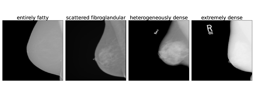
Breast density was assessed according to BI-RADS criteria into one of four categories: “entirely fatty”, “scattered fibroglandular”, “heterogeneously dense”, or “extremely dense” (Liberman & Menell, 2002).
To assess the performance of breast density rating task, we use linearly weighted Cohen’s Kappa score, , which is commonly used to measure agreement between two raters, where is the proportion of observed agreement and is the agreement due to random chance.
| (4) |
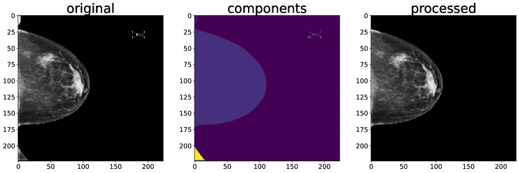
To avoid confounding bias from burnt in imaging artifacts, we process each image by taking connected components and keeping only the largest component (see Figure2). Additionally, this processing allows a simple quantification of breast volume to be the sum of non-zero pixels in the image. We scale pixel intensities to , apply random vertical flipping during training for data augmentation, and repeat across the channel dimension times in order to initialize each model with pretrained ImageNet weights (Deng et al., 2009). To account for class imbalance, we train weighted cross entropy using inversely proportional class ratios.
To form prediction sets, we use the nonconformality score described above from Sadinle et al. (2018), which provably has the smallest average set size. We calibrate the empirical quantile on the internal validation set. We train each model for 50 epochs and average over 5 training runs.
4.2 Characterizing Distribution Shift
Generalizability to natural distribution shifts is one of the major challenges of machine learning for clinical applications. Models trained and evaluated on one healthcare institution, geographic regions, or scanner type have difficulty maintaining performance to other institutions, regions, and scanners (Rajpurkar et al., 2020; Kaushal et al., 2020; Mårtensson et al., 2020). As CP assumes exchangability in distribution, we would expect most distribution shifts to violate this assumption.
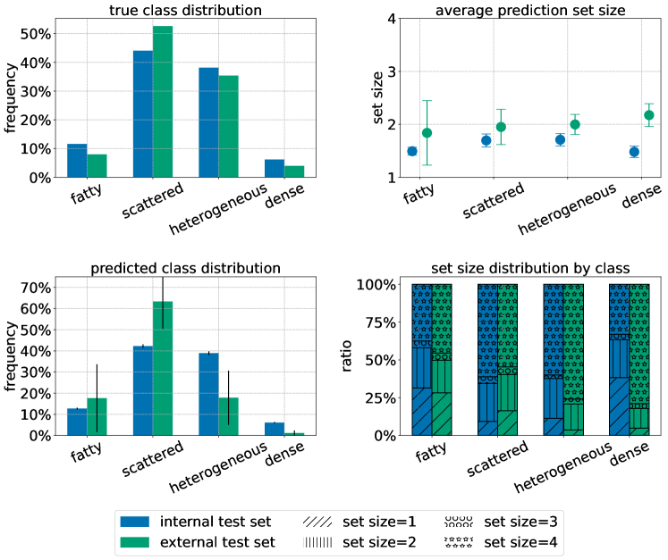
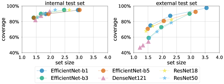
When comparing our internal test set (DMIST) to our external test set (MGH), we observe much higher coverage violations and larger set sizes on the external test set across a variety of architectures, shown in Figure 4.
We can further characterize the differences by examining the distribution of set sizes between internal and external prediction sets in Figure3. While the scattered and heterogeneous classes comprise the majority of the breast density ratings, they are more difficult and ambiguous to rate (even for human experts) compared to the fatty and extremely dense classes. This inherent uncertainty is reflected in the higher average set sizes for scattered and heterogeneous classes in the internal test set. However in the external test set, the dense class has the highest average set size and both the heterogeneous and dense classes have significantly more sets of size 4 (all classes contained) compared the prediction sets on the internal test set.
This analysis of conformal prediction set provides an alternative insight of distribution shift, that may be helpful in developing techniques and strategies for effective detection and mitigation of different forms of distribution shift.
4.3 Improving Prediction Quality
Deferring predictions and selective classification can help make deep learning systems more robust in many clinical scenarios where manual review would be preferable to wrong automated predictions (Madras et al., 2018; Geifman & El-Yaniv, 2017)
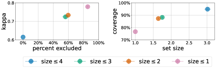
A second application of CP is the improvement of prediction quality by filtering out low-quality predictions as shown in Figure 5. For example, at , we can improve kappa from to by removing the highest uncertainty prediction of set size 4 at a cost of of total predictions and a coverage violation of from the ideal .
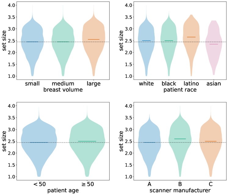
4.4 Quantifying Cohort Disparity
Fairness and algorithmic bias has been highlighted as a challenge in medical AI (Seyyed-Kalantari et al., 2020; Ahmad et al., 2020; Gichoya et al., 2022; Chen et al., 2021). We believe CP may be highly useful for quantifying disparity between different cohorts to measure efficiency and fairness between clinically relevant demographics.
Using CP calibrated per subgroup, as described in Lu et al. (2021), we examine the cohorts relevant to breast density: breast volume, patient age, and patient race, in addition to scanner type which has been show to affect performance of deep learning for breast density rating (Gupta et al., 2021). We observe several statistically significant () differences in Figure 6, specifically higher average set sizes for large breasts, Latino and/or Hispanic patients, patients older than 50 years of age, and scanner type B. Age and breast size are known to be inversely correlated with breast density; however the difference in uncertainty for scanner type B merit additional investigation.
5 Discussion
Currently, DL algorithms, despite their high promise, has lagged in translation into clinical practice. One key challenge is the inability to know the trustworthiness of a given model’s output. Specifically, whether a prediction for a patient is reliable or should be flagged for further review by experts. Given the high stakes nature of clinical medicine, gaining the trust of the user is crucial. CP methods present a solution by providing a distribution-free measure of uncertainty. Importantly, conformal outputs are highly intuitive to the users while providing theoretical guarantees. Up until this point, conformal predictions have mostly been used in simulated settings. In this paper, we utilize a large multi-institutional medical imaging dataset to show three realistic possible applications. Specifically, we show how CP can be used for three different tasks: distribution shift characterization, prediction quality improvement, and subgroup fairness analysis.
There are several possible limitations to this study. First, we only focus on one medical application within radiology. Further study can evaluate the performance of CP for other diseases and other medical specialities.
References
- Advani et al. (2021) Advani, S. M., Zhu, W., Demb, J., Sprague, B. L., Onega, T., Henderson, L. M., Buist, D. S. M., Zhang, D., Schousboe, J. T., Walter, L. C., Kerlikowske, K., Miglioretti, D. L., Braithwaite, D., and Consortium, B. C. S. Association of Breast Density With Breast Cancer Risk Among Women Aged 65 Years or Older by Age Group and Body Mass Index. JAMA Network Open, 4(8):e2122810–e2122810, 08 2021. ISSN 2574-3805. doi: 10.1001/jamanetworkopen.2021.22810. URL https://doi.org/10.1001/jamanetworkopen.2021.22810.
- Ahmad et al. (2020) Ahmad, M. A., Patel, A., Eckert, C., Kumar, V., and Teredesai, A. Fairness in machine learning for healthcare. In Proceedings of the 26th ACM SIGKDD International Conference on Knowledge Discovery & Data Mining, pp. 3529–3530, 2020.
- Angelopoulos et al. (2020) Angelopoulos, A. N., Bates, S., Malik, J., and Jordan, M. I. Uncertainty sets for image classifiers using conformal prediction. arXiv preprint arXiv:2009.14193, 2020.
- Arun et al. (2021) Arun, N., Gaw, N., Singh, P., Chang, K., Aggarwal, M., Chen, B., Hoebel, K., Gupta, S., Patel, J., Gidwani, M., Adebayo, J., Li, M. D., and Kalpathy-Cramer, J. Assessing the trustworthiness of saliency maps for localizing abnormalities in medical imaging. Radiology: Artificial Intelligence, 3(6):e200267, 2021. doi: 10.1148/ryai.2021200267. URL https://doi.org/10.1148/ryai.2021200267.
- Begoli et al. (2019) Begoli, E., Bhattacharya, T., and Kusnezov, D. The need for uncertainty quantification in machine-assisted medical decision making. Nature, 01 2019. doi: 10.1038/s42256-018-0004-1.
- Boyd et al. (2011) Boyd, N., Martin, L., Yaffe, M., and Minkin, S. Mammographic density and breast cancer risk: Current understanding and future prospects. Breast Cancer Res., 13:1–12, 01 2011.
- Chang et al. (2020) Chang, K., Beers, A., Brink, L., Patel, J., Singh, P., Arun, N., Hoebel, K., Gaw, N., Shah, M., Pisano, E., Tilkin, M., Coombs, L., Dreyer, K., Allen, B., Agarwal, S., and Kalpathy-Cramer, J. Multi-institutional assessment and crowdsourcing evaluation of deep learning for automated classification of breast density. Journal of the American College of Radiology, 17, 06 2020. doi: 10.1016/j.jacr.2020.05.015.
- Chen et al. (2021) Chen, R. J., Chen, T. Y., Lipkova, J., Wang, J. J., Williamson, D. F. K., Lu, M. Y., Sahai, S., and Mahmood, F. Algorithm fairness in ai for medicine and healthcare, 2021. URL https://arxiv.org/abs/2110.00603.
- Deng et al. (2009) Deng, J., Dong, W., Socher, R., Li, L.-J., Li, K., and Fei-Fei, L. Imagenet: A large-scale hierarchical image database. In 2009 IEEE conference on computer vision and pattern recognition, pp. 248–255. Ieee, 2009.
- Freer (2015) Freer, P. E. Mammographic breast density: Impact on breast cancer risk and implications for screening. RadioGraphics, 35(2):302–315, 2015. doi: 10.1148/rg.352140106. URL https://doi.org/10.1148/rg.352140106. PMID: 25763718.
- Geifman & El-Yaniv (2017) Geifman, Y. and El-Yaniv, R. Selective classification for deep neural networks. In Guyon, I., Luxburg, U. V., Bengio, S., Wallach, H., Fergus, R., Vishwanathan, S., and Garnett, R. (eds.), Advances in Neural Information Processing Systems, volume 30. Curran Associates, Inc., 2017. URL https://proceedings.neurips.cc/paper/2017/file/4a8423d5e91fda00bb7e46540e2b0cf1-Paper.pdf.
- Gichoya et al. (2022) Gichoya, J. W., Banerjee, I., Bhimireddy, A. R., Burns, J. L., Celi, L. A., Chen, L.-C., Correa, R., Dullerud, N., Ghassemi, M., Huang, S.-C., et al. Ai recognition of patient race in medical imaging: a modelling study. The Lancet Digital Health, 2022.
- Gupta et al. (2021) Gupta, S., Singh, P., Chang, K., Qu, L., Aggarwal, M., Arun, N. T., Vaswani, A., Raghavan, S., Agarwal, V., Gidwani, M., Hoebel, K., Patel, J. B., Lu, C., Bridge, C. P., Rubin, D. L., and Kalpathy-Cramer, J. Addressing catastrophic forgetting for medical domain expansion. CoRR, abs/2103.13511, 2021. URL https://arxiv.org/abs/2103.13511.
- Kaushal et al. (2020) Kaushal, A., Altman, R., and Langlotz, C. Geographic Distribution of US Cohorts Used to Train Deep Learning Algorithms. JAMA, 324(12):1212–1213, 09 2020. ISSN 0098-7484. doi: 10.1001/jama.2020.12067. URL https://doi.org/10.1001/jama.2020.12067.
- Kohli & Jha (2018) Kohli, A. and Jha, S. Why cad failed in mammography. Journal of the American College of Radiology, 15, 02 2018. doi: 10.1016/j.jacr.2017.12.029.
- Kompa et al. (2021a) Kompa, B., Snoek, J., and Beam, A. Second opinion needed: communicating uncertainty in medical machine learning. npj Digital Medicine, 4, 12 2021a. doi: 10.1038/s41746-020-00367-3.
- Kompa et al. (2021b) Kompa, B., Snoek, J., and Beam, A. Second opinion needed: communicating uncertainty in medical machine learning. npj Digital Medicine, 4, 12 2021b. doi: 10.1038/s41746-020-00367-3.
- Kyono et al. (2019) Kyono, T., Gilbert, F. J., and van der Schaar, M. Improving workflow efficiency for mammography using machine learning. Journal of the American College of Radiology : JACR, 2019.
- Larrazabal et al. (2020) Larrazabal, A. J., Nieto, N., Peterson, V., Milone, D. H., and Ferrante, E. Gender imbalance in medical imaging datasets produces biased classifiers for computer-aided diagnosis. Proceedings of the National Academy of Sciences, 117(23):12592–12594, 2020.
- Lehman et al. (2019) Lehman, C. D., Yala, A., Schuster, T., Dontchos, B., Bahl, M., Swanson, K., and Barzilay, R. Mammographic breast density assessment using deep learning: Clinical implementation. Radiology, 290(1):52–58, 2019. doi: 10.1148/radiol.2018180694. URL https://doi.org/10.1148/radiol.2018180694. PMID: 30325282.
- Lei et al. (2013) Lei, J., Robins, J., and Wasserman, L. Distribution-free prediction sets. Journal of the American Statistical Association, 108, 03 2013. doi: 10.1080/01621459.2012.751873.
- Liberman & Menell (2002) Liberman, L. and Menell, J. H. Breast imaging reporting and data system (bi-rads). Radiologic clinics of North America, 40:409–430, 3 2002. doi: 10.1016/s0033-8389(01)00017-3.
- Lu et al. (2021) Lu, C., Lemay, A., Chang, K., Hoebel, K., and Kalpathy-Cramer, J. Fair conformal predictors for applications in medical imaging, 2021. URL https://arxiv.org/abs/2109.04392.
- Madras et al. (2018) Madras, D., Pitassi, T., and Zemel, R. Predict responsibly: Improving fairness and accuracy by learning to defer. In Bengio, S., Wallach, H., Larochelle, H., Grauman, K., Cesa-Bianchi, N., and Garnett, R. (eds.), Advances in Neural Information Processing Systems, volume 31. Curran Associates, Inc., 2018. URL https://proceedings.neurips.cc/paper/2018/file/09d37c08f7b129e96277388757530c72-Paper.pdf.
- McCarthy et al. (2016) McCarthy, A. M., Keller, B. M., Pantalone, L. M., Hsieh, M.-K., Synnestvedt, M., Conant, E. F., Armstrong, K., and Kontos, D. Racial Differences in Quantitative Measures of Area and Volumetric Breast Density. JNCI: Journal of the National Cancer Institute, 108(10), 04 2016. ISSN 0027-8874. doi: 10.1093/jnci/djw104. URL https://doi.org/10.1093/jnci/djw104. djw104.
- McKinney et al. (2020) McKinney, S. M., Sieniek, M., Godbole, V., Godwin, J., Antropova, N., Ashrafian, H., Back, T., Chesus, M., Corrado, G., Darzi, A., Etemadi, M., Garcia-Vicente, F., Gilbert, F. J., Halling-Brown, M. D., Hassabis, D., Jansen, S., Karthikesalingam, A., Kelly, C. J., King, D., Ledsam, J. R., Melnick, D. S., Mostofi, H., Peng, L. H., Reicher, J. J., Romera-Paredes, B., Sidebottom, R., Suleyman, M., Tse, D., Young, K. C., Fauw, J. D., and Shetty, S. International evaluation of an ai system for breast cancer screening. Nature, 577:89–94, 2020.
- Mårtensson et al. (2020) Mårtensson, G., Ferreira, D., Granberg, T., Cavallin, L., Oppedal, K., Padovani, A., Rektorova, I., Bonanni, L., Pardini, M., Kramberger, M. G., Taylor, J.-P., Hort, J., Snædal, J., Kulisevsky, J., Blanc, F., Antonini, A., Mecocci, P., Vellas, B., Tsolaki, M., Kłoszewska, I., Soininen, H., Lovestone, S., Simmons, A., Aarsland, D., and Westman, E. The reliability of a deep learning model in clinical out-of-distribution mri data: A multicohort study. Medical Image Analysis, 66:101714, 2020. ISSN 1361-8415. doi: https://doi.org/10.1016/j.media.2020.101714. URL https://www.sciencedirect.com/science/article/pii/S1361841520300785.
- Nishikawa & Bae (2018) Nishikawa, R. M. and Bae, K. T. Importance of better human-computer interaction in the era of deep learning: Mammography computer-aided diagnosis as a use case. Journal of the American College of Radiology : JACR, 15 1 Pt A:49–52, 2018.
- Papadopoulos et al. (2007) Papadopoulos, H., Vovk, V., and Gammerman, A. Conformal prediction with neural networks. In 19th IEEE International Conference on Tools with Artificial Intelligence(ICTAI 2007), volume 2, pp. 388–395, 2007. doi: 10.1109/ICTAI.2007.47.
- Pisano et al. (2005) Pisano, E. D., Gatsonis, C., Hendrick, E., Yaffe, M., Baum, J. K., Acharyya, S., Conant, E. F., Fajardo, L. L., Bassett, L., D’Orsi, C., Jong, R., and Rebner, M. Diagnostic Performance of Digital versus Film Mammography for Breast-Cancer Screening. New England Journal of Medicine, 353(17):1773–1783, 10 2005. ISSN 0028-4793. doi: 10.1056/NEJMoa052911. URL http://www.nejm.org/doi/abs/10.1056/NEJMoa052911.
- Rajpurkar et al. (2020) Rajpurkar, P., Joshi, A., Pareek, A., Chen, P., Kiani, A., Irvin, J., Ng, A. Y., and Lungren, M. P. Chexpedition: Investigating generalization challenges for translation of chest x-ray algorithms to the clinical setting, 2020. URL https://arxiv.org/abs/2002.11379.
- Romano et al. (2020) Romano, Y., Sesia, M., and Candès, E. J. Classification with valid and adaptive coverage. In Proceedings of the 34th International Conference on Neural Information Processing Systems, NIPS’20, Red Hook, NY, USA, 2020. Curran Associates Inc. ISBN 9781713829546.
- Sadinle et al. (2018) Sadinle, M., Lei, J., and Wasserman, L. Least ambiguous set-valued classifiers with bounded error levels. Journal of the American Statistical Association, 114(525):223–234, jun 2018. doi: 10.1080/01621459.2017.1395341. URL https://doi.org/10.1080%2F01621459.2017.1395341.
- Seyyed-Kalantari et al. (2020) Seyyed-Kalantari, L., Liu, G., McDermott, M. B. A., and Ghassemi, M. Chexclusion: Fairness gaps in deep chest x-ray classifiers. CoRR, abs/2003.00827, 2020. URL https://arxiv.org/abs/2003.00827.
- Seyyed-Kalantari et al. (2021) Seyyed-Kalantari, L., Zhang, H., McDermott, M., Chen, I. Y., and Ghassemi, M. Underdiagnosis bias of artificial intelligence algorithms applied to chest radiographs in under-served patient populations. Nature medicine, 27(12):2176–2182, 2021.
- Shashikumar et al. (2021) Shashikumar, S., Wardi, G., Malhotra, A., and Nemati, S. Artificial intelligence sepsis prediction algorithm learns to say “i don’t know”. npj Digital Medicine, 4, 12 2021. doi: 10.1038/s41746-021-00504-6.
- Shen et al. (2019) Shen, L., Margolies, L., Rothstein, J., Fluder, E., McBride, R., and Sieh, W. Deep learning to improve breast cancer detection on screening mammography. Scientific Reports, 9:1–12, 08 2019. doi: 10.1038/s41598-019-48995-4.
- Siu & Force (2016) Siu, A. L. and Force, U. P. S. T. Screening for breast cancer: Us preventive services task force recommendation statement. Annals of internal medicine, 164(4):279–296, 2016.
- Sprague et al. (2016) Sprague, B. L., Conant, E. F., Onega, T., Garcia, M. P., Beaber, E. F., Herschorn, S. D., Lehman, C. D., Tosteson, A. N., Lacson, R., Schnall, M. D., et al. Variation in mammographic breast density assessments among radiologists in clinical practice: a multicenter observational study. Annals of internal medicine, 165(7):457–464, 2016.
- Topol (2019) Topol, E. J. High-performance medicine: the convergence of human and artificial intelligence. Nature medicine, 25(1):44–56, 2019.
- Vazquez & Facelli (2022) Vazquez, J. and Facelli, J. Conformal prediction in clinical medical sciences. Journal of Healthcare Informatics Research, 01 2022. doi: 10.1007/s41666-021-00113-8.
- Vovk et al. (2005) Vovk, V., Gammerman, A., and Shafer, G. Algorithmic Learning in a Random World. Springer-Verlag, Berlin, Heidelberg, 2005. ISBN 0387001522.
- Wang et al. (2016) Wang, J., Yang, X., Cai, H., Tan, W. Y., Jin, C., and Li, L. Discrimination of breast cancer with microcalcifications on mammography by deep learning. Scientific Reports, 6, 2016.
- Wu et al. (2018) Wu, N., Geras, K. J., Shen, Y., Su, J., Kim, S. G., Kim, E., Wolfson, S., Moy, L., and Cho, K. Breast density classification with deep convolutional neural networks. In 2018 IEEE International Conference on Acoustics, Speech and Signal Processing (ICASSP), pp. 6682–6686, 2018. doi: 10.1109/ICASSP.2018.8462671.
- Yala et al. (2019) Yala, A., Lehman, C., Schuster, T., Portnoi, T., and Barzilay, R. A deep learning mammography-based model for improved breast cancer risk prediction. Radiology, 292(1):60–66, 2019. doi: 10.1148/radiol.2019182716. URL https://doi.org/10.1148/radiol.2019182716. PMID: 31063083.
- Yala et al. (2021) Yala, A., Mikhael, P. G., Strand, F., Lin, G., Smith, K., Wan, Y.-L., Lamb, L., Hughes, K., Lehman, C., and Barzilay, R. Toward robust mammography-based models for breast cancer risk. Science Translational Medicine, 13(578):eaba4373, 2021.
- Łukasiewicz et al. (2021) Łukasiewicz, S., Czeczelewski, M., Forma, A., Baj, J., Sitarz, R., and Stanisławek, A. Breast cancer—epidemiology, risk factors, classification, prognostic markers, and current treatment strategies—an updated review. Cancers, 13, 2021.