Automated GI tract segmentation using deep learning
Abstract
The job of Radiation oncologists is to deliver x-ray beams pointed towards the tumor and at the same time avoid the stomach and intestines. With newer technologies such as MR-Linacs, oncologists can visualize the position of the tumor and allow for precise dose according to tumor cell presence which can vary from day to day. The current job of outlining the position of the stomach and intestines to adjust the X-ray beam’s direction for the dose delivery to the tumor while avoiding the organs. This is a time-consuming and labor-intensive process that can easily prolong treatments from 15 minutes to an hour a day unless deep learning methods can automate the segmentation process.
This paper studies semantic segmentation on the GI Tract scans using deep learning to make this process faster and allow more patients to get effective treatment.
Keywords:
GI Tract segmentation, instance segmentation, U-Net1 Introduction
In 2019, an estimated 5 million people were diagnosed with cancer of the gastro-intestinal tract worldwide [1].
radiation therapy (RT) has the potential to improve the rates of cure of 3.5 million people and provide palliative relief for an additional 3.5 million people [2].
The Radiation oncologists deliver x-ray beams pointed toward the tumor and at the same time avoid the stomach and intestines. With MR-Linacs (magnetic resonance imaging and linear accelerator systems) [3], oncologists can visualize the position of the tumor and monitor for precise dose according to tumor cell presence which can vary from day to day. The current job is to manually outline the position of the stomach and intestines for adjustments to the X-ray beam’s direction to increase the dose delivery to the tumor while avoiding the organs. This is a time-consuming and labor-intensive process that can easily prolong treatments from 15 minutes to an hour a day unless deep learning methods can be applied and could help automate the segmentation process.
Deep learning can help by automating the segmentation process to reduce manual labor and help more patients to get effective treatment. In this paper, we study the segmentation task on GI Tract scans using different backbones for the UNet architecture. Our best model yielded an IoU score of 85.3% using the Efficientnet backbone.
2 Related work
Many researchers have used different deep learning architectures in medical imaging to create both semantic and instance segmentation and achieved excellent results. For instance, in medical images, deep learning approaches have been applied for brain tumor segmentation [4], colon polyp segmentation [5] and pancreas segmentation [6]. In 2016, V-Net [7] network architecture was proposed with the aim of segmenting volumetric data such as 3D MRI scans. This approach was similar to U-Net network architecture which was proposed in 2015 [8] for biomedical image segmentation. U-Net relied on the strong use of data augmentation due to the fact that data in biomedical applications is not present in abundance. The effectiveness of U-Net architecture has led to the development of different architectures like U-Net++ [9] and TMD-Net [10] which have reported significant performance gains over U-Net in different biomedical segmentation tasks.
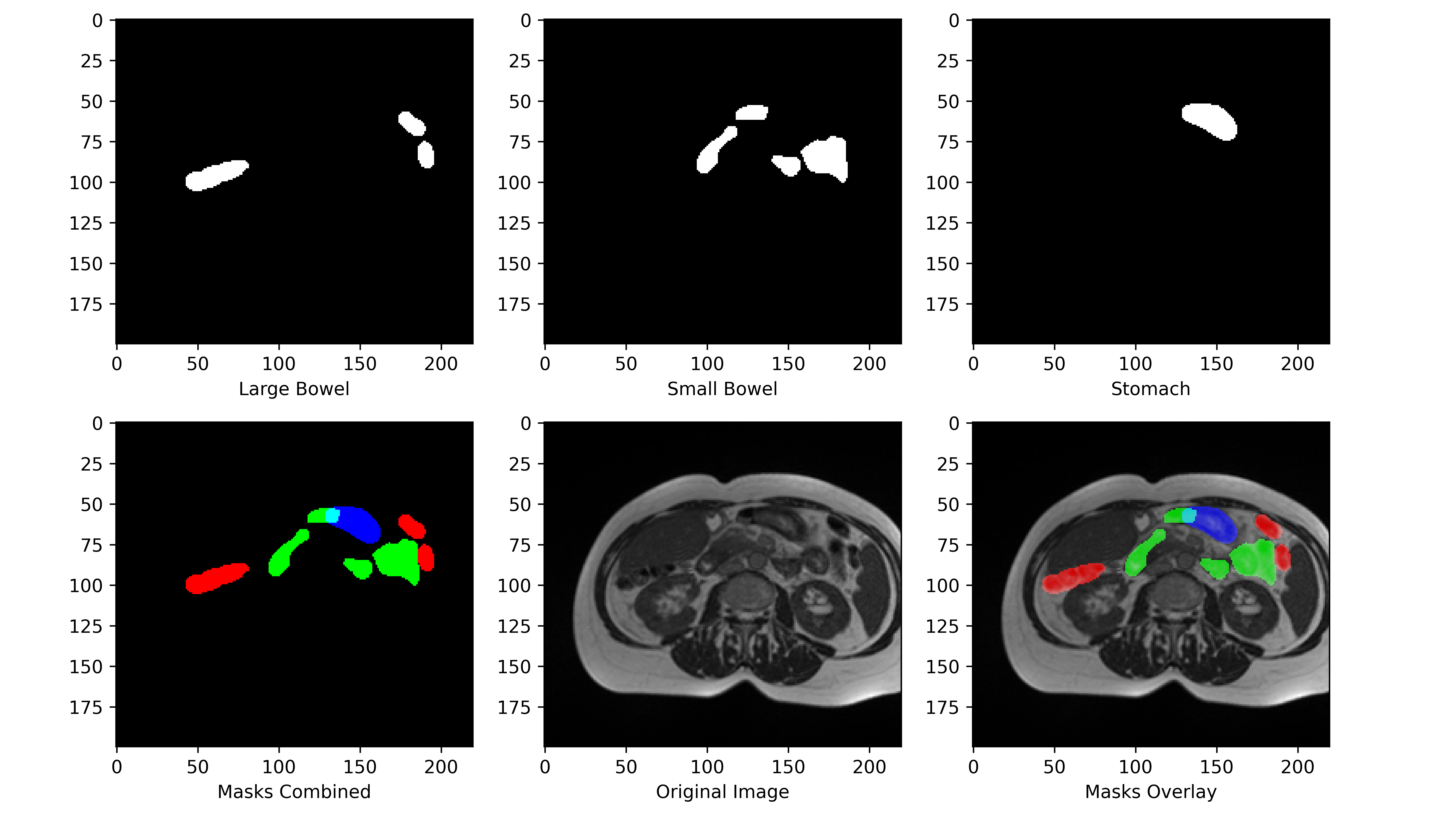
The aim of this study is to use the existing knowledge and apply it to the problem of GI tract segmentation. There is a lack of research done on the segmentation of GI tract organs. This study aims to provide a new baseline using different backbones of U-Net architectures in order to segment organs such as stomach, large Bowel and small Bowel (Fig. 1). The task of this research is to find the best architecture to create masks for a scan that highlights the Large Bowel, Small Bowel, and Stomach. Generally, the process of applying deep learning to such tasks starts with collecting the data and effectively pre-prossessing the data to be fed into the model to create useful predictions. The next section discusses the dataset used in this research.
3 Dataset and Preprocessing
The dataset used in this research is available on the Kaggle platform for free. This dataset uses anonymized scans of patients provided by the UWMadison Carbone Cancer Center. The recent competition ”UW-Madison GI Tract Image Segmentation” hosted on Kaggle challenges researchers to apply Deep learning models to perform semantic segmentation of the GI tract scans. These scans are taken over a period of six days and the masks for training are provided as Run-length encoding to save memory and bandwidth. The input images are the float16 PNG slices of the scans.
The problem with these scans is the inconsistent widths and heights (Fig. 3). Few images are square and while others are rectangular. To match the input shape of the model (288x288), zero padding is done where the length is less than 288, and pixel trimming was done where the length is greater than 288. The cropping was done in such a way that the black/empty pixels were trimmed first. Same pre-processing was done for the masks as well.
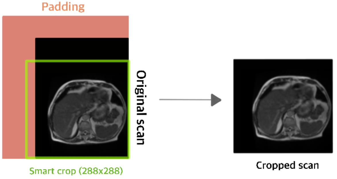
Resizing was not considered as this can create abnormal artifacts which can hinder the correct mask of the organ (this is crucial as the organs can be very close to each other).
The same padding step will be done for the test image lower than 288x288 and then the sliding window patches of 288x288 will be generated for each image for images larger in size to make patch-wise predictions. The predicted patches will be stitched to get the image mask.
The input images were converted into tensors before being fed to the network and each image was also normalized to help the model in learning the patterns [11].

4 Structure of Model
A convolutional neural network or CNN consists of a stack of three main neural layers: convolutional layer, pooling layer, and fully connected layer [12][13].
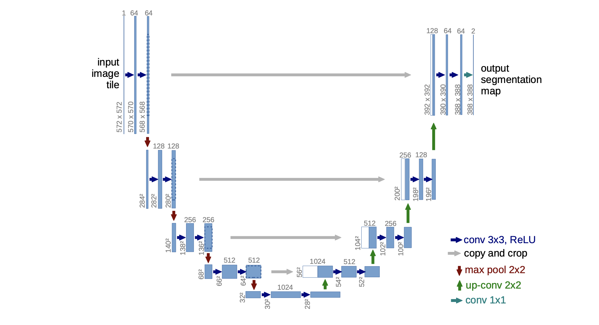
The U-NET is a deep learning convolutional network that create segmentation masks from an input image. The U-Net consist of an encoder part that converts -channel image to a dense encoding and a decoder part that converts the dense encoding back to the -channel output [8].
In this paper, we used different encoders (ResNet[14], EfficientNet [15], VGG16 [16], MobileNet [17]) for U-Net having 1 input channel and 3 output channel and compare the results. Each of the 3 output channels corresponds to a different segmentation mask. Each channel in the output tensor is a mask for one of the three classes (Large Bowl, Small Bowel, and Stomach).
The encoder part of the U-Net classifies each pixel in the image into a particular class. The information is compressed using a down-sampling (max-pooling) approach and the resulting segmentation masks are reconstructed by using up-sampling (Up convolution/Convolution transpose) instead of pooling layers to improve the resolution of the output. A successive convolution layer in the decoder part can then learn to assemble a more precise output based on this information [8].
5 Performance Metrics
There are many performance metrics that can be applied to segmentation models to measure their prediction performance. The performance metric used in this research for measuring accuracy is ”Intersection over Union” and the loss functions are derived from the combinations of ”Intersection over Union Loss [18]”, ”Binary Cross Entropy Loss”, ”Tversky Loss [19]”.
Performance Metric (For single sample) :
| (1) |
Loss functions used :
| (2) | |||
| (3) | |||
| (4) |
6 Training
The input images along with their segmentation masks are used to train the network with the Adam optimizer [20] implementation of PyTorch with initial learning rate of . Consine Annealing [21] was used to decay the learning rate as after 30 epochs the model loss was not decreasing and was fluctuating within the range. To minimize the overhead and make maximum use of the GPU memory, we used 288x288 image for input with borders cropped as the borders held no information for training. For similar reasons the batch size of 32 was used and the models were trained for 80 epochs. The dataset was divided into training data and validation data using 80/20 split. The models were trained using training data and the performance was evaluated using validation data.
The output of the model was passed through a sigmoid unit and the output was clamped to 0 for values less than 0.5 and 1 otherwise.
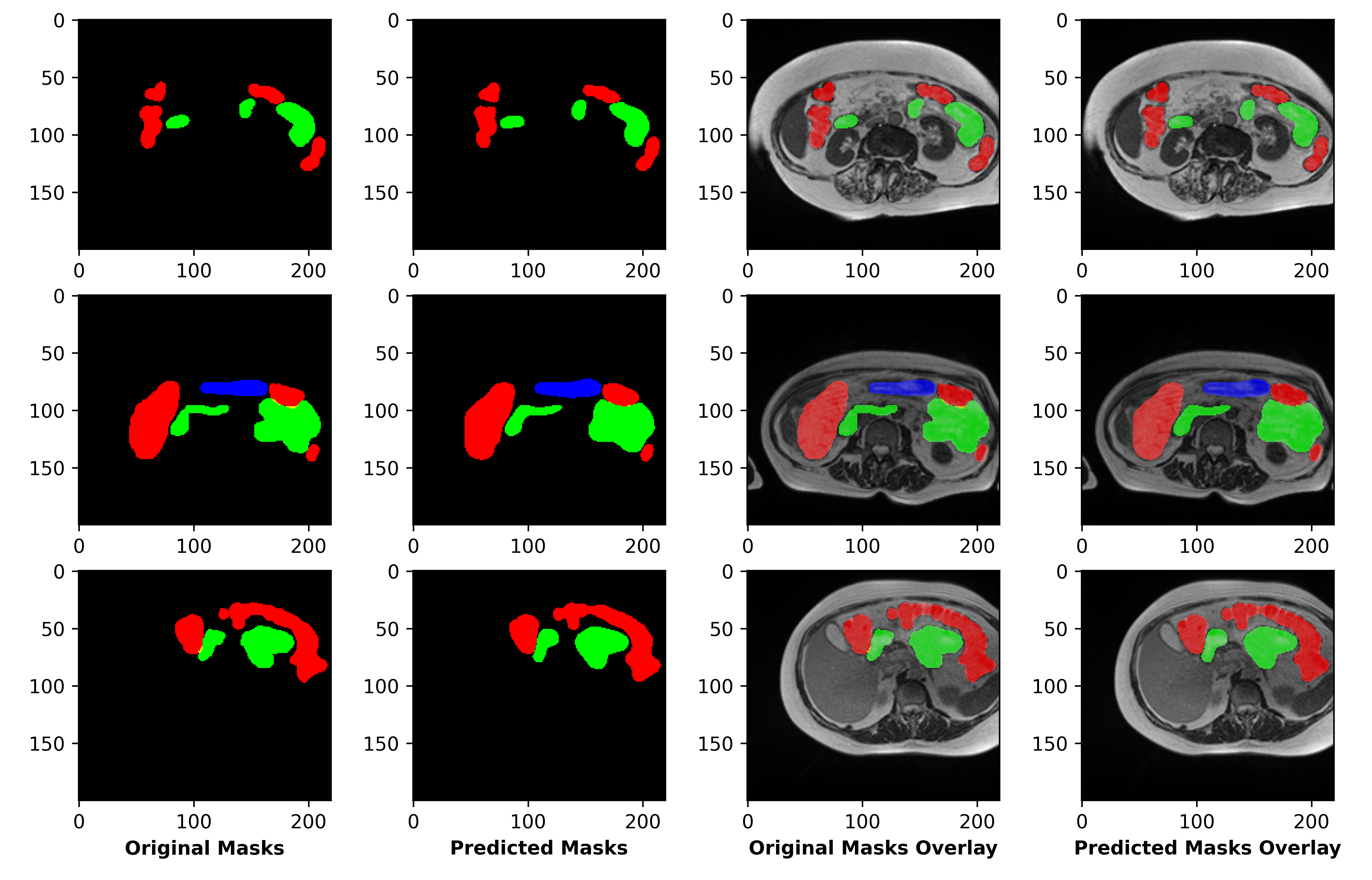
7 Experimentation Results
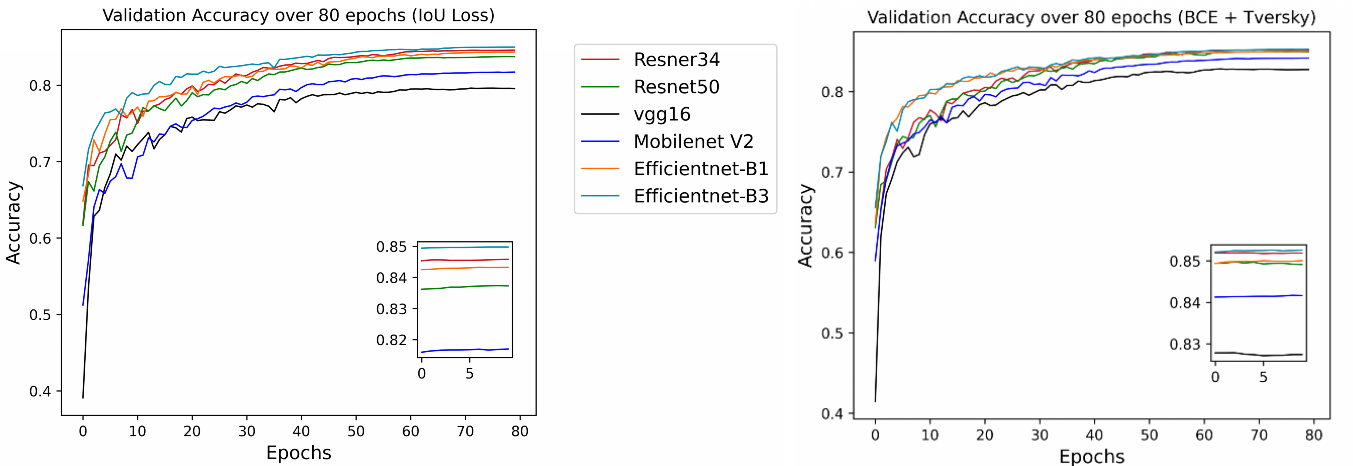
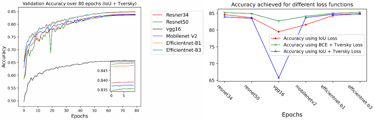
The results of the experimentations are very clear from the table 1. The Efficientnet encoders outperformed all the other other encoders. The models were trained for 80 epochs. No data augmentation other than Horizontal flipping and vertical flipping were used. Different augmentation techniques like ElasticTransform and ShiftScaleRotate were tried but they had no visible effect on the performance of the model. Although VGG16 was the least performing encoder among other encoders, yet it managed to achieve over 80% accuracy using (Tversky + BCE) Loss function. Upon experimentations it is clear that the best choice of loss function for this task is (BCE + Tversky) Loss (Fig. 7).
| Validation Accuracy over after 80 Epochs | |||
|---|---|---|---|
| Encoder | IoU Loss | BCE + Tversky Loss | IoU + Tversky Loss |
| Efficientnet-B3 | 84.9% | 85.3% | 84.8% |
| Efficientnet-B1 | 84.3% | 85% | 84.5% |
| Resnet34 | 84.6% | 85.2% | 83.9% |
| Resnet50 | 83.7% | 84.9% | 83.5% |
| Mobilenet V2 | 81.6% | 84.1% | 83.7% |
| VGG16 | 79.5% | 82.7% | 65.7% |
8 Conclusion
This research used classic U-Net architecture with different encoder. There are more advanced algorithms available that have achieved excellent results in different classification tasks. These algorithms can be applied as an encoder to create a new U-Net flavor and achieve better results. Though the loss functions used in this research are IoU Loss, (BCE + Tversky) Loss, (IoU + Tversky) Loss, different loss functions can be applied to experiment with the results. A few of them are Binary cross entropy Loss, Dice Loss, and Focal Loss (For imbalanced masks) and combination of these to create new loss functions. This research can serve as a baseline for new research in the same criteria. Also, scans for the same patients on the same day can be stacked to make a high channel input and predict the mask based on the channel. channels can serve as past history for the prediction.
9 Acknowledgements
This study was made possible with the anonymized data provided by The UW-Madison Carbone Cancer Center on Kaggle platform. This research was made possible by the repository ”Segmentation models.PyTorch” which provides a high level API for different encoder implementation for UNet [22].
10 Data availability
Data used in this research is available on kaggle platform under the competition ”UW-Madison GI Tract Image Segmentation” and can be downloaded from https://www.kaggle.com/competitions/uw-madison-gi-tract-image-segmentation.
11 Statements & Declarations
11.1 Funding
The author declare that no funds, grants, or other support were received during the preparation of this manuscript.
11.2 Competing Interests
The author has no relevant financial or non-financial interests to disclose.
11.3 Author Contributions
Material preparation, data collection and analysis was performed by Manhar Sharma. The author read and approved the final manuscript.
References
- [1] Prashanth Rawla and Adam Barsouk “Epidemiology of gastric cancer: global trends, risk factors and prevention” In Gastroenterology Review/Przeglad Gastroenterologiczny 14.1 Termedia, 2019, pp. 26–38
- [2] David A Jaffray and Mary K Gospodarowicz “Radiation therapy for cancer” In Cancer: disease control priorities 3 International Bank for ReconstructionDevelopmentWorld Bank …, 2015, pp. 239–248
- [3] Jan JW Lagendijk, Bas W Raaymakers and Marco Van Vulpen “The magnetic resonance imaging–linac system” In Seminars in radiation oncology 24.3, 2014, pp. 207–209 Elsevier
- [4] Mohammad Havaei et al. “Brain tumor segmentation with deep neural networks” In Medical image analysis 35 Elsevier, 2017, pp. 18–31
- [5] Nguyen Thanh Duc et al. “ColonFormer: An Efficient Transformer based Method for Colon Polyp Segmentation” In arXiv preprint arXiv:2205.08473, 2022
- [6] Ozan Oktay et al. “Attention u-net: Learning where to look for the pancreas” In arXiv preprint arXiv:1804.03999, 2018
- [7] Fausto Milletari, Nassir Navab and Seyed-Ahmad Ahmadi “V-net: Fully convolutional neural networks for volumetric medical image segmentation” In 2016 fourth international conference on 3D vision (3DV), 2016, pp. 565–571 IEEE
- [8] Olaf Ronneberger, Philipp Fischer and Thomas Brox “U-net: Convolutional networks for biomedical image segmentation” In International Conference on Medical image computing and computer-assisted intervention, 2015, pp. 234–241 Springer
- [9] Zongwei Zhou, Md Mahfuzur Rahman Siddiquee, Nima Tajbakhsh and Jianming Liang “Unet++: A nested u-net architecture for medical image segmentation” In Deep learning in medical image analysis and multimodal learning for clinical decision support Springer, 2018, pp. 3–11
- [10] Song-Toan Tran et al. “TMD-Unet: Triple-Unet with multi-scale input features and dense skip connection for medical image segmentation” In Healthcare 9.1, 2021, pp. 54 MDPI
- [11] Lei Huang et al. “Normalization techniques in training dnns: Methodology, analysis and application” In arXiv preprint arXiv:2009.12836, 2020
- [12] Priyanka Malhotra, Sheifali Gupta and Deepika Koundal “Computer aided diagnosis of pneumonia from chest radiographs” In Journal of Computational and Theoretical Nanoscience 16.10 American Scientific Publishers, 2019, pp. 4202–4213
- [13] Shaveta Dargan, Munish Kumar, Maruthi Rohit Ayyagari and Gulshan Kumar “A survey of deep learning and its applications: a new paradigm to machine learning” In Archives of Computational Methods in Engineering 27.4 Springer, 2020, pp. 1071–1092
- [14] Kaiming He, Xiangyu Zhang, Shaoqing Ren and Jian Sun “Deep residual learning for image recognition” In Proceedings of the IEEE conference on computer vision and pattern recognition, 2016, pp. 770–778
- [15] Mingxing Tan and Quoc Le “Efficientnet: Rethinking model scaling for convolutional neural networks” In International conference on machine learning, 2019, pp. 6105–6114 PMLR
- [16] Anindya Apriliyanti Pravitasari et al. “UNet-VGG16 with transfer learning for MRI-based brain tumor segmentation” In TELKOMNIKA (Telecommunication Computing Electronics and Control) 18.3, 2020, pp. 1310–1318
- [17] Andrew G. Howard et al. “MobileNets: Efficient Convolutional Neural Networks for Mobile Vision Applications” arXiv, 2017 DOI: 10.48550/ARXIV.1704.04861
- [18] Dingfu Zhou et al. “IoU Loss for 2D/3D Object Detection” arXiv, 2019 DOI: 10.48550/ARXIV.1908.03851
- [19] Seyed Sadegh Mohseni Salehi, Deniz Erdogmus and Ali Gholipour “Tversky loss function for image segmentation using 3D fully convolutional deep networks” arXiv, 2017 DOI: 10.48550/ARXIV.1706.05721
- [20] Diederik P. Kingma and Jimmy Ba “Adam: A Method for Stochastic Optimization”, 2017 arXiv:1412.6980 [cs.LG]
- [21] Ilya Loshchilov and Frank Hutter “SGDR: Stochastic Gradient Descent with Warm Restarts”, 2017 arXiv:1608.03983 [cs.LG]
- [22] Pavel Yakubovskiy “Segmentation Models Pytorch” In GitHub repository GitHub, https://github.com/qubvel/segmentation_models.pytorch, 2020