Degeneracy in epilepsy: Multiple Routes to Hyperexcitable Brain Circuits and their Repair
Tristan Manfred Stöbera, Danylo Batulina, Jochen Triescha,1, Rishikesh Narayananb,1, Peter Jedlickaa,c,d,1,*
a Frankfurt Institute for Advanced Studies, 60438 Frankfurt am Main, Germany
b Cellular Neurophysiology Laboratory, Molecular Biophysics Unit, Indian Institute of Science, Bangalore 560012, India
c ICAR3R – Interdisciplinary Centre for 3Rs in Animal Research, Faculty of Medicine, Justus Liebig University Giessen, 35390 Giessen, Germany
d Institute of Clinical Neuroanatomy, Neuroscience Center, Goethe University, 60590 Frankfurt am Main, Germany
1 Joint senior authors
*peter.jedlicka@informatik.med.uni-giessen.de
Keywords
ion channel degeneracy, hippocampal sclerosis, temporal lobe epilepsy, multiscale modeling, population modeling, epileptor, cell loss, multitarget therapy, personalized medicine, neurogenesis, mossy fiber sprouting, tonic inhibition, evolutionary trade-offs, Pareto optimality
1 Preface
Due to its complex and multifaceted nature, developing effective treatments against epilepsy is still a major challenge. To deal with this complexity we introduce the concept of degeneracy to the field of epilepsy research: the ability of disparate elements to cause an analogous (mal)function. Here, we review examples of epilepsy-related degeneracy at multiple levels of brain organization, ranging from the cellular to the network and systems level. Based on these insights, we outline new approaches to disentangle the complex web of interactions underlying epilepsy and to design personalized multitarget therapies.
2 Abstract
Developing effective therapies against epilepsy remains a challenge. The complex and multifaceted nature of this disease still fuels controversies about its origin. In this perspective article, we argue that conflicting hypotheses can be reconciled by taking into account the degeneracy of the brain, which manifests in multiple routes leading to similar function or dysfunction. We exemplify degeneracy at three different levels, ranging from the cellular to the network and systems level. First, at the cellular level, we describe the relevance of ion channel degeneracy for epilepsy and discuss its interplay with dendritic morphology. Second, at the network level, we provide examples for the degeneracy of synaptic and intrinsic neuronal properties that supports the robustness of neuronal networks but also leads to diverse responses to ictogenic and epileptogenic perturbations. Third, at the system level, we provide examples for degeneracy in the intricate interactions between the immune and nervous system. Finally, we show that computational approaches including multiscale and so called population neural circuit models help disentangle the complex web of physiological and pathological adaptations. Such models may contribute to identifying the best personalized multitarget strategies for directing the system towards a physiological state.
3 Introduction
Degeneracy, “the ability of elements that are structurally different to perform the same function or yield the same output” (Edelman and Gally, 2001), is a general principle, present in most complex adaptive biological systems (Tononi et al., 1999; Edelman and Gally, 2001). Degeneracy should be distinguished from redundancy, which results from multiple identical elements performing the same function (Edelman and Gally, 2001; Mason et al., 2015; Man et al., 2016; Kamaleddin, 2022; but see also Mizusaki and O’Donnell, 2021). In contrast to redundant components, degenerate components may generate dissimilar outputs in different contexts (Tononi et al., 1999; Morozova et al., 2022). Therefore, although degeneracy is sometimes called also ’partial redundancy’ (Whitacre and Bender, 2010), we follow the terminology of Edelman and Gally (2001) and distinguish degeneracy from redundancy.
The ubiquity of degeneracy is rooted in the advantages it entails for organisms’ evolvability (Whitacre, 2010; Whitacre and Bender, 2010) and robustness (Wagner, 2005; Mellen, 2008; Whitacre, 2012). Degeneracy allows organisms to satisfy the two seemingly contradictory goals of preserving already evolved functions and concurrently searching for and evolving new functions (Whitacre, 2010). In this way degeneracy is linked to both robustness and innovation in evolution (Edelman and Gally, 2001; Whitacre, 2010).
It is becoming increasingly clear that also the brain exhibits degeneracy (Edelman and Gally, 2001; Mason et al., 2015; Cropper et al., 2016; Ratté and Prescott, 2016; Seifert et al., 2016; Marder et al., 2017), with similar physiological states arising from a multitude of different subcellular, cellular and synaptic mechanisms (Prinz et al., 2004; Marder and Taylor, 2011; Rathour and Narayanan, 2019; Goaillard and Marder, 2021; Kamaleddin, 2022, for recent reviews see). However, not only similar physiological but also similar pathological brain states may arise from structurally disparate mechanisms (Neymotin et al., 2016; Ratté and Prescott, 2016; O’Leary, 2018; Kamaleddin, 2022). Therefore, the existence of degeneracy in the brain has important implications for understanding and treating complex brain disorders such as epilepsy.
Despite considerable progress in epilepsy research (reviewed by Bui et al., 2015; Symonds et al., 2017; Demarest and Brooks-Kayal, 2018), its complex multicausal and variable nature (Duncan et al., 2006; Scharfman, 2007; Lytton, 2008) has made it difficult to disentangle the underlying mechanisms. This has hindered the development of effective therapies. Apparently contradictory observations still fuel deep controversies about the origin of epilepsy. In this review, we argue that competing hypotheses can be reconciled by taking into account the concept of degeneracy. Moreover, we emphasize that degeneracy can help explain why multiple minor changes can sometimes induce pathological phenotypes, while at other times their effects cancel, preserving healthy/physiological circuit states.
Our aim is to review examples for epilepsy-related degeneracy at three different levels of brain organization: the cellular, network and systems level. We propose that epilepsy is a group of multiscale disorders with a potential involvement of degenerative mechanisms at each level (see Figure 1).
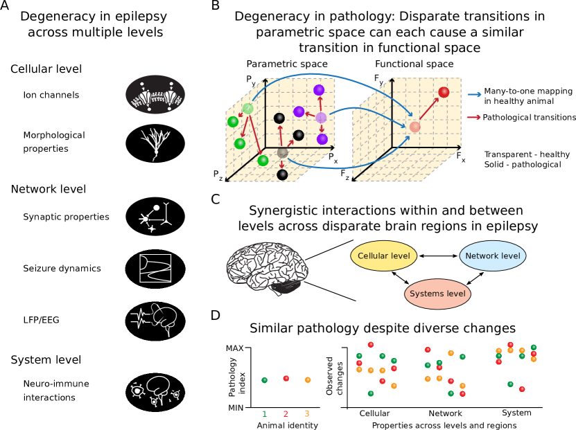
(A) In this article we exemplify degeneracy in epilepsy across the cellular, network and system level. Levels and sub-components are highlighted. (B) The concept of degeneracy, e.g. different changes leading to a similar outcome, can be visualized by a many-to-one relationship between the parametric and the functional space. Diverse changes in the parametric space relating to different properties brain regions can lead to a similar pathological outcome in the functional space. Transparent spheres represent properties in a healthy brain. Red arrows reflect transitions to pathological states, represented by solid spheres. (C) Epilepsy arises due to pathological changes in the cellular, network and system level across multiple brain regions. (D) Thus, similar pathology indices in multiple animals can be caused by degenerate modifications of multiple properties across various levels.
4 Degeneracy in epilepsy at the cellular level
4.1 Intrinsic properties and ion channel degeneracy
It is now well established that indistinguishable single-cell (and network) activity can result from a variety of parameter combinations of different ion channels (Prinz et al., 2004; Marder and Goaillard, 2006; Calabrese, 2018). This phenomenon has been termed ion channel degeneracy (Drion et al., 2015; Mishra and Narayanan, 2021a; O’Leary, 2018; Goaillard and Marder, 2021, for recent reviews see). Ion channel degeneracy means that “distinct channel types overlap in their biophysical properties and can thus contribute collectively to specific physiological phenotypes”(O’Leary, 2018).
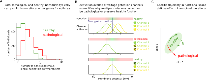
There have been extensive studies of epilepsy-relevant changes in ion channels (Lerche et al., 2013; Wolfart and Laker, 2015; Oyrer et al., 2018). However, their role in the context of ion channel degeneracy and epilepsy has not yet been studied extensively. Neuronal hyperexcitability and associated epilepsy often arise as a result of combinatorial effects of multiple ion channel mutations. This has been demonstrated by an important sequencing study that examined over 200 ion channel genes in epilepsy patients and healthy controls (Klassen et al., 2011). The study found a highly complex pattern of gene changes in single individuals. Even multiple nonsynonymous mutations in known monogenic risk genes for epilepsy were frequently identified in healthy individuals (Klassen et al., 2011, Figure 2A). This suggests that, due to ion channel degeneracy, different channel types are able to partially compensate each other’s defects (O’Leary, 2018). When a mutation affects a function of a given ion channel, other channels may rescue normal excitability (O’Leary, 2018; Figure 2B). This applies both to loss-of-function as well as gain-of-function changes in ion channels. Accordingly, individuals with epilepsy typically have more than one mutation in human epilepsy genes (Klassen et al., 2011).
The results of Klassen et al. (2011) indicate that epilepsies are more often caused by an accumulation of mutations in multiple ion channels than by mutation in one particular ion channel (Kaplan et al., 2016). This is supported also by computational modeling showing that combinations of small changes in the voltage activation of two or three ion channels can lead to network hyperexcitability (Thomas et al., 2009). Ion channel degeneracy may also explain why in some individuals a lower number of mutations (hits) leads to a pathological phenotype (epilepsy) while in other individuals a higher number of hits does not make them sick (O’Leary, 2018). The net effect of combined mutations will depend, first, on the initial (pre-mutation) distance of a given individual (i.e. a given set of ion channel parameters) from the pathological (hyperexcitable) region of the possible functional space. Second, the net effect will depend on the specific trajectory in functional space determined by single or multiple mutations (Figure 2C; see also Figure 4). Multiple mutations in ion channels can sometimes cancel each other out, paradoxically preserving physiological firing behavior of affected neurons (O’Leary, 2018). In line with this, the degeneracy between ion channel types leads to high-dimensional parameter spaces, which support robustness (O’Leary, 2018; Onasch and Gjorgjieva, 2020; Schneider et al., 2021; Goaillard and Marder, 2021).
The effects of multiple mutations in ion channels appear even more complex if one considers that excitability of neurons depends not only on axonal or somatic but also on dendritic ion channels. The interplay of dendritic and somatic channels is known to affect dendritic and somatic spikes (Larkum et al., 2022; Manita et al., 2017; Johnston and Narayanan, 2008; Sjostrom et al., 2008; Basak and Narayanan, 2018). There is a first, epilepsy-related line of indirect evidence for the role of ion-channel degeneracy in shaping somato-dendritic properties. Accordingly, with specific reference to acquired channelopathies and epilepsy, a growing body of evidence implicates several dendritic ion channels in enhanced neuronal excitability (Oyrer et al., 2018). Several dendritic ion channels, including the A-type potassium and hyperpolarization-activated cyclic nucleotide gated (HCN) channels, have been implicated in mediating such hyperexcitability across different neuronal subtypes. These changes are dependent on cell-type and location, for example in the hippocampus with differences appearing within the same cell type along the dorso-ventral axis (Johnston et al., 2000; Su et al., 2002; Bernard et al., 2004; Shah et al., 2004; Jung et al., 2007; Shin et al., 2008; Jung et al., 2010, 2011; Poolos and Johnston, 2012; Lerche et al., 2013; Arnold et al., 2019).
In parallel, there is a second, epilepsy-unrelated but complementary line of direct evidence for the manifestation of ion-channel degeneracy in the emergence of characteristic somato-dendritic properties, which shape normal dendritic and somatic excitability of neurons (Rathour and Narayanan, 2014; Rathour et al., 2016; Basak and Narayanan, 2018; Migliore et al., 2018; Basak and Narayanan, 2020; Goaillard and Marder, 2021; Roy and Narayanan, 2021). The convergence of these two lines of research directly probing degeneracy in the manifestation of pathological excitability states of neuronal dendrites, however, has been much less explored.
Nevertheless, although epilepsy research has not directly and explicitly addressed ion channel degeneracy, there is an increasing amount of indirect and implicit experimental evidence. Neuronal recordings in animal models and in the tissue of epileptic patients have generated a lot of data on the diversity and degeneracy of ion channel changes associated with epilepsy (Wolfart and Laker, 2015; Oyrer et al., 2018). This is the case for different cell types. For example, hippocampal CA1 pyramidal cells show changes in multiple ion channel types, including upregulation of T-type calcium channels (Sanabria et al., 2001; Su et al., 2002; Yaari et al., 2007), downregulation of A-type potassium channels (Bernard et al., 2004), and HCN channels (Jung et al., 2007). These changes are thought to lead to increased excitability (Wolfart and Laker, 2015) in the form of enhanced firing and bursting (Beck and Yaari, 2008). In addition, gain-of-function mutations in the sodium channels have been identified as a cause of hyperexcitability in CA1 pyramidal neurons (Lopez-Santiago et al., 2017).
Similarly, changes in multiple ion channel types of neocortical pyramidal cells have been observed during epilepsy and some of them identified as a cause of pathologically increased excitability. These include epilepsy-associated changes in KCNQ2 potassium channels (Niday et al., 2017; Soh et al., 2014), BK potassium channels (Shruti et al., 2008), HCN cation channels (Kole et al., 2007; Albertson et al., 2011), and Nav1.6 sodium channels (Ottolini et al., 2017; Szulczyk and Nurowska, 2017).
Viewed together, these studies suggest that there are multiple different routes towards hyperexcitability (and its prevention or reversal) in hippocampal and neocortical pyramidal neurons. Ion channel degeneracy implies that the effect of changes in one channel type will depend on the biophysical context, i.e. on the activation other intrinsic or synaptic channels in a given neuron. Indeed, for example, depending on their synaptic and intrinsic context, sodium (Kispersky et al., 2012), A-type potassium (Drion et al., 2015; Mishra and Narayanan, 2021a), SK (Bock et al., 2019; Oyrer et al., 2018) and HCN channels (Dyhrfjeld-Johnsen et al., 2009; Mäki-Marttunen and Mäki-Marttunen, 2022) can contribute to a suppression or an enhancement of neuronal spiking. Therefore, predicting how a specific channel alteration affects neuronal excitability will require detailed models and experimental analyses of ion channel degeneracy. We would like to encourage future studies on the role of ion channel degeneracy in epilepsy. We believe it is a promising direction for epilepsy research. For example in pain research, recent studies have directly shown that different configurations of ion channels can result in hyperexcitability that underlies chronic pain (Rho and Prescott, 2012; Ratté et al., 2014; Ratté and Prescott, 2016).
4.2 Morphological properties
In response to epileptogenic changes in the circuitry, neurons change the structure of their dendritic trees. Altered dendritic branching has been reported in different animal models of epilepsy (Multani et al., 1994; Arisi and Garcia-Cairasco, 2007; Vannini et al., 2016; Narayanan and Chattarji, 2010; Dhupia et al., 2015). The impact of such epilepsy-related dendritic remodeling on neuronal function is poorly understood.
Computational and experimental studies have shown that morphological changes of dendrites are able to affect neuronal excitability (e.g., Mainen and Sejnowski, 1996; Bekkers and Häusser, 2007). Even if one keeps electrotonic properties in neuronal models unchanged, spiking behavior of neurons changes strongly depending on their size and topology (Mainen and Sejnowski, 1996; Vetter et al., 2001; Van Ooyen et al., 2002; van Elburg and van Ooyen, 2010). This is the case for spiking behavior driven by somatic current injections.
In contrast, the situation may be different, if spikes are triggered by distributed synaptic inputs instead of somatic current injections. In such synaptic stimulation scenarios, spike rates (but not spike patterns) are largely independent of dendritic size and topology, provided synaptic density is preserved (Cuntz et al., 2021). This has been generalized to different cell types as a universal dendritic constancy principle (Cuntz et al., 2021). In a degeneracy-like manner, different morphological shapes help make firing rates (but not patterns) more similar.
Likewise, with active dendritic trees, diversity in neuronal morphologies has been found to be a sloppy parameter (i.e. parameter with little influence; Gutenkunst et al., 2007) in models of hippocampal CA1 pyramidal cells driven by distributed synapses (Basak and Narayanan, 2020). CA1 pyramidal cell models remained functionally similar despite the cell-to-cell variability in dendrites because the variability was compensated by ion channel degeneracy and synaptic democracy (Basak and Narayanan, 2020). On the other hand, in contrast to spike rate, spiking pattern, for instance the presence of bursting in healthy (Mainen and Sejnowski, 1996; Cuntz et al., 2021) or epileptic cells (Beck and Yaari, 2008), depends strongly on dendrite morphology and intrinsic as well as synaptic ion channels.
Overall, these studies imply that the net impact of dendritic changes in epileptic tissue will depend on the interplay of morphological changes with changes in synaptic (densities, strength, etc.) and nonsynaptic parameters in epileptic circuitry. Indeed, this has been supported by computational modeling of epilepsy-related dendrite changes in adult-born dentate granule cells. The models have shown that isolated morphological changes, observed in epileptic animals, made neurons less excitable (Tejada et al., 2012). However, when placed in a network context with pathologically altered excitatory inputs (in the form of synaptic sprouting and synapse loss), altered morphologies either did not change network hyperexcitability or enhanced it (Tejada et al., 2014). Taken together, the degeneracy of morphological and biophysical parameters makes it unlikely that morphological changes of dendrites can monocausally explain epileptic hyperexcitability or its reversal.
5 Degeneracy in epilepsy at the network level
5.1 Degenerate synaptic (and intrinsic) properties
Degeneracy can be found not only at the single cell level but also at the network level of synaptically connected neurons. Analogous to multiple configurations of intrinsic ion channels leading to indistinguishable electrical behavior of single neurons (Taylor et al., 2009), multiple configurations of synaptic properties can support indistinguishable electrical behavior of neuronal networks (Prinz et al., 2004; Marder and Goaillard, 2006; Medlock et al., 2022; Mishra and Narayanan, 2019; Mishra and Narayanan, 2021b). Moreover, the circuit degeneracy is also enhanced by the fact that intrinsic and synaptic channels can (not only in isolation but also) jointly contribute to indistinguishable network activity (Prinz et al., 2004; Marder and Goaillard, 2006; Grashow et al., 2010). Hence both extrinsic (synaptic) as well as intrinsic properties belong to important degenerate parameters of the circuit (Goaillard et al., 2009). Therefore synaptic mechanisms can in principle compensate the impaired function of intrinsic ion channels and vice versa (Marder and Goaillard, 2006; Grashow et al., 2010), thus increasing the robustness of the nervous system (O’Leary, 2018).
Degeneracy of synaptic and intrinsic properties is directly relevant in the context of epilepsy. For example, degenerate synaptic and intrinsic properties are important for the spatially and temporally sparse firing of dentate granule cells (Mishra and Narayanan, 2021a; Schneider et al., 2021). The sparsely active dentate gyrus is considered a protective gate for the spread of epilepsy-related hyperexcitability in the hippocampus (Lothman et al., 1992; Heinemann et al., 1992; Krook-Magnuson et al., 2015). The degeneracy hypothesis predicts that effective protection of the dentate gate is supported by degenerate intrinsic and synaptic mechanisms mediating default but also inducible (homeostatic) maintenance of firing behavior of dentate granule cells. If one set of protective mechanisms were impaired, the other set would compensate and keep the gate intact.
There exists ample experimental evidence for degenerate intrinsic and synaptic mechanisms to protect the dentate gate (Dengler and Coulter, 2016). It is well established that even strong excitatory input from the entorhinal cortex leads only to limited activity in dentate granule cells. Their relative inertia to perforant path input is partly due to specific intrinsic biophysical properties such as hyperpolarized resting potential, spike rate adaptation and a low expression of active channels in dendrites (Krueppel et al., 2011; Dengler and Coulter, 2016). Moreover, dentate granule cells have been reported to upregulate their leak (Kir, Kv1.1) and HCN channels and thereby further decrease their excitability in temporal lobe epilepsy (TLE Stegen et al., 2009, 2012; Young et al., 2009; Kirchheim et al., 2013). This can be seen as an antiepileptic compensatory reaction (Wolfart and Laker, 2015) to proepileptic changes such as the enhancement of main excitatory (perforant path) synapses of dentate granule cells (Janz et al., 2017). This suggests that a compensatory recruitment of three different ion channels can support robust maintenance of firing rate homeostasis, which would otherwise be impaired by synaptic pathology in epileptic tissue. This might partially explain the robustness of dentate granule cells against excitotoxic cell death in hippocampal sclerosis, which is associated with TLE (Wolfart and Laker, 2015). Intriguingly, ion channels that get upregulated in dentate granule cells in epilepsy might be suitable candidates for pharmacological or genetic antiepileptic treatment (Dey et al., 2014; Wolfart and Laker, 2015).
In addition to intrinsic channels, also extrinsic network mechanisms contribute to the protection of the dentate gate. For example synaptic (phasic) as well as extrasynaptic (tonic) GABAergic inhibition is known to be exceptionally strong in the dentate gyrus (Coulter and Carlson, 2007). Moreover, even synaptic inhibition itself is supported by structurally diverse mechanisms. Specifically, the existence of a stunning diversity in GABAergic interneurons of the dentate gyrus (Freund and Buzsáki, 1996; Houser, 2007; Hainmueller and Bartos, 2020; Degro et al., 2022; Dudok et al., 2021) suggests significant degeneracy in maintaining synaptic dendritic and somatic inhibition controlling the recruitment of dentate granule cells (Lee et al., 2016). And if, despite its degeneracy and robustness, synaptic inhibition becomes impaired, tonic extrasynaptic inhibition can still provide some protection. The evidence shows namely that extrasynaptic inhibition remains preserved or even becomes enhanced in some animal models of epilepsy (Walker and Kullmann, 2012; Li et al., 2013). Hence the increase in extrasynaptic inhibition in case of impaired synaptic inhibition suggests that degeneracy mechanisms protect the dentate gate at the network level.
Degenerate protection mechanisms allow for multiple routes to failure. Because multiple mechanisms protect the dentate gate, there are different ways to make it fail. This is reflected in the variety of animal models of epilepsy, ranging from kindling or status-epilepticus models induced by pilocarpine, kainate or electrical stimulation (Sloviter et al., 2012; Dengler and Coulter, 2016) to traumatic brain injury (TBI) models (Neuberger et al., 2019). Stereotypical changes in the dentate gyrus network in all these different animal models of TLE have been well characterized (Scharfman, 2019). They include loss of mossy cells and hilar GABAergic neurons, sprouting of inhibitory synapses and changes in GABA currents and their reversal potentials, increased recurrent excitation (due to mossy fiber sprouting), enhanced adult neurogenesis and astrocytic gliosis (Dudek and Sutula, 2007; Sloviter et al., 2012; Scharfman and Brooks-Kayal, 2014; Dengler and Coulter, 2016; Alexander et al., 2016; Neuberger et al., 2019).
However, despite similarities between distinct animal models of TLE, there are also differences that are still not well understood. For example, intrinsic properties of granule cells are largely unaltered in TBI models (Neuberger et al., 2019) in contrast to intrahippocampal kainate injection models (Young et al., 2009). Also changes in dendritic and somatic GABAergic synaptic inhibition are complex and difficult to interpret (Scharfman and Brooks-Kayal, 2014). This is further complicated by depolarizing shifts in GABA reversal potentials caused by chloride dysregulation (reviewed in Moore et al., 2017). We believe that future research guided by the degeneracy framework might help reconciliate some controversies regarding seemingly conflicting findings of intact (Wittner et al., 2001), reduced (Kobayashi and Buckmaster, 2003; Hofmann et al., 2016), enhanced (Wittner and Maglóczky, 2017) or first reduced and later enhanced (Sloviter et al., 2006) GABAergic inhibition in the TLE. The concept of degeneracy could help elucidate how changes in inhibition in early and later stages of epileptogenesis interact with changes in excitation (including mossy fibre sprouting), cell loss and neurogenesis. The challenge is to determine, which of these changes are adaptive or maladaptive (Dengler and Coulter, 2016).
In early epileptogenesis induced by TBI (first days to weeks upon injury), an integrated picture starts emerging for synaptic and cellular changes in the dentate circuitry (Neuberger et al., 2019). Interestingly, this picture seems to include degeneracy at the molecular, cellular and network levels. For example, convergent mTOR, TLR4 and VEGF receptor signaling has been found to be involved in synaptic changes (i.e. enhanced excitatory AMPA currents, altered inhibition, chloride pump changes and mossy fiber sprouting) as well as in cellular changes (i.e. cell loss, neurogenesis and astrogliosis; see also a recent review on neuroinflammatory cellular components of TBI-induced epileptogenesis in Mukherjee et al., 2020). At the network level, these synaptic and cellular changes drive jointly dentate gate disruption and contribute to epileptogenesis (Neuberger et al., 2019). The details still need to be clarified but multiscale network models of the dentate gyrus emerge as a promising tool to reveal the conditions and limits under which these synaptic and cellular changes are adaptive or maladaptive (Morgan et al., 2007; Yu et al., 2013; Proddutur et al., 2013; Bui et al., 2015). Their combination with multiscale experimental approaches will help resolve controversies concerning the question to what extent do altered excitation (Janz et al., 2017) and inhibition (Scharfman and Brooks-Kayal, 2014), neurogenesis (Jessberger and Parent, 2015; Danzer, 2019), mossy fiber sprouting (Sutula and Dudek, 2007; Buckmaster, 2014; Cavarsan et al., 2018) or mossy cell loss (Ratzliff et al., 2002; Sloviter et al., 2003; Scharfman, 2016) contribute to dentate hyperexcitability and epileptogenesis.
The framework of degeneracy may help reconcile the ‘impaired inhibition’ with the ’enhanced excitation’ hypotheses not only in TLE but also for example in Dravet syndrome. A Dravet-like epileptic phenotype can arise as a consequence of a Nav1.1 sodium channel deletion in GABAergic interneurons. Such an interneuron-specific Nav1.1 deletion reduces GABAergic inhibition due to impaired firing of GABAergic interneurons and is sufficient to cause Dravet-like seizures (Cheah et al., 2012; Dutton et al., 2013; Rubinstein et al., 2015; but see also a surprising robustness of CA1 pyramidal cells in Chancey and Howard, 2022). However, in line with the concept of multiple degenerate routes toward epilepsy, the Dravet-like phenotype can also result from enhanced excitability of excitatory neurons (Jiao et al., 2013; Liu et al., 2013) or enhanced synaptic excitation (Mattis et al., 2022) in spite of mostly intact GABAergic inhibition. Consequently, the efficiency of personalized therapy for Dravet syndrome may, to some extent, depend on the underlying circuit changes. In patients with deficient sodium channels in GABAergic inhibitory neurons, a pharmacological block of sodium channels would be ineffective or even counterproductive (cf. Hawkins et al., 2017; Oyrer et al., 2018) whereas in patients with hyperactive sodium channels in excitatory neurons it may be an effective therapy. Similarly, in TLE patients with chloride dysregulation (leading to excitatory GABA reversal potentials) enhancement of GABAergic signaling (e.g., by benzodiazepines) might worsen rather then suppress seizures, despite pathologially diminished GABA-A conductances (Burman et al., 2019; see also Codadu et al., 2019). Thus, by drawing our attention to multiple causal links and their context, degeneracy can account for therapy failures and suggest new therapy approaches.
5.2 Similar seizure dynamics despite diverse biophysical mechanisms
There is an astounding variety of causes and features of epileptic seizures. However, surprisingly, computational analysis has revealed that seizure dynamics display certain invariant properties. Dynamical systems analysis of transitions occurring at seizure onset and offset showed that most seizures are associated with two types of bifurcations: the so called saddle-node bifurcation at seizure onset and homoclinic bifurcation at seizure offset (Jirsa et al., 2014; see also Kramer et al., 2012).
Unexpectedly, this kind of dynamics was found in different brain regions and in three different species including human, zebrafish and mouse (Jirsa et al., 2014). A combination of experiments and dynamical modeling in the study by Jirsa et al. (2014) revealed that seizure dynamics share general fundamental properties (Raikov and Soltesz, 2015) described in more detail below. However, this does not mean that biophysical mechanisms of seizure generation are identical across distinct brain regions and species. On the contrary, it is likely that multiple different molecular, cellular and circuit mechanisms are capable of generating the observed seizure dynamics. Therefore, the authors have proposed that “there exists a wide array of possible biophysical mechanisms for seizure genesis, while preserving central invariant properties” (Jirsa et al., 2014). This proposal is in agreement with the concept of degeneracy in epilepsy.
The authors have generated a phenomenological dynamical model, called Epileptor, that captures invariant dynamical properties of seizure events using only five state variables. A slow variable, the permittivity variable, determines the emergence of seizure onsets and offsets. Several different biophysical mechanisms could underlie this slow variable such as accumulation of extracellular potassium (Lux et al., 1986; Fröhlich et al., 2008; Raimondo et al., 2015) or changes in energy metabolism (Jirsa et al., 2014). Jirsa et al. (2014) experimentally showed that potassium accumulation and metabolic changes (oxygen and ATP consumption) were indeed correlated with the slow onset of seizure-like events. However, other slow processes, operating on a similar time scale, e.g. short-term synaptic depression (Chizhov et al., 2018) or concentration changes of other ion species such as chloride (Raimondo et al., 2015) might also contribute to the biophysical implementation of the slow permittivity variable.
This is the case also for the remaining four faster variables. Based on electrophysiological recordings, the authors have linked the first state variable of the Epileptor to excitatory glutamatergic synaptic activity and the second state variable to inhibitory (GABAergic) activity (Jirsa et al., 2014). Nevertheless, they showed in their own experiments that invariant dynamics of seizure-like events remain preserved even after dramatic changes of conditions such as disrupting synaptic release. This is another example showing that the Epileptor state variables, which underlie the invariant properties of simulated seizures, can be instantiated by different underlying physiological mechanisms. In agreement with this interpretation and fully in the spirit of degeneracy, the authors concluded that the above mentioned physiological correlates of the five state variables (potassium, ATP, glutamatergic and GABAergic activity) “may be only valid for (…) very specific experimental conditions” and emphasized that many other parameter configurations and trajectories could lead to the conserved system dynamics and behaviour (Jirsa et al., 2014; see also Codadu et al., 2019).
5.3 Similar LFP and EEG discharges despite diversity of underlying mechanisms
An important example for degeneracy in epilepsy at the border between macro- and micro-circuit scales is related to extracellular electrical recordings, including local field potentials (LFP), electrocorticogram (ECoG), and electroencephalogram (EEG). These extracellular recordings constitute a crucial tool to discern physiological and pathological patterns of network activity at different spatial scales (Kajikawa and Schroeder, 2011; Linden et al., 2011; Buzsaki et al., 2012; Einevoll et al., 2013; Leski et al., 2013; Hagen et al., 2018; Pesaran et al., 2018; Martinez-Canada et al., 2021; Sinha and Narayanan, 2022).
With specific reference to LFPs, it is clear from the analyses above that epileptic disorders are associated with changes in ion channel densities and synchrony (correlation) in activity patterns. LFP and its frequency-dependent characteristics are critically reliant on single-neuron properties and on input correlations (Leski et al., 2013; Reimann et al., 2013; Sinha and Narayanan, 2015, 2022; Ness et al., 2016, 2018). Together these imply that changes in LFP recordings are expected with epileptic disorders, which have indeed been shown across different brain regions (Gibbs et al., 1935; Dichter, 1997; Bragin et al., 1999a, b; McCormick and Contreras, 2001; Uhlhaas and Singer, 2006; Urrestarazu et al., 2007; Muldoon et al., 2015; Liu and Parvizi, 2019; Sparks et al., 2020). Importantly, these observations also imply that the interpretations and analyses of extracellular recordings, their frequency-dependent properties, and spatial spread must account for pathological changes in ion channel distributions and input synchrony.
There are several lines of support for degeneracy in the generation of similar LFP and EEG patterns (Farrell et al., 2019; Rathour and Narayanan, 2019; Sinha and Narayanan, 2022; Codadu et al., 2019; El Houssaini et al., 2020; Weng et al., 2020). For instance, although EEG can be macroscopically similar across different subjects, the firing patterns and the ion-channel compositions of the distinct neurons can still be very different. In addition, LFP oscillations in the theta frequency range, which are observed in the hippocampus during exploratory behavior, have been linked both to intrinsic intra-hippocampal activation (Buzsaki, 2002, 2006; Goutagny et al., 2009; Colgin, 2013; Bezaire et al., 2016; Colgin, 2016) as well as to the activation of several afferent areas including entorhinal cortex (Pernia-Andrade and Jonas, 2014) and the medial septal-diagonal band of Broca (Buzsaki, 2002, 2006; Colgin, 2013, 2016). Similar observations have been reported about the differential origins of gamma oscillations as well (Colgin and Moser, 2010; Buzsaki and Wang, 2012; Colgin, 2016). Thus, physiological LFP oscillations might emerge due to intra-regional but also inter-regional circuit mechanisms.
Degeneracy suggests that not only physiological patterns of activity such as theta and gamma oscillations but also pathological patterns might emerge due to multiple combinations of disparate mechanisms. For instance, hypersynchronous seizure-associated oscillations can arise from altered activity in several brain regions due to changes in disparate network components such as excitatory and inhibitory transmission or cell-specific intrinsic properties (Farrell et al., 2019; Rathour and Narayanan, 2019; Sinha and Narayanan, 2022; Codadu et al., 2019; El Houssaini et al., 2020; Weng et al., 2020).
Epileptologists are becoming increasingly aware of the fact that similar macro- and mesoscopic activity (as measured e.g., by EEG) can be brought about by different underlying microscopic activities. As suggested in a recent review on micro-macro relationships in seizure networks, there are possible (although not yet experimentally shown) scenarios in which similar EEG in different patients could emerge from different activities of distinct hippocampal cell types (Farrell et al., 2019). If validated in experiments, this would be an important lesson teaching us that “ostensibly similar epilepsy expression at the macroscopic scale can originate from a variety of mechanisms at the microscopic scale” (Farrell et al., 2019). In a similar vein, the authors of a recent study on ”divergent paths to seizure-like events” concluded that ”ostensibly similar pathological discharges can arise from different sources” (Codadu et al., 2019).
The expression of such degeneracy, and the consequent macro-micro disconnect has critical implications for how extracellular recordings are interpreted and how therapeutic targets are designed (Krook-Magnuson and Soltesz, 2015; Codadu et al., 2019; Farrell et al., 2019; Weng et al., 2020). Failure to recognize degeneracy or the manifestation of the many-to-one mappings between neural circuit components and extracellular recordings might result in negative side effects of drugs that are ineffectual as well (Krook-Magnuson and Soltesz, 2015; Farrell et al., 2019). Thus, analyses and interpretation of extracellular potentials must carefully account for the manifestation of considerable heterogeneity in the parametric space of neural circuits expressing degeneracy in the generation of these potentials. Basic research at the microcircuit and cellular scales is essential to elucidate the complex one-to-many mapping between similar electrographic recordings of seizures and distinct local network mechanisms (Krook-Magnuson and Soltesz, 2015; Codadu et al., 2019; Farrell et al., 2019; Weng et al., 2020).
Together, we emphasize the need to recognize degeneracy in the emergence of disrupted extracellular signatures associated with epilepsy and the divergent paths to similar seizure events. These observations caution against extrapolation of similarities in extracellular signatures at the macroscopic scale to imply similarities of sources and mechanisms at the microscopic scale, involving disparate combinations of cellular and synaptic components (Krook-Magnuson and Soltesz, 2015; Codadu et al., 2019; Farrell et al., 2019; Weng et al., 2020; El Houssaini et al., 2020).
6 Degeneracy in epileptogenesis at the systems level
Similarly to the network and single-cell levels, also at the systems level, many interacting components are known to contribute to the development of epilepsy. Here we highlight the immune system, the blood brain barrier and their interactions with neural circuits.
Data from clinical and animal model studies show that similar epilepsy phenotypes can be associated with distinct underlying neuronal and inflammatory mechanisms. For example, seizures are accompanied by neuronal death in some (Zhang et al., 2015) but not in other (Weissberg et al., 2015) animal models of epilepsy. This has led to controversies and questions whether seizures cause neuronal loss, and whether neuronal death is necessary and/or sufficient for triggering epileptogenesis (Dingledine et al., 2014). Current evidence indicates that traumatic brain injury and associated neuronal death (Kenny et al., 2019) are capable of triggering epilepsy development in humans (Pitkänen and Immonen, 2014) and model animals (Ostergard et al., 2016). However, other experimental evidence suggests that epileptogenesis is possible also without apparent signs of neuronal loss when triggered by BBB breakdown (Weissberg et al., 2015). Thus, neuronal loss seems sufficient but not necessary for triggering epileptogenesis. This can be understood by looking at epileptogenesis in the context of degeneracy.
One origin of degeneracy in epileptogenesis (Figure 3) lies in the complexity of neuroimmunity (Xanthos and Sandkühler, 2014) and its crosstalk to multiple processes in the central nervous system (Vezzani et al., 2019). Recent studies emphasize the key role of the immune system in the development of epilepsy (Li et al., 2011; Klein et al., 2018; Vezzani et al., 2019; Mukherjee et al., 2020). This is unsurprising given that nervous and innate immune systems have close developmental trajectories with extensive mechanistic overlap (Zengeler and Lukens, 2021).
The interconnectedness of nervous and immune systems can be illustrated with functions of microglia, the major player of brain innate immunity (González et al., 2014; Deczkowska et al., 2018). This cell type is involved not only in neuroinflammation, but also in inhibiting neuronal excitability in an activity-dependent fashion (Badimon et al., 2020). Furthermore, microglia regulates the secretion of, among others, IL-1 and TNF that are known to modulate neuronal excitability and seizure threshold (Stellwagen and Malenka, 2006; Li et al., 2011; Nikolic et al., 2018; Vezzani et al., 2019). Moreover, microglia are only one among a growing list of neuroimmunity-associated players involved in epileptogenesis.
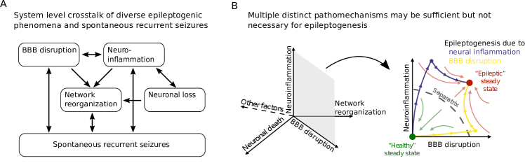
A) Complex interactions at the system level illustrate why multiple distinct pathomechanisms may be sufficient but not necessary for epileptogenesis. B) Experiments and simulations (Batulin et al., 2022) show that different causes, such as blood-brain barrier (BBB) disruption or neuroinflammation, can lead to a similar epileptic outcome.
Glial activity and associated spectrum of neuroinflammatory reactions have been shown to be in close interplay with blood-brain barrier (BBB) permeability (Shlosberg et al., 2010), neuronal activity (Stellwagen and Malenka, 2006; Badimon et al., 2020), neuronal loss (Dingledine et al., 2014) and network reorganization (Yong et al., 2019). Additional complexity is constituted by the fact that neuroimmune responses can be triggered not only by neurological injury. They can also result from downstream pathological changes that are characteristic for epilepsy and the ictal activity itself. For instance, enhanced BBB permeability was shown to be induced by epileptic seizures (Van Vliet et al., 2007; Rüber et al., 2018). And vice versa, the spillover of brain-born substances, following BBB leakage, can cause secondary neuroinflammation (Kim et al., 2012). In this way, seizures may develop via multiple mechanisms including BBB disruption, neuroinflammation and/or neuronal loss and network remodeling (Figure 3).
We have recently created a phenomenological model of these major epileptogenic processes and their interactions at realistic time scales (Batulin et al., 2022). We have described the neuro-immune crosstalk in the context of neurological injury using a dynamical systems approach. In agreement with experimental data from three animal models (Kirkman et al., 2010; Weissberg et al., 2015; Zhang et al., 2015; Brackhan et al., 2016; Patel et al., 2017), simulations showed that neuronal loss can be sufficient but is not necessary to drive epileptogenesis. Overall, computational modeling supports the concept that in the brain with its degenerate mechanisms, multiple different pathological mechanisms may contribute to epileptogenesis requiring different kinds of interventions for successful treatment (Batulin et al., 2022).
7 Multiscale and population modeling of degenerate circuits and epilepsy
Degeneracy is a multiscale phenomenon present at multiple levels of brain structure. Degeneracy implies that different processes on a lower level can lead to a certain phenomenon on a higher level. If phenomena on different scales interact, multiscale modeling becomes necessary. Therefore, deeper understanding of degenerate excitable systems requires multiscale computational approaches (Lytton et al., 2017). Creating models of neural circuits that can bridge two or more scales is a challenging task because of the nested hierarchy of molecular, cellular and supracellular networks with many feedforward and feedback loops between the scales. However, in well studied systems, multiscale models can provide new insights, which are relevant for epilepsy in the context of degeneracy.
A simulation study of the thalamo-cortical network in childhood absence epilepsy (Knox et al., 2018) is an intriguing example of multiscale modeling connecting the ion channel, cellular and microcircuit scales. The model showed that enhancing T-type calcium channel activation or reducing inhibitory GABA-A synaptic channel activation — in isolation as well as in combination — converted physiological network activity to seizure-like discharges. The results were in agreement with clinical observations of multiple different mutations in GABA-A receptors and T-type calcium channels in human patients and animals. The model predicted that these different mutations can lead to the same phenotype of absence epilepsy. In this way, the simulations have linked individual genetic variability in patients (simulated as the variability in the parameters of GABA-A and T-type calcium channels) to childhood absence epilepsy. Moreover, the model suggested plausible explanations for the failure or success of pharmacological medications targeting GABA-A and/or T-type calcium channels. Importantly, modeling predicted the necessity of multitarget therapy, simultaneously enhancing GABA-A transmission and suppressing T-type calcium channel activation, for patients with mutations in both ion channels (see also Figure 4). Furthermore, these simulations highlight the need for personalized epilepsy therapy in patients with different genetic backgrounds.
Another recent multiscale simulation study, although not focusing on epilepsy, has provided novel insights on degeneracy in the context of pathological perturbations linked to hyperexcitability associated with chronic pain (Medlock et al., 2022). In network simulations of spinal dorsal horn, the authors have demonstrated that, under physiological conditions, similar circuit activity can emerge in different models with disparate configurations of synaptic properties. However, following identical pathological perturbations (such as a reduction of inhibition or cell type diversity), these models displayed heterogeneous circuit responses. This is in agreement with previous work showing that perturbations in degenerate, superficially similar circuits can reveal hidden variability in synaptic and intrinsic properties leading to heterogeneous responses (Sakurai et al., 2014; Haddad and Marder, 2018; Onasch and Gjorgjieva, 2020; Morozova et al., 2022). Such modeling insights are highly relevant also for epilepsy. Different individuals with similar physiological circuit output may exhibit different resilience to ictogenic and epileptogenic perturbations, due to hidden variability of circuit parameters. Likewise, for the same reason, different patients with similar hyperexcitable circuit output may exhibit different susceptibility to different therapies.
In degenerate systems, instead of a single model, a large population of models with different parameter combinations is able to generate similar behavior. Consequently, in addition to multiscale modeling, degeneracy requires population modeling (Marder and Taylor, 2011; also called ensemble or database modeling, Günay et al., 2008; Rathour and Narayanan, 2014, 2019; Basak and Narayanan, 2018, 2020; Mishra and Narayanan, 2019; Mishra and Narayanan, 2021a; Sekulic et al., 2014) of neurons and neural circuits. One downside of population modeling is that it is unclear, which models/parameter combinations (from the large theoretically possible parameter space) exist in real brains. However, it is plausible to assume that evolution removed suboptimal models from the parameter space (Shoval et al., 2012). Indeed, evolutionary optimization principles might be useful to greatly simplify the parameter space by restricting it to the models that are (Pareto) optimal for the evolutionary trade-offs between multiple biologically plausible objectives (e.g., Szekely et al., 2013; Remme et al., 2018). Pareto optimality for the trade-off between energy efficiency and functional effectiveness has been suggested as a guiding principle for modeling neurons and neural circuits (Pallasdies et al., 2021; Jedlicka et al., 2022) and for reducing their degenerate parameter space (including ion channel space) to low-dimensional manifolds (Jedlicka et al., 2022). Hence Pareto theory might be used to improve population modeling of healthy but also epileptic neurons and neuronal circuits.
The upside of using populations of (single scale or multiscale) models is the ability to predict novel multicausal treatment options. Currently many computer models of epilepsy treatment simulate only mono-causal pharmacological effects on one type of ion channels. Using populations of conductance-based models of neurons and their circuits, implementing synaptic and intrinsic channel variability would enable predictions for a combination of multiple therapeutic targets (multitarget therapy) in silico. The use of such population-based in silico models could contribute to the discovery of new antiepileptic multitarget drug cocktails (Figure 4, Goaillard and Dufour, 2014). A recent simulation study provided a successful example for using population neuronal models as a tool for designing new multitarget holistic virtual drugs, which rescue pathologically increased excitability in Huntington’s disease (Allam et al., 2021). A similar approach could be adopted in epilepsy research for finding therapeutically efficient sets of perturbations of multiple ion channels that would switch hyperexcitable neuronal phenotypes to control phenotypes with normal excitability.
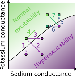
Epileptic hyperexcitability or restoration of normal excitability can be achieved by individual or combined changes in ion channels. The transition between normal excitation (green) and pathological hyperexcitability (violet) occurs when a tipping line is crossed. This transition can be induced for example by variation of sodium (horizontal axis) and potassium (vertical axis) conductance. Decrease in potassium conductance (1), increase in sodium conductance (3), or combining both (2) induces a transition from normal to pathological firing behavior of neurons. Increasing both conductances (4) maintains normal excitability. Conversely, reversal of epileptic hyperexcitability can be achieved by increasing potassium conductance (5), decreasing sodium conductance (6), or applying both changes simultaneously (7). Combined modification (7) moves the system farther away from the dangerous tipping point than isolated modification (5) or (6). For this reason, drugs might treat certain forms of epilepsy better if they modulate two or more types of ion channels simultaneously. This illustrates the potential advantage of multi-target therapy. (Modified from Goaillard and Dufour, 2014 and Ratté and Prescott, 2016.)
8 Degeneracy and its consequences for the pathophysiology and therapy of epilepsy
From the disease-etiology as well as therapeutic standpoints, the expression of degeneracy in pathological hyperexcitability states has two critical implications: (i) The source of hyperexcitability could manifest animal-to-animal and circuit-to-circuit variability, thus precluding the targeting of individual ion-channels or receptors across all animals and circuits. (ii) As hyperexcitability could emerge through disparate routes, it is equally possible that the reversal of hyperexcitability could be achieved through disparate routes (Figure 4, Goaillard and Dufour, 2014; Ratté and Prescott, 2016). The use of inhibitory receptor agonists as anticonvulsants, in many cases irrespective of the mechanistic origins of synchronous firing, constitutes an example of the latter point. Thus, it is not always necessary that the reversal of neuronal hyperexcitability is achieved by reversing the mechanisms that resulted in the hyperexcitability. Such reversal could be achieved through other routes without probing the mechanistic origins behind the pathological characteristic. On the other hand, under some conditions, the knowledge of the precise mechanistic route towards hyperexcitability in an individual patient may be necessary for the choice of effective personalized treatment. For example, the therapeutic effects of benzodiazepines and sodium channel blockers can be strongly dependent on the context of specific network pathology (see above the discussion of benzodiazepines and sodium channel blockers in the section on network level degeneracy).
The recognition of degeneracy in epileptic pathology could enable simultaneous use of multiple disparate components and routes to reverse hyperexcitability. Hence there is no need for sticking to one specific drug target. In fact, to repair a failed degenerate system, it is often not enough to restore a single target mechanism (Figure 4). One reason for this is that, due to degeneracy, many different pathologically altered mechanisms are sufficient, but usually none of them is by itself necessary for pathological malfunction (Kamaleddin, 2022). In addition, a degenerate nervous system sometimes displays compensatory adaptations, which undermine therapeutic interventions focusing on a single target (Ratté et al., 2014; Ratté and Prescott, 2016). Therefore, multitarget strategies in pharmacology or in neuromodulatory stimulation might be more promising than monotarget strategies (although carrying a higher risk for side effects). In fact, many already approved antiseizure drugs affect multiple targets (Kwan et al., 2001; Löscher, 2021). Degeneracy offers rationale also for another currently considered option of multitarget pharmacology, namely using combinations of drugs with different single targets instead of single multitarget drugs (Ratté and Prescott, 2016; Löscher, 2021) or even using combinations of multitarget drugs. However, an intense basic and clinical research is still needed to find drug combinations with synergistic (supraadditive) therapeutic effects and low (infraadditive) toxicity (Brodie and Sills, 2011; Brigo et al., 2013; Verrotti et al., 2020; Löscher, 2021). Similarly, future clinical research is needed to provide evidence for beneficial effects of neurostimulation targeting multiple brain areas. Such evidence is currently sparse (Li and Cook, 2018).
As there are many potential drug targets, the choice of specific drugs could be allowed to take advantage of circuit-specific differences in target expression profiles. For instance, if the hyperexcitability is specific to a neuronal subtype in a given brain region, this could be reversed by identifying mechanisms that are abundant in that neuronal subtype but not others. However, due to degeneracy, it is sometimes impossible to determine which neuron subtype (or ion channel subtype) is more crucial than others since it might depend on other (e.g., synaptic) parameters (O’Leary, 2018; Medlock et al., 2022).
Degeneracy of the brain creates opportunities but also challenges for the precision or personalized medicine, which tries to develop therapies targeted to the specific etiology and pathophysiology of individual patients. One challenge in the basic and clinical research of degeneracy is measuring multiple hyperexcitability-relevant parameters in the same individual (animal or human). Ideally, in a denegerate circuit, one would need to know most or all contributing components/mechanisms of hyperexcitability including the knowledge of most relevant (hub) mechanisms with highest ”cruciality score” of their involvement (Kamaleddin, 2022). In other words, reductionist monocausal research strategies focusing on isolated components are only partially suitable for studying complex degenerate systems (Regenmortel, 2004; Kamaleddin, 2022). Moreover, the conceptual framework of degeneracy with its emphasis on multitarget and multicausal thinking has consequences not only for the therapy development but also for the basic research and its perturbation and lesion strategies. For example, multitarget lesion experiments (Kamaleddin, 2022) or multi-knockout studies might be necessary to evoke a dysfunction of degenerate neural or molecular networks. Measuring and perturbing multiple parameters and mechanisms simultaneously is challenging but can be facilitated by computational approaches such as multiscale and population modeling (see above).
9 Costs and benefits of degeneracy
In summary, we argue that the evolution of the complex nervous system led to the emergence of multiple protective mechanisms against neural hyperexcitability but at the same time multiple potential routes towards protection failure. In a healthy state, multiple compensatory mechanisms may act jointly with a potential for compensating each other’s failure thereby supporting the robustness of physiological neural excitability. The compensation may be immediate (”default” or ”constitutive” compensation) or it can be recruited on demand with a time delay as a form of (”inducible”) homeostatic plasticity (”homeostatic compensation”). Notably, degeneracy in homeostatic plasticity mechanisms has been demonstrated in visual cortex (Maffei and Turrigiano, 2008) and elsewhere (Watt and Desai, 2010; Turrigiano, 2011). The authors of visual cortex studies have explicitly stated that ”multiple, partially redundant forms of homeostatic plasticity may ensure that network compensation can be achieved in response to a wide range of sensory perturbations” (Maffei and Turrigiano, 2008). As we mentioned before partial redundancy is a term that is sometimes used in the literature instead of degeneracy. We believe that similar research in the context of epilepsy will reveal degeneracy in homeostatic plasticity mechanisms protecting the hippocampus and other brain regions against hyperexcitability and epileptogenesis. Support for this idea comes also from computational models (Lazar et al., 2009) and control theory (Cannon and Miller, 2016), which indicate that degenerate homeostatic mechanisms provide functional benefits. For example, an effective control of neuronal firing rate (its mean and variance) can be achieved by degeneracy of cooperative synaptic and intrinsic homeostatic plasticity (Cannon and Miller, 2016).
Degeneracy in biological systems is closely linked to their complexity in terms of the number of (mutually interacting) individual components and mechanisms (Edelman and Gally, 2001; Mason, 2010). The higher the number of system’s interacting mechanisms, the higher is the system’s complexity, flexibility and robustness. At the same time, the higher are also the system’s energy costs linked to the extent of functional redundancy of identical components (Kamaleddin, 2022). If a system has multiple identical components performing one function A (redundancy) then it does not have the benefit of multiple (structurally) different components performing not only function A but also contributing to function B. So degeneracy entails more functionality due to diversity of components. Since energy and material resources are limited, complex biological organisms display universal trade-offs between (1) functional performance, its (2) robustness and (3) flexibility and (4) energy costs (Del Giudice and Crespi, 2018). Degeneracy facilitates the robustness and flexibility of functional behavior (Whitacre, 2010, 2012). In contrast, pure redundancy increases the energy costs (Kamaleddin, 2022). Through degeneracy evolution seems to have optimized biological systems for these (multi-objective) trade-offs (Shoval et al., 2012; Szekely et al., 2013; Alon, 2020). A biological system tries to maximize functionality, flexibility and robustness but minimize energy expenditure (Laughlin and Sejnowski, 2003; Sterling and Laughlin, 2015). Therefore, it is plausible that degenerate living systems evolved to become (Pareto) optimal for these multiple objectives. Therefore, one would expect biological systems, including nervous systems (Pallasdies et al., 2021; Jedlicka et al., 2022) to exist close to an optimal compromise between low energy costs and high functionality (as reflected in functional effectiveness, and its robustness and flexibility). In line with these ideas, a recent study proposed that ”degeneracy affords a flexibility that offsets the cost of redundancy” (Sajid et al., 2020).
The conceptual link between degeneracy and evolutionary optimization opens many interesting questions. For example, counterintuitively, expressing multiple complementary protective mechanisms may paradoxically help reduce the overall cost of protection. This could be the case if activation of multiple protective mechanisms allows for optimal cost sharing among them (Stearns and Medzhitov, 2016). That would open the possibility for every mechanism being expressed at the lower (and hence cheaper) end of its dynamical range (Stearns and Medzhitov, 2016). In this way, the function (homeostasis of normal excitability) would be preserved at a lower cost of its protection. Currently such optimal cost sharing is only a hypothesis that needs to be tested in experiments and simulations. On the other hand, it is also possible that the relatively high fragility of the nervous system with respect to hyperexcitability is a high evolutionary price that we have to pay for the enormous computational capabilities of our energy-efficient brains (Laughlin and Sejnowski, 2003; Sterling and Laughlin, 2015), which operate close to criticality that maximizes information-processing. This would mean that the powerful neural computation was bought at the expense of suboptimal protection against hyperexcitability.
Another largely unexplored area is the potential trade-off between immune defense and homeostasis of neural excitability. Immune defense of the brain is costly and under threatening conditions it might get activated at the expense of neural homeostasis (cf. Stearns and Medzhitov, 2016). Brain injuries (e.g., trauma, infection) may shift the balance and energy expenditure towards immune defense (cf. Casaril et al., 2022). Future research might clarify whether epileptogenesis can be understood as a consequence of (chronically) enhanced immune defense (with its collateral damage) at the expense of suppressed excitability homeostasis.
Backmatter
Ethics approval and consent to participate
Not applicable
Consent for publication
Not applicable
Availability of data and materials
Not applicable
Funding
The work was supported by the grant LOEWE CePTER – Center for Personalized Translational Epilepsy Research (DB, TS, JT, PJ). DB is supported by the International Max Planck Research School (IMPRS) for Neural Circuits. JT is supported by the Johanna Quandt Foundation. RN is supported by a senior fellowship (IA/S/16/2/502727) from the DBT-Wellcome Trust India Alliance. PJ is supported by the BMBF grant (No. 031L0229) and funds from the von Behring Röntgen Foundation.
Competing interests
The authors declare that they have no competing interests.
Author’s contributions
All authors contributed to conceptualizing and writing the manuscript. RN, DB and TMS created the figures.
References
- Albertson et al. (2011) Albertson AJ, Yang J, Hablitz JJ (2011) Decreased hyperpolarization-activated currents in layer 5 pyramidal neurons enhances excitability in focal cortical dysplasia. Journal of neurophysiology 106:2189–2200.
- Alexander et al. (2016) Alexander A, Maroso M, Soltesz I (2016) Organization and control of epileptic circuits in temporal lobe epilepsy. Progress in brain research 226:127–154.
- Allam et al. (2021) Allam SL, Rumbell TH, Hoang-Trong T, Parikh J, Kozloski JR (2021) Neuronal population models reveal specific linear conductance controllers sufficient to rescue preclinical disease phenotypes. Iscience 24:103279.
- Alon (2020) Alon U (2020) Multi-objective optimality in biology In Alon U, editor, An Introduction to Systems Biology: Design Principles of Biological Circuits, chapter 14, pp. 249–272. Chapman and Hall/CRC, Boca Raton, London, New York, 2 edition.
- Arisi and Garcia-Cairasco (2007) Arisi GM, Garcia-Cairasco N (2007) Doublecortin-positive newly born granule cells of hippocampus have abnormal apical dendritic morphology in the pilocarpine model of temporal lobe epilepsy. Brain research 1165:126–134.
- Arnold et al. (2019) Arnold EC, McMurray C, Gray R, Johnston D (2019) Epilepsy-induced reduction in hcn channel expression contributes to an increased excitability in dorsal, but not ventral, hippocampal ca1 neurons. eNeuro 6.
- Badimon et al. (2020) Badimon A, Strasburger HJ, Ayata P, Chen X, Nair A, Ikegami A, Hwang P, Chan AT, Graves SM, Uweru JO et al. (2020) Negative feedback control of neuronal activity by microglia. Nature 586:417–423.
- Basak and Narayanan (2018) Basak R, Narayanan R (2018) Spatially dispersed synapses yield sharply-tuned place cell responses through dendritic spike initiation. J Physiol 596:4173–4205.
- Basak and Narayanan (2020) Basak R, Narayanan R (2020) Robust emergence of sharply tuned place-cell responses in hippocampal neurons with structural and biophysical heterogeneities. Brain Struct Funct 225:567–590.
- Batulin et al. (2022) Batulin D, Lagzi F, Vezzani A, Jedlicka P, Triesch J (2022) A phenomenological model of neuroimmune interactions in epileptogenesis. iScience .
- Beck and Yaari (2008) Beck H, Yaari Y (2008) Plasticity of intrinsic neuronal properties in cns disorders. Nature Reviews Neuroscience 9:357–369.
- Bekkers and Häusser (2007) Bekkers JM, Häusser M (2007) Targeted dendrotomy reveals active and passive contributions of the dendritic tree to synaptic integration and neuronal output. Proceedings of the National Academy of Sciences 104:11447–11452.
- Bernard et al. (2004) Bernard C, Anderson A, Becker A, Poolos NP, Beck H, Johnston D (2004) Acquired dendritic channelopathy in temporal lobe epilepsy. Science 305:532–5.
- Bezaire et al. (2016) Bezaire MJ, Raikov I, Burk K, Vyas D, Soltesz I (2016) Interneuronal mechanisms of hippocampal theta oscillations in a full-scale model of the rodent ca1 circuit. Elife 5.
- Bock et al. (2019) Bock T, Honnuraiah S, Stuart GJ (2019) Paradoxical excitatory impact of sk channels on dendritic excitability. Journal of Neuroscience 39:7826–7839.
- Brackhan et al. (2016) Brackhan M, Bascuñana P, Postema JM, Ross TL, Bengel FM, Bankstahl M, Bankstahl JP (2016) Serial quantitative tspo-targeted pet reveals peak microglial activation up to 2 weeks after an epileptogenic brain insult. Journal of Nuclear Medicine 57:1302–1308.
- Bragin et al. (1999a) Bragin A, Engel J J, Wilson CL, Fried I, Buzsaki G (1999a) High-frequency oscillations in human brain. Hippocampus 9:137–42.
- Bragin et al. (1999b) Bragin A, Engel J J, Wilson CL, Fried I, Mathern GW (1999b) Hippocampal and entorhinal cortex high-frequency oscillations (100–500 hz) in human epileptic brain and in kainic acid–treated rats with chronic seizures. Epilepsia 40:127–37.
- Brigo et al. (2013) Brigo F, Ausserer H, Tezzon F, Nardone R (2013) When one plus one makes three: the quest for rational antiepileptic polytherapy with supraadditive anticonvulsant efficacy. Epilepsy & Behavior 27:439–442.
- Brodie and Sills (2011) Brodie MJ, Sills GJ (2011) Combining antiepileptic drugs—rational polytherapy? Seizure 20:369–375.
- Buckmaster (2014) Buckmaster PS (2014) Does mossy fiber sprouting give rise to the epileptic state? Issues in clinical epileptology: a view from the bench pp. 161–168.
- Bui et al. (2015) Bui A, Kim HK, Maroso M, Soltesz I (2015) Microcircuits in Epilepsy: Heterogeneity and Hub Cells in Network Synchronization. Cold Spring Harbor Perspectives in Medicine 5:a022855.
- Burman et al. (2019) Burman RJ, Selfe JS, Lee JH, van den Berg M, Calin A, Codadu NK, Wright R, Newey SE, Parrish RR, Katz AA et al. (2019) Excitatory gabaergic signalling is associated with benzodiazepine resistance in status epilepticus. Brain 142:3482–3501.
- Buzsaki (2002) Buzsaki G (2002) Theta oscillations in the hippocampus. Neuron 33:325–40.
- Buzsaki (2006) Buzsaki G (2006) Rhythms of the brain Oxford University Press, New York.
- Buzsaki et al. (2012) Buzsaki G, Anastassiou CA, Koch C (2012) The origin of extracellular fields and currents–eeg, ecog, lfp and spikes. Nat Rev Neurosci 13:407–20.
- Buzsaki and Wang (2012) Buzsaki G, Wang XJ (2012) Mechanisms of gamma oscillations. Annu Rev Neurosci 35:203–25.
- Calabrese (2018) Calabrese RL (2018) Inconvenient Truth to Principle of Neuroscience. Trends in Neurosciences 41:488–491.
- Cannon and Miller (2016) Cannon J, Miller P (2016) Synaptic and intrinsic homeostasis cooperate to optimize single neuron response properties and tune integrator circuits. Journal of neurophysiology 116:2004–2022.
- Casaril et al. (2022) Casaril AM, Katsalifis A, Schmidt RM, Bas-Orth C (2022) Activated glia cells cause bioenergetic impairment of neurons that can be rescued by knock-down of the mitochondrial calcium uniporter. Biochemical and biophysical research communications .
- Cavarsan et al. (2018) Cavarsan CF, Malheiros J, Hamani C, Najm I, Covolan L (2018) Is mossy fiber sprouting a potential therapeutic target for epilepsy? Frontiers in Neurology p. 1023.
- Chancey and Howard (2022) Chancey JH, Howard MA (2022) Synaptic integration in ca1 pyramidal neurons is intact despite deficits in gabaergic transmission in the scn1a haploinsufficiency mouse model of dravet syndrome. Eneuro .
- Cheah et al. (2012) Cheah CS, Frank HY, Westenbroek RE, Kalume FK, Oakley JC, Potter GB, Rubenstein JL, Catterall WA (2012) Specific deletion of nav1. 1 sodium channels in inhibitory interneurons causes seizures and premature death in a mouse model of dravet syndrome. Proceedings of the National Academy of Sciences 109:14646–14651.
- Chizhov et al. (2018) Chizhov AV, Zefirov AV, Amakhin DV, Smirnova EY, Zaitsev AV (2018) Minimal model of interictal and ictal discharges “Epileptor-2”. PLOS Computational Biology 14:e1006186.
- Codadu et al. (2019) Codadu NK, Graham RT, Burman RJ, Jackson-Taylor RT, Raimondo JV, Trevelyan AJ, Parrish RR (2019) Divergent paths to seizure-like events. Physiol Rep 7:e14226.
- Colgin (2013) Colgin LL (2013) Mechanisms and functions of theta rhythms. Annual Review of Neuroscience, Vol 36 36:295–312.
- Colgin (2016) Colgin LL (2016) Rhythms of the hippocampal network. Nat Rev Neurosci 17:239–49.
- Colgin and Moser (2010) Colgin LL, Moser EI (2010) Gamma oscillations in the hippocampus. Physiology (Bethesda) 25:319–29.
- Coulter and Carlson (2007) Coulter DA, Carlson GC (2007) Functional regulation of the dentate gyrus by gaba-mediated inhibition. Progress in brain research 163:235–812.
- Cropper et al. (2016) Cropper EC, Dacks AM, Weiss KR (2016) Consequences of degeneracy in network function. Current Opinion in Neurobiology 41:62–67.
- Cuntz et al. (2021) Cuntz H, Bird AD, Mittag M, Beining M, Schneider M, Mediavilla L, Hoffmann FZ, Deller T, Jedlicka P (2021) A general principle of dendritic constancy: A neuron’s size-and shape-invariant excitability. Neuron 109:3647–3662.
- Danzer (2019) Danzer SC (2019) Adult neurogenesis in the development of epilepsy. Epilepsy currents 19:316–320.
- Deczkowska et al. (2018) Deczkowska A, Amit I, Schwartz M (2018) Microglial immune checkpoint mechanisms. Nature neuroscience 21:779–786.
- Degro et al. (2022) Degro CE, Bolduan F, Vida I, Booker SA (2022) Interneuron diversity in the rat dentate gyrus: An unbiased in vitro classification. Hippocampus .
- Del Giudice and Crespi (2018) Del Giudice M, Crespi BJ (2018) Basic functional trade-offs in cognition: An integrative framework. Cognition 179:56–70.
- Demarest and Brooks-Kayal (2018) Demarest ST, Brooks-Kayal A (2018) From molecules to medicines: the dawn of targeted therapies for genetic epilepsies. Nature Reviews Neurology 14:735–745.
- Dengler and Coulter (2016) Dengler CG, Coulter DA (2016) Chapter 6 - Normal and epilepsy-associated pathologic function of the dentate gyrus In Rossignol E, Carmant L, Lacaille JC, editors, Neurobiology of Epilepsy, Vol. 226, pp. 155–178. Elsevier.
- Dey et al. (2014) Dey D, Eckle VS, Vitko I, Sullivan KA, Lasiecka ZM, Winckler B, Stornetta RL, Williamson JM, Kapur J, Perez-Reyes E (2014) A potassium leak channel silences hyperactive neurons and ameliorates status epilepticus. Epilepsia 55:203–213.
- Dhupia et al. (2015) Dhupia N, Rathour RK, Narayanan R (2015) Dendritic atrophy constricts functional maps in resonance and impedance properties of hippocampal model neurons. Frontiers in Cellular Neuroscience 8:456.
- Dichter (1997) Dichter MA (1997) Basic mechanisms of epilepsy: targets for therapeutic intervention. Epilepsia 38 Suppl 9:S2–6.
- Dingledine et al. (2014) Dingledine R, Varvel NH, Dudek FE (2014) When and how do seizures kill neurons, and is cell death relevant to epileptogenesis? Issues in clinical epileptology: a view from the bench pp. 109–122.
- Drion et al. (2015) Drion G, O’Leary T, Marder E (2015) Ion channel degeneracy enables robust and tunable neuronal firing rates. Proceedings of the National Academy of Sciences 112:E5361–E5370.
- Dudek and Sutula (2007) Dudek FE, Sutula TP (2007) Epileptogenesis in the dentate gyrus: a critical perspective In Scharfman HEBTPiBR, editor, The dentate gyrus: A comprehensive guide to structure, function, and clinical implications, Vol. 163, pp. 755–773. Elsevier.
- Dudok et al. (2021) Dudok B, Klein PM, Soltesz I (2021) Toward understanding the diverse roles of perisomatic interneurons in epilepsy. Epilepsy Currents p. 15357597211053687.
- Duncan et al. (2006) Duncan JS, Sander JW, Sisodiya SM, Walker MC (2006) Adult epilepsy. The Lancet 367:1087–1100.
- Dutton et al. (2013) Dutton SB, Makinson CD, Papale LA, Shankar A, Balakrishnan B, Nakazawa K, Escayg A (2013) Preferential inactivation of scn1a in parvalbumin interneurons increases seizure susceptibility. Neurobiology of disease 49:211–220.
- Dyhrfjeld-Johnsen et al. (2009) Dyhrfjeld-Johnsen J, Morgan RJ, Soltesz I (2009) Double trouble? potential for hyperexcitability following both channelopathic up-and downregulation of ih in epilepsy. Frontiers in neuroscience 3:5.
- Edelman and Gally (2001) Edelman GM, Gally JA (2001) Degeneracy and complexity in biological systems. Proceedings of the National Academy of Sciences 98:13763–13768.
- Einevoll et al. (2013) Einevoll GT, Kayser C, Logothetis NK, Panzeri S (2013) Modelling and analysis of local field potentials for studying the function of cortical circuits. Nat Rev Neurosci 14:770–85.
- El Houssaini et al. (2020) El Houssaini K, Bernard C, Jirsa VK (2020) The epileptor model: A systematic mathematical analysis linked to the dynamics of seizures, refractory status epilepticus, and depolarization block. eNeuro 7.
- Farrell et al. (2019) Farrell JS, Nguyen QA, Soltesz I (2019) Resolving the micro-macro disconnect to address core features of seizure networks. Neuron 101:1016–1028.
- Freund and Buzsáki (1996) Freund TF, Buzsáki G (1996) Interneurons of the hippocampus. Hippocampus 6:347–470.
- Fröhlich et al. (2008) Fröhlich F, Bazhenov M, Iragui-Madoz V, Sejnowski TJ (2008) Potassium dynamics in the epileptic cortex: new insights on an old topic. The Neuroscientist : a review journal bringing neurobiology, neurology and psychiatry 14:422–33.
- Gibbs et al. (1935) Gibbs FA, Davis H, Lennox WG (1935) The electro-encephalogram in epilepsy and in conditions of impaired consciousness. Arch. Neurol. Psychiatry 34:1133–1148.
- Goaillard and Dufour (2014) Goaillard JM, Dufour MA (2014) Neuropathic pain: The pros and cons of degeneracy. Elife 3:e02615.
- Goaillard and Marder (2021) Goaillard JM, Marder E (2021) Ion channel degeneracy, variability, and covariation in neuron and circuit resilience. Annual review of neuroscience 44:335–357.
- Goaillard et al. (2009) Goaillard JM, Taylor AL, Schulz DJ, Marder E (2009) Functional consequences of animal-to-animal variation in circuit parameters. Nature neuroscience 12:1424–1430.
- González et al. (2014) González H, Elgueta D, Montoya A, Pacheco R (2014) Neuroimmune regulation of microglial activity involved in neuroinflammation and neurodegenerative diseases. Journal of neuroimmunology 274:1–13.
- Goutagny et al. (2009) Goutagny R, Jackson J, Williams S (2009) Self-generated theta oscillations in the hippocampus. Nat Neurosci 12:1491–3.
- Grashow et al. (2010) Grashow R, Brookings T, Marder E (2010) Compensation for variable intrinsic neuronal excitability by circuit-synaptic interactions. Journal of Neuroscience 30:9145–9156.
- Günay et al. (2008) Günay C, Edgerton JR, Jaeger D (2008) Channel density distributions explain spiking variability in the globus pallidus: a combined physiology and computer simulation database approach. The Journal of Neuroscience 28:7476–7491.
- Gutenkunst et al. (2007) Gutenkunst RN, Waterfall JJ, Casey FP, Brown KS, Myers CR, Sethna JP (2007) Universally sloppy parameter sensitivities in systems biology models. PLoS computational biology 3:e189.
- Haddad and Marder (2018) Haddad SA, Marder E (2018) Circuit robustness to temperature perturbation is altered by neuromodulators. Neuron 100:609–623.e3.
- Hagen et al. (2018) Hagen E, Naess S, Ness TV, Einevoll GT (2018) Multimodal modeling of neural network activity: Computing lfp, ecog, eeg, and meg signals with lfpy 2.0. Front Neuroinform 12:92.
- Hainmueller and Bartos (2020) Hainmueller T, Bartos M (2020) Dentate gyrus circuits for encoding, retrieval and discrimination of episodic memories. Nature Reviews Neuroscience 21:153–168.
- Hawkins et al. (2017) Hawkins NA, Anderson LL, Gertler TS, Laux L, George Jr AL, Kearney JA (2017) Screening of conventional anticonvulsants in a genetic mouse model of epilepsy. Annals of clinical and translational neurology 4:326–339.
- Heinemann et al. (1992) Heinemann U, Beck H, Dreier J, Ficker E, Stabel J, Zhang C (1992) The dentate gyrus as a regulated gate for the propagation of epileptiform activity. Epilepsy research. Supplement 7:273–280.
- Hofmann et al. (2016) Hofmann G, Balgooyen L, Mattis J, Deisseroth K, Buckmaster PS (2016) Hilar somatostatin interneuron loss reduces dentate gyrus inhibition in a mouse model of temporal lobe epilepsy. Epilepsia 57:977–983.
- Houser (2007) Houser CR (2007) Interneurons of the dentate gyrus: an overview of cell types, terminal fields and neurochemical identity. Progress in brain research 163:217–811.
- Janz et al. (2017) Janz P, Savanthrapadian S, Häussler U, Kilias A, Nestel S, Kretz O, Kirsch M, Bartos M, Egert U, Haas CA (2017) Synaptic Remodeling of Entorhinal Input Contributes to an Aberrant Hippocampal Network in Temporal Lobe Epilepsy. Cerebral Cortex 27:2348–2364.
- Jedlicka et al. (2022) Jedlicka P, Bird A, Cuntz H (2022) Pareto optimality, economy-effectiveness trade-offs and ion channel degeneracy: Improving population models of neurons. arXiv preprint arXiv:2203.06391 .
- Jessberger and Parent (2015) Jessberger S, Parent JM (2015) Epilepsy and adult neurogenesis. Cold Spring Harbor Perspectives in Biology 7:a020677.
- Jiao et al. (2013) Jiao J, Yang Y, Shi Y, Chen J, Gao R, Fan Y, Yao H, Liao W, Sun XF, Gao S (2013) Modeling dravet syndrome using induced pluripotent stem cells (ipscs) and directly converted neurons. Human molecular genetics 22:4241–4252.
- Jirsa et al. (2014) Jirsa VK, Stacey WC, Quilichini PP, Ivanov AI, Bernard C (2014) On the nature of seizure dynamics. Brain 137:2210–2230.
- Johnston et al. (2000) Johnston D, Hoffman DA, Poolos NP (2000) Potassium channels and dendritic function in hippocampal pyramidal neurons. Epilepsia 41:1072–3.
- Johnston and Narayanan (2008) Johnston D, Narayanan R (2008) Active dendrites: colorful wings of the mysterious butterflies. Trends in neurosciences 31:309–316.
- Jung et al. (2010) Jung S, Bullis JB, Lau IH, Jones TD, Warner LN, Poolos NP (2010) Downregulation of dendritic hcn channel gating in epilepsy is mediated by altered phosphorylation signaling. J Neurosci 30:6678–88.
- Jung et al. (2007) Jung S, Jones TD, Lugo JN J, Sheerin AH, Miller JW, D’Ambrosio R, Anderson AE, Poolos NP (2007) Progressive dendritic hcn channelopathy during epileptogenesis in the rat pilocarpine model of epilepsy. J Neurosci 27:13012–21.
- Jung et al. (2011) Jung S, Warner LN, Pitsch J, Becker AJ, Poolos NP (2011) Rapid loss of dendritic hcn channel expression in hippocampal pyramidal neurons following status epilepticus. J Neurosci 31:14291–5.
- Kajikawa and Schroeder (2011) Kajikawa Y, Schroeder CE (2011) How local is the local field potential? Neuron 72:847–58.
- Kamaleddin (2022) Kamaleddin MA (2022) Degeneracy in the nervous system: from neuronal excitability to neural coding. BioEssays 44:2100148.
- Kaplan et al. (2016) Kaplan DI, Isom LL, Petrou S (2016) Role of Sodium Channels in Epilepsy. Cold Spring Harbor Perspectives in Medicine 6:a022814.
- Kenny et al. (2019) Kenny EM, Fidan E, Yang Q, Anthonymuthu TS, New LA, Meyer EA, Wang H, Kochanek PM, Dixon CE, Kagan VE et al. (2019) Ferroptosis contributes to neuronal death and functional outcome after traumatic brain injury. Critical care medicine 47:410.
- Kim et al. (2012) Kim SY, Buckwalter M, Soreq H, Vezzani A, Kaufer D (2012) Blood–brain barrier dysfunction–induced inflammatory signaling in brain pathology and epileptogenesis. Epilepsia 53:37–44.
- Kirchheim et al. (2013) Kirchheim F, Tinnes S, Haas CA, Stegen M, Wolfart J (2013) Regulation of action potential delays via voltage-gated potassium kv1. 1 channels in dentate granule cells during hippocampal epilepsy. Frontiers in cellular neuroscience 7:248.
- Kirkman et al. (2010) Kirkman NJ, Libbey JE, Wilcox KS, White HS, Fujinami RS (2010) Innate but not adaptive immune responses contribute to behavioral seizures following viral infection. Epilepsia 51:454–464.
- Kispersky et al. (2012) Kispersky TJ, Caplan JS, Marder E (2012) Increase in sodium conductance decreases firing rate and gain in model neurons. Journal of Neuroscience 32:10995–11004.
- Klassen et al. (2011) Klassen T, Davis C, Goldman A, Burgess D, Chen T, Wheeler D, McPherson J, Bourquin T, Lewis L, Villasana D, Morgan M, Muzny D, Gibbs R, Noebels J (2011) Exome Sequencing of Ion Channel Genes Reveals Complex Profiles Confounding Personal Risk Assessment in Epilepsy. Cell 145:1036–1048.
- Klein et al. (2018) Klein P, Dingledine R, Aronica E, Bernard C, Blümcke I, Boison D, Brodie MJ, Brooks-Kayal AR, Engel Jr J, Forcelli PA et al. (2018) Commonalities in epileptogenic processes from different acute brain insults: Do they translate? Epilepsia 59:37–66.
- Knox et al. (2018) Knox AT, Glauser T, Tenney J, Lytton WW, Holland K (2018) Modeling pathogenesis and treatment response in childhood absence epilepsy. Epilepsia 59:135–145.
- Kobayashi and Buckmaster (2003) Kobayashi M, Buckmaster PS (2003) Reduced inhibition of dentate granule cells in a model of temporal lobe epilepsy. The Journal of Neuroscience 23:2440 – 2452.
- Kole et al. (2007) Kole MH, Bräuer AU, Stuart GJ (2007) Inherited cortical hcn1 channel loss amplifies dendritic calcium electrogenesis and burst firing in a rat absence epilepsy model. The Journal of physiology 578:507–525.
- Kramer et al. (2012) Kramer MA, Truccolo W, Eden UT, Lepage KQ, Hochberg LR, Eskandar EN, Madsen JR, Lee JW, Maheshwari A, Halgren E, Chu CJ, Cash SS (2012) Human seizures self-terminate across spatial scales via a critical transition. Proceedings of the National Academy of Sciences 109:21116–21121.
- Krook-Magnuson and Soltesz (2015) Krook-Magnuson E, Soltesz I (2015) Beyond the hammer and the scalpel: selective circuit control for the epilepsies. Nat Neurosci 18:331–8.
- Krook-Magnuson et al. (2015) Krook-Magnuson E, Armstrong C, Bui A, Lew S, Oijala M, Soltesz I (2015) In vivo evaluation of the dentate gate theory in epilepsy. The Journal of physiology 593:2379–2388.
- Krueppel et al. (2011) Krueppel R, Remy S, Beck H (2011) Dendritic integration in hippocampal dentate granule cells. Neuron 71:512–28.
- Kwan et al. (2001) Kwan P, Sills GJ, Brodie MJ (2001) The mechanisms of action of commonly used antiepileptic drugs. Pharmacology & therapeutics 90:21–34.
- Larkum et al. (2022) Larkum ME, Wu J, Duverdin SA, Gidon A (2022) The guide to dendritic spikes of the mammalian cortex in vitro and in vivo. Neuroscience .
- Laughlin and Sejnowski (2003) Laughlin SB, Sejnowski TJ (2003) Communication in neuronal networks. Science 301:1870–4.
- Lazar et al. (2009) Lazar A, Pipa G, Triesch J (2009) Sorn: a self-organizing recurrent neural network. Frontiers in computational neuroscience 3:23.
- Lee et al. (2016) Lee CT, Kao MH, Hou WH, Wei YT, Chen CL, Lien CC (2016) Causal evidence for the role of specific gabaergic interneuron types in entorhinal recruitment of dentate granule cells. Scientific reports 6:1–13.
- Lerche et al. (2013) Lerche H, Shah M, Beck H, Noebels J, Johnston D, Vincent A (2013) Ion channels in genetic and acquired forms of epilepsy. The Journal of Physiology 591:753–764.
- Leski et al. (2013) Leski S, Linden H, Tetzlaff T, Pettersen KH, Einevoll GT (2013) Frequency dependence of signal power and spatial reach of the local field potential. PLoS Comput Biol 9:e1003137.
- Li et al. (2011) Li G, Bauer S, Nowak M, Norwood B, Tackenberg B, Rosenow F, Knake S, Oertel WH, Hamer HM (2011) Cytokines and epilepsy. Seizure 20:249–256.
- Li and Cook (2018) Li MC, Cook MJ (2018) Deep brain stimulation for drug-resistant epilepsy. Epilepsia 59:273–290.
- Li et al. (2013) Li ZX, Yu HM, Jiang KW (2013) Tonic gaba inhibition in hippocampal dentate granule cells: its regulation and function in temporal lobe epilepsies. Acta Physiologica 209:199–211.
- Linden et al. (2011) Linden H, Tetzlaff T, Potjans TC, Pettersen KH, Grun S, Diesmann M, Einevoll GT (2011) Modeling the spatial reach of the lfp. Neuron 72:859–72.
- Liu and Parvizi (2019) Liu S, Parvizi J (2019) Cognitive refractory state caused by spontaneous epileptic high-frequency oscillations in the human brain. Sci Transl Med 11.
- Liu et al. (2013) Liu Y, Lopez-Santiago LF, Yuan Y, Jones JM, Zhang H, O’Malley HA, Patino GA, O’Brien JE, Rusconi R, Gupta A, Thompson RC, Natowicz MR, Meisler MH, Isom LL, Parent JM (2013) Dravet syndrome patient-derived neurons suggest a novel epilepsy mechanism. Annals of Neurology 74:128–139.
- Lopez-Santiago et al. (2017) Lopez-Santiago LF, Yuan Y, Wagnon JL, Hull JM, Frasier CR, O’Malley HA, Meisler MH, Isom LL (2017) Neuronal hyperexcitability in a mouse model of scn8a epileptic encephalopathy. Proceedings of the National Academy of Sciences 114:2383–2388.
- Löscher (2021) Löscher W (2021) Single-target versus multi-target drugs versus combinations of drugs with multiple targets: Preclinical and clinical evidence for the treatment or prevention of epilepsy. Frontiers in pharmacology 12.
- Lothman et al. (1992) Lothman EW, Stringer JL, Bertram EH (1992) The dentate gyrus as a control point for seizures in the hippocampus and beyond. Epilepsy research. Supplement 7:301–313.
- Lux et al. (1986) Lux HD, Heinemann U, Dietzel I (1986) Ionic changes and alterations in the size of the extracellular space during epileptic activity. Advances in neurology 44:619–39.
- Lytton et al. (2017) Lytton WW, Arle J, Bobashev G, Ji S, Klassen TL, Marmarelis VZ, Schwaber J, Sherif MA, Sanger TD (2017) Multiscale modeling in the clinic: diseases of the brain and nervous system. Brain Inform 4:219–230.
- Lytton (2008) Lytton WW (2008) Computer modelling of epilepsy. Nature reviews. Neuroscience 9:626–37.
- Maffei and Turrigiano (2008) Maffei A, Turrigiano GG (2008) Multiple modes of network homeostasis in visual cortical layer 2/3. Journal of Neuroscience 28:4377–4384.
- Mainen and Sejnowski (1996) Mainen ZF, Sejnowski TJ (1996) Influence of dendritic structure on firing pattern in model neocortical neurons. Nature 382:363–366.
- Mäki-Marttunen and Mäki-Marttunen (2022) Mäki-Marttunen T, Mäki-Marttunen V (2022) Excitatory and inhibitory effects of hcn channel modulation on excitability of layer v pyramidal cells. bioRxiv .
- Man et al. (2016) Man M, Zhang Y, Ma G, Friston K, Liu S (2016) Quantification of degeneracy in hodgkin–huxley neurons on newman–watts small world network. Journal of theoretical biology 402:62–74.
- Manita et al. (2017) Manita S, Miyakawa H, Kitamura K, Murayama M (2017) Dendritic spikes in sensory perception. Frontiers in cellular neuroscience 11:29.
- Marder and Goaillard (2006) Marder E, Goaillard JM (2006) Variability, compensation and homeostasis in neuron and network function. Nature Reviews Neuroscience 7:563–574.
- Marder et al. (2017) Marder E, Gutierrez GJ, Nusbaum MP (2017) Complicating connectomes: Electrical coupling creates parallel pathways and degenerate circuit mechanisms. Developmental neurobiology 77:597–609.
- Marder and Taylor (2011) Marder E, Taylor AL (2011) Multiple models to capture the variability in biological neurons and networks. Nature neuroscience 14:133–8.
- Martinez-Canada et al. (2021) Martinez-Canada P, Ness TV, Einevoll GT, Fellin T, Panzeri S (2021) Computation of the electroencephalogram (eeg) from network models of point neurons. PLoS Comput Biol 17:e1008893.
- Mason (2010) Mason PH (2010) Degeneracy at multiple levels of complexity. Biological Theory 5:277–288.
- Mason et al. (2015) Mason P, Domínguez D. J, Winter B, Grignolio A (2015) Hidden in plain view: degeneracy in complex systems. Biosystems 128:1–8.
- Mattis et al. (2022) Mattis J, Somarowthu A, Goff KM, Jiang E, Yom J, Sotuyo N, Mcgarry LM, Feng H, Kaneko K, Goldberg EM (2022) Corticohippocampal circuit dysfunction in a mouse model of Dravet syndrome. eLife 11:e69293.
- McCormick and Contreras (2001) McCormick DA, Contreras D (2001) On the cellular and network bases of epileptic seizures. Annu Rev Physiol 63:815–46.
- Medlock et al. (2022) Medlock L, Sekiguchi K, Hong S, Dura-Bernal S, Lytton WW, Prescott SA (2022) Multiscale computer model of the spinal dorsal horn reveals changes in network processing associated with chronic pain. The Journal of Neuroscience .
- Mellen (2008) Mellen NM (2008) Belt-and-Suspenders as a Biological Design Principle In Advances in experimental medicine and biology, Vol. 605, pp. 99–103.
- Migliore et al. (2018) Migliore R, Lupascu CA, Bologna LL, Romani A, Courcol JD, Antonel S, Van Geit WAH, Thomson AM, Mercer A, Lange S, Falck J, Rossert CA, Shi Y, Hagens O, Pezzoli M, Freund TF, Kali S, Muller EB, Schurmann F, Markram H, Migliore M (2018) The physiological variability of channel density in hippocampal ca1 pyramidal cells and interneurons explored using a unified data-driven modeling workflow. PLoS Comput Biol 14:e1006423.
- Mishra and Narayanan (2019) Mishra P, Narayanan R (2019) Disparate forms of heterogeneities and interactions among them drive channel decorrelation in the dentate gyrus: Degeneracy and dominance. Hippocampus 29:378–403.
- Mishra and Narayanan (2021a) Mishra P, Narayanan R (2021a) Ion-channel degeneracy: Multiple ion channels heterogeneously regulate intrinsic physiology of rat hippocampal granule cells. Physiological Reports 9:e14963.
- Mishra and Narayanan (2021b) Mishra P, Narayanan R (2021b) Ion-channel regulation of response decorrelation in a heterogeneous multi-scale model of the dentate gyrus. Current research in neurobiology 2:100007.
- Mizusaki and O’Donnell (2021) Mizusaki BE, O’Donnell C (2021) Neural circuit function redundancy in brain disorders. Current Opinion in Neurobiology 70:74–80 Computational Neuroscience.
- Moore et al. (2017) Moore YE, Kelley MR, Brandon NJ, Deeb TZ, Moss SJ (2017) Seizing control of KCC2: A new therapeutic target for epilepsy. Trends in Neurosciences 40:555–571.
- Morgan et al. (2007) Morgan RJ, Santhakumar V, Soltesz I (2007) Modeling the dentate gyrus. Progress in brain research 163:639–658.
- Morozova et al. (2022) Morozova E, Newstein P, Marder E (2022) Reciprocally inhibitory circuits operating with distinct mechanisms are differently robust to perturbation and modulation. Elife 11:e74363.
- Mukherjee et al. (2020) Mukherjee S, Arisi GM, Mims K, Hollingsworth G, O’Neil K, Shapiro LA (2020) Neuroinflammatory mechanisms of post-traumatic epilepsy. Journal of neuroinflammation 17:193.
- Muldoon et al. (2015) Muldoon SF, Villette V, Tressard T, Malvache A, Reichinnek S, Bartolomei F, Cossart R (2015) Gabaergic inhibition shapes interictal dynamics in awake epileptic mice. Brain 138:2875–90.
- Multani et al. (1994) Multani P, Myers R, Blume H, Schomer D, Sotrel A (1994) Neocortical dendritic pathology in human partial epilepsy: a quantitative golgi study. Epilepsia 35:728–736.
- Narayanan and Chattarji (2010) Narayanan R, Chattarji S (2010) Computational analysis of the impact of chronic stress on intrinsic and synaptic excitability in the hippocampus. Journal of neurophysiology 103:3070–3083.
- Ness et al. (2016) Ness TV, Remme MWH, Einevoll GT (2016) Active subthreshold dendritic conductances shape the local field potential. Journal of Physiology-London 594:3809–3825.
- Ness et al. (2018) Ness TV, Remme MWH, Einevoll GT (2018) h-type membrane current shapes the local field potential from populations of pyramidal neurons. J Neurosci 38:6011–6024.
- Neuberger et al. (2019) Neuberger EJ, Gupta A, Subramanian D, Korgaonkar AA, Santhakumar V (2019) Converging early responses to brain injury pave the road to epileptogenesis. Journal of neuroscience research 97:1335–1344.
- Neymotin et al. (2016) Neymotin SA, Dura-Bernal S, Lakatos P, Sanger TD, Lytton WW (2016) Multitarget multiscale simulation for pharmacological treatment of dystonia in motor cortex. Frontiers in Pharmacology 7.
- Niday et al. (2017) Niday Z, Hawkins VE, Soh H, Mulkey DK, Tzingounis AV (2017) Epilepsy-associated kcnq2 channels regulate multiple intrinsic properties of layer 2/3 pyramidal neurons. Journal of Neuroscience 37:576–586.
- Nikolic et al. (2018) Nikolic L, Shen W, Nobili P, Virenque A, Ulmann L, Audinat E (2018) Blocking tnf-driven astrocyte purinergic signaling restores normal synaptic activity during epileptogenesis. Glia 66:2673–2683.
- O’Leary (2018) O’Leary T (2018) Homeostasis, failure of homeostasis and degenerate ion channel regulation. Current Opinion in Physiology 2:129–138.
- Onasch and Gjorgjieva (2020) Onasch S, Gjorgjieva J (2020) Circuit stability to perturbations reveals hidden variability in the balance of intrinsic and synaptic conductances. Journal of Neuroscience 40:3186–3202.
- Ostergard et al. (2016) Ostergard T, Sweet J, Kusyk D, Herring E, Miller J (2016) Animal models of post-traumatic epilepsy. Journal of neuroscience methods 272:50–55.
- Ottolini et al. (2017) Ottolini M, Barker BS, Gaykema RP, Meisler MH, Patel MK (2017) Aberrant sodium channel currents and hyperexcitability of medial entorhinal cortex neurons in a mouse model of scn8a encephalopathy. Journal of Neuroscience 37:7643–7655.
- Oyrer et al. (2018) Oyrer J, Maljevic S, Scheffer IE, Berkovic SF, Petrou S, Reid CA (2018) Ion channels in genetic epilepsy: From genes and mechanisms to disease-targeted therapies. Pharmacological Reviews 70:142–173.
- Pallasdies et al. (2021) Pallasdies F, Norton P, Schleimer JH, Schreiber S (2021) Neural optimization: Understanding trade-offs with pareto theory. Current opinion in neurobiology 71:84–91.
- Patel et al. (2017) Patel DC, Wallis G, Dahle EJ, McElroy PB, Thomson KE, Tesi RJ, Szymkowski DE, West PJ, Smeal RM, Patel M et al. (2017) Hippocampal tnf signaling contributes to seizure generation in an infection-induced mouse model of limbic epilepsy. Eneuro 4.
- Pernia-Andrade and Jonas (2014) Pernia-Andrade AJ, Jonas P (2014) Theta-gamma-modulated synaptic currents in hippocampal granule cells in vivo define a mechanism for network oscillations. Neuron 81:140–52.
- Pesaran et al. (2018) Pesaran B, Vinck M, Einevoll GT, Sirota A, Fries P, Siegel M, Truccolo W, Schroeder CE, Srinivasan R (2018) Investigating large-scale brain dynamics using field potential recordings: analysis and interpretation. Nat Neurosci 21:903–919.
- Pitkänen and Immonen (2014) Pitkänen A, Immonen R (2014) Epilepsy related to traumatic brain injury. Neurotherapeutics 11:286–296.
- Poolos and Johnston (2012) Poolos NP, Johnston D (2012) Dendritic ion channelopathy in acquired epilepsy. Epilepsia 53 Suppl 9:32–40.
- Prinz et al. (2004) Prinz AA, Bucher D, Marder E (2004) Similar network activity from disparate circuit parameters. Nature Neuroscience 7:1345–1352.
- Proddutur et al. (2013) Proddutur A, Yu J, Elgammal FS, Santhakumar V (2013) Seizure-induced alterations in fast-spiking basket cell gaba currents modulate frequency and coherence of gamma oscillation in network simulations. Chaos: An Interdisciplinary Journal of Nonlinear Science 23:046109.
- Raikov and Soltesz (2015) Raikov I, Soltesz I (2015) A Master Plan for the Epilepsies? Toward a General Theory of Seizure Dynamics. Epilepsy Currents 15:133–135.
- Raimondo et al. (2015) Raimondo JV, Burman RJ, Katz AA, Akerman CJ (2015) Ion dynamics during seizures. Frontiers in cellular neuroscience 9:419.
- Rathour et al. (2016) Rathour RK, Malik R, Narayanan R (2016) Transient potassium channels augment degeneracy in hippocampal active dendritic spectral tuning. Sci Rep 6:24678.
- Rathour and Narayanan (2014) Rathour RK, Narayanan R (2014) Homeostasis of functional maps in active dendrites emerges in the absence of individual channelostasis. Proc Natl Acad Sci U S A 111:E1787–96.
- Rathour and Narayanan (2019) Rathour RK, Narayanan R (2019) Degeneracy in hippocampal physiology and plasticity. Hippocampus 29:980–1022.
- Ratté and Prescott (2016) Ratté S, Prescott SA (2016) Afferent hyperexcitability in neuropathic pain and the inconvenient truth about its degeneracy. Current opinion in neurobiology 36:31–37.
- Ratté et al. (2014) Ratté S, Zhu Y, Lee KY, Prescott SA (2014) Criticality and degeneracy in injury-induced changes in primary afferent excitability and the implications for neuropathic pain. Elife 3:e02370.
- Ratzliff et al. (2002) Ratzliff AH, Santhakumar V, Howard A, Soltesz I (2002) Mossy cells in epilepsy: rigor mortis or vigor mortis? Trends in neurosciences 25:140–144.
- Regenmortel (2004) Regenmortel MHV (2004) Reductionism and complexity in molecular biology: Scientists now have the tools to unravel biological complexity and overcome the limitations of reductionism. EMBO reports 5:1016–1020.
- Reimann et al. (2013) Reimann MW, Anastassiou CA, Perin R, Hill SL, Markram H, Koch C (2013) A biophysically detailed model of neocortical local field potentials predicts the critical role of active membrane currents. Neuron 79:375–90.
- Remme et al. (2018) Remme MW, Rinzel J, Schreiber S (2018) Function and energy consumption constrain neuronal biophysics in a canonical computation: Coincidence detection. PLoS Computational Biology 14:e1006612.
- Rho and Prescott (2012) Rho YA, Prescott SA (2012) Identification of molecular pathologies sufficient to cause neuropathic excitability in primary somatosensory afferents using dynamical systems theory. PLoS computational biology 8:e1002524.
- Roy and Narayanan (2021) Roy A, Narayanan R (2021) Spatial information transfer in hippocampal place cells depends on trial-to-trial variability, symmetry of place-field firing, and biophysical heterogeneities. Neural Netw 142:636–660.
- Rüber et al. (2018) Rüber T, David B, Lüchters G, Nass RD, Friedman A, Surges R, Stöcker T, Weber B, Deichmann R, Schlaug G et al. (2018) Evidence for peri-ictal blood–brain barrier dysfunction in patients with epilepsy. Brain 141:2952–2965.
- Rubinstein et al. (2015) Rubinstein M, Han S, Tai C, Westenbroek RE, Hunker A, Scheuer T, Catterall WA (2015) Dissecting the phenotypes of dravet syndrome by gene deletion. Brain 138:2219–2233.
- Sajid et al. (2020) Sajid N, Parr T, Hope TM, Price CJ, Friston KJ (2020) Degeneracy and redundancy in active inference. Cerebral Cortex 30:5750–5766.
- Sakurai et al. (2014) Sakurai A, Tamvacakis AN, Katz PS (2014) Hidden synaptic differences in a neural circuit underlie differential behavioral susceptibility to a neural injury. eLife 3:e02598.
- Sanabria et al. (2001) Sanabria ER, Su H, Yaari Y (2001) Initiation of network bursts by ca2+-dependent intrinsic bursting in the rat pilocarpine model of temporal lobe epilepsy. The Journal of Physiology 532:205–216.
- Scharfman (2007) Scharfman HE (2007) The neurobiology of epilepsy. Current neurology and neuroscience reports 7:348–354.
- Scharfman (2016) Scharfman HE (2016) The enigmatic mossy cell of the dentate gyrus. Nature Reviews Neuroscience 17:562–575.
- Scharfman (2019) Scharfman HE (2019) The dentate gyrus and temporal lobe epilepsy: an “exciting” era. Epilepsy Currents 19:249–255.
- Scharfman and Brooks-Kayal (2014) Scharfman HE, Brooks-Kayal AR (2014) Is plasticity of gabaergic mechanisms relevant to epileptogenesis? Issues in clinical epileptology: a view from the bench pp. 133–150.
- Schneider et al. (2021) Schneider M, Gidon A, Triesch J, Jedlicka P, Cuntz H (2021) Biological complexity facilitates tuning of the neuronal parameter space. bioRxiv .
- Seifert et al. (2016) Seifert L, Komar J, Araújo D, Davids K (2016) Neurobiological degeneracy: A key property for functional adaptations of perception and action to constraints. Neuroscience & Biobehavioral Reviews 69:159–165.
- Sekulic et al. (2014) Sekulic V, Lawrence JJ, Skinner FK (2014) Using multi-compartment ensemble modeling as an investigative tool of spatially distributed biophysical balances: application to hippocampal oriens-lacunosum/moleculare (O-LM) cells. PloS One 9:e106567.
- Shah et al. (2004) Shah MM, Anderson AE, Leung V, Lin X, Johnston D (2004) Seizure-induced plasticity of h channels in entorhinal cortical layer iii pyramidal neurons. Neuron 44:495–508.
- Shin et al. (2008) Shin M, Brager D, Jaramillo TC, Johnston D, Chetkovich DM (2008) Mislocalization of h channel subunits underlies h channelopathy in temporal lobe epilepsy. Neurobiol Dis .
- Shlosberg et al. (2010) Shlosberg D, Benifla M, Kaufer D, Friedman A (2010) Blood–brain barrier breakdown as a therapeutic target in traumatic brain injury. Nature Reviews Neurology 6:393–403.
- Shoval et al. (2012) Shoval O, Sheftel H, Shinar G, Hart Y, Ramote O, Mayo A, Dekel E, Kavanagh K, Alon U (2012) Evolutionary trade-offs, pareto optimality, and the geometry of phenotype space. Science 336:1157–1160.
- Shruti et al. (2008) Shruti S, Clem RL, Barth AL (2008) A seizure-induced gain-of-function in bk channels is associated with elevated firing activity in neocortical pyramidal neurons. Neurobiology of disease 30:323–330.
- Sinha and Narayanan (2015) Sinha M, Narayanan R (2015) Hcn channels enhance spike phase coherence and regulate the phase of spikes and lfps in the theta-frequency range. Proc Natl Acad Sci U S A 112:E2207–16.
- Sinha and Narayanan (2022) Sinha M, Narayanan R (2022) Active dendrites and local field potentials: Biophysical mechanisms and computational explorations. Neuroscience .
- Sjostrom et al. (2008) Sjostrom PJ, Rancz EA, Roth A, Hausser M (2008) Dendritic excitability and synaptic plasticity. Physiological reviews 88:769–840.
- Sloviter et al. (2012) Sloviter RS, Bumanglag AV, Schwarcz R, Frotscher M (2012) Abnormal dentate gyrus network circuitry in temporal lobe epilepsy. Jasper’s Basic Mechanisms of the Epilepsies [Internet]. 4th edition .
- Sloviter et al. (2003) Sloviter RS, Zappone CA, Harvey BD, Bumanglag AV, Bender RA, Frotscher M (2003) “dormant basket cell” hypothesis revisited: relative vulnerabilities of dentate gyrus mossy cells and inhibitory interneurons after hippocampal status epilepticus in the rat. Journal of Comparative Neurology 459:44–76.
- Sloviter et al. (2006) Sloviter RS, Zappone CA, Harvey BD, Frotscher M (2006) Kainic acid-induced recurrent mossy fiber innervation of dentate gyrus inhibitory interneurons: possible anatomical substrate of granule cell hyperinhibition in chronically epileptic rats. Journal of comparative neurology 494:944–960.
- Soh et al. (2014) Soh H, Pant R, LoTurco JJ, Tzingounis AV (2014) Conditional deletions of epilepsy-associated kcnq2 and kcnq3 channels from cerebral cortex cause differential effects on neuronal excitability. Journal of Neuroscience 34:5311–5321.
- Sparks et al. (2020) Sparks FT, Liao Z, Li W, Grosmark A, Soltesz I, Losonczy A (2020) Hippocampal adult-born granule cells drive network activity in a mouse model of chronic temporal lobe epilepsy. Nat Commun 11:6138.
- Stearns and Medzhitov (2016) Stearns SC, Medzhitov R (2016) Evolutionary medicine Sinauer Associates, Incorporated, Publishers.
- Stegen et al. (2012) Stegen M, Kirchheim F, Hanuschkin A, Staszewski O, Veh RW, Wolfart J (2012) Adaptive intrinsic plasticity in human dentate gyrus granule cells during temporal lobe epilepsy. Cerebral cortex (New York, N.Y. : 1991) 22:2087–101.
- Stegen et al. (2009) Stegen M, Young CC, Haas CA, Zentner J, Wolfart J (2009) Increased leak conductance in dentate gyrus granule cells of temporal lobe epilepsy patients with Ammon’s horn sclerosis. Epilepsia 50:646–653.
- Stellwagen and Malenka (2006) Stellwagen D, Malenka RC (2006) Synaptic scaling mediated by glial tnf-. Nature 440:1054–1059.
- Sterling and Laughlin (2015) Sterling P, Laughlin S (2015) Principles of neural design MIT press.
- Su et al. (2002) Su H, Sochivko D, Becker A, Chen J, Jiang Y, Yaari Y, Beck H (2002) Upregulation of a t-type ca2+ channel causes a long-lasting modification of neuronal firing mode after status epilepticus. J Neurosci 22:3645–55.
- Sutula and Dudek (2007) Sutula TP, Dudek FE (2007) Unmasking recurrent excitation generated by mossy fiber sprouting in the epileptic dentate gyrus: an emergent property of a complex system. Progress in brain research 163:541–563.
- Symonds et al. (2017) Symonds JD, Zuberi SM, Johnson MR (2017) Advances in epilepsy gene discovery and implications for epilepsy diagnosis and treatment. Current Opinion in Neurology 30:193–199.
- Szekely et al. (2013) Szekely P, Sheftel H, Mayo A, Alon U (2013) Evolutionary tradeoffs between economy and effectiveness in biological homeostasis systems. PLoS computational biology 9:e1003163.
- Szulczyk and Nurowska (2017) Szulczyk B, Nurowska E (2017) Valproic acid inhibits ttx-resistant sodium currents in prefrontal cortex pyramidal neurons. Biochemical and biophysical research communications 491:291–295.
- Taylor et al. (2009) Taylor AL, Goaillard JM, Marder E (2009) How multiple conductances determine electrophysiological properties in a multicompartment model. Journal of Neuroscience 29:5573–5586.
- Tejada et al. (2012) Tejada J, Arisi GM, Garcia-Cairasco N, Roque AC (2012) Morphological alterations in newly born dentate gyrus granule cells that emerge after status epilepticus contribute to make them less excitable. PloS one 7:e40726.
- Tejada et al. (2014) Tejada J, Garcia-Cairasco N, Roque AC (2014) Combined role of seizure-induced dendritic morphology alterations and spine loss in newborn granule cells with mossy fiber sprouting on the hyperexcitability of a computer model of the dentate gyrus. PLoS computational biology 10:e1003601.
- Thomas et al. (2009) Thomas EA, Reid CA, Berkovic SF, Petrou S (2009) Prediction by modeling that epilepsy may be caused by very small functional changes in ion channels. Archives of neurology 66:1225–1232.
- Tononi et al. (1999) Tononi G, Sporns O, Edelman GM (1999) Measures of degeneracy and redundancy in biological networks. Proceedings of the National Academy of Sciences 96:3257–3262.
- Turrigiano (2011) Turrigiano G (2011) Too many cooks? intrinsic and synaptic homeostatic mechanisms in cortical circuit refinement. Annual review of neuroscience 34:89–103.
- Uhlhaas and Singer (2006) Uhlhaas PJ, Singer W (2006) Neural synchrony in brain disorders: relevance for cognitive dysfunctions and pathophysiology. Neuron 52:155–68.
- Urrestarazu et al. (2007) Urrestarazu E, Chander R, Dubeau F, Gotman J (2007) Interictal high-frequency oscillations (100-500 hz) in the intracerebral eeg of epileptic patients. Brain 130:2354–66.
- van Elburg and van Ooyen (2010) van Elburg RA, van Ooyen A (2010) Impact of dendritic size and dendritic topology on burst firing in pyramidal cells. PLoS computational biology 6:e1000781.
- Van Ooyen et al. (2002) Van Ooyen A, Duijnhouwer J, Remme MW, van Pelt J (2002) The effect of dendritic topology on firing patterns in model neurons. Network: Computation in neural systems 13:311.
- Van Vliet et al. (2007) Van Vliet E, da Costa Araujo S, Redeker S, van Schaik R, Aronica E, Gorter J (2007) Blood–brain barrier leakage may lead to progression of temporal lobe epilepsy. Brain 130:521–534.
- Vannini et al. (2016) Vannini E, Restani L, Pietrasanta M, Panarese A, Mazzoni A, Rossetto O, Middei S, Micera S, Caleo M (2016) Altered sensory processing and dendritic remodeling in hyperexcitable visual cortical networks. Brain Structure and Function 221:2919–2936.
- Verrotti et al. (2020) Verrotti A, Tambucci R, Di Francesco L, Pavone P, Iapadre G, Altobelli E, Matricardi S, Farello G, Belcastro V (2020) The role of polytherapy in the management of epilepsy: suggestions for rational antiepileptic drug selection. Expert review of neurotherapeutics 20:167–173.
- Vetter et al. (2001) Vetter P, Roth A, Häusser M (2001) Propagation of action potentials in dendrites depends on dendritic morphology. Journal of neurophysiology 85:926–937.
- Vezzani et al. (2019) Vezzani A, Balosso S, Ravizza T (2019) Neuroinflammatory pathways as treatment targets and biomarkers in epilepsy. Nature Reviews Neurology 15:459–472.
- Wagner (2005) Wagner A (2005) Distributed robustness versus redundancy as causes of mutational robustness. Bioessays 27:176–88.
- Walker and Kullmann (2012) Walker MC, Kullmann DM (2012) Tonic gabaa receptor-mediated signaling in epilepsy. Jasper’s Basic Mechanisms of the Epilepsies [Internet]. 4th edition .
- Watt and Desai (2010) Watt AJ, Desai NS (2010) Homeostatic plasticity and stdp: keeping a neuron’s cool in a fluctuating world. Frontiers in synaptic neuroscience 2:5.
- Weissberg et al. (2015) Weissberg I, Wood L, Kamintsky L, Vazquez O, Milikovsky DZ, Alexander A, Oppenheim H, Ardizzone C, Becker A, Frigerio F et al. (2015) Albumin induces excitatory synaptogenesis through astrocytic tgf-/alk5 signaling in a model of acquired epilepsy following blood–brain barrier dysfunction. Neurobiology of disease 78:115–125.
- Weng et al. (2020) Weng Y, Lariviere S, Caciagli L, Vos de Wael R, Rodriguez-Cruces R, Royer J, Xu Q, Bernasconi N, Bernasconi A, Thomas Yeo BT, Lu G, Zhang Z, Bernhardt BC (2020) Macroscale and microcircuit dissociation of focal and generalized human epilepsies. Commun Biol 3:244.
- Whitacre and Bender (2010) Whitacre J, Bender A (2010) Degeneracy: A design principle for achieving robustness and evolvability. Journal of Theoretical Biology 263:143–153.
- Whitacre (2010) Whitacre JM (2010) Degeneracy: a link between evolvability, robustness and complexity in biological systems. Theoretical Biology and Medical Modelling 7:1–17.
- Whitacre (2012) Whitacre JM (2012) Biological robustness: paradigms, mechanisms, and systems principles. Frontiers in genetics 3:67.
- Wittner et al. (2001) Wittner L, Maglóczky Z, Borhegyi Z, Halász P, Tóth S, Erőss L, Szabó Z, Freund TF (2001) Preservation of perisomatic inhibitory input of granule cells in the epileptic human dentate gyrus. Neuroscience 108:587–600.
- Wittner and Maglóczky (2017) Wittner L, Maglóczky Z (2017) Synaptic reorganization of the perisomatic inhibitory network in hippocampi of temporal lobe epileptic patients. BioMed Research International 2017.
- Wolfart and Laker (2015) Wolfart J, Laker D (2015) Homeostasis or channelopathy? Acquired cell type-specific ion channel changes in temporal lobe epilepsy and their antiepileptic potential. Frontiers in Physiology 6:168.
- Xanthos and Sandkühler (2014) Xanthos DN, Sandkühler J (2014) Neurogenic neuroinflammation: inflammatory cns reactions in response to neuronal activity. Nature Reviews Neuroscience 15:43–53.
- Yaari et al. (2007) Yaari Y, Yue C, Su H (2007) Recruitment of apical dendritic t-type ca2+ channels by backpropagating spikes underlies de novo intrinsic bursting in hippocampal epileptogenesis. The Journal of Physiology 580:435–450.
- Yong et al. (2019) Yong HY, Rawji KS, Ghorbani S, Xue M, Yong VW (2019) The benefits of neuroinflammation for the repair of the injured central nervous system. Cellular & molecular immunology 16:540–546.
- Young et al. (2009) Young CC, Stegen M, Bernard R, Müller M, Bischofberger J, Veh RW, Haas CA, Wolfart J (2009) Upregulation of inward rectifier K+ (Kir2) channels in dentate gyrus granule cells in temporal lobe epilepsy. Journal of Physiology 587:4213–4233.
- Yu et al. (2013) Yu J, Proddutur A, Elgammal FS, Ito T, Santhakumar V (2013) Status epilepticus enhances tonic gaba currents and depolarizes gaba reversal potential in dentate fast-spiking basket cells. Journal of neurophysiology 109:1746–1763.
- Zengeler and Lukens (2021) Zengeler KE, Lukens JR (2021) Innate immunity at the crossroads of healthy brain maturation and neurodevelopmental disorders. Nature Reviews Immunology pp. 1–15.
- Zhang et al. (2015) Zhang L, Guo Y, Hu H, Wang J, Liu Z, Gao F (2015) Fdg-pet and neun-gfap immunohistochemistry of hippocampus at different phases of the pilocarpine model of temporal lobe epilepsy. International journal of medical sciences 12:288.