Emergence of Tension Chains and Active Force Patterning
Abstract
Viewed under a fluorescence microscope, the actomyosin cytoskeleton presents vivid streaks of lines together with persistent oscillatory waves. Using an active hydrodynamic approach, we show how a uniform distribution of single or mixture of contractile stresslets spontaneously segregate, followed by the formation of singular structures of high contractility (tension chains) in finite time. Simultaneously, the collection of stresslets exhibit travelling waves and swapping as a consequence of nonreciprocity. In the finite geometry of the cell, the collection of active tension chains can form an active web held together by specific anchoring at the cell boundary. On the other hand, preferential wetting at the cell boundary can reinforce active segregation in a mixture of stresslets leading to stratification.
Introduction
The distribution of forces across the scale of the cell is dynamically templated by the active cytoskeleton, in particular the cell spanning assemblies of a variety of myosins together with actin filaments and their crosslinkers PollardGoldman2016 . Cytoskeletal organisation and remodelling give the cell its dynamical shape and form Tanejaetal2020 , as well as its adaptive mechanical response Matthewsetal2006 ; banerjeeetal2020 . In addition, it sets up a global scaffold for the patterning of mesoscale condensates Brangwynne2021 and the relative positioning of subcellular organelles Marshall2020 , such as the centrosome position jimenezetal2021 and nuclear localization Makhija2015 ; Sunetal2020 .
The active cytoskeleton is put together by the nonequilibrium self-assembly of its active components, that both exert and sense forces, the latter via their strain dependent turnover Kovacsetal2007 ; Fernandez-Gonzalezetal2009 ; Mullaetal2022 . We will refer to these units of mechanotransduction as stresslets; the spatiotemporal patterning of these stresslets will then mark the patterning of forces. High resolution images show distinct actomyosin line patterns (stress fibres) HotulainenLappalainen2006 ; Bershadsky2017 ; Weissenbruchetal2021 and web-like structures Bershadsky2017 ; Weissenbruchetal2021 , with different stresslet species displaying different cellular localisations Beachetal2014 ; Weissenbruchetal2021 . More recently, there have been systematic studies of spatial patterning of cytoskeletal structures within cells and tissues Vicente-Manzanaresetal2008 , in vitro reconstitutions silvaetal2011 ; koenderink-paluch2018 , and in cell extracts on micropatterned substrates jimenezetal2021 ; zhangetal2021 . In vivo, these structures appear to coexist with persistent waves of oscillation reminiscent of an excitable system zallen2011 ; munjal2015 ; debsankar2017 ; Lehtimakietal2021 . A physical basis for these pervasive phenomena is lacking, as is a dynamical theory for the establishment of the cellular framework, the emergent force patterning and their homeostatic response Weissenbruchetal2021 .
Here we address these issues using an active hydrodynamic description Marchettietal2013 for single and mixtures of contractile stresslets, such as myosin IIA and IIB Bershadsky2017 ; Shutovaetal2017 ; Weissenbruchetal2021 , on an (actin) elastomer, while allowing turnover BanerjeeLiverpoolMarchetti2011 ; BanerjeeMarchetti2011 ; debsankar2017 . This is the natural setting for experiments such as Bershadsky2017 . We next extend the analysis to the case where the cell background is a fluid at long time scales. In both situations, a slight difference between the contractile activities or turnover rates of the individual stresslets, leads to spontaneous segregation – stresslets with the higher contractile activity come together, resulting in force patterns at macroscopic scales much larger than the scale of the stresslets. Unlike conventional segregation driven by gradients in chemical potential Bray2002 , the spontaneous segregation of the stresslets is driven by an effective elastic stress relaxation. The breaking of time reversal symmetry (TRS) leads to a non-Hermitian dynamical matrix which exhibits striking nonreciprocal features Fruchartetal2021 such as exceptional points Kato1984 which presage the travelling wave Youetal2020 and swap phases Fruchartetal2021 . At later times, the linearly segregated configurations evolve into well separated, singular structures of enhanced contractility, in striking departure from conventional coarsening. We derive the scaling behaviour of these finite time singular structures eggersfontelos-book2015 and verify them with careful numerics. The amplification of contractile stresses along singular tension lines recalls the study in Roncerayetal2016 ; Roncerayetal2019 . We find that these tension lines can be static or moving; we derive equations for their mass and force balance and analyse conditions for their merger and phantom crossings. In the finite geometry of the cell, more complex active webs of these tension lines can be shaped and stabilised by cell geometry and cell surface anchors. This is reminiscent of the patterning of actomyosin networks between cadherin-mediated adherens junctions (AJ) and the integrin-mediated focal adhesion (FA), with properties that depend on the geometry of anchoring GuptonWaterman-Storer2006 ; GeigerSpatzBershadsky2009 . Finally, when coupled with preferential wetting to substrates, such as the cell membrane, this provides a driving force for the differential cellular localisations and stratification of a mixture of contractile stresslets Vicente-Manzanaresetal2008 ; Weissenbruchetal2021 .
Hydrodynamic Equations
We start with a description of the cellular background as an elastomer of mass density embedded in the cytosol, whose displacement relative to an unstrained reference state is . The hydrodynamic linear momentum balance of the elastomer is, , where is the friction of the elastomer with respect to the fluidic cytosol (whose dynamics we neglect since the volume fraction of the mesh is high), and is the total stress in the elastomer, dependent on the densities of the bound stresslets.
The constitutive equation for the total stress in the long time limit is the summation of elastic stress , the viscous stress , and active stress : BanerjeeLiverpoolMarchetti2011 ; BanerjeeMarchetti2011 ; debsankar2017 . The elastic stress associated with the linearized strain , comes from a free-energy functional , describing an isotropic, linear elastic material, , where and are the isotropic and deviatoric strain, respectively, and are the passive bulk and shear moduli, respectively, is the local deviation of from its state value , and is the isothermal compressibility at constant strain. The passive viscous stress of the elastomer is , where and are the bulk and shear viscosities, respectively. We take the active contractile stress , to be isotropic and of the form: , where ; and is a sigmoidal function which captures the dependence of the active stress on the local mesh density, hence, on the local meshwork strain (as is enslaved to ).
The bound active contractile stresslets exert different contractile stresses and undergo different turnovers, which can depend on the local strain. Here we have chosen the unbinding to be strain dependent with a Hill form: , where , are the strain independent parts of the respective rates, and are dimensionless numbers; represent catch bonds where local contraction (extension) will decrease (increase) the unbinding of the stresslets, while represent slip bonds where local extension (contraction) will decrease (increase) the unbinding of the stresslets Kovacsetal2007 ; Fernandez-Gonzalezetal2009 ; Mullaetal2022 ; debsankar2017 .
For a binary mixture, the physics of segregation, and the subsequent force patterning, is explored by casting the hydrodynamic equations (see SI Sec. 1D) in terms of the average density , and relative density ( being the more contractile species), which in the overdamped limit reduces to,
| (1a) | |||
| (1b) | |||
| (1c) | |||
made dimensionless by setting the units of time, length and density, as , and , where , are the average and relative binding (unbinding) rates of the stresslets, respectively.
With enslaved to , the stress , can be recast as
| (2) |
with a purely active back pressure where , are the average and relative contractility, respectively, an activity renormalized bulk modulus and activity generated nonlinear elastic bulk moduli that are linearly dependent on and (see SI Sec. 1E for details).
The material model in (1) is dynamic, renewable and active, i.e., breaks time reversal symmetry (TRS). As a consequence, the effective elastic constitutive relation (2) exhibits dynamical compression-weakening and extension-stiffening through differential activity . Indeed, for , where is the (compressibility renormalized) passive bulk modulus, i.e., local accumulation (depletion) of stronger contractile stresslets decreases (increases) the bulk modulus, hence, triggering a linear elastic instability () in the extremely contractile regimes (where ). The material, in turn, actively generates a stabilising back pressure and nonlinear saturation thresholds of appropriate signs for this ensuing instability. This is reminiscent of other nonlinear, although passive, elastic models for biological fiber networks, such as the ‘bucklable’ elastic material which ‘yields’ when the compressive stress locally saturates at a specified threshold Roncerayetal2019 , and a ‘compression-weakening’ uniaxial elastic material with smaller stiffness for contractile strains than extensile strains Rosakisetal2015 ; Grekasetal2021 . Interestingly, its extreme dilational softness makes the material highly auxetic Keelingetal2017 , i.e., the 2D Poisson’s ratio .
The linear elastic instability may manifest either as failure of elliptic elasticity in the bulk when KnowlesSternberg1978 , or as failure of the ‘complementing condition’ for finite bodies when a free boundary is present (this gives rise to surface instability) SimpsonSpector1985 ; EgorovPalencia2011 . Hence, with suitable boundary conditions, our active material would exhibit force propagation along the ‘characteristic’ lines in the parabolic and hyperbolic elasticity regimes of extreme contractile activity. This is reminiscent of isostatic elasticity model for cytoskeletal long range force propagation along chains in Blumenfeld2006 , and compression chains in jammed granular media Catesetal1998 ; Bouchaud2002 ; Otto2003 . Note that, as a consequence of enhanced catch bond response at the sites of extreme contractile activity, tension gets amplified along the chains, as in Roncerayetal2016 . The connection between non-elliptic elasticity, isostaticity and force chain formation, will be discussed in a forthcoming article rct22 . Here, we will see how these force chains emerge in our active elastomer supported by tension, together with excitable behaviour.
Linear Stability Analysis
The stability about the homogeneous unstrained steady state of the system (considering perturbation along the strain direction to be purely isotropic), starting from a symmetric mixture of stresslets , is described by the linear dynamical system, (see SI Sec. 2C), where depends on the wave vector and the dynamical matrix is non-Hermitian due to TRS breaking. As a consequence, the eigenvectors along which the perturbations propagate are no longer orthogonal to each other and may even co-align for some parameter values, as we will see later.
We make a further simplifying assumption, that the bare (strain independent) unbinding rates and binding rates are identical. With this, the instabilities are determined solely by the maximum eigenvalue
| (3) |
where
| (4a) | |||
| (4b) | |||
, is the activity renormalized bulk modulus of the homogeneous symmetric mixture, is the average bare unbinding rate, and and are the average and relative (strain dependent) unbinding rates, respectively.
Contractile Instability
Starting from a stable elastomer, we see that large enough average activity drives the renormalized bulk modulus of the symmetric mixture to negative values, a linear elastic instability (and ellipticity loss) of the underlying elastomer that affects all modes (see dispersion curve in SI Fig. S5(a)). This shows up as self-penetration and subsequent collapse (halted by steric effects) of the uniform contractile mixture, unless constrained by appropriate boundary conditions.
As we will see, force patterning of the mixture is achieved through entrapping this (system spanning) contractile instability into segregated pockets of the cell body. In the linear theory, this segregation, and other nonequilibrium phases, show up in the mechanically stable regime of the active elastomer, .
Segregation Instability
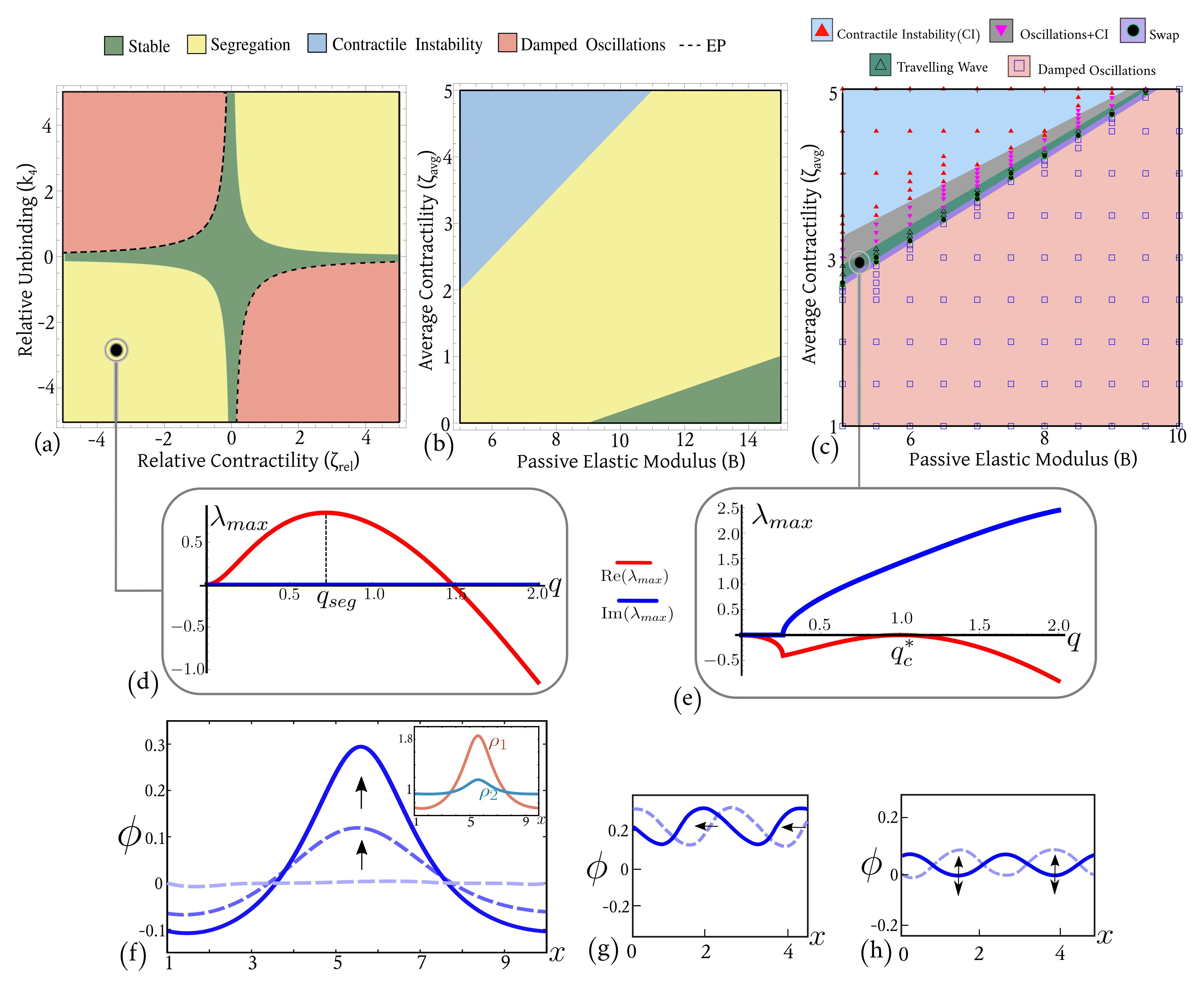
As increases beyond , goes from being negative (stable) to positive, leading to a long-wavelength instability in , with a fastest growing wave-vector which sets the characteristic width of the segregated pattern (Fig. 1(d), SI-Eq. (46)), provided is bounded between,
| (5) |
This linear segregation regime is typically realised when the relative activity and relative strain dependent unbinding have the same sign, which since the stresslets are contractile, implies . To drive segregation, the stresslet with stronger contractile activity must have a lower strain dependent unbinding rate. Note that the density peaks of the individual stresslets colocalise (Fig. 1(f) inset, also SI-Movie S1) unlike in conventional phase separation, which is reminiscent of the study in Beachetal2014 . This occurs when both stresslets exhibit catch-bond behaviour (); for slip-bond response (), the individual density peaks separate as in usual segregation (SI-Movie S2). It is worth emphasizing that even a small difference in contractility or strain dependent unbinding rate manifests as a large segregation width in the real space (SI-Fig. S2).
What is the driving force for this segregation in the linear theory? Since the stresslets do not directly interact with each other, the driving force must come from their indirect interaction through the elastomer strain. We find that to linear order, the power density
associated with the effective elastic energy density ( is the system size) is negative in the segregated phase (SI-Fig. S3), that is to say is a Lyapunov functional driving segregation of the stresslets. The appearance of micro-domains within the (more contractile) domain, is a consequence of the interplay between the strain dependent catch-bond turnover and this driving force. Note that the value of strain in the linearly segregated domains of high contractility is set by the minima of , , that depends directly on the active back pressure . Hence, the active back pressure is significant in keeping the segregated domains of stronger contractile stresslets well-separated, preventing them from clumping.
Travelling Waves
From the form of ((3)), we see that characterizes the various oscillatory phases (stable and unstable pulsations and/or waves), with frequency , and decay/growth rate . For negative values of , the oscillations grow with a fastest growing mode at a wave vector (SI-Eq. (S55)). However, as first touches (Fig. 1(e)), we get travelling waves with wave vector and frequency (SI-Eq. (S59)), whose speed is set by (See Fig. 1(g), SI-Movie S3).
Swap
As we have seen, the rate of the contractile instability is determined by the time scale , while the time scale of unbinding of the stronger stresslet is . Within the oscillatory phase, i.e., when , if the stronger stresslet unbinds before the contractile instability sets in, i.e., if , then the contracting domain bounces back. This is the swap phase, a standing wave that breaks time translation symmetry Fruchartetal2021 (See Fig. 1(h), SI-Movie S4). The swap phase does not appear as a distinct phase in the linear stability phase diagrams based on the dispersion curves. However, it appears in the full phase diagram, at the boundary between the damped oscillations and contractile instability phase, as discussed below.
Exceptional Points
So far our discussion of the instabilities has been based on the behaviour of the maximum eigenvalue . However, since the dynamical matrix is non-Hermitian (see SI Eq. (S36)), the nature of the instabilities depends crucially on the angle between the eigenvectors, in particular on exceptional points (EPs), where two eigenvalues coincide and the corresponding eigenvectors co-align Kato1984 ; Fruchartetal2021 . In general, eigenvalue based linear stability analysis gives robust predictions only about asymptotic phases, i.e., for . In the vicinity of EPs, however, short time ‘transient growth’ becomes several orders of magnitude large so that linearity fails and the system ‘bootstraps’ into nonlinear phases TrefethenEmbree2005 ; Trefethenetal93 . We defer a detailed analysis and classification of these exceptional points to a later study. Here we only mention that in the linear stability phase diagrams (Fig. 1(a)), the only EPs present are on the boundary between stable and damped oscillation phases. Hence, the system goes into the oscillatory phase through an EP Youetal2020 .
Numerical phase diagram
The swap and the travelling wave phases show up distinctly in the full phase diagram (Fig. 1(c)) obtained from a numerical analysis of the scalar version of (1) using our own code based on finite difference Euler scheme with a stencil adaptive algorithm, and the spectral methods based pde solver Dedalus dedalus2020 , with periodic boundary conditions (see SI Sec. 6 for details). For , we observe from numerical phase diagram Fig. 1(c) that there is a region where travelling waves and swap are coexisting phases in time (SI-Movie S5). Starting with small random perturbations about the uniform, symmetric unstrained steady state in the parameter regime , sustained oscillations appear at the unstable oscillations and damped oscillations phase boundary, either as a travelling wave train, or as a standing wave (i.e., swap). Typically the system exhibits a long transient, where it first goes into a swap phase, then a coexistence between swap and travelling wave, and then finally transitions into a travelling wave Lehtimakietal2021 . The transient time decreases with an increase in the average contractility. We draw the phase diagrams by making note of the configuration at a fixed large time starting from statistically identical initial conditions. The numerical code shows an eventual blowup at a very large run time in the travelling wave phase, due to the sharpness of the slopes of the travelling front. In a later publication, we will study in detail the instabilities through which swap phase transitions into the travelling wave phase. At the boundary between the oscillatory phases and the contractile instability, we see a travelling wave train with amplitude that grows indefinitely, giving rise to an array of moving tension lines.
Single stresslets
Our results for a mixture of stresslets carry over to the case of a single stresslet too, provided the dependence of the active stress on stresslet density () is steep (see SI Sec. 4), resulting in a phase separation between regions of low and high stresslet density, akin to a gas-liquid phase separation (SI-Movie S6). In the mixture of stresslets, the segregation depends on the profiles of both the average and relative contractility, and thus appears over a wider parameter range compared to the single stresslet.
Stresslets in fluid
In case of fluid mediated interaction between the stresslets, non-monotonic dependence of the active stress on the stresslet density is necessary for segregation (here, a monotonic with steep positive slope results in a clumping instability instead GowrishankarRao2016 ; HusainRao2017 . This non-monotonicity in naturally arises from the binding of contractile stresslets on finite patches of actin mesh with free boundaries embedded in a fluid, where the elastic response of the patches is taken to be fast. The crucial role of the active back pressure in driving this segregation, is played by the negative slope branch of the non-monotonic , that separates the positive slope branches corresponding to low and high stresslet density (SI Sec. 5, SI-Movie S7).
Nonlinear Effects: emergence of tension chains
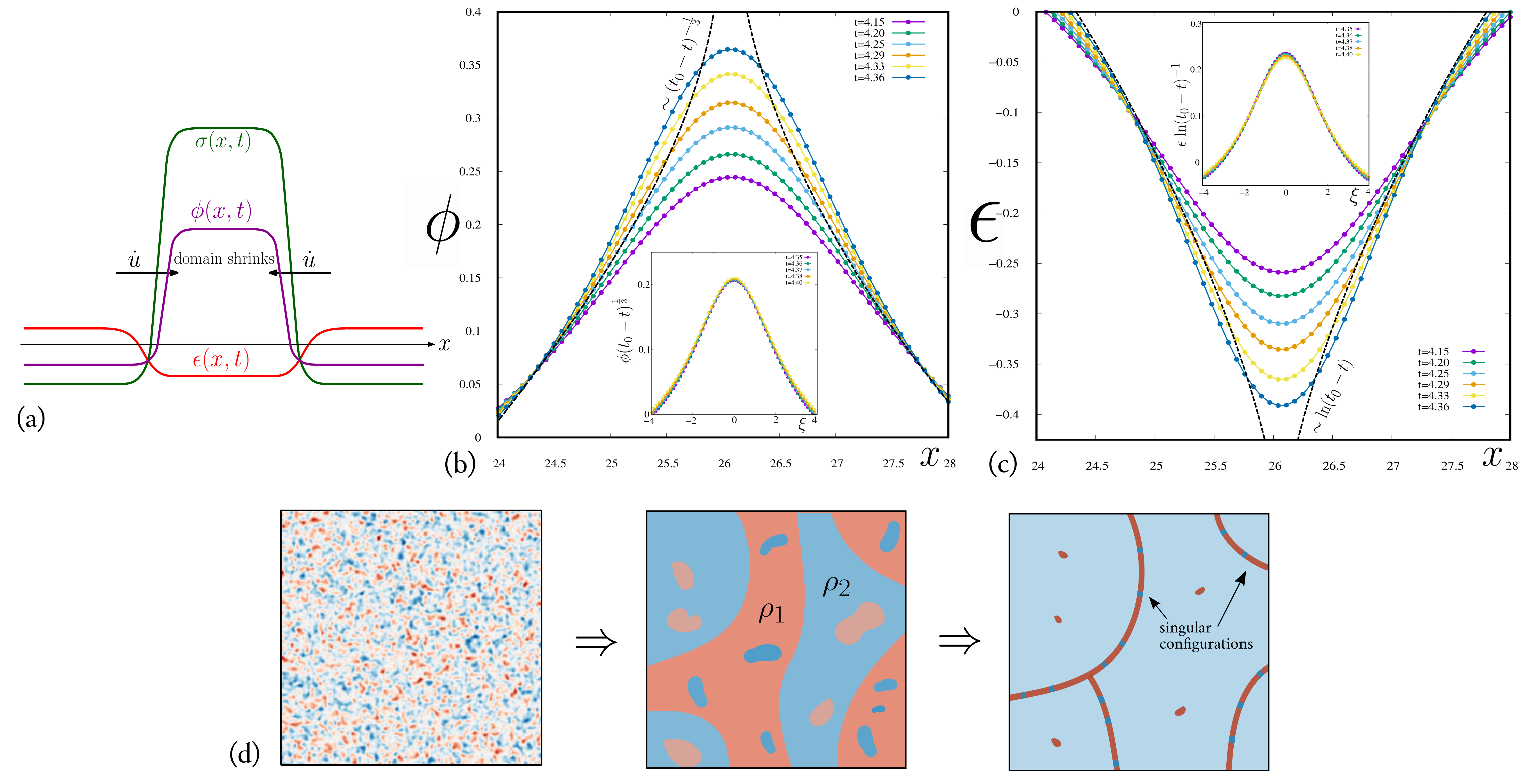
The exponential growth of the linear segregation instability quickly leads to a stage where nonlinear effects become significant. However unlike usual segregation, where nonlinearities temper the exponential growth to a slower power-law Bray2002 , here the effect of nonlinearities is to drive it to form singular structures in finite time eggersfontelos-book2015 . This happens through a feedback mechanism where a contractile instability rides atop the segregation instability. To see this, we note that the typical order parameter profile of a segregation after the linear instability regime would look like Fig. 2(a), with width . In the region between the two fronts, and so the strain (i.e., the stress is highly tensile). Outside this region and so and the stress is compressive, though of relatively low magnitude, Fig. 2(a). Since , the stress jumps across the fronts, cause them to move towards each other, resulting in an ever increasing concentration of within the shrinking domain. Enhanced catch bond response accelerates this shrinking. Eventually, this shrinking domain enters the contractile instability regime when , where there is no escape from collapse, leading to the formation of singular structures in finite time! These tensile structures remain well-separated in space through the actively produced back pressure. This is very different from the algebraically growing domains in usual phase segregation Bray2002 .
To compute the scaling behaviour as one approaches the finite-time singularity, we find it convenient to turn off the contributions from stresslet turnover, thus making and conserved. In this situation, the term in the effective strain energy density is the dominant driver of the concentration of the stresslet densities towards a singularity in a finite time at spatial location . Using the method of dominant balance eggersfontelos-book2015 in the vicinity of the singularity, we find that , and exhibit self-similar forms, , , (see SI Sec. 7A). For an initial segregating domain of width , dimensional analysis suggests that the domain width goes to zero at time , where and are the characteristic time and length scales, with . We verify these self-similar forms in a careful numerical study of the scalar version of (1) (Fig. 2(b,c)). These singularities are physical in that their resolution involves incorporation of additional physical effects such as steric hindrance (represented by the term in ). Thus the singularity is never reached, resulting in highly concentrated tensile regions of finite width, nm, the length of myosin-II bipolar filament Bershadsky2017 .
Evidently the geometry of these singular structures depends on dimensionality – in 1D the singular regions are punctae, in 2D the singular regions appear as tension chains and punctae, while in 3D, they would manifest as sheets, lines and punctae. In a finite system, these singular structures would need to be stabilized by anchoring conditions at the boundary.
Note that, these actively generated chains and sheets of tension are anisotropic structures that emerge through an unconventional spontaneous breaking of the underlying isotropic symmetry of the active elastomer. The anisotropy is a consequence of the local nature of the nonlinear effects, viz. the entrapment of contractile instability within the segregated domains of high contractility, since the effective elastic moduli depend on and . As increases in the linearly segregated domains, crosses zero (where the linear elastic response loses positive definiteness) and quickly becomes negative. As soon as the local elasticity crosses its ellipticity threshold at , ‘characteristic’ lines of the ensuing parabolic, and eventually hyperbolic response emerge; this characteristic direction sets the local tangent direction of the tension chain. This emergent anisotropy is encoded in the uniaxial nature of the singular stress field along the tension chain: SimhaBhattacharya1998 , where is the tension in the chain and is the local tangent vector specifying the orientation of the anisotropy. The uniaxial form is equivalent to the existence of a local fabric tensor Kanatani1984 , symmetric and traceless, such that . Evolution of this fabric tensor, hence the anisotropy, depends on the local history of formation and motion of the singular structure that will come from solving the complete initial-boundary-value problem, as discussed below.
Mechanics of Tension Chains
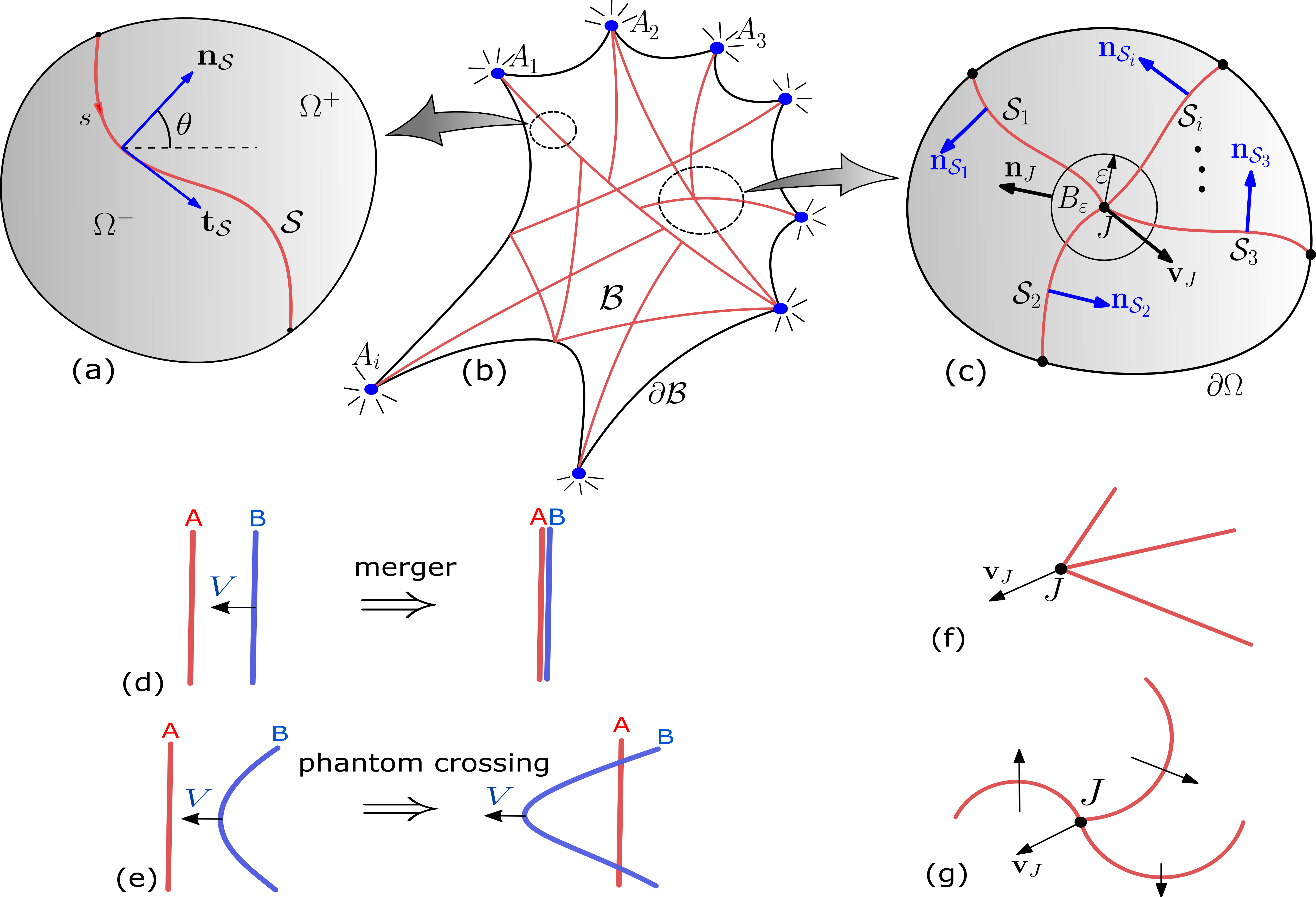
The initial profile of the two fronts bounding the segregating domain in Fig. 2 was taken to be symmetric, suggesting that there is no pressure difference on either side of the domain. In general this is not guaranteed, leading to the possibility of moving tension chains Shutovaetal2017 . A moving tension chain in a fixed 2D domain is represented by a smooth evolving curve , see Fig. 3(a). is a material curve consisting predominantly of the stronger stresslet species, across which bulk fields of the predominant weaker stresslet species suffer jump discontinuities. , in the reference configuration, has a local parametrization , where is an arc length parameter. The unit tangent and normal fields on are and , respectively, and the normal speed is . We denote the jump in a bulk discontinuous field across by , where are the limiting values of as one approaches from . We also define the average value of at as . Then, using the divergence and transport theorems for fields with line singularities in a 2D domain (details in SI Sec. 8), one can derive the governing system of equations for fields in the bulk and fields defined on the singular structure. The singular counterparts of the mass balance equations (1b) and (1c) relate the rate of change of the average and relative densities, and , of the two stresslets on the tension chain to the jump in the bulk mass flux of the stresslet species across the chain and their intrinsic flux along the chain, with contributions due to its curvature and intrinsic turnover (Eq. (S126) in SI). On the other hand, the singular counterpart of the force balance (1a) yields
| (6) |
where, is the intrinsic velocity field, is the tension, and is the curvature of the tension chain SimhaBhattacharya1998 . If we assume, for simplicity, that both the passive and the active parts of the bulk stress are isotropic, i.e., , then the normal and tangential components of the above equation along gives
| (7a) | |||
| (7b) | |||
The static tension chain, for which , gives the active Young-Laplace law, with the following consequences: (1) the tension chain is straight () if the active pressure jump counter-balances the (passive) elastic pressure jump (i.e., ); (2) in the absence of a passive pressure jump (i.e., ), active pressure jump gives rise to a curved tension chain (); and (3) tension along the chain is constant.
It can be readily shown (SI Sec. 8G) that, if two tension chains are moving relatively towards each other (Fig. 3(d,e)), they merge into a single chain if they are straight and parallel and they scatter if they are curved towards each other.
Finite geometry – surface anchoring and wetting
In the finite geometry of the cell, one needs to specify appropriate boundary anchoring conditions or boundary interactions at the cell surface. A natural choice is to declare surface anchoring at the locations of integrin-based focal adhesions and cadherin-based adherens junctions. These molecular complexes embedded in the cell surface bind strongly to the actomyosin filaments via linker proteins on the intracellular side and to the substrate or adjoining cells on the extracellular side.
The complete initial boundary value problem for a cell body consists of the dynamical (1), supplemented by the anchoring conditions and at a finite number of boundary points , and no flux conditions and on the rest of the boundary , together with appropriate initial condition respecting the boundary data (Fig. 3(b)). Due to contractile activity, the stress fields naturally concentrate near , and stable tension chains emerge from these anchoring points that span the whole system .
One may deduce the force-balance condition at these rigid anchoring sites to be,
| (8) |
where tension chains meet at the anchoring point . Here, is a singular force at coming from the bulk stress field , is a small half-ball, centered at , of radius and unit normal pointing into the bulk; is the tension on the boundary at acting along the tangent (coming from surface elasticity of the membrane); and is the reaction force due to the anchoring condition (Fig. 3(c)). Supported by these stable anchoring sites at the cell surface, the intracellular side may support a tension web, viz. a web of tension lines with multi-valent junctions. For tension chains meeting at a junction , force balance at the junction gives
| (9) |
where is a singular force at coming from the bulk stress field ; here, is a small ball of radius and unit normal containing the junction SimhaBhattacharya1998 , is the velocity and is the friction coefficient at the junction (Fig. 3(b)). If is isotropic, . Using the compatibility condition for velocity at the junction Fischeretal2012 : , , and the identities and , where and are the angles that and , respectively, make with a fixed global axis (say, the -axis), and , the force balance equation gives
| (10) |
for ; here, . This is the generalized Young-Dupré equation for junctions of force chains embedded in an elastic medium. In striking contrast to the usual Young-Dupré equations, these junctions can display polarity and hence move, or can support spiral junctions which will rotate owing to active stresses (Fig. 3(f,g)).
Since the effective elasticity is hyperbolic when the tension chain formation takes place, the standard boundary conditions for conventional elliptic elasticity discussed above may become incompatible for the hyperbolic regimes. This is because hyperbolic equations require half of the boundary conditions than its elliptic counterpart, as the rest are determined by ‘propagating’ the boundary data along the characteristics to the other half Bouchaud2002 . Such incompatible boundary conditions, or more interestingly, perturbations of compatible boundary conditions (thereby altering the geometry of focal adhesions), for a given set of steady state tension chains, would make the web unstable, resulting in dynamical remodelling of the web through binding-unbinding, so that the boundary conditions become compatible again for the remodelled network Bouchaud2002 ; RamaswamyRao2007 . The steady state configuration of a web of tension chains is, hence, fragile in this precise sense Catesetal1998 .
Alternatively, one may also specify boundary interactions, either at the inner leaflet of the cell membrane or on the membranes of intracellular organelles, such as the Golgi, endosomes or nucleus. A moving actomyosin web can then adhere to the inner surface of the cell membrane or wrap over organelles. Considering a mixture of stresslets, one species may have a preferential wetting to a substrate, say the inner leaflet of the cell membrane. In SI Sec. 9, we demonstrate that active bulk segregation together with preferential wetting to the substrate will ensure macroscopic segregation where one of the stresslets wets the substrate (SI-Movie S8 and S9), leading to a stratified layering of the two stresslets.
Discussion
To summarize, we have shown using a hydrodynamic approach, how active cytoskeletal stresslets that act as molecular force generators and sensors, give rise to striking singular patterns of nonequilibrium tension chains within the cell. It is the nonlinear coupling between the linear segregation and contractile instability that leads to the formation of these singular tension patterns. The associated anisotropy emerges via local departure from elliptic elasticity. In the finite geometry of the cell, these patterns are stabilised by cell surface anchors such as focal adhesions and adherens junctions. These fragile force patterns are sensitive to boundary conditions, and thus naturally shaped by the current geometry of the system Matthewsetal2006 ; banerjeeetal2020 . Thus, different FA and AJ geometries would result in different networks with different mechanical properties GeigerSpatzBershadsky2009 ; GuptonWaterman-Storer2006 . Simultaneously, the system exhibits a variety of oscillatory force patterns such as travelling waves, swap, and their temporal coexistence, that are accessed through an exceptional point of this underlying non-reciprocal dynamics.
An immediate extension of this work is to analyse the force patterning in 3D, where we expect both tension lines and sheets to emerge. Further, we will study the implications of nonequilibrium force patterning for force adaptation and strain homeostasis and analyse the frequency dependent rheological response of the stable steady state of tension chains with rigid anchors at the boundary. In an ongoing study, we find that segregation is substantially enhanced in a mixture of contractile and extensile stresslets due to presence of both attraction and repulsion mediated through the elastomer jimenezetal2021 , which, with nonlinear feedback, result in singular force patterns that include both tension and compression chains (the latter endogeneously stabilizes the former, together with boundary anchoring) – with implications for an active hydrodynamic theory of cellular tensegrity ingberetal2014 and the nonequilibrium assembly of active metamaterials PishvarHarne2020 ; Qietal2022 .
Acknowledgement. We thank T. van Zanten for a careful reading and pointing us to relevant experimental references, A. Nagilla for help in the initial numerical implementation and the members of Simons Centre for critical discussions. We acknowledge support from the Department of Atomic Energy (India), under project no. RTI4006, and the Simons Foundation (Grant No. 287975), and computational facilities at NCBS. MR acknowledges a JC Bose Fellowship from DST-SERB (India)
References
- (1) T. D. Pollard and R. D. Goldman (Editors), The Cytoskeleton, Cold Spring Harbor Laboratory Press (2016).
- (2) N. Taneja, M. R. Bersi, S. M. Baillargeon, A. M. Fenix, J. A. Cooper, R. Ohi, V. Gama, W. D. Merryman and D. T. Burnette, Precise Tuning of Cortical Contractility Regulates Cell Shape during Cytokinesis, Cell Reports 31, 107477 (2020).
- (3) B. D. Matthews, D. R. Overby, R. Mannix and D. E. Ingber, Cellular adaptation to mechanical stress: role of integrins, Rho, cytoskeletal tension and mechanosensitive ion channels, Journal of Cell Science 119, 508–518 (2006).
- (4) S. Banerjee, M. L. Gardel and U. S. Schwarz, The actin cytoskeleton as an active adaptive material, Annual Review of Condensed Matter Physics 11, 421–439 (2020).
- (5) S. F. Shimobayashi, P. Ronceray, D. W. Sanders, M. P. Haataja and C. P. Brangwynne, Nucleation landscape of biomolecular condensates, Nature 599, 503–506 (2021).
- (6) W. F. Marshall, Pattern formation and complexity in single cells, Current Biology 30, R544–R552 (2020).
- (7) A. J. Jimenez, A. Schaeffer, C. D. Pascalis, G. Letort, B. Vianay, M. Bornens, M. Piel, L. Blanchoin and M. Théry, Acto-myosin network geometry defines centrosome position, Current Biology 31, 1206–1220 (2021).
- (8) E. Makhija, D. S. Jokhun and G. V. Shivashankar, Nuclear deformability and telomere dynamics are regulated by cell geometric constraints, The Proceedings of the National Academy of Sciences 113, E32–E40 (2015).
- (9) X. Sun, D. Y. Z. Phua, L. Axiotakis Jr., M. A. Smith, E. Blankman, R. Gong, R. C. Cail, S. E. de los Reyes, M. C. Beckerle, C. M. Waterman and G. M.Alushin, Mechanosensing through Direct Binding of Tensed F-Actin by LIM Domains, Developmental Cell 55, 468–482 (2020).
- (10) M. S. Shutova, S. B. Asokan, S. Talwar, R. K. Assoian, J. E. Bear and T. M. Svitkina, Self-sorting of nonmuscle myosins IIA and IIB polarizes the cytoskeleton and modulates cell motility, Journal of Cell Biology 216, 2877–2889 (2017).
- (11) M. Kovács, K. Thirumurugan, P. J. Knight, and J. R. Sellers, Load-dependent mechanism of nonmuscle myosin 2, The Proceedings of the National Academy of Sciences 104, 9994–9999 (2007).
- (12) R. Fernandez-Gonzalez, S. M. Simoes, J-C Röper, S. Eaton, and J. A. Zallen, Myosin II dynamics are regulated by tension in intercalating cells, Developmental Cell 17, 736–743 (2009)
- (13) Y. Mulla, M. J. Avellaneda, A. Roland, L. Baldauf, S. J. Tans and G. H. Koenderink, Weak catch bonds make strong networks, bioRxiv preprint doi: https://doi.org/10.1101/2020.07.27.219618 (2022).
- (14) J. R. Beach, L. Shao, K. Remmert, D. Li, E. Betzig and J. A. Hammer III, Nonmuscle Myosin II Isoforms Coassemble in Living Cells, Current Biology 24, 1160–1166 (2014).
- (15) P. Hotulainen and P. Lappalainen, Stress fibers are generated by two distinct actin assembly mechanisms in motile cells, The Journal of Cell Biology 173, 383–394 (2006).
- (16) S. Hu, K. Dasbiswas, Z. Guo, Y-H. Tee, V. Thiagarajan, P. Hersen, T-L. Chew, S. A. Safran, R. Zaidel-Bar, and A. D. Bershadsky , Long-range self-organization of cytoskeletal myosin II filament stacks, Nature Cell Biology 19, 133–141 (2017).
- (17) K. Weißenbruch, J. Grewe, M. Hippler, M. Fladung, M. Tremmel, K. Stricker, U. S. Schwarz and M. Bastmeyer, Distinct roles of nonmuscle myosin II isoforms for establishing tension and elasticity during cell morphodynamics, eLife 10, e71888 (2021).
- (18) M. Vicente-Manzanares, M. A. Koach, L. Whitmore, M. L. Lamers and A. F. Horwitz, Segregation and activation of myosin IIB creates a rear in migrating cells, Journal of Cell Biology 183, 543–554 (2008).
- (19) M. S. de Silva, M. Depken, B. Stuhrmann, M. Korsten, F. C. MacKintosh and G. H. Koenderink, Active multistage coarsening of actin networks driven by myosin motors, The Proceedings of the National Academy of Sciences 108, 9408–9413 (2011).
- (20) G. H. Koenderink and E. K. Paluch, Architecture shapes contractility in actomyosin networks, Current Opinion in Cell Biology 50, 79–85 (2018).
- (21) R. Zhang, S. A. Redford, P. V. Ruijgrok, N. Kumar, A. Mozaffari, S. Zemsky, A. R. Dinner, V. Vitelli, Z. Bryant, M. L. Gardel and J. J. de Pablo, Spatiotemporal control of liquid crystal structure and dynamics through activity patterning, Nature Materials 20, 875–882 (2021).
- (22) R. Fernandez-Gonzalez, and J.A. Zallen, Oscillatory behaviors and hierarchical assembly of contractile structures in intercalating cells, Physical Biology 8, 045005 (2011).
- (23) A. Munjal, J-M. Philippe, E. Munro and T. Lecuit, A self-organized biomechanical network drives shape changes during tissue morphogenesis, Nature 524, 351–355 (2015).
- (24) D. S. Banerjee, A. Munjal, T. Lecuit and M. Rao, Actomyosin pulsation and flows in an active elastomer with turnover and network remodeling, Nature Communications 8, 1121 (2017).
- (25) J. I. Lehtimäki, E. K. Rajakylä, S. Tojkander and Pekka Lappalainen, Generation of stress fibers through myosin-driven re-organization of the actin cortex, eLife 10, e60710 (2021).
- (26) M. C. Marchetti, J.-F. Joanny, S. Ramaswamy, T. B. Liverpool, J. Prost, M. Rao and R.A. Simha, Hydrodynamics of Soft Active Matter, Review of Modern Physics 85, 1143–1189 (2013).
- (27) S. Banerjee, T.B. Liverpool and M.C. Marchetti, Generic phases of cross-linked active gels: Relaxation, oscillation and contractility, Europhysics Letters 96, 58004 (2011).
- (28) S. Banerjee and M.C. Marchetti, Instabilities and oscillations in isotropic active gels, Soft Matter 7, 463 (2011).
- (29) A. J. Bray, Theory of phase ordering kinetics, Advances in Physics 51, 481–587 (2002).
- (30) M. Fruchart, R. Hanai, P. B. Littlewood, and V. Vitelli, Non-reciprocal phase transitions, Nature 592, 363–369 (2021).
- (31) T. Kato, Perturbation Theory for Linear Operators, 2nd edn, Springer (1984).
- (32) Z. You, A. Baskaran and M. C. Marchetti, Nonreciprocity as a generic route to traveling states, The Proceedings of the National Academy of Sciences 117, 19767–19772 (2020).
- (33) P. Ronceray, C. P. Boedersz and M. Lenz, Fiber networks amplify active stress, The Proceedings of the National Academy of Sciences 113, 2827–2832 (2016).
- (34) P. Ronceray, C. P. Boedersz and M. Lenz, Stress-dependent amplification of active forces in nonlinear elastic media, Soft Matter 15, 331–338 (2019).
- (35) B. Geiger, J. P. Spatz and A. D. Bershadsky, Environmental sensing through focal adhesions, Nature Reviews Molecular Cell Biology 10, 21–33 (2009).
- (36) S. L. Gupton and C. M. Waterman-Storer, Spatiotemporal Feedback between Actomyosin and Focal-Adhesion Systems Optimizes Rapid Cell Migration, Cell 125, 1361 (2006).
- (37) P. Rosakis, J. Notbohm, and G. Ravichandran, A model for compression-weakening materials and the elastic fields due to contractile cells, Journal of The Mechanics and Physics of Solids, 85, 16–32 (2015).
- (38) G. Grekas, M. Proestaki, P. Rosakis, J. Notbohm, C. Makridakis and G. Ravichandran, Cells exploit a phase transition to mechanically remodel the fibrous extracellular matrix, Journal of The Royal Society Interface, 18, 20200823 (2021).
- (39) M. C. Keeling, L. R. Flores, A. H. Dodhy, E. R. Murray and N. Gavara, Actomyosin and vimentin cytoskeletal networks regulate nuclear shape, mechanics and chromatin organization,Scientific Reports 7, 5219 (2017).
- (40) J. K. Knowles and E. Sternberg, On the failure of ellipticity and the emergence of discontinuous deformation gradients in plane finite elastostatics, Journal of Elasticity, 8, 329–379 (1978).
- (41) H. C. Simpson and S. J. Spector, On failure of the complementing condition and nonuniqueness in linear elastostatics, Journal of Elasticity, 15, 229–231 (1985).
- (42) Yu. V. Egorov and E. Sanchez-Palencia, On ill-posedness of free-boundary problems for highly compressible two-dimensional elastic bodies, St. Petersburg Math. J. 22, 913–926 (2011).
- (43) R. Blumenfeld, Isostaticity and controlled force transmission in the cytoskeleton: A model awaiting experimental evidence, Biophysical Journal 91, 1970–1983 (2006).
- (44) M. E. Cates, J. P. Wittmer, J. P. Bouchaud, and P. Claudin, Jamming, Force Chains, and Fragile Matter, Physical Review Letters 81, 1841 (1998).
- (45) J. P. Bouchaud, Granular Media: Some ideas from Statistical Physics, in Slow Relaxations and Nonequilibrium Dynamics in Condensed Matter, NATO Advanced Study Institute, Les Houches, Session LXXVII, eds. J.L. Barrat, M. Feigelman, J. Kurchan and J. Dalibard. (2002).
- (46) M. Otto, J.P. Bouchaud, P. Claudin and J.E.S. Socolar, Anisotropy in granular media: Classical elasticity and directed-force chain network, Physical Review E 67, 031302 (2003).
- (47) A. Roychowdhury and L. Truskinovsky, Towards Marginal Elascity, to be submitted.
- (48) L. N. Trefethen, A. E. Trefethen, S. C. Reddy and T A Driscoll, Hydrodynamic Stability Without Eigenvalues, Science 261, 578–584 (1993).
- (49) L. N. Trefethen and M. Embree, Spectra and Pseudospectra, Princeton University Press (2005).
- (50) K. J. Burns, G. M. Vasil, J. S. Oishi, D. Lecoanet and B. P. Brown, Dedalus: A Flexible Framework for Numerical Simulations with Spectral Methods, Physical Review Research 2, 023068 (2020).
- (51) K. Gowrishankar and M. Rao, Nonequilibrium phase transitions, fluctuations and correlations in an active contractile polar fluid, Soft matter 12, 2040–2046 (2016).
- (52) K. Husain and M. Rao, Emergent structures in an active polar fluid: Dynamics of shape, scattering, and merger, Physical Review Letters 118, 078104 (2017).
- (53) J. Eggers and M. A. Fontelos, Singularities: Formation, Structure, and Propagation, Cambridge University Press, Cambridge, UK (2015).
- (54) K-I. Kanatani, Distribution of directional data and fabric tensors, International Journal of Engineering Science 22, 149–164 (1984).
- (55) N. K. Simha and K. Bhattacharya, Kinetics of Phase Boundaries with Edges and Junctions, Journal of The Mechanics and Physics of Solids 46, 2323–2359 (1998).
- (56) F. D. Fischer, J. Svoboda and K. Hackl, Modelling the kinetics of a triple junction, Acta Materiala 60, 4704–4711 (2012).
- (57) S. Ramaswamy and M. Rao, Active-filament hydrodynamics: instabilities, boundary conditions and rheology, New Journal of Physics 9, 423 (2007).
- (58) D. E. Ingber, N. Wang and D. Stamenović, Tensegrity, cellular biophysics, and the mechanics of living systems, Reports on Progress in Physics 77, 046603 (2014).
- (59) M. Pishvar and R. L. Harne, Foundations for Soft, Smart Matter by Active Mechanical Metamaterials, Advanced Science 7, 2001384 (2020).
- (60) J. Qi, Z. Chen, P. Jiang, W. Hu, Y. Wang, Z. Zhao, X. Cao, S. Zhang, R. Tao, Y. Li and D. Fang, Recent Progress in Active Mechanical Metamaterials and Construction Principles, Advanced Science 9, 2102662 (2022).