Can autism be diagnosed with AI? A narrative review
Abstract
Radiomics with deep learning models have become popular in computer-aided diagnosis and have outperformed human experts on many clinical tasks. Specifically, radiomic models based on artificial intelligence (AI) are using medical data (i.e., images, molecular data, clinical variables, etc.) for predicting clinical tasks like Autism Spectrum Disorder (ASD). In this review, we summarized and discussed the radiomic techniques used for ASD analysis. Currently, the limited radiomic work of ASD is related to variation of morphological features of brain thickness that is different from texture analysis. These techniques are based on imaging shape features that can be used with predictive models for predicting ASD. This review explores the progress of ASD-based radiomics with a brief description of ASD and the current non-invasive technique used to classify between ASD and Healthy Control (HC) subjects. With AI, new radiomic models using the deep learning techniques will be also described. To consider the texture analysis with deep CNNs, more investigations are suggested to be integrated with additional validation steps on various MRI sites.
1 Introduction
Autism spectrum disorder (ASD) is a pervasive developmental disorder with cognitive abilities that are below normal for their age group. Its core symptoms are categorized by social communication deficits, repetitive stereotypical interests, and persistent patterns of behavior Hyman et al. (2020). For example, ASD patients have an inability to understand other intentions properly, reduced interactive eye contact, etc. Specifically, ASD endangers the physical and mental health of children, placing a burden on patients’ social interaction, learning, life, employment, family, and society van’t Hof et al. (2021). In this context, early diagnosis and early intervention for children with ASD can greatly improve the lives of those affected Tao (2020). Unfortunately, ASD has unclear direct indicators, and many studies have suggested the genetic factors Toma (2020), immunological Al-Ameen et al. (2020), and neuropsychological associations Haigh et al. (2020). The prevalence of mental disabilities among children aged six and below in China is 1/1000, and ASD accounts for 36.9% of them Center (2015). According to the World Health Organization, the prevalence of ASD is gradually increasing worldwide, with a global average prevalence of 62/10,000 (0.62%), equivalent to one child with ASD in every 160 children. It has become a major global public health problem Lord (2020).
Most of the diagnostic tools and methods are based on various tests. The present research work on ASD symptoms is not accurate Kim et al. (2018). It is generally believed that children with ASD will have many problems in their growth and development Crowell et al. (2019); Arbanas (2015). So far, there are incomplete diagnostic tools for ASD. In this context, a multi assessment is usually considered. ASD could be related to developmental delays and abnormalities. This assessment considers the growth history, parental interview Crowell et al. (2019), medical examinations if necessary, and many other things. According to American Psychiatric Association’s Diagnostic and Statistical Manual of Mental Disorders, 5th edition (DSM-5), the diagnostic criteria for ASD consist of persistent defects in social communication, social interaction, restricted and repetitive behavior patterns Arbanas (2015). In Lord et al. (2000), the Autism Diagnostic Observation Schedule (ADOS) is considered the gold standard test because of its reliability, validity and usefulness. Unfortunately, this test-based screening method can only diagnose children when they have the ability to communicate. We note that there is a great advantage when ASD could be identified at an earlier age. However, according to traditional methods, ASD is difficult to identify at an early age Farooq and Ahmed (2020) due to the gaps in cognitive abilities in infants at 24 months or older Miller et al. (2021). Even with clinical investigation, deep neurological assessment seems to be more needed. ASD diagnosis is improved by involving many neurological techniques/features (e.g., brain waves Jayawardana et al. (2019); Heunis et al. (2018), magnetic resonance images (MRI) Chaddad et al. (2017a) and eye-tracking techniques Vacas et al. (2021), etc.). Another aspect of assessment is related to many genes for predicting ASD Cederquist et al. (2020). For example, SHANK3 Soorya et al. (2013) and PTCHD1 Noor et al. (2010) are two genes involved in the pathogenesis of ASD through regulation of the nervous system. ITGB3 Napolioni et al. (2011), is associated with the pathogenesis of similar disorders. Like genomic analysis, imaging analysis is a promising technique that leads to identify ASD patients.
Imaging like MRI is used to show the anatomy of brain (e.g., ASD patients) Nordahl et al. (2016). Two types are the most considered identifying ASD patients: 1) functional magnetic resonance imaging (fMRI) and 2) structural magnetic resonance imaging (sMRI or MRI) Sen et al. (2018a). The fMRI can show brain function, like active brain regions. While sMRI shows the structure variations (e.g., growth, deformation, atrophy, etc.). In addition, sMRI/MRI is currently the most used technique for imaging the brain structure due to its fast and high-resolution 3D volume imaging. For ASD, sMRI can describe structural brain changes by analyzing gray matter volume, cortical thickness, cortical complexity, and co-variance networks. While fMRI relies on the oxygen content of local tissue vessels (blood oxygen levels depend on functional brain MRI imaging) and can track signal changes in real time. So far, MRI or fMRI provide relevant imaging features that are related to ASD Sen et al. (2018a).
Imaging features (or radiomics) are widely used in medical image analysis. Among the imaging feature techniques, the following features (radiomics) are the most features used for ASD, namely, 1) colour features, 2) texture, 3) shape/morphology, and 4) spatial relationship features. Briefly, describe the surface properties of the image. It is not affected by image rotation and translation; can better describe the structure image; can effectively describe the geometrical area (i.e., region of interest); can enhance the ability to describe and distinguish the content of the image. These features have been used for many clinical applications such as cancer Chaddad et al. (2019a, 2018a, 2018b, 2016, 2014, b, c, 2017b), neuroimaging Zhang et al. (2021); Chaddad et al. (2018c), segmentation Chaddad and Tanougast (2016), etc. As ASD examples, MRI regional features were computed to study the abnormalities in brain development of ASD patients YangLiu et al. (2020). In Xiuyan (2018), shape features are considered to predict ASD. In Smith et al. (2016); Wolff et al. (2018), multiple ASD brain developmental abnormalities are detected during infancy. Moreover, many studies have shown that young children with ASD have a much larger brain size compared to their normally developing peers Lainhart et al. (1997). The overall volume and density are significantly larger than those of normal children (or healthy control) Sacco et al. (2015). Briefly, we will describe the brain differences between ASD and HC as follows:
Surface area: Studies have shown that the early cerebral cortex of children with autism expands rapidly between six and twelve months. This atypical expansion leads to problems such as visual receptivity deficits and neglect of social cues Elison et al. (2013). Cortical surface area increases with an accelerated rate between one and two years of age Hazlett et al. (2017). It coincides with problems of social deficits. Another longitudinal study found that the white matter of the temporal lobe of the brain increased in autistic children between two and four to five years old. However, the brain grows at a similar rate to normal during this age interval Nassar et al. (2009). As a result, the overgrowth of the temporal and frontal lobes is an indicator of ASD Lord (2020); Hazlett et al. (2017); Nassar et al. (2009).
Cerebrospinal fluid: An excessive increase in cerebrospinal fluid can also occur in young children with ASD. During the first few months of life (i.e., within 24 months), infants with ASD have a high volume of inter-axial extra-cerebrospinal fluid Wang . Specifically, the increase in the volume of extra-axial cerebrospinal fluid is more significant at 6 months, which is about 25% higher than ordinary babies. This is related to movement, communication, and the condition of ASD. When the extra-axial cerebrospinal fluid continues to rise, the communication disorder will become more serious Huang (2018).
Structural abnormalities in the white matter: The corpus callosum develops abnormally at six months. Its area and volume increase significantly Wolff et al. (2015). This abnormality is positively correlated with the stereotypical behavior of children with autism Wolff et al. (2017). Therefore, structural abnormalities in the white matter are likely to be an important causal factor in the core social deficits (especially emotional disturbances) of later autism.
For ASD diagnosis, clinicians have been committed to using neuroimaging tools. It can automatically distinguish patients with brain diseases from HC or other patients. This can be achieved using features (e.g., imaging, genes, clinical, omics, etc.) with machine learning (ML). ML consists of many methods to classify between classes (e.g., neural networks, support vector machines, random forests, etc.). It learns how to identify the features associated with ASD and then constructs a relevant model. The accuracy of a classifier/predictive model is improved by training the model on large datasets. Eventually, the model can be relied upon to diagnose the presence of ASD. Its accuracy is measured by how well it is able to predict the true class (e.g., ASD). In this context, we aim in this paper to discuss the general radiomics/features and AI/ML model for predicting ASD.
The rest of this paper is structured as the following: Section 2 contains a review of the literature on ASD diagnostic methods. We then discuss the general radiomic methodology for predicting ASD in Section 3. In Section 4, we present the recent explainable artificial intelligence (XAI) literatures that are related to ASD. Section 5 discusses the strengths and limitations of ASD predictive models and summarizes the main findings of this study. Last, Section 6 concludes the paper with future recommendations.
2 Related works
Increasing attention has been remarkable for ASD, when Leo Kanner has talked about ASD in 1943, and has mentioned that the ASD is related to the brain Piven et al. (1997); Courchesne et al. (2001); 29 (2020); Reinhardt et al. (2020). In Piven et al. (1997), children with ASD show larger brain volume than HC. In Courchesne et al. (2001), ASD was related to large and small brain white matter hyperplasia and early gray matter hyperplasia, respectively. Compared to HC, ASD children have a larger volume in the amygdala 29 (2020) and hippocampus Reinhardt et al. (2020); Guannan et al. (2019). Most of these studies consider the classifications between ASD and HC according to the difference in brain volume or thickness. While, texture feature based on gray-scale co-occurrence matrix (GLCM) and Laplacian filter was firstly appeared by Chaddad et al. to compare Chaddad et al. (2017a) and classify Chaddad et al. (2017c) between ASD and HC. As the prevalence of autism increases year by year, effective ASD diagnostic methods have become a major concern worldwide. We summarize three main diagnostic methods as follows:
Electroencephalography (EEG): EEG measures neural activity and can detect children at risk of developing ASD and, thus, provide an opportunity for early diagnosis. For example, EEG data is used to compare between ASD and HC Bosl et al. (2018); Heunis et al. (2018); Vicnesh et al. (2020). In Peya et al. (2020), used CNN model for classification after converting the data into 2D form. Although EEG can be used to diagnose ASD, it still has limitations in a number of conditions (e.g., signal noises).
Eye tracking: It is based on characteristic changes of eyes, like periphery and iris. In Moriuchi et al. (2017), they studied 86 two-year-olds (26 ASD, 38 HC and 22 Developmentally delayed children). It shows that eyes with ASD were associated with passive insensitivity to social signals. In Pino et al. (2021), they selected 29 ASD children aged between 5-11 years. Through the visualization of real faces and avatars, it was possible to study how children with ASD recognize emotions. For ASD patients, eye tracking is not an optimal method because it takes long time for children to cooperate, in addition that it is not flexible for clinical diagnosis.
MRI/fMRI scans: Data quality of MR imaging is improving in function with the advanced technology. Previous studies have shown that brain structures in patients with ASD can differ significantly in terms of volume, thickness, and texture Albajara Sáenz et al. (2020); Chaddad et al. (2017c). However, this scenario still under radiomic and AI investigation for ASD diagnosis. Yet, no clear tools have been involved in the clinical system. However, we find many works prove that classifier models using extracted features from MRI/fMRI images have the ability to predict ASD patients. For example, a support vector machine (SVM) model has shown an accuracy value of 66.8% to predict ASD images Sen et al. (2018b). In Mellema et al. (2019), 12 classifiers are compared, namely, six nonlinear shallow ML models, three linear shallow models, and three deep learning models. A dense feedforward network provides the best results among the 12 models with AUC value of 80%. This demonstrates that even when using features derived from imaging data, deep learning methods like dense feedforward network can provide higher predictive accuracy over classical ML methods Mellema et al. (2019). To let the AI models be feasible for ASD prediction, more investigation is recommended since the performance metrics are still limited. In addition, no clear study is considered when the images from MRI scanner consist of 7 Tesla or above. We believe that more resolution of images will let the radiomic with texture analysis be more informative for predicting ASD Chaddad et al. (2017a, c).
3 Radiomic methodology
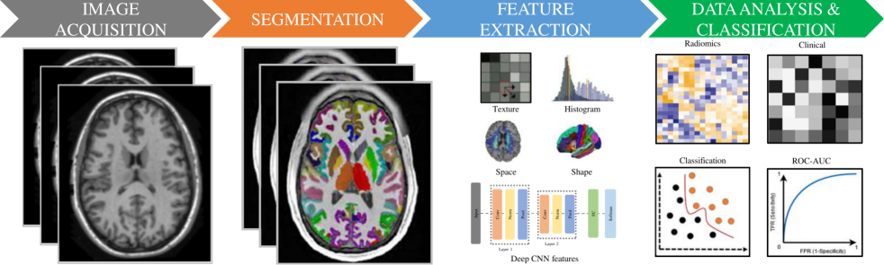
To provide a wider perspective to the readers, radiomic pipeline is simply given in Figure 1. It illustrates the processing steps for radiomic pipeline that consist of image acquisition and preprocessing, segmentation, feature extraction, statistical analysis, and classifications. We described below a detailed review of each step.
3.1 Image acquisition and preprocessing
The purpose of preprocessing is to improve the visual effect of the image. It can purposely emphasize the overall or partial characteristics of the image for various scenarios Wang et al. (2020). For example, improve the color, brightness, and contrast of the image. There are two main methods for image enhancement: a) Spatial domain that includes image gray-scale transformation, histogram correction Salem et al. (2019), local statistics method, image smoothing and image sharpening, etc. As in Bao et al. (2019), fast non-rigid registration can improve the contrast of the brain structures. In Zhao et al. (2019), image enhancement method based on brightness level and gradient modulation. This method reduces the dynamic range of the brightness level and enhances the details (i.e., texture) of the image. b) frequency domain that transforms an image into the frequency domain for filtering using Fourier analysis. The image is then inversely transformed back into the spatial domain. The most used frequency-domain methods are the homomorphic filtering Yugander et al. (2020) and wavelet transform Sagheer and George (2020). Therefore, image denoising plays a major role for texture analysis. It can be described by probability distribution function and probability density function. We note that the texture analysis could be related to the scale of image filtering and then to ASD Park et al. (2017).
3.2 Normalization/standardization
Image normalization is one of the preprocessing steps to avoid image distortions (i.e., translation, rotation, scaling, and skew). However, the main challenge for ASD studies is related to MRI images derived from multisite Yankowitz et al. (2020); Hoeksma et al. (2005); Delisle et al. (2021). For example, scans from multisite lead to high differences of texture features between these sites. In Ahammed et al. (2021a), Min-Max normalization is used to overcome the image variation, and convert the image values to a range of [0-1]. Normalization of images here improves the learning rate, reduces the dependence on initialization, and reduces the training time to overcome the overfitting problem. In Dekhil et al. (2020), non-parametric models are used to correct intensity inhomogeneities and avoid the scanner distortions. In Mendes et al. (2021), MRI was normalized using voxel-based morphometry (VBM) that is available in the Statistical Parametric Mapping software 111. VBM technique is spatially normalizing the MRI scans to the same stereotactic space to correct nonuniform intensity variations Ashburner and Friston (2000). Recent work shows that domain adaptation can effectively reduce the site variation using the CNN models Delisle et al. (2021). However, more work on image normalization for multisite variation is needed to extract the texture features for radiomic analysis.
| Work | Data Source | Cases Number | Data Type | FEM | Classifer Model | Acc | Sen | Spec | AUC |
|---|---|---|---|---|---|---|---|---|---|
| Haweel et al. (2021a) | FSL | 50 ASD and 50 HC | TfMRI | SPF | DWT-CNN | 80% | 84% | 76% | - |
| K and Murthy Oruganti (2021) | ABIDE-I+II | 23 ASD and 15 HC | Rs-fMRI | SPF | SVM | 80.76% | - | - | - |
| Dekhil et al. (2021a) | NDAR | 185 subjects | sMRI-fMRI | SF | RF | 80.8% | 84.9% | 79.2% | 81.92% |
| Yang et al. (2019) | ABIDE | 505 ASD and 530 HC | Rs-fMRI | SPF | Ridge Return | 71.98% | - | - | - |
| Gao et al. (2021) | ABIDE | 518 ASD and 567 HC | rs-fMRI | SF | CNN | 71.8% | 81.25% | 68.75% | 67% |
| Squarcina et al. (2021) | Private | 40 ASD and 36 HC | MRI | SF | SVM | 84.2% | 80% | 88.9% | - |
| Heinsfeld et al. (2018) | ABIDE-I | 505 ASD and 530 HC | rs-fMRI | OF | DNN | 70% | 74% | 63% | - |
| Sen et al. (2018c) | ADHD-200 | 279 ASD and 279 HC | fMRI | TF | SVM | 64.91% | 44.16% | 81.91% | - |
| Alvarez-Jimenez et al. (2020) | ABIDE-I+II | 76 ASD and 75 HC | MRI | TF and SF | SVM | 64.3% | 77% | 82% | 69% |
| Madine et al. (2020) | ABIDE-I | 155 ASD and 186 HC | T1-MRI | SF | HGNN | 76.7% | - | - | - |
| Wang et al. (2019) | ABIDE-I+II | 255 ASD and 276 HC | rs-fMRI | SF | SVM | 75.00%–95.23% | 90.62% | 90.58% | - |
| Fu et al. (2021) | ABIDE | 539 ASD and 573 HC | T1-MRI | SF | 6 classifiers | 80% | - | - | - |
| Shi et al. (2020a) | ABIDE | 539 ASD and 573 HC | rs-fMRI | OF | SVM | 86.7% | 87.5% | 85.7% | - |
| You et al. (2020a) | ABIDE | 99 ASD and 85 HC | fMRI | SPF | CNN | 68.54% | 69.49% | 67.58% | - |
| Byeon et al. (2020) | ABIDE-I | 270 ASD and 305 HC | rs-fMRI | SPF | ANN | 74.54% | 63.46% | 84.33% | - |
| Soeiro et al. | ABIDE-I | 48 ASD and 24HC | MRI | TF and SF | RF | 98% | - | - | 52.5%- 53% |
| Ma et al. (2021a) | ABIDE | 49 ASD and 41 HC | rs-fMRI | SF | SVM | 78.89% | 85.71% | 70.73% | - |
| Husna et al. (2021) | ABIDE | 539 ASD and 573 HC | fMRI | SF | CNN | 87% | - | - | - |
| Sherkatghanad et al. (2020) | ABIDE-I | 505 ASD and 530 HC | fMRI | SF | CNN | 70.22% | 77.46% | 61.82% | 74.86% |
| Ahammed et al. (2021b) | ABIDE-I | 79 ASD and 105 HC | 3D-fMRI | OF | CNN | 94.7% | - | - | 94.703% |
| Wang et al. (2019) | ABIDE-I+II | 255 ASD and 276 HC | rs-fMRI | SF | SVM-RFECV | 75.0%-95.23% | 90.62% | 90.58% | - |
| Raki et al. (2020) | ABIDE-I | 368 ASD and 449 HC | sMRI | SF | AE, MLP | 85.06% | - | - | - |
| Thomas et al. (2020a) | ABIDE-I+II | 620 ASD and 542 HC | rs-fMRI | SF | 3D-CNN,SVM | 72.3% | - | - | - |
| Shrivastava et al. (2020) | ABIDE-I | 505 ASD and 530 HC | rs-fMRI | OF | CNN | 82.69% | 88.23% | 88.67% | - |
| Liu et al. (2020) | ABIDE-I | 403 ASD and 468 HC | fMRI | OF | SVM | 76.8% | 72.5% | 79.9% | 81% |
| Zhang and Zheng (2020) | ABIDE-II | 26 ASD and 26 HC | MRI | SF | SVM-RFE | 73% | 71% | 75% | 81% |
| Bayram et al. (2021) | ABIDE-I | 403 ASD and 468 HC | rs-fMRI | SF | RNN-LSTM | 74.74% | 72.95% | - | - |
| Almuqhim and Saeed (2021) | ABIDE-I | 505 ASD and 530 HC | fMRI | SF | SAE | 70.8% | 62.2% | 79.1% | - |
| Ali et al. (2021a) | ABIDE-I | 505 ASD and 530 HC | sMRI | SF | RFE+RF | 72% | - | - | - |
| Rakić et al. (2020) | ABIDE-I | 368 ASD and 449 HC | sMRI | SF | AE | - | - | - | |
| Lu et al. (2021) | NDAR | 47 ASD and 24 HC | rs-fMRI | OF | SVM-RFE | 86% | 81% | 88% | - |
| Chaitra et al. (2020) | ABIDE | 539 ASD and 573 HC | fMRI | SF | RCE-SVM | 70.01% | - | - | - |
| Shi et al. (2020b) | ABIDE-I | 539 ASD and573 HC | rs-fMRI | SF | SVM | 86.7% | 87.5% | 85.7% | - |
| Haweel et al. (2021b) | NDAR | 33 ASD and 33 HC | fMRI | SF | 1D-CNN | 77.2% | 78.1% | 76.5% | - |
| Al-Hiyali et al. (2021) | ABIDE | 41 ASD and 41 HC | rs-fMRI | OF | KNN | 85.9% | 79.3% | 92.6% | - |
SPF: spatial feature, TF: texture features, SF: shape/morphological feature, OF: other features, rs-fMRI: resting state-functional magnetic resonance imaging, T1-MRI: T1-weighted magnetic resonance imaging, ABIDE I and II: autism brain imaging data exchange I and II, NDAR: national database for autism research, FSL: fMRI software library, SVM-RFECV: support vector machine-recursive feature elimination with a stratified-4-fold cross-validation, AE: autoencoder, SAE: stacked autoencoder, DWT-CNN: discrete wavelet transform-convolutional neural network, MLP:muti-layer perceptron, DNN: deep neural networks, KNN: kohonen neural network, RNN: recurrent neural networks, LSTM: long short-term memory, SVM-RFE: support vector machines-recursive feature elimination, RCE-SVM: recursive cluster elimination-support vector machines, FEM: feature extraction method, Acc: accuracy, Sen: sensitivity, Spec: specificity, AUC: area under curve, HGNN: hypergraph neural network, +: combination, >: is greater than, ±: plus or minus.
3.3 Segmentation/labeling
ASD Segmentation aims to label the brain regions (e.g., region of interest). An image is divided into several regions with similar properties. Currently, clustering and deep learning techniques are the main methods for segmenting brain MRI images Li (2020). For example, deep learning is able to segment properly the corpus callosum (CC) Yang et al. (2020). This technique reduces the need for manual or semi-automatic segmentation of neuroanatomical. Manual and semiautomatic segmentation can be performed on brain MRI using 3D Slicer tool Pieper et al. (2004). However, deep learning based segmentation offered significant algorithms for labeling brain regions in automatic fashion Dolz et al. (2018). Actually, the most used tool for ASD image preprocessing, standardization and brain region labeling is the Freesurfer Fischl (2012). Specifically, it processes the 3D structure brain image, performs automatic cortical and subcortical labeling. By generating accurate gray and white matter, and cerebrospinal fluid regions, it can compute cortical thickness and other surface characteristics. Specifically for ASD, FreeSurfer is widely used in the preprocessing of MRI images. For example, it is used to preprocess and extract features from MRI images of ASD patients Dekhil et al. (2021b). While it is analyzed, high-quality MRI images in Hegarty et al. (2020). It is also considered generating brain morphological features, including regional volume, surface area, average cortical thickness and Gaussian curvature Wu et al. (2020).
3.4 Features Extraction
Image consists of many features that define the behavior of an image Kumar and Bhatia (2014). Specifically, feature extraction techniques aim to find the most important information to save computational work and data storage. Briefly, we summarize three types of image features that used for predicting ASD: 1) shape features, 2) spatial features, and 3) texture features.
Shape features: This type of features is related to the geometric and morphological region of interest (e.g., brain subcortical regions). For example, many studies consider the shape features to predict ASD patients Courchesne et al. (2001); 29 (2020); Reinhardt et al. (2020). The shape feature problem is represented by unreliable results when the target is deformed, in addition to distortion due to changes in viewpoint. We note that Hough transform and Fourier shape descriptors are classical methods to extract shape features. Despite the wide use of shape features, this type of features do not describe the content of the image. Then, in combination with other informative features, may improve performance metrics Chaddad et al. (2017a, c)
Spatial features: It refers to the spatial position or relative direction. It can strengthen the description and distinction of image content. While, the rotation and change of the scale can affect the spatial characteristics. There are two ways to extract the spatial relationship: 1) extract the features using the automatic segmentation (objects, colors, etc.) and 2) by generating an index. Alternatively, you can segment the image uniformly, extract the features from each image separately, and consider the index. What’s more, spatial features have advantages in diagnosing ASD patients of different ages and genders. For example, spatial filter can provide highly discriminative features between ASD patients and neurotypical subjects Subbaraju et al. (2017). However, there are few studies that used spatial features for ASD diagnosis due to the constraint of high-dimensional data and a relatively small data set El-Gazzar et al. (2019).
Texture feature: Most of the current literatures for predicting ASD based on images are based on shape features. However, the potential of MRI images has not been fully developed. Fortunately, studies like Chaddad et al. (2017a, c) have proved that texture features can classify patients with ASD. Specifically, texture features reflect the homogeneity of the image Chaddad et al. (2017a, c). These features describe the surface properties of the image Kou (2019). Specifically, the texture is based on a statistical order that is widely used for many topics. For example, gray-level co-occurrence matrix (GLCM) is currently one of the best statistical techniques for computing image texture. Computation of GLCM reflects comprehensive information about the direction, adjacent interval, and gray level of an image. In addition, the local patterns and their arrangement rules are analyzed using this technique.
Table 1 reports the performance metrics in predicting the ASD using the current feature extraction techniques and various image sources (ABIDE 222, NDAR 333 and FSL 444). We found that shape/morphological and texture features lead generally to higher accuracy rate comparing to shape or texture features. We noted that the use of texture features is still limited due to the limited resolution of MRI/fMRI scans with 1.5 or 3 Tesla. Thus, more investigation related to texture analysis is recommended for improving the performance metrics.
| Work | Feature group | Feature selection type | Technique |
|---|---|---|---|
| Squarcina et al. (2021) | SF | WM | Identify the feature group that achieves the best performance through greedy forward feature selection. |
| Shi et al. (2020c) | OF | WM | A feature selection algorithm based on a minimum spanning tree is proposed to find the optimal feature set. |
| Ali et al. (2021b) | SF | WM | Use recursion to perform feature selection. |
| Ronicko et al. (2020) | OF | FM | Use Pearson correlation coefficient to filter redundant features. |
| Haweel et al. (2020) | OF | WM | Use recursive feature elimination (RFE) to rank the importance of features and then remove irrelevant features recursively. |
| Mostafa et al. (2019) | OF | WM | Use the reverse order feature selection algorithm. |
| Huang et al. (2020) | OF | WM | Adopt a restricted path depth-first search algorithm (RP-DFS). |
| Abdullah et al. (2019) | OF | FM | Chi-Square is used to remove non-significant features. |
| Ma et al. (2021b) | SF | EM | Use principal component analysis (PCA) to select the principal components. |
| Zhuang et al. (2019) | SF | EM | Use the sure independence screening (SIS) method. Multiple features are removed in each iteration. |
SF: shape/morphological feature, OF: other features, FM: Filter method, WM: Wrapper method, EM: Embedded method
3.5 Feature selection
Due to the high-dimensional nature of MRI data, features may consist of redundant information Shi et al. (2020c). Feature selection is a procedure to choose the dominant features. Specifically, feature selection algorithms aim to find the most predictive features by removing irrelevant or redundant features. This procedure improves the classifier model performance and reduces the running time Zebari et al. (2020). These algorithms can be classified to three methods, namely, filter, wrapper, and embedded Urbanowicz et al. (2018); Chen et al. (2020).
Filter method (FM): The features of each dimension are given weights, which represent the importance of the feature. These features are then ranked according to the weights Bommert et al. (2020). A number of features are selected using a threshold. The typical methods are Pearson correlation coefficient and chi-square test. This kind of feature selection algorithm has low algorithmic complexity and is suitable for large-scale data sets. However, it has a lower classification performance compared to wrapper algorithms.
Wrapper method (WM): It divides the features into different combinations, evaluates the combinations, and compares them with other combinations. Typical methods are represented by recursive feature elimination (REF), step-wise selection, backward elimination, etc. These algorithms are convenient with some studies. Despite the advantage of wrapper methods, more investigations to generalize these algorithms are needed Solorio-Fernández et al. (2020).
Embedded method (EM): The feature selection algorithm itself is embedded in the learning algorithm as a component Urbanowicz et al. (2018). ML models are used for training, then obtain the weight coefficients of each feature. Features are selected based on coefficients from the largest to smallest (similar to the filter method, except that the coefficients are trained). This method is considered as an efficient technique to select predictive features.
Table 2 reports the techniques used recently for feature selection in ASD studies. For example, wrapper methods are generally more used comparing to filtering and embedding. As expected that the predictive feature derived WM is higher performance than FM Rahman et al. (2020). Due to the difficulty of setting parameters, the use of EM is limited. More details about feature selection techniques are reported in Pavithra and Jayalakshmi (2020).
3.6 Statistical analysis and classification models
To predict the ASD images, features extracted are used as input to a classifier model. Many ML models could be used as predictive models. The ML models are generally divided into two types: supervised and unsupervised Nielsen et al. (2020). An algorithm based on supervised learning uses labeled input and output data, while an unsupervised learning algorithm does not. We summarize two groups of ML methods: conventional methods (i.e., SVM, KNN, RF, etc.) and deep learning (e.g., CNN, RNN, LSTM, etc.).
Conventional method: The most popular is the SVM. It is in many neuroimaging tasks Chen et al. (2016); Squarcina et al. (2021); Jahedi (2020). However, SVM is not recommended when the samples less than the features number due to the overfitting. In this context, random forest can solve this problem by selecting automatically the features to build the classifier model Grossard et al. (2020). RF combines random feature selection and bootstrap aggregation to build a collection of decision trees that exhibit controlled variation Amit and Geman (1997). To tune the parameters of conventional models, a grid search on a validation set is considered. In addition, many types of validation steps like cross-validation are used to test the classifier model Berrar (2019).
Deep learning: Deep learning is a part of ML models, advanced with new hardware technology like graphics processing unit (GPU). Recently, deep learning demonstrates remarkable classification results in clinical applications Choi (2018). In You et al. (2020b), a hybrid model consists of CNN with brain features are used to improve the performance metrics. In addition, some literature combined the conventional methods and deep learning to improve the performance and overcome overfitting Thomas et al. (2020b).
Statistical and performance metrics: For evaluating the classifier models, many performance metrics are considered. However, the common measurements are the area under the receiver operating characteristic (ROC) curves (AUC), accuracy, sensitivity, and specificity. To compare between classes (e.g., ASD versus HC), significance tests are used to measure the . Correction of significant values (e.g., ) is recommended following the Holm-Bonferroni correction (or using other correction techniques) Aickin and Gensler (1996). For example, we note that the range value of classification accuracy is 70.01-94.7% depends on the features extracted, classifier model and data source (i.e., see Table 1). We observed that the most common models are CNNs and SVMs. However, CNN demonstrates higher performance metrics comparing to other models Raj and Masood (2020). Moreover, ML or deep learning algorithms require a large data set to generalize a reliable predictive model, which is not available currently in a medical field. To get benefit from deep learning, a transfer learning technique is used to overcome the overfitting and time computation Dominic et al. (2021). Although the potential of deep learning for clinical tasks, more work is required to understand the mechanism of such algorithms (e.g., information flow of CNN for classifications Goldfeld and Polyanskiy (2020)).
4 Explainable Artificial Intelligence
Recent literatures for reporting clinical research that involves deep learning will realize the full potential of machine learning tools Mateen et al. (2020); Chen et al. (2021). Unfortunately, these models (i.e., algorithms) work as a black box in the medical field Castelvecchi (2016). It is not explained how to correlate inputs and outputs or the mechanism of information flow in the hidden layers Jean-Marc et al. (2019). XAI provides interpretability for algorithms, models, and tools. It aims to make AI algorithms more transparent to improve human understanding of these models. For example, CNNs can automatically extract features based on their convolutional layers, its interpretability is crucial for personalized diagnosis (e.g., ASD Ruan et al. (2021), Coronavirus Chaddad et al. (2021), etc.). The output can be mapped back to the input space to see which parts of the input are discriminative Zeiler et al. (2011). In Zhang et al. (2018), loss function is considered for each filter within the high-level convolutional layer to produce interpretable activation patterns. In Chaddad et al. (2019d), convolutional layers of CNN models are quantified to understand the information flow from input to output of architecture for predicting Alzheimer disease using MRI images. EEG data were used to detect emotions in ASD patients, and an interpretable deep learning technique (SincNet) was investigated Mayor-Torres et al. (2021). In addition, an explainable SVM model for ASD identification was studied by demonstrating a link between the dominant features and the model outcome Biswas et al. (2021).
Applying such XAI models in predicting ASD image will provide more details about the brain subcortical regions related to ASD. Most of the XAI is focused on model-agnostic post-hoc explainability algorithms due to their easier integration and wider reach Lundberg and Lee (2017). Interpretable AI techniques can be generally characterized from a different perspective Tjoa and Guan (2020). While the former strategies are easier to grasp and hence adopt, their effectiveness is often limited, necessitating the deployment of more sophisticated procedures. Deep radiomic analysis, in which the CNN layers are encoded and utilized as input into a classifier model, is one of the most active study areas in XAI Chaddad et al. (2021, 2019d, 2018c). In this context, deep radiomic analysis seeks to provide high-level transparency of deep learning algorithms in the health data (e.g., images). Despite gaining traction of XAI, evaluating these methods is still a challenge and poses an open question in the future of XAI research in clinical tasks.
5 Discussions
Using radiomics with AI models is considered a pioneering development of precision medicine work Chaddad et al. (2019d); Parekh and Jacobs (2019) like in mental disorders (e.g., ASD) Cui et al. (2021). This is motivating to make a systematic overview of the radiomic application for ASD diagnosis. There are only two classes (ASD and HC) available in the public domain, which are not able to investigate all subtypes of ASD. Although fMRI and sMRI data are public available in the ABIDE dataset, the results of combining these multisite data for ASD diagnosis using radiomics and deep learning models have not yet been investigated. As we previously mentioned that the texture features depend on the MRI sites that lead to bias when we combined all ABIDE sites. Nowadays, assistive tools using domain adaptation algorithms can reduce this issue; however, the problems still dominate when implementing these algorithms in real-world scenarios.
This study demonstrated the various uses of radiomic models in diagnosing and classifying ASD, along with their strengths and limitations. Critical examples of radiomic pipelines for ASD with classification accuracy, different evaluation measures, and essential feature selection and their techniques and dataset sources have been discussed and analyzed. However, certain problems prevailing need to be addressed, such as learning from limited data, considering inappropriate sampling methods, classification between imbalanced datasets and how we involve the XAI in radiomic analysis. Integrating AI in clinical settings would not only improve our knowledge of ASD, but will also allow healthcare practitioners to employ these methods as clinical decision support systems for screening and diagnostic processes. To sum up, we summarize the main findings of this study on ASD as follows:
-
•
MRI-based models for the diagnosis of ASD are more suitable for clinical trials than eye-tracking and CT image analysis. MRI can provide more detail of the brain.
-
•
The brain of ASD patients can be heterogeneous in many locations (e.g., hippocampus, amygdala, etc.). The variation could be captured by shape features (e.g., volume, thickness, etc.).
-
•
Deep learning is still challenging to diagnose ASD patients due to the lack of benchmark datasets Thabtah (2019).
-
•
XAI could be the solution as a diagnostic model for ASD. However, it needs more investigation in real-world scenarios.
-
•
The public data set needs to be continually expanded to avoid inappropriate studies due to insufficient data. In addition, ensure that there is no error in results due to age, gender, etc.de Belen et al. (2020).
6 Conclusions
In this paper, we present a survey of AI related to ASD using MRI/fMRI scans. We discussed the general radiomic features and classifier models that are used for predicting the ASD images. Recent studies show that the texture features are informative features. Among the deep learning models, CNN demonstrates the highest metrics. However, more investigation is needed in the context of XAI. For future work, high-precision and high-transparency models can be established by quantifying the deep texture from CNN models to predict early ASD patients.
References
- Hyman et al. [2020] Susan L Hyman, Susan E Levy, Scott M Myers, et al. Identification, evaluation, and management of children with autism spectrum disorder. Pediatrics, 145(1), 2020.
- van’t Hof et al. [2021] Maarten van’t Hof, Chanel Tisseur, Ina van Berckelear-Onnes, Annemyn van Nieuwenhuyzen, Amy M Daniels, Mathijs Deen, Hans W Hoek, and Wietske A Ester. Age at autism spectrum disorder diagnosis: A systematic review and meta-analysis from 2012 to 2019. Autism, 25(4):862–873, 2021.
- Tao [2020] Liang Tao. Research progress on early recognition of childhood autism spectrum disorder. China Maternal and Child Health Care 2020, Volume 35, Issue 8, Page 1554-1558 ISTIC CA, 2020.
- Toma [2020] Claudio Toma. Genetic variation across phenotypic severity of autism. Trends in Genetics, 36(4):228–231, 2020.
- Al-Ameen et al. [2020] Safaa A Al-Ameen, Fadwa KH Tawfeeq, and Khawla A Shihab. Estimation of some biochemical and immunological parameters of autism spectrum disorder. Biochemical and Cellular Archives, 20(1):1601–1604, 2020.
- Haigh et al. [2020] Sarah M Haigh, Timothy A Keller, Nancy J Minshew, and Shaun M Eack. Reduced white matter integrity and deficits in neuropsychological functioning in adults with autism spectrum disorder. Autism Research, 13(5):702–714, 2020.
- Center [2015] WuCaiLu Children’s Behavior Modification Center. China’s autism education and rehabilitation industry development report. China Autism Education and Rehabilitation Industry Development Report, 2015.
- Lord [2020] Catherine Lord. The future of autism: Global & local achievements & challenges. The Indian Journal of Medical Research, 151(4):263, 2020.
- Kim et al. [2018] So Hyun Kim, Vanessa H Bal, Nurit Benrey, Yeo Bi Choi, Whitney Guthrie, Costanza Colombi, and Catherine Lord. Variability in autism symptom trajectories using repeated observations from 14 to 36 months of age. Journal of the American Academy of Child & Adolescent Psychiatry, 57(11):837–848, 2018.
- Crowell et al. [2019] Judith A Crowell, Jennifer Keluskar, and Amanda Gorecki. Parenting behavior and the development of children with autism spectrum disorder. Comprehensive psychiatry, 90:21–29, 2019.
- Arbanas [2015] G. Arbanas. Diagnostic and statistical manual of mental disorders (dsm-5). Codas, 25, 2015.
- Lord et al. [2000] Catherine Lord, Susan Risi, Linda Lambrecht, Edwin H Cook, Bennett L Leventhal, Pamela C DiLavore, Andrew Pickles, and Michael Rutter. The autism diagnostic observation schedule—generic: A standard measure of social and communication deficits associated with the spectrum of autism. Journal of autism and developmental disorders, 30(3):205–223, 2000.
- Farooq and Ahmed [2020] Anum Farooq and Shoaib Ahmed. Sociocultural barriers to early diagnosis of autism spectrum disorder. Life and Science, 1(4):6–6, 2020.
- Miller et al. [2021] Lauren E Miller, Yael G Dai, Deborah A Fein, and Diana L Robins. Characteristics of toddlers with early versus later diagnosis of autism spectrum disorder. Autism, 25(2):416–428, 2021.
- Jayawardana et al. [2019] Yasith Jayawardana, Mark Jaime, and Sampath Jayarathna. Analysis of temporal relationships between asd and brain activity through eeg and machine learning. In 2019 IEEE 20th International Conference on Information Reuse and Integration for Data Science (IRI), pages 151–158. IEEE, 2019.
- Heunis et al. [2018] T Heunis, Chris Aldrich, JM Peters, SS Jeste, M Sahin, C Scheffer, and PJ De Vries. Recurrence quantification analysis of resting state eeg signals in autism spectrum disorder–a systematic methodological exploration of technical and demographic confounders in the search for biomarkers. BMC medicine, 16(1):1–17, 2018.
- Chaddad et al. [2017a] Ahmad Chaddad, Christian Desrosiers, and Matthew Toews. Multi-scale radiomic analysis of sub-cortical regions in mri related to autism, gender and age. Scientific reports, 7(1):1–17, 2017a.
- Vacas et al. [2021] Julia Vacas, Adoración Antolí, Araceli Sánchez-Raya, and Carolina Pérez-Dueñas. Eye tracking methodology for studying emotional competence in children with autism spectrum disorder (asd) and specific language impairment (sli): a comparative research review. Review Journal of Autism and Developmental Disorders, pages 1–15, 2021.
- Cederquist et al. [2020] Gustav Y Cederquist, Jason Tchieu, Scott J Callahan, Kiran Ramnarine, Sean Ryan, Chao Zhang, Chelsea Rittenhouse, Nadja Zeltner, Sun Young Chung, Ting Zhou, et al. A multiplex human pluripotent stem cell platform defines molecular and functional subclasses of autism-related genes. Cell Stem Cell, 27(1):35–49, 2020.
- Soorya et al. [2013] Latha Soorya, Alexander Kolevzon, Jessica Zweifach, Teresa Lim, Yuriy Dobry, Lily Schwartz, Yitzchak Frank, A Ting Wang, Guiqing Cai, Elena Parkhomenko, et al. Prospective investigation of autism and genotype-phenotype correlations in 22q13 deletion syndrome and shank3 deficiency. Molecular autism, 4(1):1–17, 2013.
- Noor et al. [2010] Abdul Noor, Annabel Whibley, Christian R Marshall, Peter J Gianakopoulos, Amelie Piton, Andrew R Carson, Marija Orlic-Milacic, Anath C Lionel, Daisuke Sato, Dalila Pinto, et al. Disruption at the ptchd1 locus on xp22. 11 in autism spectrum disorder and intellectual disability. Science translational medicine, 2(49):49ra68–49ra68, 2010.
- Napolioni et al. [2011] Valerio Napolioni, Federica Lombardi, Roberto Sacco, Paolo Curatolo, Barbara Manzi, Riccardo Alessandrelli, Roberto Militerni, Carmela Bravaccio, Carlo Lenti, Monica Saccani, et al. Family-based association study of itgb3 in autism spectrum disorder and its endophenotypes. European journal of human genetics, 19(3):353–359, 2011.
- Nordahl et al. [2016] Christine Wu Nordahl, Melissa Mello, Audrey M Shen, Mark D Shen, Laurie A Vismara, Deana Li, Kayla Harrington, Costin Tanase, Beth Goodlin-Jones, Sally Rogers, et al. Methods for acquiring mri data in children with autism spectrum disorder and intellectual impairment without the use of sedation. Journal of neurodevelopmental disorders, 8(1):1–10, 2016.
- Sen et al. [2018a] Bhaskar Sen, Neil C Borle, Russell Greiner, and Matthew RG Brown. A general prediction model for the detection of adhd and autism using structural and functional mri. PloS one, 13(4):e0194856, 2018a.
- Chaddad et al. [2019a] Ahmad Chaddad, Michael Jonathan Kucharczyk, Paul Daniel, Siham Sabri, Bertrand J Jean-Claude, Tamim Niazi, and Bassam Abdulkarim. Radiomics in glioblastoma: current status and challenges facing clinical implementation. Frontiers in oncology, 9:374, 2019a.
- Chaddad et al. [2018a] Ahmad Chaddad, Paul Daniel, Christian Desrosiers, Matthew Toews, and Bassam Abdulkarim. Novel radiomic features based on joint intensity matrices for predicting glioblastoma patient survival time. IEEE journal of biomedical and health informatics, 23(2):795–804, 2018a.
- Chaddad et al. [2018b] Ahmad Chaddad, Paul Daniel, and Tamim Niazi. Radiomics evaluation of histological heterogeneity using multiscale textures derived from 3d wavelet transformation of multispectral images. Frontiers in oncology, 8:96, 2018b.
- Chaddad et al. [2016] Ahmad Chaddad, Christian Desrosiers, and Matthew Toews. Radiomic analysis of multi-contrast brain mri for the prediction of survival in patients with glioblastoma multiforme. In 2016 38th Annual International Conference of the IEEE Engineering in Medicine and Biology Society (EMBC), pages 4035–4038, 2016. doi:10.1109/EMBC.2016.7591612.
- Chaddad et al. [2014] Ahmad Chaddad, Pascal O Zinn, and Rivka R Colen. Brain tumor identification using gaussian mixture model features and decision trees classifier. In 2014 48th annual conference on information sciences and systems (CISS), pages 1–4. IEEE, 2014.
- Chaddad et al. [2019b] Ahmad Chaddad, Paul Daniel, Siham Sabri, Christian Desrosiers, and Bassam Abdulkarim. Integration of radiomic and multi-omic analyses predicts survival of newly diagnosed idh1 wild-type glioblastoma. Cancers, 11(8):1148, 2019b.
- Chaddad et al. [2019c] Ahmad Chaddad, Christian Desrosiers, Bassam Abdulkarim, and Tamim Niazi. Predicting the gene status and survival outcome of lower grade glioma patients with multimodal mri features. IEEE Access, 7:75976–75984, 2019c.
- Chaddad et al. [2017b] Ahmad Chaddad, Christian Desrosiers, Matthew Toews, and Bassam Abdulkarim. Predicting survival time of lung cancer patients using radiomic analysis. Oncotarget, 8(61):104393, 2017b.
- Zhang et al. [2021] Mingli Zhang, Fan Zhang, Jianxin Zhang, Ahmad Chaddad, Fenghua Guo, Wenbin Zhang, Ji Zhang, and Alan Evans. Autoencoder for neuroimage. In International Conference on Database and Expert Systems Applications, pages 84–90. Springer, 2021.
- Chaddad et al. [2018c] Ahmad Chaddad, Christian Desrosiers, and Tamim Niazi. Deep radiomic analysis of mri related to alzheimer’s disease. Ieee Access, 6:58213–58221, 2018c.
- Chaddad and Tanougast [2016] Ahmad Chaddad and Camel Tanougast. Quantitative evaluation of robust skull stripping and tumor detection applied to axial mr images. Brain informatics, 3(1):53–61, 2016.
- YangLiu et al. [2020] YangLiu, Zhijun Zhu, Manrui Cao, Bingguang Liu, Jimin Guo, and Guobin Wan. Smri study of early brain overdevelopment in children with autism. Magnetic Resonance Imaging, 11(04):264–269, 2020.
- Xiuyan [2018] Wang Xiuyan. Prediction Research on Autism Based on Structural Magnetic Resonance Imaging. PhD thesis, Beijing Jiaotong University, 2018.
- Smith et al. [2016] Elizabeth Smith, Audrey Thurm, Deanna Greenstein, Cristan Farmer, Susan Swedo, Jay Giedd, and Armin Raznahan. Cortical thickness change in autism during early childhood. Human brain mapping, 37(7):2616–2629, 2016.
- Wolff et al. [2018] Jason J Wolff, Suma Jacob, and Jed T Elison. The journey to autism: insights from neuroimaging studies of infants and toddlers. Development and psychopathology, 30(2):479–495, 2018.
- Lainhart et al. [1997] Janet E Lainhart, Joseph Piven, Maryann Wzorek, Rebecca Landa, Susan L Santangelo, Hilary Coon, and Susan E Folstein. Macrocephaly in children and adults with autism. Journal of the American Academy of Child & Adolescent Psychiatry, 36(2):282–290, 1997.
- Sacco et al. [2015] Roberto Sacco, Stefano Gabriele, and Antonio M Persico. Head circumference and brain size in autism spectrum disorder: a systematic review and meta-analysis. Psychiatry Research: Neuroimaging, 234(2):239–251, 2015.
- Elison et al. [2013] Jed T Elison, Sarah J Paterson, Jason J Wolff, J Steven Reznick, Noah J Sasson, Hongbin Gu, Kelly N Botteron, Stephen R Dager, Annette M Estes, Alan C Evans, et al. White matter microstructure and atypical visual orienting in 7-month-olds at risk for autism. American Journal of Psychiatry, 170(8):899–908, 2013.
- Hazlett et al. [2017] Heather Cody Hazlett, Hongbin Gu, Brent C Munsell, Sun Hyung Kim, Martin Styner, Jason J Wolff, Jed T Elison, Meghan R Swanson, Hongtu Zhu, Kelly N Botteron, et al. Early brain development in infants at high risk for autism spectrum disorder. Nature, 542(7641):348–351, 2017.
- Nassar et al. [2009] Natasha Nassar, Glenys Dixon, Jenny Bourke, Carol Bower, Emma Glasson, Nick De Klerk, and Helen Leonard. Autism spectrum disorders in young children: effect of changes in diagnostic practices. International journal of epidemiology, 38(5):1245–1254, 2009.
- [45] Runshi Wang. Morphological Brain Network Research on Childhood Autism. PhD thesis, University of Electronic Science and Technology of China.
- Huang [2018] Yan Huang. Documentary research on communication disorders in children with autism. Primary School Science: Teacher, 000(003):81–81, 2018.
- Wolff et al. [2015] Jason J Wolff, Guido Gerig, John D Lewis, Takahiro Soda, Martin A Styner, Clement Vachet, Kelly N Botteron, Jed T Elison, Stephen R Dager, Annette M Estes, et al. Altered corpus callosum morphology associated with autism over the first 2 years of life. Brain, 138(7):2046–2058, 2015.
- Wolff et al. [2017] Jason J Wolff, Meghan R Swanson, Jed T Elison, Guido Gerig, John R Pruett, Martin A Styner, Clement Vachet, Kelly N Botteron, Stephen R Dager, Annette M Estes, et al. Neural circuitry at age 6 months associated with later repetitive behavior and sensory responsiveness in autism. Molecular autism, 8(1):1–12, 2017.
- Piven et al. [1997] Joseph Piven, Khalil Saliba, James Bailey, and Stephan Arndt. An mri study of autism: the cerebellum revisited. Neurology, 49(2):546–551, 1997.
- Courchesne et al. [2001] Eric Courchesne, CM Karns, HR Davis, R Ziccardi, RA Carper, ZD Tigue, HJ Chisum, P Moses, K Pierce, C Lord, et al. Unusual brain growth patterns in early life in patients with autistic disorder: an mri study. Neurology, 57(2):245–254, 2001.
- 29 [2020] High psychopathology subgroup in young children with autism: Associations with biological sex and amygdala volume. Journal of the American Academy of Child and Adolescent Psychiatry, 59(12):1353–1363.e2, 2020.
- Reinhardt et al. [2020] Vanessa P Reinhardt, Ana-Maria Iosif, Lauren Libero, Brianna Heath, Sally J Rogers, Emilio Ferrer, Christine Nordahl, Simona Ghetti, David Amaral, and Marjorie Solomon. Understanding hippocampal development in young children with autism spectrum disorder. Journal of the American Academy of Child & Adolescent Psychiatry, 59(9):1069–1079, 2020.
- Guannan et al. [2019] Guannan, Meng-Hsiang, Chen, Gang, Quansen, Sun, Dinggang, Shen, and Wang. A preliminary volumetric mri study of amygdala and hippocampal subfields in autism during infancy. Proceedings. IEEE International Symposium on Biomedical Imaging, 2019:1052–1056, 2019.
- Chaddad et al. [2017c] Ahmad Chaddad, Christian Desrosiers, Lama Hassan, and Camel Tanougast. Hippocampus and amygdala radiomic biomarkers for the study of autism spectrum disorder. BMC neuroscience, 18(1):1–12, 2017c.
- Bosl et al. [2018] William J Bosl, Helen Tager-Flusberg, and Charles A Nelson. Eeg analytics for early detection of autism spectrum disorder: a data-driven approach. Scientific reports, 8(1):1–20, 2018.
- Vicnesh et al. [2020] Jahmunah Vicnesh, Joel Koh En Wei, Shu Lih Oh, N Arunkumar, Enas Abdulhay, Edward J Ciaccio, U Rajendra Acharya, et al. Autism spectrum disorder diagnostic system using hos bispectrum with eeg signals. International journal of environmental research and public health, 17(3):971, 2020.
- Peya et al. [2020] Zahrul Jannat Peya, MAH Akhand, Jannatul Ferdous Srabonee, and N Siddique. Eeg based autism detection using cnn through correlation based transformation of channels’ data. In 2020 IEEE Region 10 Symposium (TENSYMP), pages 1278–1281. IEEE, 2020.
- Moriuchi et al. [2017] Jennifer M Moriuchi, Ami Klin, and Warren Jones. Mechanisms of diminished attention to eyes in autism. American Journal of Psychiatry, 174(1):26–35, 2017.
- Pino et al. [2021] Maria Chiara Pino, Roberto Vagnetti, Marco Valenti, and Monica Mazza. Comparing virtual vs real faces expressing emotions in children with autism: An eye-tracking study. Education and Information Technologies, pages 1–16, 2021.
- Albajara Sáenz et al. [2020] Ariadna Albajara Sáenz, Peter Van Schuerbeek, Simon Baijot, Mathilde Septier, Nicolas Deconinck, Pierre Defresne, Véronique Delvenne, Gianfranco Passeri, Hubert Raeymaekers, Hichem Slama, et al. Disorder-specific brain volumetric abnormalities in attention-deficit/hyperactivity disorder relative to autism spectrum disorder. PloS one, 15(11):e0241856, 2020.
- Sen et al. [2018b] Bhaskar Sen, Neil C Borle, Russell Greiner, and Matthew RG Brown. A general prediction model for the detection of adhd and autism using structural and functional mri. PloS one, 13(4):e0194856, 2018b.
- Mellema et al. [2019] Cooper Mellema, Alex Treacher, Kevin Nguyen, and Albert Montillo. Multiple deep learning architectures achieve superior performance diagnosing autism spectrum disorder using features previously extracted from structural and functional mri. In 2019 IEEE 16th International Symposium on Biomedical Imaging (ISBI 2019), pages 1891–1895, 2019. doi:10.1109/ISBI.2019.8759193.
- Wang et al. [2020] Wencheng Wang, Xiaojin Wu, Xiaohui Yuan, and Zairui Gao. An experiment-based review of low-light image enhancement methods. IEEE Access, 8:87884–87917, 2020.
- Salem et al. [2019] Nema Salem, Hebatullah Malik, and Asmaa Shams. Medical image enhancement based on histogram algorithms. Procedia Computer Science, 163:300–311, 2019.
- Bao et al. [2019] Shunxing Bao, Camilo Bermudez, Yuankai Huo, Prasanna Parvathaneni, William Rodriguez, Susan M Resnick, Pierre-François D’Haese, Maureen McHugo, Stephan Heckers, Benoit M Dawant, et al. Registration-based image enhancement improves multi-atlas segmentation of the thalamic nuclei and hippocampal subfields. Magnetic resonance imaging, 59:143–152, 2019.
- Zhao et al. [2019] Chenyi Zhao, Zeqi Wang, Huanyu Li, Xiaoyang Wu, Shuang Qiao, and Jianing Sun. A new approach for medical image enhancement based on luminance-level modulation and gradient modulation. Biomedical Signal Processing and Control, 48:189–196, 2019.
- Yugander et al. [2020] P Yugander, CH Tejaswini, J Meenakshi, BVN Suresh Varma, M Jagannath, et al. Mr image enhancement using adaptive weighted mean filtering and homomorphic filtering. Procedia Computer Science, 167:677–685, 2020.
- Sagheer and George [2020] Sameera V Mohd Sagheer and Sudhish N George. A review on medical image denoising algorithms. Biomedical signal processing and control, 61:102036, 2020.
- Park et al. [2017] Woon Ju Park, Kimberly B Schauder, Ruyuan Zhang, Loisa Bennetto, and Duje Tadin. High internal noise and poor external noise filtering characterize perception in autism spectrum disorder. Scientific reports, 7(1):1–12, 2017.
- Yankowitz et al. [2020] Lisa D Yankowitz, John D Herrington, Benjamin E Yerys, Joseph A Pereira, Juhi Pandey, and Robert T Schultz. Evidence against the “normalization” prediction of the early brain overgrowth hypothesis of autism. Molecular autism, 11(1):1–17, 2020.
- Hoeksma et al. [2005] Marco R Hoeksma, J Leon Kenemans, Chantal Kemner, and Herman van Engeland. Variability in spatial normalization of pediatric and adult brain images. Clinical neurophysiology, 116(5):1188–1194, 2005.
- Delisle et al. [2021] Pierre-Luc Delisle, Benoit Anctil-Robitaille, Christian Desrosiers, and Herve Lombaert. Realistic image normalization for multi-domain segmentation. Medical Image Analysis, page 102191, 2021.
- Ahammed et al. [2021a] Md Shale Ahammed, Sijie Niu, Md Rishad Ahmed, Jiwen Dong, Xizhan Gao, and Yuehui Chen. Darkasdnet: Classification of asd on functional mri using deep neural network. Frontiers in Neuroinformatics, page 20, 2021a.
- Dekhil et al. [2020] Omar Dekhil, Mohamed Ali, Reem Haweel, Yaser Elnakib, Mohammed Ghazal, Hassan Hajjdiab, Luay Fraiwan, Ahmed Shalaby, Ahmed Soliman, Ali Mahmoud, et al. A comprehensive framework for differentiating autism spectrum disorder from neurotypicals by fusing structural mri and resting state functional mri. In Seminars in Pediatric Neurology, volume 34, page 100805. Elsevier, 2020.
- Mendes et al. [2021] Sergio Leonardo Mendes, Walter Hugo Lopez Pinaya, Pedro Pan, and João Ricardo Sato. Estimating gender and age from brain structural mri of children and adolescents: A 3d convolutional neural network multitask learning model. Computational intelligence and neuroscience, 2021, 2021.
- Ashburner and Friston [2000] John Ashburner and Karl J Friston. Voxel-based morphometry—the methods. Neuroimage, 11(6):805–821, 2000.
- Haweel et al. [2021a] Reem Haweel, Ahmed Shalaby, Ali Mahmoud, Noha Seada, Said Ghoniemy, Mohammed Ghazal, Manuel F Casanova, Gregory N Barnes, and Ayman El-Baz. A robust dwt–cnn-based cad system for early diagnosis of autism using task-based fmri. Medical Physics, 48(5):2315–2326, 2021a.
- K and Murthy Oruganti [2021] Devika K and V Ramana Murthy Oruganti. A machine learning approach for diagnosing neurological disorders using longitudinal resting-state fmri. In 2021 11th International Conference on Cloud Computing, Data Science Engineering (Confluence), pages 494–499, 2021. doi:10.1109/Confluence51648.2021.9377173.
- Dekhil et al. [2021a] Omar Dekhil, Mohamed Ali, Yaser El-Nakieb, Ahmed Shalaby, Ahmed Soliman, Andrew Switala, Ali Mahmoud, Mohammed Ghazal, Hassan Hajjdiab, Manuel F Casanova, et al. A personalized autism diagnosis cad system using a fusion of structural mri and resting-state functional mri data. Frontiers in psychiatry, 10:392, 2021a.
- Yang et al. [2019] Xin Yang, Mohammad Samiul Islam, and A M Arefin Khaled. Functional connectivity magnetic resonance imaging classification of autism spectrum disorder using the multisite abide dataset. In 2019 IEEE EMBS International Conference on Biomedical Health Informatics (BHI), pages 1–4, 2019. doi:10.1109/BHI.2019.8834653.
- Gao et al. [2021] Jingjing Gao, Mingren Chen, Yuanyuan Li, Yachun Gao, Yanling Li, Shimin Cai, and Jiaojian Wang. Multisite autism spectrum disorder classification using convolutional neural network classifier and individual morphological brain networks. Frontiers in Neuroscience, 14:1473, 2021.
- Squarcina et al. [2021] Letizia Squarcina, Guido Nosari, Riccardo Marin, Umberto Castellani, Marcella Bellani, Carolina Bonivento, Franco Fabbro, Massimo Molteni, and Paolo Brambilla. Automatic classification of autism spectrum disorder in children using cortical thickness and support vector machine. Brain and behavior, 11(8):e2238, 2021.
- Heinsfeld et al. [2018] Anibal Sólon Heinsfeld, Alexandre Rosa Franco, R Cameron Craddock, Augusto Buchweitz, and Felipe Meneguzzi. Identification of autism spectrum disorder using deep learning and the abide dataset. NeuroImage: Clinical, 17:16–23, 2018.
- Sen et al. [2018c] Bhaskar Sen, Neil C Borle, Russell Greiner, and Matthew RG Brown. A general prediction model for the detection of adhd and autism using structural and functional mri. PloS one, 13(4):e0194856, 2018c.
- Alvarez-Jimenez et al. [2020] Charlems Alvarez-Jimenez, Nicolas Munera Garzon, Maria A Zuluaga, Nelson F Velasco, and Eduardo Romero. Autism spectrum disorder characterization in children by capturing local regional brain changes in mri. Medical physics, 47(1):119–131, 2020.
- Madine et al. [2020] Mohammad Madine, Islem Rekik, and Naoufel Werghi. Diagnosing autism using t1-w mri with multi-kernel learning and hypergraph neural network. In 2020 IEEE International Conference on Image Processing (ICIP), pages 438–442, 2020. doi:10.1109/ICIP40778.2020.9190924.
- Wang et al. [2019] Canhua Wang, Zhiyong Xiao, and Jianhua Wu. Functional connectivity-based classification of autism and control using svm-rfecv on rs-fmri data. Physica Medica, 65:99–105, 2019.
- Fu et al. [2021] Yu Fu, Jie Zhang, Yuan Li, Jie Shi, Ying Zou, Hanning Guo, Yongchao Li, Zhijun Yao, Yalin Wang, and Bin Hu. A novel pipeline leveraging surface-based features of small subcortical structures to classify individuals with autism spectrum disorder. Progress in Neuro-Psychopharmacology and Biological Psychiatry, 104:109989, 2021.
- Shi et al. [2020a] Chunlei Shi, Jiacai Zhang, and Xia Wu. An fmri feature selection method based on a minimum spanning tree for identifying patients with autism. Symmetry, 12(12):1995, 2020a.
- You et al. [2020a] Yang You, Hongjin Liu, Shaolin Zhang, and Lizhen Shao. Classification of autism based on fmri data with feature-fused convolutional neural network. In Cyberspace Data and Intelligence, and Cyber-Living, Syndrome, and Health, pages 77–88. Springer, 2020a.
- Byeon et al. [2020] Kyoungseob Byeon, Junmo Kwon, Jisu Hong, and Hyunjin Park. Artificial neural network inspired by neuroimaging connectivity: Application in autism spectrum disorder. In 2020 IEEE International Conference on Big Data and Smart Computing (BigComp), pages 575–578, 2020. doi:10.1109/BigComp48618.2020.00013.
- [92] Joana Soeiro, Lília Dias, Augusto Silva, and Ana Tomé. Radiomic analysis of brain mri: A case study in autism spectrum disorder.
- Ma et al. [2021a] Xueke Ma, Xun-Heng Wang, and Lihua Li. Identifying individuals with autism spectrum disorder based on the principal components of whole-brain phase synchrony. Neuroscience Letters, 742:135519, 2021a.
- Husna et al. [2021] R Nur Syahindah Husna, AR Syafeeza, Norihan Abdul Hamid, YC Wong, and R Atikah Raihan. Functional magnetic resonance imaging for autism spectrum disorder detection using deep learning. Jurnal Teknologi, 83(3):45–52, 2021.
- Sherkatghanad et al. [2020] Zeinab Sherkatghanad, Mohammadsadegh Akhondzadeh, Soorena Salari, Mariam Zomorodi-Moghadam, Moloud Abdar, U Rajendra Acharya, Reza Khosrowabadi, and Vahid Salari. Automated detection of autism spectrum disorder using a convolutional neural network. Frontiers in neuroscience, 13:1325, 2020.
- Ahammed et al. [2021b] Md Shale Ahammed, Sijie Niu, Md Rishad Ahmed, Jiwen Dong, Xizhan Gao, and Yuehui Chen. Darkasdnet: Classification of asd on functional mri using deep neural network. Frontiers in Neuroinformatics, 15, 2021b.
- Raki et al. [2020] Mladen Raki, Mariano Cabezas, Kaisar Kushibar, Arnau Oliver, and Xavier Llad. Improving the detection of autism spectrum disorder by combining structural and functional mri information. NeuroImage: Clinical, 25:102181, 2020.
- Thomas et al. [2020a] Rajat Mani Thomas, Selene Gallo, Leonardo Cerliani, Paul Zhutovsky, Ahmed El-Gazzar, and Guido van Wingen. Classifying autism spectrum disorder using the temporal statistics of resting-state functional mri data with 3d convolutional neural networks. Frontiers in psychiatry, 11:440, 2020a.
- Shrivastava et al. [2020] Siddharth Shrivastava, Upasana Mishra, Nitisha Singh, Anjali Chandra, and Shrish Verma. Control or autism - classification using convolutional neural networks on functional mri. In 2020 11th International Conference on Computing, Communication and Networking Technologies (ICCCNT), pages 1–6, 2020. doi:10.1109/ICCCNT49239.2020.9225506.
- Liu et al. [2020] Jin Liu, Yu Sheng, Wei Lan, Rui Guo, Yufei Wang, and Jianxin Wang. Improved asd classification using dynamic functional connectivity and multi-task feature selection. Pattern Recognition Letters, 138:82–87, 2020.
- Zhang and Zheng [2020] Zhe Zhang and Weihao Zheng. The discriminative power of white matter microstructures for autism diagnosis. IFAC-PapersOnLine, 53(5):446–451, 2020.
- Bayram et al. [2021] Muhammed Ali Bayram, ÖZER İlyas, and Feyzullah Temurtaş. Deep learning methods for autism spectrum disorder diagnosis based on fmri images. Sakarya University Journal of Computer and Information Sciences, 4(1):142–155, 2021.
- Almuqhim and Saeed [2021] Fahad Almuqhim and Fahad Saeed. Asd-saenet: A sparse autoencoder, and deep-neural network model for detecting autism spectrum disorder (asd) using fmri data. Frontiers in Computational Neuroscience, 15:27, 2021.
- Ali et al. [2021a] Mohamed T. Ali, Yaser A. Elnakieb, Ahmed Shalaby, Ali Mahmoud, Andy Switala, Mohammed Ghazal, Adel Khelifi, Luay Fraiwan, Georgy Barnes, and Ayman El-Baz. Autism classification using smri: A recursive features selection based on sampling from multi-level high dimensional spaces. In 2021 IEEE 18th International Symposium on Biomedical Imaging (ISBI), pages 267–270, 2021a. doi:10.1109/ISBI48211.2021.9433973.
- Rakić et al. [2020] Mladen Rakić, Mariano Cabezas, Kaisar Kushibar, Arnau Oliver, and Xavier Lladó. Improving the detection of autism spectrum disorder by combining structural and functional mri information. NeuroImage: Clinical, 25:102181, 2020.
- Lu et al. [2021] Peixin Lu, Xin Li, Lianting Hu, and Long Lu. Integrating genomic and resting state fmri for efficient autism spectrum disorder classification. Multimedia Tools and Applications, pages 1–12, 2021.
- Chaitra et al. [2020] N Chaitra, PA Vijaya, and Gopikrishna Deshpande. Diagnostic prediction of autism spectrum disorder using complex network measures in a machine learning framework. Biomedical Signal Processing and Control, 62:102099, 2020.
- Shi et al. [2020b] Chunlei Shi, Jiacai Zhang, and Xia Wu. An fmri feature selection method based on a minimum spanning tree for identifying patients with autism. Symmetry, 12(12):1995, 2020b.
- Haweel et al. [2021b] Reem Haweel, Ahmed Shalaby, Ali Mahmoud, Mohammed Ghazal, Noha Seada, Said Ghoniemy, Gregory Barnes, and Ayman El-Baz. A novel dwt-based discriminant features extraction from task-based fmri: An asd diagnosis study using cnn. In 2021 IEEE 18th International Symposium on Biomedical Imaging (ISBI), pages 196–199, 2021b. doi:10.1109/ISBI48211.2021.9433768.
- Al-Hiyali et al. [2021] Mohammed I. Al-Hiyali, Norashikin Yahya, Ibrahima Faye, Zia Khan, and Khaled Alsaih. Classification of bold fmri signals using wavelet transform and transfer learning for detection of autism spectrum disorder. In 2020 IEEE-EMBS Conference on Biomedical Engineering and Sciences (IECBES), pages 94–98, 2021. doi:10.1109/IECBES48179.2021.9398803.
- Li [2020] Zimeng Li. Segmentation and recognition of MRI images of the brain. PhD thesis, Jilin University, 2020.
- Yang et al. [2020] Xulei Yang, Xin Zhao, Gabriel Tjio, Cen Chen, Li Wang, Bihan Wen, and Yi Su. Opencc–an open benchmark data set for corpus callosum segmentation and evaluation. In 2020 IEEE International Conference on Image Processing (ICIP), pages 3020–3024. IEEE, 2020.
- Pieper et al. [2004] Steve Pieper, Michael Halle, and Ron Kikinis. 3d slicer. In 2004 2nd IEEE international symposium on biomedical imaging: nano to macro (IEEE Cat No. 04EX821), pages 632–635. IEEE, 2004.
- Dolz et al. [2018] Jose Dolz, Christian Desrosiers, and Ismail Ben Ayed. 3d fully convolutional networks for subcortical segmentation in mri: A large-scale study. NeuroImage, 170:456–470, 2018.
- Fischl [2012] Bruce Fischl. Freesurfer. Neuroimage, 62(2):774–781, 2012.
- Dekhil et al. [2021b] Omar Dekhil, Mohamed Ali, Yaser El-Nakieb, Ahmed Shalaby, Ahmed Soliman, Andrew Switala, Ali Mahmoud, Mohammed Ghazal, Hassan Hajjdiab, Manuel F Casanova, et al. A personalized autism diagnosis cad system using a fusion of structural mri and resting-state functional mri data. Frontiers in psychiatry, 10:392, 2021b.
- Hegarty et al. [2020] John P Hegarty, Laura C Lazzeroni II, Mira M Raman, Joachim F Hallmayer, Sue C Cleveland, Olga N Wolke, Jennifer M Phillips, Allan L Reiss, and Antonio Y Hardan. Genetic and environmental influences on corticostriatal circuits in twins with autism. Journal of psychiatry & neuroscience: JPN, 45(3):188, 2020.
- Wu et al. [2020] Chenqing Wu, Hui Zheng, Haoting Wu, Yun Tang, Fei Li, and Dengbin Wang. Age-related brain morphological alteration of medication-naive boys with high functioning autism. Academic Radiology, 2020.
- Kumar and Bhatia [2014] Gaurav Kumar and Pradeep Kumar Bhatia. A detailed review of feature extraction in image processing systems. In 2014 Fourth International Conference on Advanced Computing Communication Technologies, pages 5–12, 2014. doi:10.1109/ACCT.2014.74.
- Subbaraju et al. [2017] Vigneshwaran Subbaraju, Mahanand Belathur Suresh, Suresh Sundaram, and Sundararajan Narasimhan. Identifying differences in brain activities and an accurate detection of autism spectrum disorder using resting state functional-magnetic resonance imaging: A spatial filtering approach. Medical image analysis, 35:375–389, 2017.
- El-Gazzar et al. [2019] Ahmed El-Gazzar, Mirjam Quaak, Leonardo Cerliani, Peter Bloem, Guido van Wingen, and Rajat Mani Thomas. A hybrid 3dcnn and 3dc-lstm based model for 4d spatio-temporal fmri data: an abide autism classification study. In OR 2.0 Context-Aware Operating Theaters and Machine Learning in Clinical Neuroimaging, pages 95–102. Springer, 2019.
- Kou [2019] Qiqi Kou. Research on image texture feature extraction method based on principal curvature. PhD thesis, China University of Mining and Technology; China University of Mining and Technology (Jiangsu), 2019.
- Shi et al. [2020c] Chunlei Shi, Jiacai Zhang, and Xia Wu. An fmri feature selection method based on a minimum spanning tree for identifying patients with autism. Symmetry, 12(12):1995, 2020c.
- Ali et al. [2021b] Mohamed T Ali, Yaser A Elnakieb, Ahmed Shalaby, Ali Mahmoud, Andy Switala, Mohammed Ghazal, Adel Khelifi, Luay Fraiwan, Georgy Barnes, and Ayman El-Baz. Autism classification using smri: A recursive features selection based on sampling from multi-level high dimensional spaces. In 2021 IEEE 18th International Symposium on Biomedical Imaging (ISBI), pages 267–270. IEEE, 2021b.
- Ronicko et al. [2020] Jac Fredo Agastinose Ronicko, John Thomas, Prasanth Thangavel, Vineetha Koneru, Georg Langs, and Justin Dauwels. Diagnostic classification of autism using resting-state fmri data improves with full correlation functional brain connectivity compared to partial correlation. Journal of Neuroscience Methods, 345:108884, 2020.
- Haweel et al. [2020] Reem Haweel, Omar Dekhil, Ahmed Shalaby, Ali Mahmoud, Mohammed Ghazal, Ashraf Khalil, Robert Keynton, Gregory Barnes, and Ayman El-Baz. A novel framework for grading autism severity using task-based fmri. In 2020 IEEE 17th International Symposium on Biomedical Imaging (ISBI), pages 1404–1407. IEEE, 2020.
- Mostafa et al. [2019] Sakib Mostafa, Lingkai Tang, and Fang-Xiang Wu. Diagnosis of autism spectrum disorder based on eigenvalues of brain networks. IEEE Access, 7:128474–128486, 2019.
- Huang et al. [2020] Zhi-An Huang, Zexuan Zhu, Chuen Heung Yau, and Kay Chen Tan. Identifying autism spectrum disorder from resting-state fmri using deep belief network. IEEE Transactions on Neural Networks and Learning Systems, 2020.
- Abdullah et al. [2019] Azian Azamimi Abdullah, Saroja Rijal, and Satya Ranjan Dash. Evaluation on machine learning algorithms for classification of autism spectrum disorder (asd). In Journal of Physics: Conference Series, volume 1372, page 012052. IOP Publishing, 2019.
- Ma et al. [2021b] Xueke Ma, Xun-Heng Wang, and Lihua Li. Identifying individuals with autism spectrum disorder based on the principal components of whole-brain phase synchrony. Neuroscience Letters, 742:135519, 2021b.
- Zhuang et al. [2019] Juntang Zhuang, Nicha C Dvornek, Qingyu Zhao, Xiaoxiao Li, Pamela Ventola, and James S Duncan. Prediction of treatment outcome for autism from structure of the brain based on sure independence screening. In 2019 IEEE 16th International Symposium on Biomedical Imaging (ISBI 2019), pages 404–408. IEEE, 2019.
- Zebari et al. [2020] Rizgar Zebari, Adnan Abdulazeez, Diyar Zeebaree, Dilovan Zebari, and Jwan Saeed. A comprehensive review of dimensionality reduction techniques for feature selection and feature extraction. Journal of Applied Science and Technology Trends, 1(2):56–70, 2020.
- Urbanowicz et al. [2018] Ryan J Urbanowicz, Melissa Meeker, William La Cava, Randal S Olson, and Jason H Moore. Relief-based feature selection: Introduction and review. Journal of biomedical informatics, 85:189–203, 2018.
- Chen et al. [2020] Chih-Wen Chen, Yi-Hong Tsai, Fang-Rong Chang, and Wei-Chao Lin. Ensemble feature selection in medical datasets: Combining filter, wrapper, and embedded feature selection results. Expert Systems, 37(5):e12553, 2020.
- Bommert et al. [2020] Andrea Bommert, Xudong Sun, Bernd Bischl, Jörg Rahnenführer, and Michel Lang. Benchmark for filter methods for feature selection in high-dimensional classification data. Computational Statistics & Data Analysis, 143:106839, 2020.
- Solorio-Fernández et al. [2020] Saúl Solorio-Fernández, J Ariel Carrasco-Ochoa, and José Fco Martínez-Trinidad. A review of unsupervised feature selection methods. Artificial Intelligence Review, 53(2):907–948, 2020.
- Rahman et al. [2020] Md Rahman, Opeyemi Lateef Usman, Ravie Chandren Muniyandi, Shahnorbanun Sahran, Suziyani Mohamed, Rogayah A Razak, et al. A review of machine learning methods of feature selection and classification for autism spectrum disorder. Brain sciences, 10(12):949, 2020.
- Pavithra and Jayalakshmi [2020] V Pavithra and V Jayalakshmi. Review of feature selection techniques for predicting diseases. In 2020 5th International Conference on Communication and Electronics Systems (ICCES), pages 1213–1217. IEEE, 2020.
- Nielsen et al. [2020] Ashley N Nielsen, Deanna M Barch, Steven E Petersen, Bradley L Schlaggar, and Deanna J Greene. Machine learning with neuroimaging: Evaluating its applications in psychiatry. Biological Psychiatry: Cognitive Neuroscience and Neuroimaging, 5(8):791–798, 2020.
- Chen et al. [2016] Heng Chen, Xujun Duan, Feng Liu, Fengmei Lu, Xujing Ma, Youxue Zhang, Lucina Q Uddin, and Huafu Chen. Multivariate classification of autism spectrum disorder using frequency-specific resting-state functional connectivity—a multi-center study. Progress in Neuro-Psychopharmacology and Biological Psychiatry, 64:1–9, 2016.
- Jahedi [2020] Afrooz Jahedi. Novel Random Forest Methods and Algorithms for Autism Spectrum Disorders Research. PhD thesis, The Claremont Graduate University, 2020.
- Grossard et al. [2020] Charline Grossard, Arnaud Dapogny, David Cohen, Sacha Bernheim, Estelle Juillet, Fanny Hamel, Stéphanie Hun, Jérémy Bourgeois, Hugues Pellerin, Sylvie Serret, et al. Children with autism spectrum disorder produce more ambiguous and less socially meaningful facial expressions: an experimental study using random forest classifiers. Molecular autism, 11(1):1–14, 2020.
- Amit and Geman [1997] Yali Amit and Donald Geman. Shape quantization and recognition with randomized trees. Neural computation, 9(7):1545–1588, 1997.
- Berrar [2019] Daniel Berrar. Cross-validation., 2019.
- Choi [2018] Joon Young Choi. Radiomics and deep learning in clinical imaging: what should we do?, 2018.
- You et al. [2020b] Yang You, Hongjin Liu, Shaolin Zhang, and Lizhen Shao. Classification of autism based on fmri data with feature-fused convolutional neural network. In Cyberspace Data and Intelligence, and Cyber-Living, Syndrome, and Health, pages 77–88. Springer, 2020b.
- Thomas et al. [2020b] Rajat Mani Thomas, Selene Gallo, Leonardo Cerliani, Paul Zhutovsky, Ahmed El-Gazzar, and Guido van Wingen. Classifying autism spectrum disorder using the temporal statistics of resting-state functional mri data with 3d convolutional neural networks. Frontiers in psychiatry, 11:440, 2020b.
- Aickin and Gensler [1996] Mikel Aickin and Helen Gensler. Adjusting for multiple testing when reporting research results: the bonferroni vs holm methods. American journal of public health, 86(5):726–728, 1996.
- Raj and Masood [2020] Suman Raj and Sarfaraz Masood. Analysis and detection of autism spectrum disorder using machine learning techniques. Procedia Computer Science, 167:994–1004, 2020.
- Dominic et al. [2021] Nicholas Dominic, Tjeng Wawan Cenggoro, Arif Budiarto, Bens Pardamean, et al. Transfer learning using inception-resnet-v2 model to the augmented neuroimages data for autism spectrum disorder classification. Commun. Math. Biol. Neurosci., 2021:Article–ID, 2021.
- Goldfeld and Polyanskiy [2020] Ziv Goldfeld and Yury Polyanskiy. The information bottleneck problem and its applications in machine learning. IEEE Journal on Selected Areas in Information Theory, 1(1):19–38, 2020.
- Mateen et al. [2020] Bilal A Mateen, James Liley, Alastair K Denniston, Chris C Holmes, and Sebastian J Vollmer. Improving the quality of machine learning in health applications and clinical research. Nature Machine Intelligence, 2(10):554–556, 2020.
- Chen et al. [2021] Xuxin Chen, Ximin Wang, Ke Zhang, Roy Zhang, Kar-Ming Fung, Theresa C Thai, Kathleen Moore, Robert S Mannel, Hong Liu, Bin Zheng, et al. Recent advances and clinical applications of deep learning in medical image analysis. arXiv preprint arXiv:2105.13381, 2021.
- Castelvecchi [2016] Davide Castelvecchi. Can we open the black box of ai? Nature News, 538(7623):20, 2016.
- Jean-Marc et al. [2019] Jean-Marc, Fellous, Guillermo, Sapiro, Andrew, Rossi, Helen, Mayberg, Michele, and Ferrante. Explainable artificial intelligence for neuroscience: Behavioral neurostimulation. Frontiers in neuroscience, 13:1346–1346, 2019.
- Ruan et al. [2021] Mindi Ruan, Paula J Webster, Xin Li, and Shuo Wang. Deep neural network reveals the world of autism from a first-person perspective. Autism Research, 14(2):333–342, 2021.
- Chaddad et al. [2021] Ahmad Chaddad, Lama Hassan, and Christian Desrosiers. Deep radiomic analysis for predicting coronavirus disease 2019 in computerized tomography and x-ray images. IEEE Transactions on Neural Networks and Learning Systems, 2021.
- Zeiler et al. [2011] Matthew D Zeiler, Graham W Taylor, and Rob Fergus. Adaptive deconvolutional networks for mid and high level feature learning. In 2011 International Conference on Computer Vision, pages 2018–2025. IEEE, 2011.
- Zhang et al. [2018] Quanshi Zhang, Ying Nian Wu, and Song-Chun Zhu. Interpretable convolutional neural networks. In Proceedings of the IEEE Conference on Computer Vision and Pattern Recognition, pages 8827–8836, 2018.
- Chaddad et al. [2019d] Ahmad Chaddad, Matthew Toews, Christian Desrosiers, and Tamim Niazi. Deep radiomic analysis based on modeling information flow in convolutional neural networks. IEEE Access, 7:97242–97252, 2019d. doi:10.1109/ACCESS.2019.2930238.
- Mayor-Torres et al. [2021] Juan Manuel Mayor-Torres, Mirco Ravanelli, Sara E Medina-DeVilliers, Matthew D Lerner, and Giuseppe Riccardi. Interpretable sincnet-based deep learning for emotion recognition from eeg brain activity. arXiv preprint arXiv:2107.10790, 2021.
- Biswas et al. [2021] Milon Biswas, M Shamim Kaiser, Mufti Mahmud, Shamim Al Mamun, Md Hossain, Muhammad Arifur Rahman, et al. An xai based autism detection: The context behind the detection. In International Conference on Brain Informatics, pages 448–459. Springer, 2021.
- Lundberg and Lee [2017] Scott M Lundberg and Su-In Lee. A unified approach to interpreting model predictions. In Proceedings of the 31st international conference on neural information processing systems, pages 4768–4777, 2017.
- Tjoa and Guan [2020] Erico Tjoa and Cuntai Guan. A survey on explainable artificial intelligence (xai): Toward medical xai. IEEE Transactions on Neural Networks and Learning Systems, pages 1–21, 2020. doi:10.1109/TNNLS.2020.3027314.
- Parekh and Jacobs [2019] Vishwa S. Parekh and Michael A. Jacobs. Radiomic synthesis using deep convolutional neural networks. In 2019 IEEE 16th International Symposium on Biomedical Imaging (ISBI 2019), pages 1114–1117, 2019. doi:10.1109/ISBI.2019.8759491.
- Cui et al. [2021] Long-Biao Cui, Xian Xu, and Feng Cao. Building the precision medicine for mental disorders via radiomics/machine learning and neuroimaging. Frontiers in Neuroscience, 15:650, 2021.
- Thabtah [2019] Fadi Thabtah. Machine learning in autistic spectrum disorder behavioral research: A review and ways forward. Informatics for Health and Social Care, 44(3):278–297, 2019.
- de Belen et al. [2020] Ryan Anthony J de Belen, Tomasz Bednarz, Arcot Sowmya, and Dennis Del Favero. Computer vision in autism spectrum disorder research: a systematic review of published studies from 2009 to 2019. Translational psychiatry, 10(1):1–20, 2020.