Radiation damage uniformity in a SiPM
Abstract
A dedicated single-cell SiPM structure is designed and measured to investigate the radiation damage effects on the gain and breakdown voltage of SiPMs exposed to a reactor neutron fluence up to = 5e13 cm-2. The cell has a pitch of 15 m. Results of the measurements and analysis of the IV-curves are presented. Impact of the self-heating effect was investigated. The radiation damage uniformity of 1 cell and 120 cells was checked up to = 1.7 V. Fluence dependence of the breakdown voltage from the current measurements was extracted and compared to that of the breakdown voltage from the gain measurements .
keywords:
Silicon photomultiplier , radiation damage , single cell SiPM1 Introduction
Silicon photomultipliers (SiPMs) [1], thanks to their excellent performance, are becoming the photodetectors of choice for many applications. One major limitation, in particular for their use at high-luminosity colliders, is the radiation damage induced by charged or neutral hadrons. As SiPMs detect single charge carriers, radiation damage is a major concern when operating these devices in harsh radiation environments (i.e. CMS and LHCb detectors at LHC, detectors at the proposed International Linear Collider (ILC), detectors for space experiments, etc.). Results on the operation of irradiated SiPMs with X-ray, gamma, electron, proton and neutron sources are reviewed in [2]. The most critical effect of radiation on SiPMs is the increase of dark count rate, which makes it impossible to resolve signals generated by a single photon from the noise. Once the single photo-electron (SPE) resolution is lost the SiPM gain cannot be directly determined as the separation of the peaks in a SPE distribution. Additionally, the breakdown voltage of the SiPM cannot be determined using the widely applied method of linear dependence of the gain versus bias voltage. It should be noted that differences in the values extracted with the two methods have been reported, for instance in Ref. [3]. For the understanding of the possible origin of this difference we refer to [4] and to recent simulation studies in [5].
We have presented the first results of radiation hardness study using SiPMs with a single-cell readout in [6], where the fluence dependence of gain and turn-off voltage are investigated. A reduction of the gain by 19% and an increase of by 0.5 V is observed after = 5e13 cm-2. Three outstanding questions related to these studies are addressed in this paper:
-
1.
Are the measurements affected by self-heating effect?
-
2.
Is the increase of with fluence correlated to that of ?
-
3.
Is the radiation damage of a single cell representative of the average radiation damage of the entire SiPM?
In particular this last point is essential to confirm the validity of the results obtained with the single cell and extend them to the whole SiPM.
In this paper we present the answers to these questions, analyzing the current-voltage curves of a single cell and of its 120 surrounding cells in a SiPM, measured on sensors irradiated with reactor neutron fluence up to = 5e13 cm-2.
2 Device and setup description
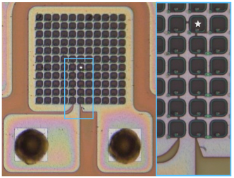
The device under test (DUT) is a Hamamatsu SiPM test structure of S14160 series [7] glued on the ceramic package. It consists of one single cell surrounded by 120 others. This single cell can be biased and read out separately.
A picture of the DUT is shown in Figure 1. It has an array of 11x11 cells with 15 m pitch. The central cell of the array is disconnected from the others and has its own output contact pad. Therefore 1 cell and 120 cells have a common cathode but separate anodes. Between the cells, trenches of 0.5 m width are implemented to reduce optical cross-talk [8].
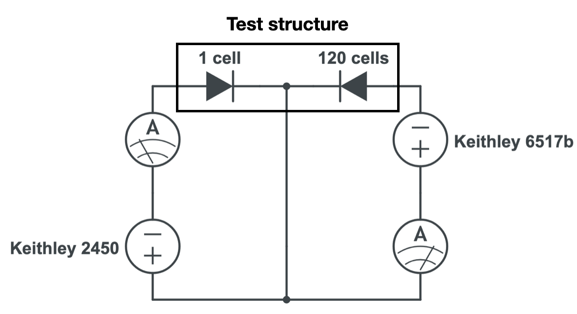
The setup for IV-measurements consists of a climate chamber, a dual-channel bias and readout board, SourceMeter Keithley 2450 used for 1 cell and SourceMeter Keithley 6517b used for 120 cells. To monitor the temperature as close to the DUT as possible, a Pt-100 is attached to the side of the ceramic package. The temperature stability in the chamber monitored by the Pt-100 readings is 0.03 ∘C and 0.2 ∘C at +20 ∘C and -30 ∘C, respectively. The accuracy of the setup on low current measurements is limited, such that currents lower than 100 pA for Keithley 2450 and 1 nA for Keithley 6517b are not reliable.
For the illumination of the device a stabilised broadband light source with a 650 nm filter is used. A filter mount placed outside the chamber is used for changing the light intensity. The light delivery system consists of two optical fibers, the first with 365 m-core and 1 m length from the light source to the filter mount, and the second with 365 m-core and 20 m length from the filter mount to the DUT. For the measurements without illumination the optical fiber was blocked in the filter mount by a beam blocker, while the light source was kept on.
The readout board consists of two channels to bias and read out 1 cell and 120 cells separately. The circuit schematic of the board is shown in Figure 2. IV-curves are measured synchronously on 1 cell and 120 cells.
Measurements were carried out for one non-irradiated device and three devices irradiated by neutrons at the TRIGA Research Reactor of the JSI, Ljubljana, to different fluences = [2e12, 1e13, 5e13] cm-2. No annealing was applied to the samples before measurement.
3 Self-heating
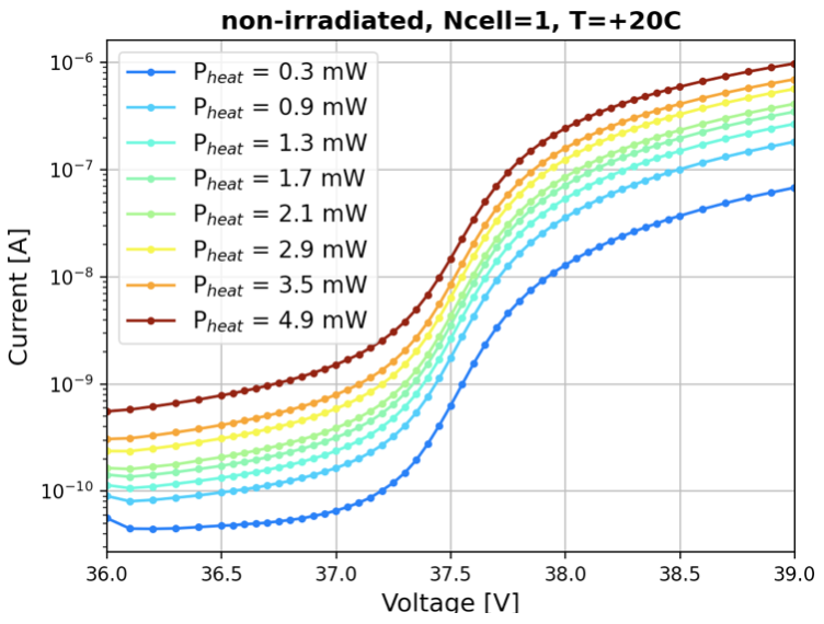
The highest heat power observed in our study was = 1.9 mW for 120 cells irradiated to = 5e13 cm-2, at T = -30 ∘C and = 4 V. In this paper the overvoltage is defined as . This power dissipated inside the SiPM could lead to a local increase of temperature and a correlated change of SiPM performance parameters, an effect denoted as self-heating. To check whether the measurements are affected by the self-heating effect the following procedure was carried out for the non-irradiated sample at T = +20 ∘C:
-
1.
Operate 120 cells at the fixed voltage above the breakdown ( = 39.1 V, = 1.5 V).
-
2.
Change the light intensity to control the heat power, thus the 120 cells serve as a heater.
-
3.
Measure the IV-curve of 1 cell and calculate , thus the 1 cell serves as a temperature sensor since strongly depends on the SiPM temperature.
Figure 3 shows the IV-curves for 1 cell, measured with different heat power generated by the other 120 cells. Using the Logarithmic Derivative method [9], is calculated for each IV-curve (see Figure 4). We conclude that the self-heating effect is negligible in our study, since no shift in of a single cell is observed up to = 4.9 mW, equivalent to the highest power measured in the highest irradiated sensor.

4 Results
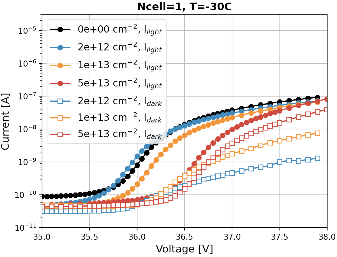
Measurements of the IV-curves for 1 cell and 120 cells were carried out for each device without illumination () and with illumination (), at the temperature T = -30 ∘C in a voltage range - 2 V + 2 V. IV-curves are presented in Figure 5. Photocurrent was calculated as a difference of and .

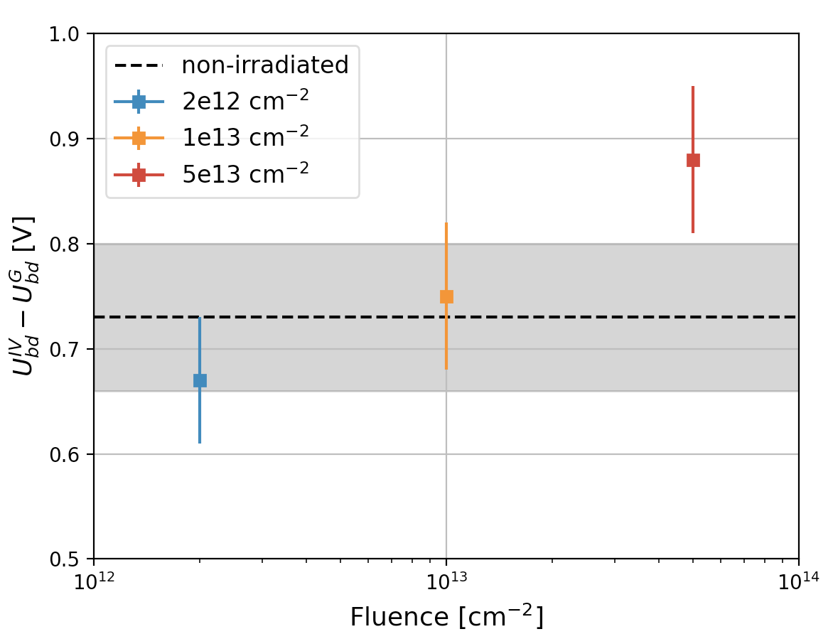
Logarithmic derivatives were calculated from the IV-curves measured with illumination for 1 cell and 120 cells. The breakdown voltage is determined as a maximum of the logarithmic derivative by approximation with the mean of a Gauss fit. The obtained values of are reported in Table 1. For all samples of a single cell is equal to of 120 cells within the errors. The table includes also the value of reported in [6]. Fig. 6 presents the answer to the second question posed in the introduction. The difference 0.7 V is approximately contact up to = 1e13 cm-2, and possibly increases for = 5e13 cm-2.
| [cm-2] | Number of cells | U [V] at -30 ∘C | U [V] at -30 ∘C |
|---|---|---|---|
| 0e00 | 1 | 35.980.03 | 35.250.04 |
| 120 | 35.940.03 | - | |
| 2e12 | 1 | 35.930.02 | 35.260.04 |
| 120 | 35.910.03 | - | |
| 1e13 | 1 | 36.160.03 | 35.410.04 |
| 120 | 36.130.03 | - | |
| 5e13 | 1 | 36.620.02 | 35.640.06 |
| 120 | 36.580.03 | - |
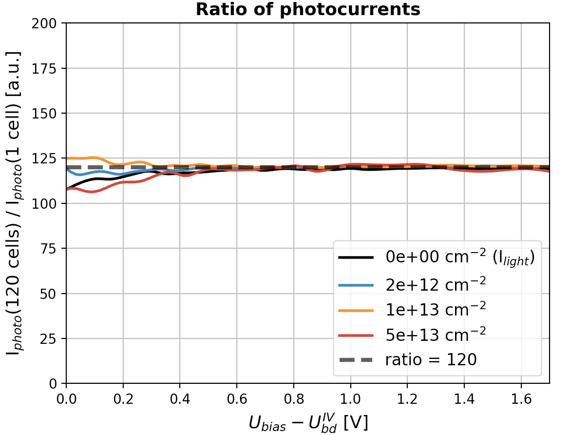
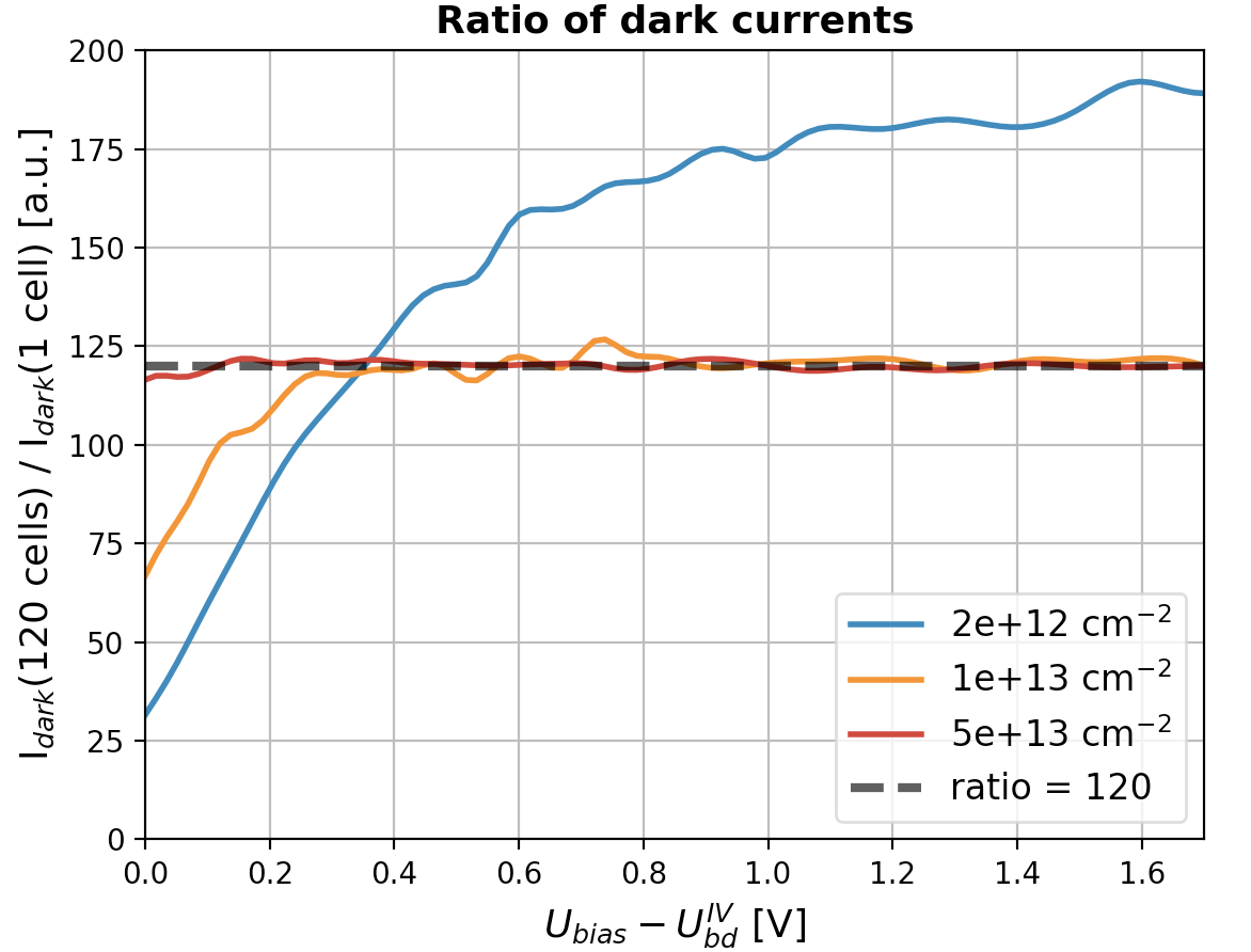
Radiation damage may produce local ”hot spots” with size smaller than or similar to a cell [10]. The probability to find a hot spot in the single cell depends on the fluence and the cell size. Our study is based on the measurements of four single cells, therefore the results could be subject to large fluctuations due to the presence of ”hot spots”. To check the uniformity of radiation damage in the SiPM cells, we calculate the ratio between the currents of 120 cells and 1 cell for and . For the non-irradiated sample only the ratio of was calculated, since is below the accuracy limit of the setup and cannot be measured. The results are presented in Fig. 7.
The expected current ratio of 120 is obtained for at all fluences, for on the non-irradiated sample, and for at the fluences = [1e13, 5e13] up to = 1.7 V. The ratio for the curves at = 2e12 does not reach a plateau and exceeds the value of 120. Possible reasons are: radiation damage non-uniformity or limited accuracy of low current measurements. For most of the studied samples the radiation damage of the single cell is comparable to the average of the surrounding 120 cells, indicating good damage uniformity both in terms of dark current change, and of change in the product .
5 Conclusions and outlook
The radiation hardness study using SiPMs with single-cell readout is ongoing. First observations from waveform measurements reported a gain reduction by 19% and an increase of by 0.5 V is observed after = 5e13 cm-2. From the analysis of the IV-curves, the fluence dependence of is extracted, which confirms the same increase of for , but with a difference in the absolute value of 0.7 V. No visible fluence dependence of the difference is seen within the uncertainties for = [2e12, 1e13] . A light increase of the difference is observed for = 5e13 . The radiation damage uniformity of 1 cell and 120 cells was checked up to = 1.7 V. A good damage uniformity both in terms of dark- and photocurrent change is confirmed on all but one sample at the lowers investigated fluence.
With a dedicated study on non-irradiated sensors it is shown that self-heating effects are negligible for the power dissipated even in the highest irradiated SiPM.
Acknowledgement
This work is supported by the Deutsche Forschungsgemeinschaft (DFG, German Research Foundation) under Germany’s Excellence Strategy, EXC 2121, Quantum Universe (390833306). The reported study was funded by RFBR and TUBITAK according to the research project 20-52-46005.
References
- [1] C. Piemonte, A. Gola, Overview on the main parameters and technology of modern silicon photomultipliers, Nucl. Instrum. Methods Phys. Res. A 926 (2019) 2–15. doi:10.1016/j.nima.2018.11.119.
- [2] E. Garutti, Y. Musienko, Radiation damage of SiPMs, Nucl. Instrum. Methods Phys. Res. A 926 (2019) 69–84. doi:10.1016/j.nima.2018.10.191.
- [3] V. Chmill, E. Garutti, R. Klanner, M. Nitschke, J. Schwandt, Study of the breakdown voltage of SiPMs, Nucl. Instrum. Methods Phys. Res. A 845 (2017) 56–59. doi:10.1016/j.nima.2016.04.047.
- [4] J. J. T. O. Marinov, J. Dean, Theory of microplasma fluctuations and noise in silicon diode in avalanche breakdown, J. Appl. Phys. 101 (064515) (2007). doi:10.1063/1.2654973.
- [5] T. Cazimajou, M. Pala, J. Saint-Martin, R. Helleboid, J. Grebot, D. Rideau, P. Dollfus, Quenching statistics of silicon single photon avalanche diodes, IEEE J. Electron Devices Soc. 9 (2021) 1098–1102. doi:10.1109/JEDS.2021.3127013.
- [6] O. Bychkova, P. Parygin, E. Garutti, A. Kaminsky, S. Martens, E. Popova, J. Schwandt, A. Stifutkin, Radiation hardness study using SiPMs with single-cell readout, Nucl. Instrum. Methods Phys. Res. A 1031C (166533) (2022). doi:10.1016/j.nima.2022.166533.
-
[7]
Datasheet
MPPC S14160 series.
URL https://www.hamamatsu.com/resources/pdf/ssd/s14160-1310ps_etc_kapd1070e.pdf -
[8]
Y. Ohashi, Current status of Hamamatsu Si
detectors for collider experiments, Calorimetry for High Energy Frontier
2019, Fukuoka, Japan, November 25–29, 2019.
URL https://indi.to/CnF9R - [9] R. Klanner, Characterisation of SiPMs, Nucl. Instrum. Methods Phys. Res. A 926 (2019) 36–56. doi:10.1016/j.nima.2018.11.083.
- [10] E. Engelmann, E. Popova, S. Vinogradov, Spatially resolved dark count rate of SiPMs (2018). arXiv:1807.04113.