Controlling the spontaneous firing behavior of a neuron with astrocyte
Abstract
Mounting evidence in recent years suggests that astrocytes, a sub-type of glial cells, not only serve metabolic and structural support for neurons and synapses but also play critical roles in regulation of proper functioning of the nervous system. In this work, we investigate the effect of astrocyte on the spontaneous firing activity of a neuron through a combined model which includes a neuron-astrocyte pair. First, we show that an astrocyte may provide a kind of multistability in neuron dynamics by inducing different firing modes such as random and bursty spiking. Then, we identify the underlying mechanism of this behavior and search for the astrocytic factors that may have regulatory roles in different firing regimes. More specifically, we explore how an astrocyte can participate in occurrence and control of spontaneous irregular spiking activity of a neuron in random spiking mode. Additionally, we systematically investigate the bursty firing regime dynamics of the neuron under the variation of biophysical facts related to the intracellular environment of the astrocyte. It is found that an astrocyte coupled to a neuron can provide a control mechanism for both spontaneous firing irregularity and burst firing statistics, i.e., burst regularity and size.
Astrocytes are the most numerous glial cells in the mature brain, and often surround neuronal somata and dendrites and provide fine enmeshment of synapses. Unlike neurons, these star-shaped cells do not elicit action potentials, yet they contribute to information processing via feedback to the cells by listening to the synaptic activity. We here investigate the their role on shaping the spontaneous firing behavior of a neuron. By using a neuron–astrocyte pair model, we identify distinct astrocyte-controlled neural activity patterns and explore their individual statistical characteristics under the variation of different biologically-plausible intrinsic astrocytic factors.
I Introduction
Spontaneous electrical activity, observed in many neurons from distinct brain regions, has long been interested and is by definition the ongoing activity of the cells when they are not subjected to an input Dehaene2005 ; Redolf2021 . This non-stimulus-evoked activity can be observed in experiments by using different measuring tools at the scales of single neuron Kubska2021 , neural microcircuits Yuan2020 as well as whole brain Fox2007 ; Calim2021 . Depending on the brain region to which it belongs and many other biological factors, the spontaneous spiking of a neuron may appear with different temporal firing patterns that are broadly categorized as "repetitive” Uddin2020 , "random" Paladini2003 and "bursty" Yang2018 . It is also possible to observe more complex patterns emerging with chaotic dynamics Yi2012 ; Dai2020 . Although it was initially considered as ”noise” and various fundamental questions concerning the meaning and features of spontaneous neural activity are still a matter of debate, mounting evidence in recent years suggests that it may reflect molecular, cellular and network dynamics of the neurons and may play roles in specific neuronal computations Ozer2007 ; Uzuntarla2012 ; Frolov2020 ; Guiyang2021 ; Martini2021 .
The spontaneous activity of a single neuron is more pronounced compared to that seen at the larger population scale. A widely observed typical feature of spontaneous spiking patterns is highly irregularity. In recent decades, there has been a great deal of effort in elucidating the functional implications of such irregularity both on the level of single cells and cell populations Ozer2006 ; Miura2007 ; Ozer2009 ; Payne2019 . Although the exact mechanism is not clear yet and the research is still in progress, the synaptic input correlations and the noisy environment have been suggested as the origin of the firing irregularity Brunel2003 ; Fellous2003 ; Burkitt2006 ; Mendonca2016 . In terms of its implications, many experimental and theoretical studies have reported that irregular spontaneous activity may reflect an underlying rich coding structure and it may be associated with several cognitive functions of the brain, such as working memory Hansel2013 , selective attention Ardid2010 and sensory coding Panzer2017 . Thus, the degree of irregularity in spontaneous firing patterns and its control may be critical to understand the underlying mechanisms of such cognitive functions or, more generally, information processing in the nervous system Kim2021 .
To date, the control of irregular firing activity has been mostly linked to internal dynamics of neurons (i.e., excitability, ion channels, membrane currents) and background activity induced by inputs from neighboring neurons Dipoppa2013 ; Peterson2017 . However, the brain is not just composed of neurons but it also includes glial cells which are generally considered to provide metabolic and structural support for neurons and synapses Barber2019 ; Patel2019 . In recent years, many experimental studies have reported evidence that glial cells influence neural activity at both cellular and network level Jakel 2017 ; Allen2018 , but have not been studied in any detail before with respect to their role in control of temporal features of neural activity.
Astrocytes are the most numerous glial cells, are ubiquitous in the mature brain, and often surround neuronal somata and dendrites and provide fine enmeshment of synapses Sofroniew2010 ; Chung2015 . Findings from experimental studies suggest that they are involved in memory formation Zorec2015 , decision making Wang2017 , attention Guimaraes2018 , cognitive Brockett2018 and behavioral processes Hwang2021 , and modulate the behavior of neurons depending on their own physiological and environmental factors Halassa2010 . On the other hand, a growing body of evidence in recent years has demonstrated that the malfunctioning of astrocytes may contribute to various neurodegenerative diseases (i.e., Alzheimer, multiple sclerosis) Preman2021 . Unlike neurons, these star-shaped cells do not elicit action potentials, yet they contribute to information processing via feedback to the cells by listening to the synaptic activity. More precisely, the released neurotransmitter molecules at synapses during a spike transmission not only activate the target neuron but also they regulate dynamics in astrocytes depending on the spike traffic at synapses. When the intensity of spike transmission at synapses is low, astrocytes respond with small transients that cannot induce any significant input to the connected neurons. With the increase in the intensity of spike transmission at synapses, the intracellular concentration exhibits oscillations, which constitutes the basis of the well-known wave propagation Bazargani2016 , and triggers the release of several gliotransmitters (i.e., glutamate and adenosine) from astrocytes to the synaptic cleft. Pre-and postsynaptic neurons consider these gliotransmitter molecules as a feedback signal which regulates their internal dynamics DePitta2009 ; yilmaz2019 ; calim2021 .
This signaling scheme in neuron–astrocyte communication suggests that astrocytes may be a significant factor in determining the level of neuronal spiking irregularity and may serve as a control mechanism Fellin2004 ; Wade2011 . Following this motivation, we here investigate the activity of a spontaneously firing neuron in a neuron–astrocyte pair, where the neuron is initially exhibiting spontaneous firing activity. More precisely, we identify distinct astrocyte-controlled neural activity patterns (eg. random, bursty) and explore their individual statistical characteristics under the variation of different biologically-plausible intrinsic astrocytic factors, i.e., the production rate of inositol trisphosphate, neuron-astrocyte coupling strength and the number of calcium channels.
II Models and Methods
We consider a coupled neuron-astrocyte pair with noise where the dynamics of the neuron is modeled with Fitzhugh-Nagumo () equations as follows FitzHugh1961 ; Yu2021 :
| (1) |
and
| (2) |
where denotes the electrical activity of the membrane and is the slow recovery variable that restores the resting state of the model. is the time scaling parameter responsible for separation of fast () and slow () variables of the model, which is fixed to 0.005. is the bifurcation parameter that determines the excitability level of the neuron. Namely, the model neuron is in the excitable regime for , with a single fixed point, while for , it exhibits oscillatory behavior. In the excitable regime, spontaneous activity of the neuron is caused by adding a Gaussian white noise source with zero mean and intensity to the equations.
The term in Eq. (2) is a depolarizing astrocytic feedback current, that is scaled with the coupling constant , induced by the dynamical changes in the concentration of two main substances in the astrocyte: inositol trisphosphate () and cytosolic calcium (). Namely, an astrocyte responds to the neuronal spiking activity by binding released glutamate in the synaptic cleft to its metabotropic glutamate receptors (). The activation of these receptors then triggers the production of , which, consequently, causes the discharge of internal stores mediated by receptor channels (). Such signaling scheme results in alteration of concentration in the astrocyte cytosol. To model these processes, we use the approximation proposed by Nadkarni and Jung nadkarni2003 for the dynamics of production in the astrocyte as follows:
| (3) |
where the first term on the right hand side refers to decay in intracellular concentration whereas the second one, defined by a sigmoid function with steepness , refers to its production which is triggered when exceeds the threshold voltage . =160nM and are the experimentally determined values for the equilibrium concentration of and the decomposition time constant, respectively. The parameter denotes the astrocyte production rate in response to a single spike emitted by the neuron amiri2011 .
The intracellular concentration is modeled based on the well-known Li-Rinzel () equations which describe the dynamics with three distinct fluxes: is flux from (Endoplasmic Reticulum) to the cytosol through channels, is the flux from cytosol into via -dependent pumps and is the leakage flux from the to intracellular space due to difference in concentrations between ER and cytosol. The model equations for the dynamics are as follows lirinzel1994 ; manninen2018 :
| (4) |
with fluxes defined as
| (5) |
| (6) |
| (7) |
where
| (8) |
and
| (9) |
Here, refers to the fraction of activated and satisfies the equation
| (10) |
where and refer the activation and inactivation rates of these receptor channels (see nadkarni2003 for the equations). In our study, stochastic dynamics for are also taken into account, and modeled with the approach where random kinetics of the are incorporated into model via an independent zero-mean Gaussian white noise source whose autocorrelation function is defined as tang2016
| (11) |
where corresponds to the number of which determines the channel noise strength. Note that and effective channel noise level are inversely related. Unless stated otherwise, the channel noise is ignored throughout our analysis in this work to understand the fundamental principles of communication between neuron and astrocyte. The values of astrocyte parameters are listed in Table I Volman2007 ; Postnow2009 .
Finally, the astrocytic current equation is obtained from Nadkarni and Jung nadkarni2003 as a function of intracellular concentration of the astrocyte according to experimental data defined by
| (12) |
where is the slow inward current which acts as a depolarizing input to the neuron. is the amount of above the threshold level and is a Heaviside function: . For statistical accuracy, each data point in the following results is obtained by averaging 50 independent realizations of the model equations for any given set of the neuron and astrocyte parameters (noise level, coupling strength, production rate).
| Parameter | Value | Description |
|---|---|---|
| Ratio of ER volume to cytosol volume | ||
| Maximum channel flux | ||
| leak flux constant | ||
| Maximum uptake | ||
| SERCA activation constant | ||
| dissociation constant | ||
| activation dissociation constant | ||
| Cytosolic free concentration |
III Results
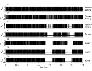
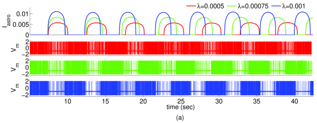
In what follows, we will systematically investigate the effects of astrocytes on the firing dynamics of a single neuron by considering the proposed neuron-astrocyte coupled model. To do so, we begin by exploring how an astrocyte changes the spontaneous spiking behavior of the proposed neuron model by changing the coupling strength . In Fig.1, we present representative voltage traces of membrane as a function of time for different values of the astrocyte-neuron pair. It is seen in the top panel that the isolated neuron () exhibits noise-induced firing activity. When the effect of the astrocyte is considered, i.e., , dramatic variation in firing profile and new firing regimes start to emerge with the increased values of as illustrated in the following panels of Fig. 1. For instance, when the coupling is weak, we observe that neuronal firing activity decreases during some specific periods of time. Further increase in results in a significant reduction in the number of spikes (see panels c and d) and, finally, the activity completely disappears during these periods (see panels e, f and g) when exceeds a certain level. These observations suggest that an astrocyte provides a kind of multistability into the neural dynamics by switching the firing behavior from one mode to the another, i.e., from random spiking to bursty firings or from silence to random spiking.
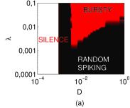
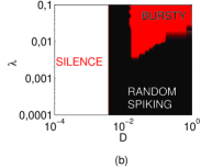
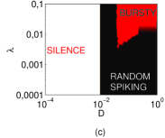
Next, we explore the effect of noise intensity for different values of coupling strength and excitability level . By changing these parameters, we observe three different neuronal phenomena, namely silence, bursty and random spike states. To classify our observations in Fig. 1, we perform a phase plane analysis in our neuron-astrocyte coupled system. Fig. 2 presents the activity mode of the neuron on (, ) plane for three different excitability levels (). It is seen that there exists three different activity mode regardless of the excitability level of the neuron: silence, random spiking (RS) and bursty behavior. Note that these activity profiles at different excitability states emerge due to the inputs from both astrocyte and noise. It is seen that if the is not high enough than a certain value, the neuron is silent for all values. The critical level of shifts to the right (large noise) as increases. This is mainly due to the fact that the closer values of to the bifurcation point () in the FHN model neuron provides more excitability where the occurrence of a spike requires less noise. On the other hand, when is sufficiently large, the neuron exhibits RS activity as well as bursty behavior. But the latter one occurs at relatively large values of and and the region of this firing regime shrinks as increases. These observations on the phase plane suggest that it is possible to switch firing mode of a neuron by fine tuning and without any change in its internal dynamics.
Following our initial motivation, we now continue to analyze the effect of astrocyte on the firing behavior of the neuron by considering the facts observed in the above phase-plane analysis. Since the prominent features of random spiking and bursty modes differ from each other, separate analyses for these two firing regimes are necessary. More precisely, we investigate the impact of astrocyte in determining the neuron’s spiking (ir)regularity which is believed to be important for information processing capacity of neurons. On the other hand, for the case of bursty mode, we study the role of neuron-astrocyte communication on the frequency and size of the bursts.
III.1 Spontaneous spiking regularity with astrocyte
Both in vivo and in vitro recordings indicate that spontaneous neural spiking activity in most brain regions is highly irregular Shadlen1998 ; Zierenberg2018 ; Uva2021 . Over the past recent decades, understanding the nature, cause and functionality of that irregularity has been attracted a great deal of attention and many hypotheses have been proposed regarding these issues. For instance, it has been suggested that the irregular timing of spikes could convey information, providing a broad information bandwidth on the neural spike train Dettner2016 ; Ventura2019 . On the other hand, the irregularity may be the reflection of noise which limits the information processing capacity of cells Pisarchik2019 ; Tischbirek2019 . So far, in terms of nature and cause, although various physiological mechanisms including inherent neural dynamics and environmental factors (e.g. stochastic dynamics of ion channels, balance between excitation and inhibition) have been proposed as the source and the controller of spiking irregularity, the underlying mechanism for this phenomenon is still not known exactly He2019 ; Ushakov2021 ; Xu2021 . Thus, we are interested in what would be the role of astrocytes in determining such irregular firing patterns.
To do so, we simulate our coupled model systematically and search the astrocytic factors that may have effects on neural activity of the neuron through the (Coefficient Variation) of spike trains. is a widely used measure of spiking irregularity which is obtained by the division of the standard deviation to mean of the interspike intervals () and defined by Schmid2001 ; Ozer2009 :
| (13) |
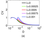
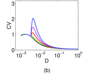
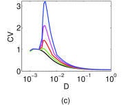
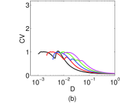
It should be noted that the larger values of the imply more irregular spike trains. First, we investigate how changes with the coupling strength as a function of and for a fixed excitability level of the neuron . Obtained results are presented in Fig. 3. It is seen that first exhibits a small increase for all at lower values of where the astrocyte does not provide any significant feedback to the neuron due to the lack of required number of arriving spikes for the calcium accumulation. More precisely, such noise levels are not enough to turn on the astrocyte, and this small increase in is not a consequence of neuron-astrocyte interaction. It appears due to the effect of noise on internal dynamics of neuron. On the other hand, for moderate noise levels, a second resonance peak emerges with the activation of feedback from astrocyte to the neuron where the increases as get larger values. Finally, for very large values of , the noise suppresses the inputs from astrocyte and the neuron fires under the influence of noise, which results in an overlap of curves for all neuron-astrocyte coupling strengths, following the trend of an isolated neuron. Fig. 3 also features the effect of production rate on behavior. It is seen that the above mentioned coupling-induced resonance peaks become more pronounced at all levels with the increased values of (see maximum values for a given in all panels). Moreover, it is also obvious that resonance curves shift to the left for higher , indicating that production rate provides less noise requirement for higher resonance peaks in .
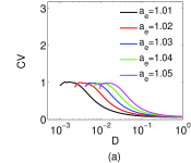
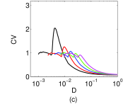
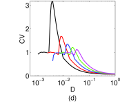
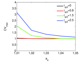
The above results clearly demonstrate that inherent dynamics of an astrocyte can induce more irregular firing patterns of spontaneous spiking activity for a particular range of noise intensity as well as provide a control mechanism for the level of such irregularity. To verify the validity of this conclusion and to investigate the dependency of astrocytic control on internal dynamics of the neuron, we perform similar analyses for different values of since the level of neuronal excitability is critical in determining the required spike inputs to activate the astrocyte. Obtained results are shown in Fig. 4, demonstrating the versus and for four different values of . In the case of isolated neuron (see Fig. 4a for ), since the astrocyte does not provide any feedback to the cell, variation of curves are determined only by the noise and . It is seen that similar trends occur for different excitability levels, which only shift to the right for increasing values of . This is due to the fact that a less excitable neuron (i.e., large ) needs more noise to fire an action potential. On the other hand, when (see Fig. 4b-d), we observe the similar effect as in our previous analysis in Fig. 3 where the required noise intensity for resonance peaks decreases as increases at all excitability levels. One can easily follow this shift by comparing curves for a given excitability level in panels of Fig. 4. It is also obvious that there exists a threshold level of for different excitability levels of the neuron to effectively control the . For instance, when (see Fig. 4b), the above mentioned astrocyte-induced second resonance peak of starts to emerge with decreasing amplitudes as increases. With the increase in , the second resonance peaks for all considered values become clearly pronounced. Fig. 5 provides a better illustration for the influence of such interaction between neuron and astrocyte parameters where we only plot maximum () values computed in Fig. 4 as a function of and . It is seen that is independent from when is less than a threshold value which is around . These findings indicate that a less excitable neuron needs astrocyte partners having large production rate to exhibit more irregular firing activity.
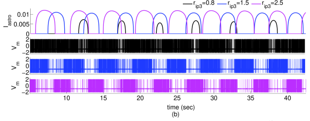
To explain the mechanism underlying neuron-astrocyte cross-talk and improve our understanding on the analysis presented above, we demonstrate the traces of the neuron’s membrane voltage and the astrocyte feedback current for different values of and (see Fig.6). Note that the color-coded traces in the top panels of Fig. 6 (a) and (b) correspond to the same color-coded membrane potential traces shown in the following panels of each figure. It is seen that the lifetime and the amplitude of the significantly change depending on the variations in and . Namely, the increase in both parameters results in amplitude enhancement as expected. However, as we increase and , the lifetime period decreases and increases, respectively. By observing the corresponding voltage traces in the following panels, one can see that there is a relevant correlation between the firing activity and where the number of spontaneous firings decrease when is strong enough. The activity reduction periods become more pronounced for increased values of both coupling strength and . It should be noted that a further increase of these astrocytic parameters induces complete cessation of firing activity during the periods of time when the astrocyte is active (data not shown). This bursty behavior will be discussed in the next section. The firing activity reductions at random periods of time arise from the fact that the feedback current decreases the excitability of the neuron. Therefore, the more frequent and long-lasting disruptions of firing activity by induce more heterogeneous interspike interval distributions resulting in increased levels of spontaneous spiking irregularity. The results concerning the regulatory role of astrocyte on spontaneous spiking statistics presented in Figs. (1-5) can be interpreted with this understanding.

.
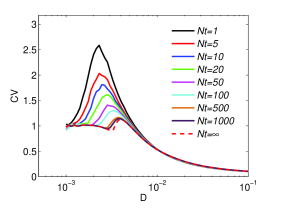
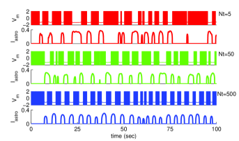
So far, the intracellular dynamics that underlies the generation of has been considered with a deterministic approach where many local and global processes taking place in the cellular environment are ignored. This approach simply models the activation of the astrocyte current. However, metabolic processes are noisy due to many external and internal factors. For instance, the astrocytic second messenger sensitizes receptor channels () mediating release from the endoplasmic reticulum. are typically distributed in clusters with several channels on the membrane of the astrocyte. The noise caused by the random opening and closing of these channels is expressed as channel noise of the astrocyte which is already included in our model (see Eq. (5)). Accordingly, if the clusters comprise a few channels, release occurs stochastically, thus generating noise, and the level of noise is dependent on the size of the cluster, i.e., the number of denoted by . Thus, we utilize a stochastic model combined with approach for release from that has proven accurate for a variety of cluster sizes nadkarni2007 . In Fig. 7, we show how astrocytic noise, which is controlled by the cluster of channels, affects the resulting behavior of as a function of neuronal noise level in our model setup. As seen in the figure, regardless of the values, the curves exhibit a bell-shaped dependence on neuronal noise, indicating the presence of an optimal for maximum firing irregularity. Moreover, values increase as the level of the astrocytic noise increases in the system (decreasing ). It can be observed that the decrease in decreases the optimal corresponding to the peak . This indicates that it requires less spike inputs from the neuron that is controlled by to obtain the same firing irregularity level in the presence of astrocytic noise in the neuron-astrocyte coupled model. Finally, for too high neuronal noise levels, the system is dominated by the neuronal noise; astrocytic noise then becomes insignificant and intense firing activity of the neuron due to high noise level causes the to decrease. These findings indicate that, for fixed parameter sets (, and ), the density of channels influences the neuronal dynamics distinctly in the coupled system.
III.2 Bursty Regime with Astrocyte
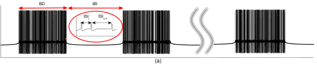
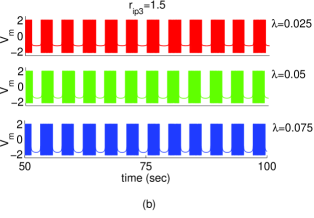
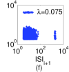
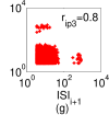
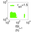
In recent decades, many researchers have suggested that the astrocyte provides an important physiological mechanism to enable bursty firing activity Morquette2015 ; Deemyad2018 . We have already showed the emergence of such a bursty mode at the proper coupling strength (see Fig. 1). In this section, based on statistical analyses, we investigate whether it is possible to control the frequency and size of bursty firings with the dynamics of the astrocyte. To do so, we first describe some statistical notations to characterize the bursty regime as illustrated in Fig. 8a. The (interburst interval) is the period between two bursts, the (burst duration) is the time period between the beginning and the end of each burst period, and finally, the denotes the i-th interspike-interval womack2004 ; fardet2018 . After providing the necessary descriptive information, we now show the effect of and on and in Fig. 8b and Fig. 8c respectively, by monitoring the membrane potential traces.
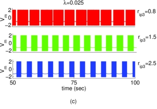
The voltage traces of the neuron in the left panel, which has the same characteristic even though the increases, clearly shows that it does not change the frequency and size of the burst window. On the other hand, with the increased values of , it is seen that increases and decreases. This indicates that can control the burst characteristic. Another point draws attention: the first period occurs earlier as increases because the astrocyte is activated in a much shorter time with larger production rate.
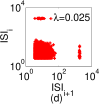
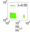
To check whether this bursty activity mode is not a transient behavior, 2D histograms are obtained for very long periods of simulation time with the same and values considered in upper panels. These charts shown in panels (d-i) present detailed information about whether the neuron exhibits bursty firing or regular spiking behavior without observing the membrane traces. Namely, the large clusters located at relatively small and values indicate the firing activity during the burst, while the other two small clusters in histograms denote the starting and the completion of the burst periods. In Figs. 8(d,e,f), it is clear that the size and the scattering of clusters do not change very much with . However, as seen in Figs. 8(g,h,i), small clusters get less noisy as increases. These observations from Fig.8 indicate that the coupling between the astrocyte and the neuron is responsible for the emergence of the bursty firing mode while it has no significant influence on bursting statistics (i.e. ). It can also be inferred that might be a critical player in the regulation of the bursty firing mode statistics.
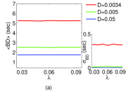
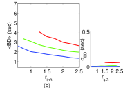
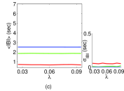
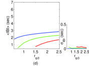
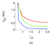
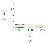
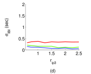
To support these findings, we systematically investigate the statistical changes in bursty regime through average and standard deviation of and measures as functions of and for three different neuronal noise levels. Fig. 9 features the obtained results. It is seen that the mean () and standard deviation () of and are approximately constant as varies for any given levels of noise (see panels (a) and (c)). This indicates that our finding from Fig. 8(d-f), i.e. that is has no role on burst statistics, is robust to neuronal noise. The most striking point here is that neuronal noise acts as a control parameter for the lifetimes of the bursts in such a neuron-astrocyte crosstalk environment. In other words, with increased values of , the emergent intense neuronal discharges result in stable (i.e. periodic and almost equal amplitude) astrocytic currents in time due to the rapid accumulation in the cytosol of the astrocyte. Thus, small and large occur as increases. Additionally, since the stable astrocytic current pulses trigger regular and consistent burst activity for large , and become quite small. On the other hand, panels (b) and (d) illustrate that has a significant role on and statistics. The decreases with at all considered levels, while the shows the opposite direction of change from the (as expected). We know from previous analyses that as increases, astrocyte current emerges with greater amplitude and lifetime. Thus, decreases for larger values of , which results in longer cessation of firing activity during the periods of time when the astrocyte is active. It is also clear from panels (b) and (d) that the influence of neuronal noise () on burst statistics under the variation of exhibits similar behavior as that observed in the case of variation (panels (a) and (c)).
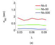
Finally, we investigate how the intrinsic noise stemming from random nature of channels affects the bursty firing mode. To do so, we compute the standard deviation of and by fixing the neuronal noise level to under the variations of and . Results are presented in Fig. 10. It is seen that as the astrocytic noise increases ( decreases), and increase. This finding is more pronounced for because the windows are larger than the windows. On the other hand, we still observe that has no role under the variation of (see panel a and c). However, can be expressed as a control parameter that allows modification of burst patterns. It is seen from panel (b) that first gradually decreases, and then, saturates to an approximately fixed level with the increase of . When the effects of and are compared on burst mode firing, it can be said that although the trends exhibit similarity, the standard deviation of burst statistics under the variation of is much larger than for the case of (around 100 times). The reason of such an extraordinary distinction arises from the fact that and shape the characteristics of the astrocytic current. Recall that the increase of results in a more stable both in time and amplitude. However, as can be observed from Fig. 11 where we illustrate and the corresponding voltage traces of the membrane (with same color-codes) for three different values of , becomes unstable with the increase of astrocytic noise (decrease of ). Namely, it is obvious that the width and position of pulses in time significantly becomes random as decreases, which results in the emergence of both long and short burst periods under the same biophysical conditions (see voltage traces in Fig.11). Thus, and reach larger values as the astrocytic noise increases compared to the case of the increase in neuronal noise.
IV Conclusion
In this work, we study the effect of an astrocyte on the spontaneous firing activity of a neuron that is initially subject to noise arising from biophysical conditions. To do so, we used a bipartite model that includes a neuron and an astrocyte and explored the parameter space that may induce intriguing neuron firing behaviors which are not present without astrocytes (isolated neurons). First, through an astrocytic parameter space analysis, it was shown that, in addition to the noise-induced spontaneous firing regime, a new bursty firing mode may emerge that does not exist in an isolated FHN neuron dynamics. We explained this astrocyte induced new firing mode through analysis of the feedback current introduced into the neuron dynamics. When there exists a crosstalk between these two different cells, the membrane voltage of the neuron acts on the intrinsic calcium dynamics of the astrocyte, which in turn affects the distance to threshold of the excitability level of the neuron through . Depending on the waveform (width and amplitude) and the frequency of the feedback current provided by astrocyte, we showed that this interaction may result in either a spontaneous or bursty firing mode.
In order to observe what would be the impact of astrocytes on these firing regimes, we restricted our analyses to the most significant features of each individual firing behavior. We first investigated the coherence of noise-induced spontaneous firing behavior of a single neuron that is coupled to an astrocyte. Compared to the isolated case, our findings revealed that the inherent dynamics of an astrocyte can induce more irregular firing patterns of spontaneous spiking activity for a particular range of noise intensity. It was also shown that the astrocytic parameters and provide a control mechanism for the level of such irregularity. We then extended our analyses by adding an additional noise source into our system, stemming from stochastic nature of receptor channels that control release inside the astrocyte. It was found that the resonance behavior of as a function of neuronal noise remains in such a coupled system. Further, the astrocytic calcium noise enhances this effect by reducing the required neuronal noise intensity for the same irregularity level.
Secondly, we studied whether it is possible to control the basic features of the bursty firing mode with astrocyte dynamics. By analyzing the width and regularity of bursty periods, we concluded that the coupling strength between astrocyte and neuron, which has a key role in determining the firing regime, does not play a significant role in modulation of bursty behavior, but the production rate in astrocyte has the ability to control all considered features of burst firing mode. Moreover, our findings suggest that, in such a neuron-astrocyte crosstalk environment, neuronal and astrocytic noise act as a control parameter for determining periodic (the same burst width) and stochastic (random burst width) characteristic of and statistics, respectively.
Finally, we would like to emphasize that our study provides an alternative theoretical framework indicating the power of astrocytes in shaping the neural activity. However, since the system under study in this work includes a single neuron-astrocyte pair, we cannot generalize our findings to a population scale. Thus, a possible extension of our study could be investigating the dynamics of spontaneous and burst firing modes in networks of neurons and astrocytes, which have been mostly studied so far in only neuron populations ma2017 ; bera2019 by considering various biophysical circumstances such as network topology and the type of gliotransmitters.
Acknowledgements.
M.U. acknowledges the support of the Scientific and Technological Research Council of Turkey (TUBITAK) BIDEB-2219 International Postdoctoral Research Fellowship Program for his research stay in Ottawa, Canada.AUTHOR DECLARATIONS
Conflict of Interest
The authors have no conflicts to disclose.
Data Availability Statement
Data sharing is not applicable to this article as no new data were created or analyzed in this study.
REFERENCES
References
- (1) S. Dehaene, J. P. Changeux, PLoS Biol 3(5), e141 (2005).
- (2) N. Redolfi and C. Lodovichi, Front. Cell. Neurosci. 15, 637536 (2021).
- (3) ZR. Kubska, J. Kamiński, Brain Sci. 11, 443 (2021).
- (4) X. Yuan, M. Schröter, MEJ. Obien, M. Fiscella, W. Gong, T. Kikuchi, et al., Nat. Commun. 11, 1-14 (2020).
- (5) M. Fox and M. Raichle, Nat. Rev. Neurosci. 8, 700–711 (2007).
- (6) A. Calim, T. Palabas, M. Uzuntarla, Philos. Trans. Royal Soc. A 379(2198), 20200236 (2021).
- (7) LQ. Uddin, Trends in Cogn. Sci. 24, 734–746 (2020).
- (8) CA. Paladini, S. Robinson, H. Morikawa, JT. Williams, RD. Palmiter, Proc. Natl. Acad. Sci. 100, 2866-2871 (2018).
- (9) Y. Yang, Y. Cui, K. Sang, et al., Nature 554, 317–322 (2018).
- (10) GS. Yi, J. Wang, CX. Han, B. Deng, and XL. Wei, Appl. Math. Modelling 36(8), 3673–3684 (2012).
- (11) Dai, X. et al. Phys. Rev. Lett. 125, 194101.
- (12) M. Ozer, LJ. Graham, O. Erkaymaz, M. Uzuntarla, NeuroReport 18, 1371-1374 (2007).
- (13) M. Uzuntarla, M. Ozer, DQ. Guo, Eur. Phys. J. B 85, 8, (2012).
- (14) L. Guiyang, N. Zhang, K. Ma, J. Weng, P. Zhu, F. Chen and G. He, Nonlinear Dyn. 104, 1475–1489 (2021).
- (15) FJ. Martini, T. Guillamon-Vivancos, V. Moreno-Juan, M. Valdeolmillos, G. Lopez-Bendito, Neuron 109, 2519–2534 (2021).
- (16) N. Frolov, V. Maksimenko, and A. Hramov, Chaos 30, 121108 (2020).
- (17) M. Ozer, M. Uzuntarla, SN. Agaoglu, Phys. Lett. A 360, 135 (2006).
- (18) K. Miura, V. Tsubo, M. Okada, T. Fukai, J. Neurosci. 27, 13802–13812 (2007).
- (19) M. Ozer, M. Perc, M. Uzuntarla, EPL 86, 40008 (2009).
- (20) HL. Payne, RL. French, CC. Guo, TDB. Nguyen-Vu, T. Manninen, JL. Raymond, Elife 8 (2019).
- (21) N. Brunel, J. X. Wang, J. Neurophysiol. 90, 415–430 (2003).
- (22) JM. Fellous, M. Rudolph, A. Destexhe and TJ. Sejnowski, Neurosci. 122, 811-829 (2003).
- (23) AN. Burkitt, Biol. Cybern. 95, 1-19 (2006).
- (24) PR. Mendonca, M. Vargas-Caballero, F. Erdelyi, G. Szabo, et al., Elife 5, e16475 (2016).
- (25) D. Hansel and G. Mato, J. Neurosci. 33, 133-149 (2013).
- (26) S. Ardid, XJ. Wang, D. Gomez-Cabrero, A. Compte, J. Neurosci. 30, 2856-2870 (2010).
- (27) S. Panzeri, CD. Harvey, E. Piasini, PE. Latham, T. Fellin, Neuron 93, 491-507 (2017).
- (28) J. Kim and GJ. Augustine, Neurosci. 462, 22–35 (2021).
- (29) M. Dipoppa and BS. Gutkin, Front. Comput. Neurosci. 7(139) (2013).
- (30) EJ. Peterson, B. Voytek, bioRxiv, 10.1101/185074 (2017).
- (31) CN. Barber and DM. Raben, Front. Cell. Neurosci. 13(212) (2019).
- (32) DC. Patel, BP. Tewari, L. Chaunsali, et al., Nat. Rev. Neurosci., 20, 282–297 (2019).
- (33) S. Jakel, L. Dimou, Front. Cell. Neurosci. 11, 24 (2017).
- (34) NJ. Allen and DA. Lyons, Science 362, 181–185 (2018).
- (35) MV. Sofroniew, and H. V. Vinters, Acta Neuropathol. 119, 7-35 (2010).
- (36) WS. Chung, NJ. Allen, C. Eroglu, Cold Spring Harb. Perspect. Biol. 7, a020370 (2015).
- (37) R. Zorec, A. Horvat, N. Vardjan, A. Verkhratsky, Front Integ. Neurosci. 9, 56 (2015).
- (38) J. Wang, J. Tu, B. Cao et al., Cell Rep. 21, 2407-2418 (2017).
- (39) K. Guimaraes, DQ. Madureira, AL. Madureira, Cogn. Syst. Res. 50, 15-28 (2018).
- (40) AT. Brockett, GA. Kane, PK. Monari et al., PloS One 13, e0195726 (2018).
- (41) SN. Hwang, JS. Lee, K. Seo and H. Lee, Cells 10(2), 296 (2021).
- (42) MM. Halassa and PG. Haydon, Annu. Rev. Physiol. 72, 335-355 (2010).
- (43) P. Preman, MA. Triguero, E. Alberdi, A. Verkhratsky, AM. Arranz, Cells 10(3), 540 (2021).
- (44) N. Bazargani, D. Attwell, Nat. Neurosci. 19, 182–189 (2016).
- (45) M. De Pitta, M. Goldberg, V. Volman, H. Berry, E. Ben-Jacob, J. Biol. Phys. 35, 383-411 (2009).
- (46) Y. Erkan, Z. Sarac and E. Yilmaz, Nonlinear Dyn., 95, 3411-3421 (2019).
- (47) A. Calim, A. Longtin and M. Uzuntarla, Philos. Trans. Royal Soc. A 379(2198), 20200267 (2021).
- (48) T. Fellin and G. Carmignoto, J. Physiol. 559, 3-15 (2004).
- (49) JJ. Wade, LJ. McDaid, J. Harkin, V. Crunelli, JAS. Kelso, PLoS One 6, e29445 (2011).
- (50) R. FitzHugh, Biophys. J. 1, 445 (1961).
- (51) D. Yu, X. Zhou, G. Wang, et al., Cogn. Neurodyn., 1-11 (2021).
- (52) S. Nadkarni, P. Jung, Phys. Rev. Lett. 91, 268101 (2003).
- (53) M. Amiri, G. Montaseri and F. Bahrami, Biol. Cybern. 105, 153-166 (2011).
- (54) Y. Li and J. Rinzel, J. Theor. Biol. 166, 461-473 (1994).
- (55) T. Manninen, R. Havela and M-L. Linne, Front. Comput. Neurosci. 12(14) 2018.
- (56) J. Tang, TB. Liu, J. Ma, et al., Commun. Nonlinear Sci. Numer. Simul. 32, 262–272 (2016).
- (57) DE. Postnov, RN. Koreshkov, NA. Brazhe, AR. Brazhe, OV. Sosnovtseva, J. Biol. Phys. 35, 425-445 (2009).
- (58) V. Volman, E. Ben-Jacob, H. Levine, Neural Comput. 19, 303-326 (2007).
- (59) MN. Shadlen and WT. Newsome, J. Neurosci. 18, 3870–3896 (1998).
- (60) J. Zierenberg, J. Wilting, V. Priesemann, Phys. Rev. X 8, 031018 (2018).
- (61) L. Uva, P. Aracri, G. Forcaia, M. de Curtis, Exp. Neurology 342, 113727 (2021).
- (62) A. Dettner, S. Muenzberg, T. Tchumatchenko, Nat. Commun. 7, 13805 (2016).
- (63) CM. Ventura and R. Kalluri, J. Neurosci. 39, 2860-2876 (2019).
- (64) AN. Pisarchik, VA. Maksimenko, AV. Andreev, et al., Sci. Rep. 9, 18325 (2019).
- (65) CH. Tischbirek, T. Noda, M. Tohmi, A. Birkner, et al., Cell Rep. 27, 1319-1326, e1315 (2019).
- (66) HY. He and HT. Cline, J. Exp. Neurosci. 13, 1179069519859371 (2019).
- (67) Y. Ushakov, A. Balanov, S. Savel’ev, Chaos, Soliton & Fractal 145, 110803 (2021).
- (68) C. Xu, X. H. Tang, H. P. Lü et al., Phys. Rev. Res. 3, 043004 (2021)
- (69) G. Schmid, I. Goychuk and P. Hanggi, Europhys. Lett. 56(1) (2001).
- (70) S. Nadkarni and P. Jung, Phys. Biol. 4, 1-9 (2007).
- (71) P. Morquette, D. Verdier, A. Kadala, J. Fethiere, et al., Nat. Neurosci. 18, 844-854 (2015).
- (72) T. Deemyad, J. Luthi, N. Spruston, Nat. Commun. 9, 4336 (2018).
- (73) MD. Womack and K. Khodakhah, J. Neurosci. 24, 3511-3521 (2004).
- (74) T. Fardet, M. Ballandras, S. Bottani, S. Métens and P. Monceau, Neurosci. 12(41) (2018).
- (75) J. Ma, J. Tang, Nonlinear Dyn. 89, 1569–1578 (2017).
- (76) BK. Bera, S. Rakshit, D. Ghosh, et al., Chaos. 29(5) (2019).