Near K-Edge Photoionization and Photoabsorption of Singly, Doubly, and Triply Charged Silicon Ions
Abstract
Experimental and theoretical results are presented for double, triple, and quadruple photoionization of Si+ and Si2+ ions and for double photoionization of Si3+ ions by a single photon. The experiments employed the photon-ion merged-beams technique at a synchrotron light source. The experimental photon-energy range 1835–1900 eV comprises resonances associated with the excitation of a electron to higher subshells and subsequent autoionization. Energies, widths, and strengths of these resonances are extracted from high-resolution photoionization measurements, and the core-hole lifetime of K-shell ionized neutral silicon is inferred. In addition, theoretical cross sections for photoabsorption and multiple photoionization were obtained from large-scale Multi-Configuration Dirac-Hartree-Fock (MCDHF) calculations. The present calculations agree with the experiment much better than previously published theoretical results. The importance of an accurate energy calibration of laboratory data is pointed out. The present benchmark results are particularly useful for discriminating between silicon absorption in the gaseous and in the solid component (dust grains) of the interstellar medium.
1 Introduction
Silicon is a relatively abundant element in the Universe and, in particular, a major component of interstellar dust. The binding energy of Si -shell electrons amounts to about 1840 eV (Deslattes et al., 2003). Correspondingly, Si -shell absorption by the interstellar medium (ISM) can be observed by x-ray satellites when directed towards distant x-ray emitting cosmic objects such as x-ray binaries (Rogantini et al., 2020). For the correct determination of the silicon abundance in the ISM it is important to know how much silicon is in the gaseous state and how much is (chemically) bound to dust grains (see, e.g., Jenkins, 2009). Since high-resolution x-ray absorption spectroscopy is sensitive to chemical shifts of characteristic absorption lines, astrophysical models of the ISM have recently been augmented by absorption coefficients of silicon-containing minerals that were obtained from laboratory measurements at a synchrotron light source (Zeegers et al., 2017, 2019). Here we provide complementary laboratory data for -shell x-ray absorption by atomic Si+, Si2+, and Si3+ ions that address the gaseous component of the ISM.
Previous experimental work on the photoabsorption of silicon ions considered only absorption by outer electronic shells (Mosnier et al., 2003; Bizau et al., 2009; Kennedy et al., 2014). With the very recent exception of negatively charged Si- (Perry-Sassmannshausen et al., 2021), -shell x-ray absorption data for silicon ions were hitherto exclusively obtained from theoretical calculations (Verner et al., 1993; Palmeri et al., 2008; Witthoeft et al., 2011; Kučas et al., 2012, 2015; Hasoglu et al., 2021). Because of the many-body nature of the theoretical problem the theoretical calculations have to resort to approximations yielding results that bear (usually unknown) uncertainties. In this situation, benchmarking by laboratory experiments (Schippers & Müller, 2020) is vital for arriving at sufficiently accurate results that allow one, in the present context, to reliably discriminate between the gaseous and solid components of the ISM.
Similar to our previous work on -shell ionization of Fe+, Fe2+, Fe3+ and Ar+ ions (Schippers et al., 2017; Beerwerth et al., 2019; Schippers et al., 2021; Müller et al., 2021), we have investigated ionization of silicon ions in different charge states. Here, we present experimental and theoretical data for -fold photoionization of Siq+ ions with primary charge state leading to the production of Sir+ ions with product charge-state
| (1) |
The paper is organized as follows. Section 2 discusses experimental issues, in particular the role of metastable ions in the silicon ion beams and the energy calibration of the photon energy scale. Section 3 describes calculations of the absorption cross-sections and of the complex deexcitation cascades that set in after the initial creation of a -shell hole. The results are presented and discussed in section 4 emphasizing their relevance for the identification of gaseous silicon in the ISM. The concluding section 5 briefly summarizes the findings and provides an outlook on future directions of laboratory astrophysics in the area of atomic inner-shell absorption.
2 Experiment
The measurements were carried out at the synchrotron light source PETRA III operated by DESY in Hamburg, Germany. More specifically, the photon-ion merged-beam technique (recently reviewed by Schippers et al., 2016a) was employed using the PIPE end-station (Schippers et al., 2014; Müller et al., 2017; Schippers et al., 2020) at the photon-beamline P04 (Viefhaus et al., 2013). The beamline’s monochromator is equipped with two diffraction gratings with different rulings of 400 and 1200 lines/mm. The latter was used for the present experiment because it offers the highest photon flux in the photon-energy region of interest. The experimental procedures have been described previously (Schippers et al., 2014; Müller et al., 2017). Therefore, we only provide the details here that are relevant for the presently-studied ion species.
Beams of Siq+ ions (=1–3) were produced by evaporating SiO powder from an electrically heated oven into an electron-cyclotron-resonance (ECR) ion source. The ion source was operated on a positive potential of 6 kV such that positively charged ions were accelerated from the ion source towards the electrically grounded ion beam-line. Downbeam, a dipole electromagnet served for selecting the desired ion species according to their mass-to-charge ratio. Subsequently, the ion beam was collimated and centered onto the counter-propagating photon beam by tuning electrostatic deflectors and lenses appropriately. Typical ion currents in the interaction region were 4.5 nA of 28Si+ and 2.5 nA of 28Si3+. The 28Si2+ ion current was significantly higher (20 nA) since it was heavily contaminated with, e.g., N+ or CO2+ ions, which have the same mass-to-charge ratio as 28Si2+. Similarly, it cannot be excluded that the Si+ beam was contaminated by N or CO+ ions. It has to be pointed out that the product ions with charge states increased by 1, 2, 3, units can all be individually separated by the dipole magnet (the demerger) in front of the ion detector so that each final channel could be individually investigated.
Relative cross sections for multiple photoionization (cf. Equation 1) were determined over a preselected range of photon energies by normalizing the photon-energy dependent product-ion count rate on the photon flux and on the ion-current, which were measured with a calibrated photodiode and a Faraday cup, respectively. For the Si3+ primary ions, the relative cross sections were put on an absolute scale by additionally accounting for the separately measured geometrical beam overlap (for details see Schippers et al., 2014; Müller et al., 2017). The uncertainty of the experimental absolute cross-section scale amounts to 15% (Schippers et al., 2014).
The measurement of absolute cross sections requires a careful adjustment of the mutual overlap of the photon and ion beams. This procedure was only applied to the Si3+ ion beam. An experimental determination of absolute cross sections for the multiple ionization of Si+ and Si2+ primary ions was not possible because of the unknown fractions of contaminating ions in these ion beams. Therefore, the Si+ and Si2+ cross sections were normalized to theoretical absorption cross sections as explained below.
2.1 Metastable primary ions
The Siq+ ion beams contained ground-level ions and possibly also unknown fractions of long-lived metastable ions with lifetimes longer than the ions’ flight time (a few microseconds) through the apparatus. For aluminium-like Si+, where we assume a statistical population of the experimentally unresolved two fine-structure components of the ground term, the long-lived metastable levels are the levels with excitation energies of 5.3 eV and lifetimes in the range 0.13–1.2 ms (Froese Fischer et al., 2006). Magnesium-like Si2+ has long-lived excited levels with excitation energies of 6.6 eV and lifetimes of 58 s (), 7.8 s (), and () with regard to single-photon emission (Froese Fischer et al., 2006). Sodium-like Si3+ does not have long-lived singly excited metastable levels. However, the core-excited level is metastable against autoionization with a lifetime in the 1–10 s range (Howald et al., 1986). Its excitation energy is 106 eV (Schmidt et al., 2007).
The fractional populations of metastable excited levels depend on their excitation energies and on the plasma conditions in the ion source. In outer-shell photoionization experiments, their presence is often revealed by characteristic threshold and resonance features in the measured photoionization cross-sections. By comparing experimental and theoretical photoionization cross-sections Kennedy et al. (2014) inferred a fraction of about 10% for a Si+ ion beam that was also produced with an ECR ion source. Similarly, Mosnier et al. (2003) arrived at a 2–3% fraction of their Si2+ ion beam. In the present inner-shell photoionization measurements we did not recognise any specific cross-section features that can be attributed to the presence of metastable levels. Nevertheless, such metastable levels cannot be excluded for the present Si+ and Si2+ beams similar as in the previous experiments by Kennedy et al. (2014) and Mosnier et al. (2003).
In view of its high excitation energy, one does not expect a significant population of the autoionizing Si3+() metastable level. However, even a tiny contamination of the Si3+ beam can lead to a noticeable production of Si4+ ions via autoionization. In the present experiment, this created a large background in the Si3+ single-ionization channel, such that no meaningful experimental result could be obtained for the production of Si4+ by photoionization of Si3+. We conclude that, apart from this limitation, metastable primary ions do not play a significant role in the present study.
2.2 Photon-energy calibration
The photon-energy scale was calibrated by a separate measurement of the krypton and absorption edges at about 1677 eV and 1730 eV (Figure 1) using a combination of a gas jet and a photoelectron spectrometer (see Müller et al., 2017, 2018, for details). The recommended values for the Kr and threshold energies are 1679.07(39) and 1730.90(50) eV (Deslattes et al., 2003). Unfortunately, it is not clear where to read these values off the measured absorption curves. Therefore, we have applied energy shifts to our measured data such that the presently measured krypton absorption edges line up with the corresponding absorption curves of Wuilleumier (1971) as displayed in Figure 1.
Wuilleumier (1971) provided wavelengths in Cu x units referring to the Cu line. For the conversion of these units to electronvolts, the CODATA 2018 set of fundamental physical constants was used (Tiesinga et al., 2021). The resulting conversion factor agrees with the one provided earlier by Deslattes et al. (2003) within its negligible (in the present context) uncertainty.
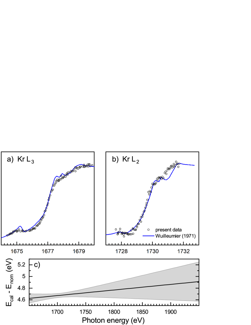
Absorption at the Kr and edges was also measured by others. The results of Kato et al. (2007) differ from those of Wuilleumier (1971) by 1.25 and 0.55 eV, respectively. Kato et al. (2007) calibrated their energy scale to the Kr excitation energy in the vicinity of the threshold as determined by Ibuki et al. (2002). These latter authors did not provide an estimate for the uncertainty of their energy scale. The same holds for the Kr absorption measurements of Schmelz et al. (1995). Nagaoka et al. (2000) and Okada et al. (2005) calibrated their energy scales by referring to the work of Wuilleumier (1971) as is also done here.
The energy shifts applied to our absorption curves were 4.65 and 4.7 eV for the edge and the edge, respectively. We attribute a read-off uncertainty of 0.05 eV to this calibration method. For the extrapolation to higher energies we used a linear fit through the two calibration points. By this extrapolation, the read-off error propagates to energy uncertainties of 0.2 eV at 1840 eV and of 0.3 eV at 1920 eV (Figure 1c). Taking the 0.5 eV uncertainty of the recommended -threshold energy (Deslattes et al., 2003) and a 0.2 eV uncertainty associated with imperfections of the photon beamline (see below) additionally into account, we arrive at an uncertainty of 1 eV of the present calibrated photon-energy scale for the photon energies in the current experimental range of 1835–1900 eV.
In calibrating the photon-energy scale of the silicon ions, we have additionally considered the Doppler shift that is caused by the unidirectional movement of the ions in the Siq+ ion beams (see, e.g., Schippers et al., 2014, for details). This does not introduce any significant additional uncertainty of the photon-energy calibration.
3 Theory
| Ion | ionization | excitation |
|---|---|---|
| Si+ | ||
| Si2+ | ||
| Si3+ | ||
| Si4+ | ||
| Si5+ | ||
The present theoretical calculations were performed in the framework of the Multi-Configuration Dirac-Hartree-Fock (MCDHF) method by applying the Grasp (Jönsson et al., 2007) and Ratip (Fritzsche, 2012) atomic structure codes for the calculation of atomic energy levels and transition rates. In particular, the Auger cascades were analyzed and calculated that follow the creation of a -shell hole in initially Siq+ ions by direct ionisation or by excitation. These cascade computations (Fritzsche et al., 2021) support quantitative estimates for the distribution of Sir+ product ions (cf. Equation 1), similar to earlier studies for other atoms and ions (Stock et al., 2017; Beerwerth & Fritzsche, 2017; Buth et al., 2018; Beerwerth et al., 2019; Schippers et al., 2017, 2021).
In these cascade computations, the Siq+ ions were assumed to be initially in their respective ground configuration with statistically populated fine-structure levels. Cross sections for the direct photoionization of these ions were obtained for each atomic subshell, while the resonant photoexcitation was considered for all shells with for Si+ and for Si2+ and Si3+. From the incoherent summation of all these direct-photoionization and resonant-excitation contributions, the total photoabsorption cross-sections were determined for Si+, Si2+, and Si3+ ions.
While different autoionization processes might contribute to the electron emission from inner-shell excited ions, only those single-step Auger processes were taken into account into the cascade, that are energetically allowed in a configuration-averaged representation of the atomic fine-structure. This simplifies the cascade computations and makes them tractable, although further contributions from the simultaneous excitation (shake-up) or ionization (shake-off) of another electron were found essential as well for accurately predicting the final charge-state distributions of light and especially negatively-charged ions (Müller et al., 2015; Schippers et al., 2016b; Perry-Sassmannshausen et al., 2020). For multiply-charged ions, however, these shake processes are typically suppressed, though not always negligible. Table 1 lists all those electron configurations that were included into the present cascade computations for Si+ ions. The number of cascade steps, i.e., the so-called depth of the cascade, were taken to be 4 for Si+, 3 for Si2+ , and 2 for Si3+. For the analysis of the cascades, therefore, the highest product-ion charge state was (cf. Equation 1), while higher charge states were not accessible within the given approach and by excluding autoionization processes with an additional excitation or ionization of electrons.
Theoretical cross sections for multiple photoionization were eventually obtained by multiplying the calculated absorption cross sections with the branching ratios from the cascade computations. These computations were carried out separately for the different core ionized and core excited configurations. For the comparison with the experimental results, the theoretical cross sections were convolved with a Gaussian with the full width at half maximum (FWHM) corresponding to the experimental photon energy spread .
4 Results and discussion
4.1 Cross sections for multiple ionization
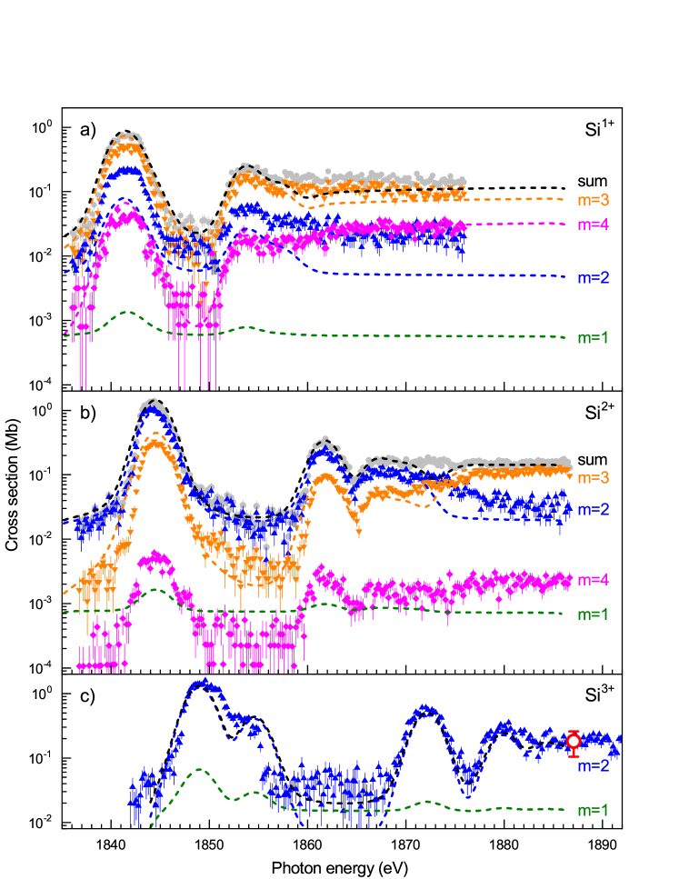
Figure 2 provides an overview over the measured and calculated cross sections for multiple photoionization of Siq+ ions (cf. Equation 1) with primary charge states together with the cross-section sums
| (2) |
The experimental energy ranges comprise the thresholds for direct ionization of one -shell electron. The experimental photon-energy spread was eV. This is sufficient for resolving the individual resonance groups for and . For Si3+, even the resonance group can be discerned.
The logarithmic cross-section scales cover up to four orders of magnitude. For Si+, the main ionization channel is triple ionization. For Si2+, triple ionization dominates only for energies above 1875 eV where direct -shell ionization becomes energetically possible. At lower energies, double ionization is stronger. For Si3+, double ionization is the dominating ionization channel in the entire experimental photon-energy range. The single-ionization channel could not be measured as explained above (Section 2.1). Other Si3+ ionization channels were scrutinized, but no detectable signal was found. This indicates that the associated cross sections are small as is also corroborated by our theoretical calculations.
Our theoretical ab-initio cross sections are in remarkable agreement with the measured ones considering the simplifications that had to be applied in order to make the calculations of the complex deexcitation cascades tractable. We like to point out that the calculations were performed independently of the measurements.
The experimental and theoretical resonances line up if energy shifts of , , and eV are applied to the Si+, Si2+, and Si3+ cross sections, respectively. These differences between our theoretical and experimental resonance positions are less than a factor of 2 larger than the 1 eV uncertainty of the experimental energy scale. Our theoretical calculations do not account for any of the metastable primary ions discussed in Section 2.1. The comparison between the theoretical and the experimental resonance structures suggests that metastable ions do indeed not play a significant role in the present study as already concluded above.
Only the theoretical and experimental cross-sections can be directly compared on an absolute scale. The mutual agreement is within the 15% experimental uncertainty over nearly the entire experimental energy range. The largest discrepancy between the experimental and present theoretical cross sections concerns the (small) Si+ double-ionization cross-section which is underestimated by a factor of 2. At energies between 1845 and 1858 eV, slight discrepancies occur which are due to a limited ability of the theory to correctly describe the experimentally observed resonance structure. There are no such obvious discrepancies for Si+ and Si2+.
We conclude that the present theoretical approach is capable of reliably predicting the multiple photoionization cross-sections of low-charged silicon ions. Similar accuracy can be expected for neighboring elements from the periodic table. The benchmarking by our experimental results leads to rather small corrections of the theoretical resonance energies which are only slightly larger than the uncertainty of the present experimental photon-energy scale.
4.2 Absorption cross-sections
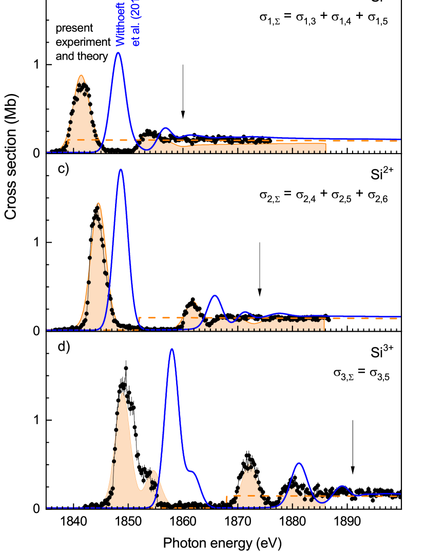
For Si+ and Si2+, the experimental cross-section sums were put on absolute scales by multiplying all individual cross sections for a given primary charge state by the same factor such that the sums line up with the respective theoretical absorption cross-sections of Verner et al. (1993, Figure 3b–3d). We have applied the same approach already earlier to the photoabsorption of negatively charged Si- ions (Perry-Sassmannshausen et al., 2021) where we used the theoretical absorption cross section for neutral silicon by Henke et al. (1993) as a reference. These data are shown in Figure 3a for comparison. Apparently, the cross sections for non-resonant absorption are nearly independent of the primary charge state. At 1890 eV the cross sections for Si0 (Henke et al., 1993) and for Si+, Si2+, and Si3+ (Verner et al., 1993) all amount to about 0.15 Mb.
For energies where there are no signatures from resonant processes, our theoretical cross-section sums for Si+, Si2+, and Si3+ agree with the results of Verner et al. (1993), which do not account for resonant photoabsorption. The same is true for the more recent theoretical results of Witthoeft et al. (2011), who also did include resonant photoabsorption in their calculations. However, their resonance positions deviate significantly by up to 9 eV from the measured ones. Our present energy-shifted (by less than 2 eV, see above) theoretical results fit much better to the experimentally observed resonance positions than the results of Witthoeft et al. (2011).
Another significant discrepancy between the present and the previous results concerns the thresholds for direct -shell photoionization. Our theoretical results are 1860, 1874, and 1891 eV for Si+, Si2+, and Si3+, respectively, with the above mentioned energy shifts applied. As can be seen from Figure 3, the threshold energies as predicted by Verner et al. (1993) are lower by more than 20 eV. This shows that our experimental benchmarks are vital for arriving at an accurate positioning of the various absorption features. From the comparison with the theoretical absorption cross sections we conclude that, within the present experimental uncertainties, the experimental cross-section sums represent the Siq+ photoabsorption cross-sections.
4.3 High-resolution measurements of resonances
The most prominent resonance feature in each absorption cross-section is the lowest-energy resonance group, which is associated with excitation (Figure 3). Its relative strength increases with increasing primary charge state. This also holds for the resonances associated with excitation to higher subshells. In order to provide accurate resonance data we have measured the most prominent resonance features at lower photon-energy spreads. This was achieved at the expense of photon flux by narrowing the monochromator exit-slit.
| Designation | ||||
|---|---|---|---|---|
| (eV) | (eV) | (Mb eV)††footnotemark: | (Mb eV) | |
| 1841.26(1) | 0.38(22) | 0.91(07) | 1.42(11) | |
| 1841.82(1) | 0.39(09) | 0.70(14) | 1.09(22) | |
| 1842.35(5) | 0.34(13) | 0.15(07) | 0.23(11) | |
| 1843.43(2) | 0.94(04) | 0.26(01) | 0.41(02) |
| Designation | ||||
|---|---|---|---|---|
| (eV) | (eV) | (Mb eV) | (Mb eV) | |
| 1845.10(01) | 0.28(03) | 2.48(16) | 3.33(22) | |
| 1845.84(15) | 0.76(14) | 0.72(16) | 0.97(22) | |
| 1862.33(24) | 0.37(08) | 0.55(10) | 0.60(11) | |
| 1863.10(20) | 1.77(37) | 0.48(18) | 0.52(20) | |
| 1867.58(08) | 0.00(20) | 0.07(05) | ||
| 1867.85(08) | 0.84(15) | 0.32(08) |
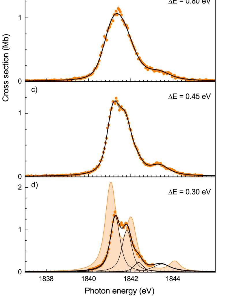
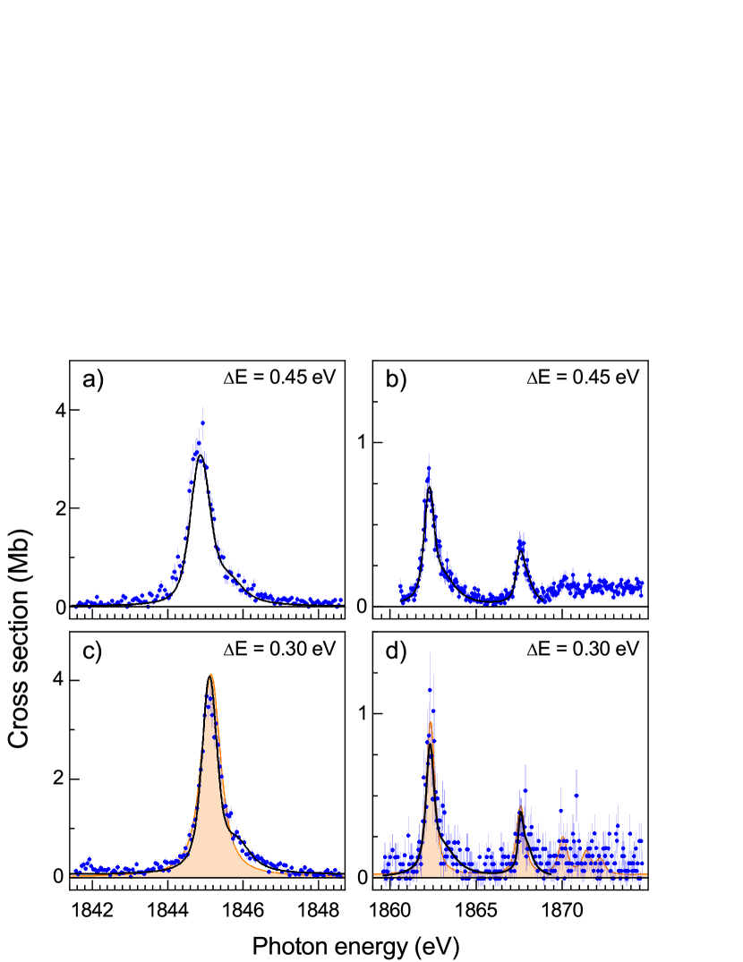
Figure 4 displays high-resolution measurements of the Si+() resonance group, which consists of the four LS terms , , , and resulting from the coupling of the single electron to the three parent terms , , and . For these measurements the dominant triple-ionization channel () was chosen. The experimental resolving power increases when going from panel a) to panel d). The numerical values of the respective photon energy spreads were obtained by a simultaneous fit of four Voigt line profiles to the four measured data sets. The resonance parameters that resulted from the fit, i.e., the resonance energies , the Lorentzian line widths , and the resonance strengths are listed in Table 2.
| Designation | ||||
|---|---|---|---|---|
| (eV) | (eV) | (Mb eV) | (Mb eV) | |
| 1849.64(01) | 0.45(02) | 5.20(09) | 5.20(09) | |
| 1853.24(01) | 0.35(04) | 1.23(06) | 1.23(06) | |
| 1871.91(02) | 0.34(05) | 1.19(08) | 1.19(08) | |
| 1872.96(04) | 0.45(12) | 0.56(08) | 0.56(08) | |
| 1880.20(05) | 1.00(44) | 0.79(06) | 0.79(06) | |
| 1883.86(04) | 0.23(12) | 0.25(04) | 0.25(04) |
In the fit, an overall energy shift was used as an additional fit parameter individually for each data set (see caption of Figure 4). The maximum shift amounts to eV which is much less than the 1 eV uncertainty of the experimental photon-energy scale. Ideally, one would expect that the resonance positions were independent of the width of the monochromator’s exit-slit. We attribute the observed shifts to a slight asymmetry of the photon-energy distribution and to mechanical imperfections of the photon-beamline’s mechanics. A pertaining 0.2 eV uncertainty has already been considered in the error budget of our photon-energy calibration (see Section 2.2).
In analogy to the work of Schlachter et al. (2004) for carbon, we note that the and levels, which are populated by excitation of Si+, can also be prepared by the removal of a electron from neutral ground-term neutral Si(). Therefore, the widths of the and levels from Table 2 correspond to the core-hole lifetime of initially neutral silicon. Both widths agree within their experimental uncertainties. From the less uncertain value we obtain fs in agreement with the present calculations and with the value of 1.47 fs calculated by Palmeri et al. (2008).
In Figure 4d, the present theoretical results for are compared with the experimental high-resolution results. For the comparison, the theoretical resonances were shifted in energy by eV and convoluted with a Gaussian with a FWHM of 0.3 eV. This resolution is a factor of 10 higher as compared to the one in Figure 3 and reveals some discrepancies between experiment and theory which are missed when looked at with larger photon-energy spread. The calculated splittings between the various terms are larger than what is found experimentally.
The computed resonance strengths of individual terms contributing to (see Figure 4d) are larger than the experimental ones by about 50%. Since the present theoretical and experimental absorption cross-sections agree with one another within the experimental uncertainty (Figure 3b) it must be concluded that the mismatch for is due to the overestimation of the corresponding branching ratios for the resonances.
The theoretical branching ratios for the triple-ionization channel are almost the same for all of the resonances, i.e., their differences are smaller than the uncertainties of the individual values. This allows us to scale the fitted individual resonance strengths to the absorption cross-section. From a fit to the peak in Figure 3b we obtain a strength of 3.15(17) Mb eV for the sum of the four tabulated resonances. The sum of the values in Table 2 amounts to 2.02(17) Mb eV. Using these values we have calculated the tabulated absorption resonance-strengths as . In the same way we have calculated separately for the [factor 4.30(3)/3.20(22)] and [factor (1.12(4)/1.03(21)] resonances of the Si2+ absorption spectrum (Table 3). For Si3+, the absorption spectrum is essentially identical with and, thus, for all resonances in Table 4.
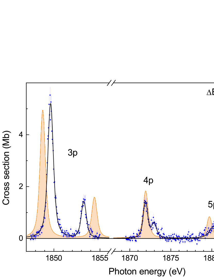
The fits to the Si2+ and Si3+ high-resolution data are shown in Figures 5 and 6, respectively. The agreement between experiment and the present theory (shifted by eV for Si2+ and by eV for Si3+, see Figure 3) is remarkable, particularly, for (Figure 5). There is less agreement for Si3+, as already noted in the low-resolution data in Figures 2 and 3. The too large separation of the first two peaks is probably caused by subtleties concerning the atomic fine structure associated with the angular momentum coupling of three open atomic subshells.
4.4 Comparison with other forms of silicon
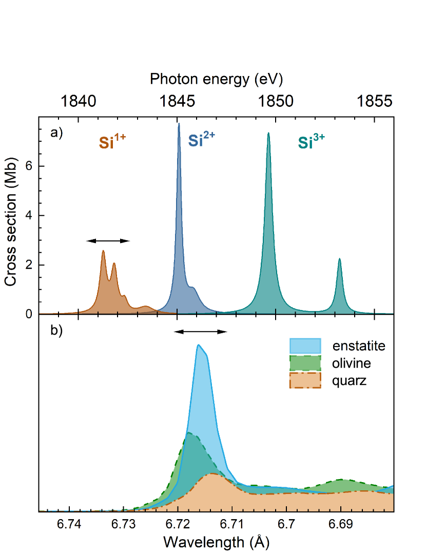
Figure 7 shows a comparison between the strongest Si -shell absorption features for Si+, Si2+, and Si3+ (panel a) with the absorption by some silicon containing minerals that potentially occur in interstellar dust (panel b, Zeegers et al., 2019). In order to be able to discriminate between the different forms of silicon in x-ray absorption spectra, the energies of the absorption features must be known with sufficient precision. The systematic uncertainty of the present experimental energy scale is eV (Section 2.2). Zeegers et al. (2019) did not quantify the uncertainty of their energy scale. Instead, they referred for their energy calibration to previous work (Li et al., 1995; Nakanishi & Ohta, 2009) which, in turn, is based on earlier investigations. The energy scale of Nakanishi & Ohta (2009) can be traced back to the work of Wong et al. (1999) who mention that their energy calibration varies by 2–3 eV depending on the operating conditions of their synchrotron light source. This suggests that the uncertainty of the energy scale of Zeegers et al. (2019, Figure 7b) is at least eV.
Different minerals exhibit different chemical shifts of the Si -shell absorption features which vary by up to 1.5 eV (Li et al., 1995). This is still less than the combined energy uncertainties of the present energy scale and the one of Zeegers et al. (2019). Therefore, we conclude that, in general, x-ray absorption spectroscopy can discriminate between absorption by gaseous silicon and absorption by silicon containing minerals. In particular, the Si+ absorption feature in Figure 7a appears at an unambiguous location ( eV). This is also true for the corresponding absorption feature of Si- which occurs at an even lower energy of 1838.4 eV (Figure 3a, Perry-Sassmannshausen et al., 2021). To the best of our knowledge, there are no such experimental data for gaseous neutral silicon. In view of the findings for Si- and Si+ its dominating absorption feature can be expected close to 1840 eV.
Gatuzz et al. (2020) investigated the gaseous component of the ISM using the theoretical absorption data of Witthoeft et al. (2011). In their data, the resonance group for neutral silicon is located at 1839.5 eV in accord with the above considerations. For the other charge states there are significant discrepancies as already noted above. The deviations between the theoretical resonance positions of Witthoeft et al. (2011) and the present experimental ones are 6.7, 4.4, and 9.1 eV for Si+, Si2+, and Si3+, respectively (Figure 3). All these differences are substantially larger than the uncertainty of the experimental energy scale and they are as large or larger than the energy differences between adjacent charge states (Figure 7a). This will lead to a wrong assignment of astronomically observed resonance features if their analysis is based on these theoretical absorption cross sections.
5 Conclusions and outlook
Using the photon-ion merged-beams technique at a synchrotron light source we have measured cross sections for multiple photoionization of low-charged Si+, Si2+, and Si3+ ions and derived precise absorption data (resonance positions, widths, and strengths) which can be directly used for the astrophysical modelling of the silicon -shell absorption by interstellar gas clouds and other cosmic objects as well as for the benchmarking the theoretical calculations. From the widths of the core excited resonances in Si+ we inferred a value for the core-hole lifetime of -ionized neutral silicon of fs.
Previously theoretically predicted absorption features deviate significantly in energy from the present experimental findings, whereas the present large-scale MCDHF calculations agree much better with the experimental results. In addition, the present computations also capture the hole-deexcitation cascades following the initial creation of a -shell hole. The obtained product charge-state distributions, which are required for an accurate modelling of the charge balance in astrophysical plasmas, agree remarkably well with the experimental results, despite of the simplifications that had to be applied to keep the calculations tractable. Current code development aims at systematically expanding such cascade calculations to improve the treatment of the deexcitation processes (Fritzsche, 2019; Fritzsche et al., 2021).
When comparing absorption data from different sources the calibration of the photon energy scales is an issue of concern. The present experimental uncertainty of eV is sufficient for discriminating between absorption by gaseous and solid silicon-containing matter in the x-ray absorption spectra from the currently operated x-ray telescopes. The accuracy demands will increase with increasing resolving power of future missions such as Athena (Barret et al., 2020). Meeting these demands will require the development of more accurate calibration standards at synchrotron light sources. Promising candidates are few-electron atomic ions which promise calibration uncertainties of less than 10 meV (see, e.g., Müller et al., 2018; Stierhof et al., 2022). Corresponding activities are under way at the PIPE setup.
References
- Barret et al. (2020) Barret, D., Decourchelle, A., Fabian, A., et al. 2020, Astron. N., 341, 224, doi: 10.1002/asna.202023782
- Beerwerth & Fritzsche (2017) Beerwerth, R., & Fritzsche, S. 2017, EPJD, 71, 253, doi: 10.1140/epjd/e2017-80064-3
- Beerwerth et al. (2019) Beerwerth, R., Buhr, T., Perry-Sassmannshausen, A., et al. 2019, ApJ, 887, 189, doi: 10.3847/1538-4357/ab5118
- Bizau et al. (2009) Bizau, J.-M., Mosnier, J.-P., Kennedy, E. T., et al. 2009, PhRvA, 79, 033407
- Buth et al. (2018) Buth, C., Beerwerth, R., Obaid, R., et al. 2018, JPhB, 51, 055602, doi: 10.1088/1361-6455/aaa39a
- Deslattes et al. (2003) Deslattes, R. D., Kessler, E. G., Indelicato, J. P., et al. 2003, RvMP, 75, 35, doi: 10.1103/RevModPhys.75.35
- Fritzsche (2012) Fritzsche, S. 2012, CoPhC, 183, 1525, doi: 10.1016/j.cpc.2012.02.016
- Fritzsche (2019) —. 2019, CoPhC, 240, 1, doi: 10.1016/j.cpc.2019.01.012
- Fritzsche et al. (2021) Fritzsche, S., Palmeri, P., & Schippers, S. 2021, Symmetry, 13, 520, doi: 10.3390/sym13030520
- Froese Fischer et al. (2006) Froese Fischer, C., Tachiev, G., & Irimia, A. 2006, ADNDT, 92, 607, doi: 10.1016/j.adt.2006.03.001
- Gatuzz et al. (2020) Gatuzz, E., Gorczyca, T. W., Hasoglu, M. F., et al. 2020, MNRAS, 498, L20, doi: 10.1093/mnrasl/slaa119
- Hasoglu et al. (2021) Hasoglu, M. F., Gorczyca, T. W., & Manson, S. T. 2021, PhyS, 96, 124024, doi: 10.1088/1402-4896/ac0b84
- Henke et al. (1993) Henke, B. L., Gullikson, E. M., & Davis, J. C. 1993, ADNDT, 54, 181, doi: 10.1006/adnd.1993.1013
- Howald et al. (1986) Howald, A. M., Gregory, D. C., Meyer, F. W., et al. 1986, PhRvA, 33, 3779, doi: 10.1103/PhysRevA.33.3779
- Ibuki et al. (2002) Ibuki, T., Okada, K., Kamimori, K., et al. 2002, SRL, 09, 85, doi: 10.1142/S0218625X02001987
- Jenkins (2009) Jenkins, E. B. 2009, ApJ, 700, 1299, doi: 10.1088/0004-637X/700/2/1299
- Jönsson et al. (2007) Jönsson, P., He, X., Froese-Fischer, C., & Grant, I. P. 2007, CoPhC, 177, 597, doi: 10.1016/j.cpc.2007.06.002
- Kato et al. (2007) Kato, M., Morishita, Y., Oura, M., et al. 2007, AIP Conference Proceedings, 879, 1121, doi: 10.1063/1.2436260
- Kennedy et al. (2014) Kennedy, E. T., Mosnier, J.-P., Van Kampen, P., et al. 2014, PhRvA, 90, 063409, doi: 10.1103/PhysRevA.90.063409
- Kučas et al. (2012) Kučas, S., Karazija, R., & Momkauskaitė, A. 2012, ApJ, 750, 90, doi: 10.1088/0004-637X/750/2/90
- Kučas et al. (2015) Kučas, S., Momkauskaitė, A., & Karazija, R. 2015, ApJ, 810, 26, doi: 10.1088/0004-637X/810/1/26
- Li et al. (1995) Li, D., Bancroft, G. M., Fleet, M. E., & Feng, X. H. 1995, PCM, 22, 115, doi: 10.1007/BF00202471
- Mosnier et al. (2003) Mosnier, J.-P., Sayyad, M. H., Kennedy, E. T., et al. 2003, PhRvA, 68, 052712, doi: 10.1103/PhysRevA.68.052712
- Müller et al. (2015) Müller, A., Borovik Jr., A., Buhr, T., et al. 2015, PhRvL, 114, 013002, doi: 10.1103/PhysRevLett.114.013002
- Müller et al. (2017) Müller, A., Bernhardt, D., Borovik Jr., A., et al. 2017, ApJ, 836, 166, doi: 10.3847/1538-4357/836/2/166
- Müller et al. (2018) Müller, A., Lindroth, E., Bari, S., et al. 2018, PhRvA, 98, 033416, doi: 10.1103/PhysRevA.98.033416
- Müller et al. (2021) Müller, A., Martins, M., Borovik, A., et al. 2021, PhRvA, 104, 033105, doi: 10.1103/PhysRevA.104.033105
- Nagaoka et al. (2000) Nagaoka, S., Ibuki, T., Saito, N., et al. 2000, JPhB, 33, L605, doi: 10.1088/0953-4075/33/17/102
- Nakanishi & Ohta (2009) Nakanishi, K., & Ohta, T. 2009, JPCM., 21, 104214, doi: 10.1088/0953-8984/21/10/104214
- Okada et al. (2005) Okada, K., Kosugi, M., Fujii, A., et al. 2005, JPhB, 38, 421, doi: 10.1088/0953-4075/38/4/009
- Palmeri et al. (2008) Palmeri, P., Quinet, P., Mendoza, C., et al. 2008, ApJS, 177, 408, doi: 10.1086/587804
- Perry-Sassmannshausen et al. (2021) Perry-Sassmannshausen, A., Buhr, T., Martins, M., et al. 2021, PhRvA, 104, 053107, doi: 10.1103/PhysRevA.104.053107
- Perry-Sassmannshausen et al. (2020) Perry-Sassmannshausen, A., Buhr, T., Borovik Jr., A., et al. 2020, PhRvL, 124, 083203, doi: 10.1103/PhysRevLett.124.083203
- Rogantini et al. (2020) Rogantini, D., Costantini, E., Zeegers, S. T., et al. 2020, 641, A149, doi: 10.1051/0004-6361/201936805
- Schippers et al. (2016a) Schippers, S., Kilcoyne, A. L. D., Phaneuf, R. A., & Müller, A. 2016a, ConPh, 57, 215, doi: 10.1080/00107514.2015.1109771
- Schippers & Müller (2020) Schippers, S., & Müller, A. 2020, Atoms, 8, 45, doi: 10.3390/atoms8030045
- Schippers et al. (2014) Schippers, S., Ricz, S., Buhr, T., et al. 2014, JPhB, 47, 115602, doi: 10.1088/0953-4075/47/11/115602
- Schippers et al. (2016b) Schippers, S., Beerwerth, R., Abrok, L., et al. 2016b, PhRvA, 94, 041401(R), doi: 10.1103/PhysRevA.94.041401
- Schippers et al. (2017) Schippers, S., Martins, M., Beerwerth, R., et al. 2017, ApJ, 849, 5, doi: 10.3847/1538-4357/aa8fcc
- Schippers et al. (2020) Schippers, S., Buhr, T., Borovik Jr., A., et al. 2020, XRS, 49, 11, doi: 10.1002/xrs.3035
- Schippers et al. (2021) Schippers, S., Beerwerth, R., Bari, S., et al. 2021, ApJ, 908, 52, doi: 10.3847/1538-4357/abcc64
- Schlachter et al. (2004) Schlachter, A. S., Sant’Anna, M. M., Covington, A. M., et al. 2004, JPhB, 37, L103, doi: 10.1088/0953-4075/37/5/L03
- Schmelz et al. (1995) Schmelz, H. C., Gaveau, M. A., Reynaud, C., et al. 1995, PhyB, 208-209, 519, doi: https://doi.org/10.1016/0921-4526(94)01038-3
- Schmidt et al. (2007) Schmidt, E. W., Bernhardt, D., Müller, A., et al. 2007, PhRvA, 76, 032717, doi: 10.1103/PhysRevA.76.032717
- Stierhof et al. (2022) Stierhof, J., Kühn, S., Winter, M., et al. 2022, EPJD, 76, 38, doi: 10.1140/epjd/s10053-022-00355-0
- Stock et al. (2017) Stock, S., Beerwerth, R., & Fritzsche, S. 2017, PhRvA, 95, 053407, doi: 10.1103/PhysRevA.95.053407
- Tiesinga et al. (2021) Tiesinga, E., Mohr, P. J., Newell, D. B., & Taylor, B. N. 2021, RvMP, 93, 025010, doi: 10.1103/RevModPhys.93.025010
- Verner et al. (1993) Verner, D. A., Yakovlev, D. G., Band, I. M., & Trzhaskovskaya., M. B. 1993, ADNDT, 55, 233, doi: 10.1006/adnd.1993.1022
- Viefhaus et al. (2013) Viefhaus, J., Scholz, F., Deinert, S., et al. 2013, NIMPA, 710, 151, doi: 10.1016/j.nima.2012.10.110
- Witthoeft et al. (2011) Witthoeft, M. C., García, J., Kallman, T. R., et al. 2011, ApJS, 1992, 7, doi: 10.1088/0067-0049/192/1/7
- Wong et al. (1999) Wong, J., Tanaka, T., Rowen, M., et al. 1999, J Snychr. Rad., 6, 1086, doi: 10.1107/S0909049599009000
- Wuilleumier (1971) Wuilleumier, F. 1971, J. Physique, 32, C4.88, doi: 10.1051/jphyscol:1971418
- Zeegers et al. (2019) Zeegers, S. T., Costantini, E., Rogantini, D., et al. 2019, 627, A16, doi: 10.1051/0004-6361/201935050
- Zeegers et al. (2017) Zeegers, S. T., Costantini, E., de Vries, C. P., et al. 2017, 599, A117, doi: 10.1051/0004-6361/201628507