Giant magnetic in-plane anisotropy and competing instabilities in \chNa_3Co_2SbO_6
Abstract
We report magnetometry data obtained on twin-free single crystals of \chNa_3Co_2SbO_6, which is considered a candidate material for realizing the Kitaev honeycomb model for quantum spin liquids. Contrary to a common belief that such materials can be modeled with the symmetries of an ideal honeycomb lattice, our data reveal a pronounced two-fold symmetry and in-plane anisotropy of over 200%, despite the honeycomb layer’s tiny orthorhombic distortion of less than 0.2%. We further use magnetic neutron diffraction to elucidate a rich variety of field-induced phases observed in the magnetometry. These phases manifest themselves in the paramagnetic state as diffuse scattering signals associated with competing ferro- and antiferromagnetic instabilities, consistent with a theory that also predicts a quantum spin liquid phase nearby. Our results call for theoretical understanding of the observed in-plane anisotropy, and render \chNa_3Co_2SbO_6 a promising ground for finding exotic quantum phases by targeted external tuning.
Frustrated magnetic systems have the potential to realize exotic quantum spin liquids (QSLs) [1, 2, 3]. The exactly solvable Kitaev model [4], which features bond-dependent Ising interactions between effective spin-1/2 nearest neighbors on a honeycomb lattice, has motivated intensive QSL research in recent years. As a guiding principle, it is believed that such interactions can be realized in spin-orbit coupled Mott insulators [5, 6, 7, 8]. Solid-state platforms for realizing the Kitaev model have evolved over the years from iridium [9] to ruthenium [10] compounds, and most recently to cobaltates [11, 12, 13, 14, 15]. Despite a potential drawback of weaker spin-orbit coupling, the cobaltates are believed to have relatively weak non-Kitaev and further-neighbor interactions compared to their and counterparts [11, 12, 15].
As a reality of nature, essentially all candidate Kitaev magnets have long-range order at low temperatures [16, 17, 18, 19, 20, 6]. This has been attributed to the presence of interactions beyond the Kitaev model [9, 21, 22, 23, 24, 25, 26, 27, 28, 29, 30], such that additional tuning is needed to overcome the ordering tendency, e.g., by using thermal disorder and external fields [31, 32, 33, 34, 35, 36, 13, 37], in order to recover QSL behaviors. To this end, it is important to know how close the microscopic model of a given system is to an anticipated QSL phase. The cobaltate \chNa_3Co_2SbO_6 is promising in this regard, as its model is inferred to situate near boundaries between ferromagnetic (FM), antiferromagnetic (AFM), and QSL phases [13]. This understanding is supported by the relatively low Néel temperature () and small saturation fields of the system compared to its sister compound \chNa_2Co_2TeO_6 [16, 38, 39, 40].
Notably, while non-Kitaev and further-neighbor terms are widely considered in theoretical constructions [21, 23, 25, 26, 24, 27, 28, 29, 30, 41, 42, 43, 44, 45], the low, monoclinic symmetry of many candidate materials, including \chNa_2IrO_3 [17, 18], -\chRuCl_3 [19, 46], and \chNa_3Co_2SbO_6 [16], is often neglected. Even though originating from inter-layer stacking, the monoclinicity also means lack of rotational symmetry of the crystal field and a transition-metal ion’s interactions with its neighbors in the same layer. Approximating the interactions with their bond-averaged values [30] is an assumption commonly taken but rarely checked. Two cobaltates, \chNa_2Co_2TeO_6 [16] and \chBaCo_2(AsO_4)_2 [20], have the symmetry, but their zero-field ground states are reported to be dissimilar to the monoclinic systems [47, 48, 20], and no consensus has been reached concerning the microscopic models [49, 50, 51, 52, 53, 54]. The lack of symmetry should result in magnetic in-plane anisotropy, as has been observed in -\chRuCl_3 [55]. However, the anisotropy is found to vary considerably [55, 56, 57], possibly due to sample-dependent monoclinic domain population.

Here, we report a systematic study of \chNa_3Co_2SbO_6 aided by the use of twin-free crystals. Magnetometry reveals at low temperatures a strong in-plane anisotropy, in both the low-field susceptibility and the critical fields for switching toward a series of field-induced states. The magnitude of the anisotropy is unprecedented, yet the field-induced transitions resemble other systems to some extent. We further use neutron diffraction to determine the wave vectors of the field-induced states. They signify a series of AFM and FM instabilities, which closely compete and produce distinct diffuse scattering above in zero field. These results render \chNa_3Co_2SbO_6 a highly intriguing system with the potential to realize exotic phases under targeted tuning.

I Magnetometry on twin-free crystals
Figure 1(a-b) presents the crystal and reciprocal-space structure of \chNa_3Co_2SbO_6, which has the same space group () as -\chRuCl_3 [19, 46]. A peculiarity common to both structures is in the stacking: adjacent honeycomb layers are offset from each other by , hence we have , with and in \chNa_3Co_2SbO_6 [39]. Similar to -\chRuCl_3 [46], the orthorhombic distortion in the honeycomb layer of \chNa_3Co_2SbO_6 is tiny: the distortion is measured as [39, 40]. Yet, we find that such a small distortion removes the overall and symmetries. We will next show the far-reaching consequences on the magnetism using a rare growth product: twin-free single crystals. Such crystals can be found by screening with Raman spectroscopy (Fig. S7 in [58]), and ultimately verified with -ray diffraction [Fig. 1(c)]. They have a well-defined of about 6.6 K (Fig. S8 in [58]) with sample-dependent variation of no more than 1 K possibly caused by structural imperfections. The variation is considerably smaller than in the literature [16, 38, 39, 40]. This is in line with the facts that our best crystals have very few stacking faults [Fig. 1(c)] compared to a previous report [39], and that the observed Bragg intensities agree well with calculation based on the ideal crystal structure [Fig. 1(d)]. Our further refinement attempts suggest that the agreement cannot be improved by introducing anti-site disorder between Co and Sb [39]. While the data do not allow us to rule out disorder in the Na layers [39], we consider its role to be minor because such disorder is expected to cause stacking faults which are rare in our crystals.

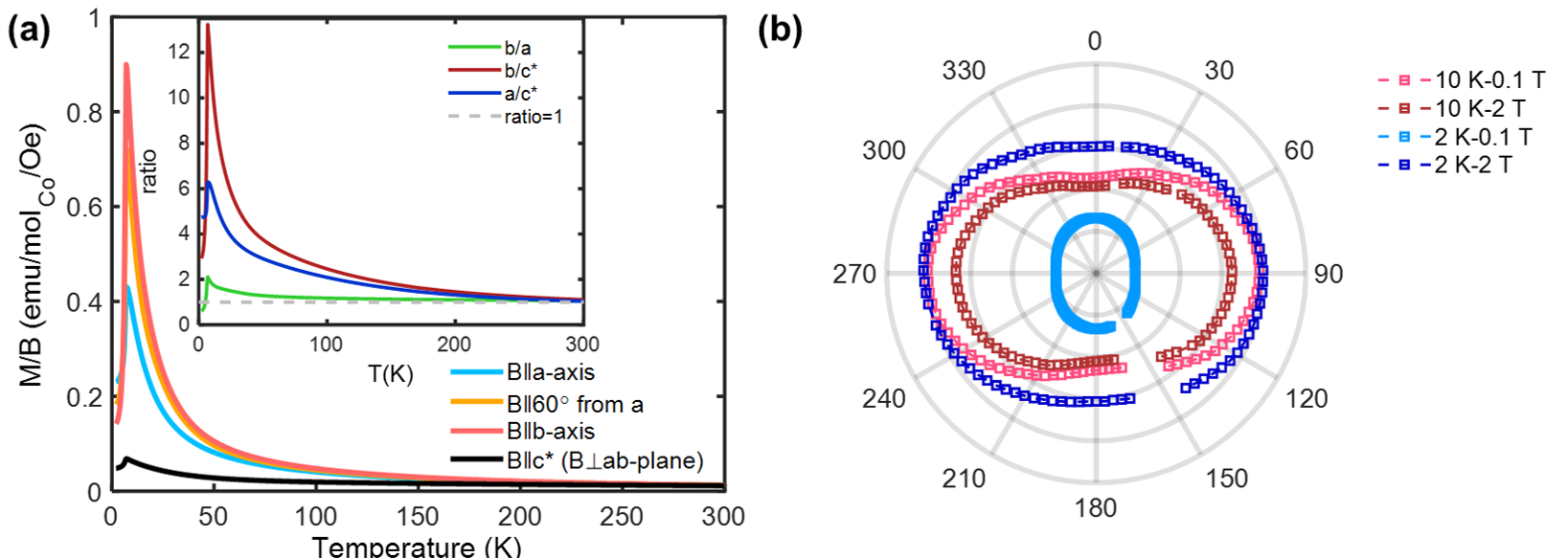
Figure 2(a) shows how a pronounced - anisotropy develops in the magnetic susceptibility upon cooling. Far above , we observe a anisotropy consistent with the anisotropy of the -factor, (Figs. S9-S10 and Table S1 in [58]), as is also seen from high-field magnetization where moments are nearly polarized [Fig. 2(b)]. The anisotropy drastically increases to over near (see Fig. S9 in [58] for out-of-plane anisotropy), which signifies the role of the fluctuations – the moments respond much more strongly in the easy direction nearly parallel to the developing order parameter [39]. This understanding also explains why the anisotropy is reversed below . The reversal is no longer observed in T (Fig. S9 in [58]) which is large enough to overcome the AFM order. We make two remarks here to relate to previous works: (1) The -axis response clearly drops below [Fig. 2(a)], suggesting that the ordered moments are not entirely along [39]. (2) No reversal is observed below in -\chRuCl_3 [55], where the anisotropy also appears to be much weaker.
The competition between anisotropic interactions and the applied field is more clearly seen in the magnetization at 2 K [Fig. 2(b-d)], where our twin-free sample reveals a wealth of remarkable features unnoticed in previous works [16, 38, 39, 40]. Two well-separated transitions are observed along both and , at critical fields [ and , Fig. 2(b)] that again differ strongly between the two directions. The lower-field transition is clearly hysteretic, as indicated by the magnetization’s dependence on the field-sweeping direction. It further splits into two hysteretic transitions, the critical fields of which we refer to as and , when the field is applied in-plane but away from the high-symmetry and axes [Fig. 2(c-d)]. The lowest value is found at about away from . The highest can approach and become no longer visible from the data, over a range of field directions between and away from . Hence, very unexpectedly, there is nearly no 6-fold symmetry in the results, including in the nearly field-polarized state at 2 T (Fig. S9 of [58]). The large magnitude of - anisotropy sharply contrasts with the -symmetric sister compound \chNa_2Co_2TeO_6, where the magnetic responses along and are reasonably similar [37, 50]. We note that quenched disorders may play a role in the experimentally observable anisotropy in \chNa_3Co_2SbO_6. By heating up a twin-free crystal to 600 ∘C at 20 ∘C/min, staying for 1 hour, and quenching the crystal in liquid nitrogen, we found the susceptibility anisotropy ratio [, see Fig. 2(a)] to change from 1.81 to 1.78 at 10 K, and from 0.53 to 0.75 at 2 K. The two field-induced transitions [Fig. 2(b)] at 2 K also became considerably smeared out. According to -ray diffraction, the crystal remained twin-free after the quenching.

II Magnetic neutron diffraction
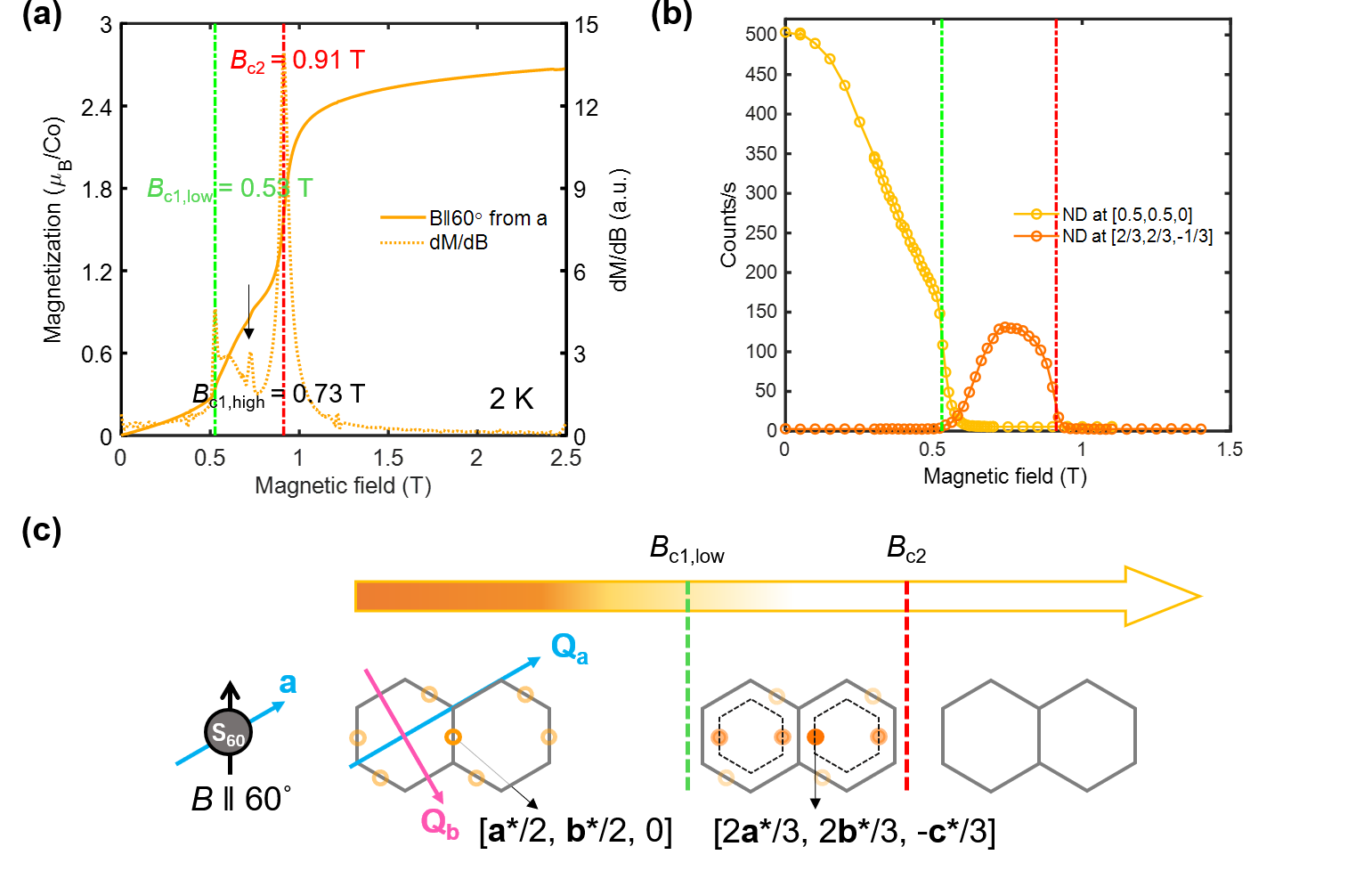
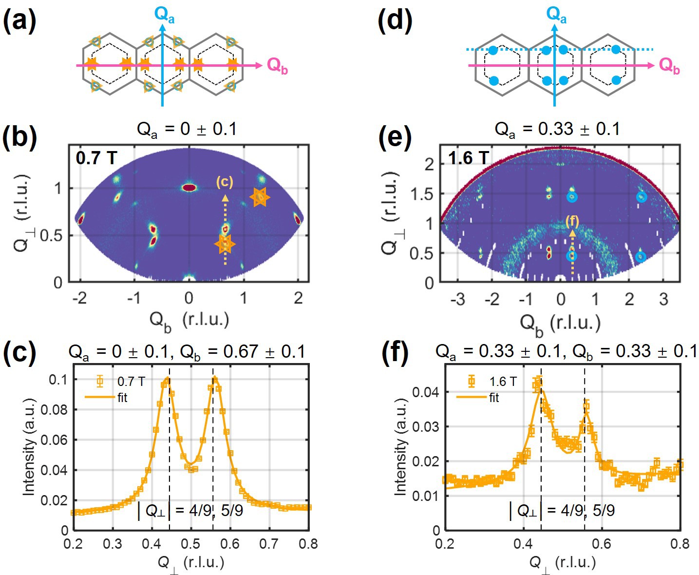
We next turn to the intermediate state(s) between and . While the step-like and hysteretic (near ) behaviors hinted at a spin-flop origin [16, 38, 39, 40], our observation of the transitions along both and (and everywhere in between) defies such an interpretation. The result in Fig. 2(c) is furthermore independent of field or temperature history, precluding the relevance of magnetic domain repopulation [59, 34]. Motivated by the fact that the magnetization above resembles “plateau” phases found in low-dimensional frustrated magnets [60, 61, 62, 63], i.e., it reaches about 1/3 and 1/2 of saturation [Fig. 2(b)] for and , respectively, we have performed neutron diffraction in magnetic fields to explore this possibility. The experiment was done on a co-aligned but twinned array of crystals with their axis horizontal, such that the vertical field was along for 1/3 of the sample (), and at 60∘ from for the rest (), see illustrations in the upper-left corner of Fig. 4. In spite of the twinning, there is no ambiguity in the domain origin ( or ) of the field-evolving signals, under the assumption that magnetization and diffraction see the same transitions (Fig. S11 in [58]). According to magnetization [Fig. 2(d)], all transitions occur below (above) 1 Tesla for (). The difference is illustrated by the thick horizontal arrow diagrams in the upper half of Fig. 4.
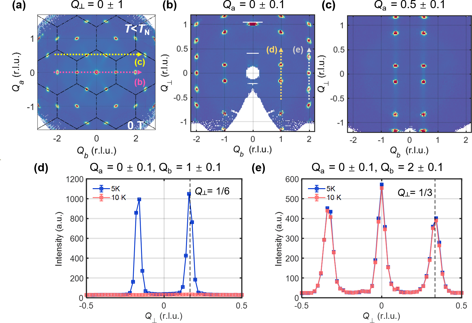
We use here a “hybrid” orthogonal coordinate system for the reciprocal space, illustrated in Fig. 1(b). Wave vectors are denoted as , with and in units of and , respectively. is in units of projected onto the real-space -axis, and it is parallel to the vertical field. This coordinate system is convenient for describing a twinned sample, because the twinning features rotations within the -plane, and mixes up and while leaving intact. We will write , , and explicitly when we refer to the (physical) monoclinic indices. A table reference for transforming between the two indexing systems can be found in Table S3 [58]. To give some examples, nuclear Bragg peaks at in the hybrid notation [Fig. 3(a-b)] are associated with physical indices of . In zero field, the AFM wave vectors [39] of transform into in the hybrid notation [Fig. 3(b-d)], producing diffractions at four locations, whereas the same diffractions from the two copies of (Fig. 4) are expected at six locations. All of these AFM wave vectors have as indicated by empty symbols in Fig. 4. While the data coverage in Fig. 4 along the direction is limited compared to that in Fig. 3), magnetic diffractions above and below the (horizontal) plane are partly observed. This is enabled by vertical focusing optics [58], which relaxes the momentum resolution and elongates diffraction features in the direction.
The results in Fig. 4 can be summarized as follows: the AFM wave vectors switch from at , to above , and eventually no AFM is left above 2.2 T (see Methods and Figs. S11-S13 in [58] for additional evidence for the peak indexing). We therefore refer to the zero-field and the intermediate states as AFM and AFM, respectively. The AFM wave vectors all have or [58], which allows the diffraction peaks to be observed separately from the AFM ones by restricting in the experiment (Fig. 4). Notably, due to the low-symmetry field direction for , the wave vectors in this part of the sample do not switch together. Instead, the switching occurs in two steps for the diffraction peaks situated on different -M lines [Fig. 1(b)] relative to the field, see the comparison of the 0, 0.7, and 0.9 T illustrations for and the associated data in the upper half of Fig. 4. This two-step switching behavior is fully consistent with the two transitions at and for the same field direction, which we have established with magnetometry [Fig. 2(d)]. We note that at each transition, the switching wave vectors remain on the same -M lines, without intermixing between the 2D momentum directions.
The wave-vector switch supports the idea that AFM is a ferrimagnetic phase with an enlarged 2D cell compared to AFM, such as “” compared to “”. With the understanding that AFM features zigzag order [39], which consists of alternating FM chains running along zigzag lines of the honeycomb lattice, AFM could feature alternating wide and narrow FM ribbons and chains. An illustration of such FM chains without the alternating correlation can be found in Fig. 6(a). The two-step transitions of and the single-step transition of introduce some restrictions on the magnetic structure, which we discuss in [58]. We further note that does not necessarily mark entrance into a field-polarized state, certainly not for , since the AFM diffraction peaks persist above (Fig. 4). The nature of will be reported elsewhere. Further above , all magnetic diffraction eventually coincide with nuclear Bragg peaks, as expected for a field-polarized state.


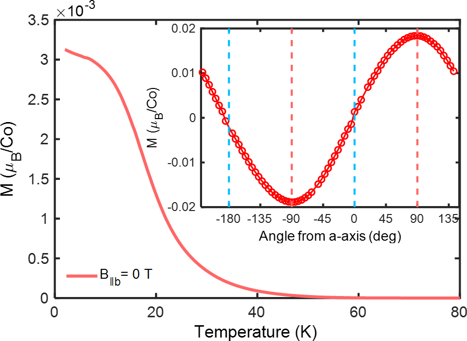
Taken together, the results show that at very low and in external in-plane fields, \chNa_3Co_2SbO_6 sequentially goes through magnetic states characterized by the M-point, the “M”-point, and eventually the zone-center -point, forming an evolution along the -M lines [Fig. 1(b)]. The direction of the field affects only when, but not whether, the transitions would occur. It is therefore tempting to think that the system possesses competing AFM-FM instabilities with wave vectors lined up along -M. In Fig. 5, we use variable- neutron diffraction to show that this is indeed the case. The experiment was performed on a twinned sample, in zero magnetic field. The most remarkable observation is found at 10 K [Fig. 5(c)]: we see distinct hexagonal-star-shaped diffuse scattering, which “flows” into the long-range magnetic Bragg peaks at the M-points upon further cooling [Fig. 5(b)]. The observed star consists of six narrow streaks which precisely cover the -M lines. In a twin-free sample, the number of streaks would likely be four [Fig. 1(b)], which would help explain the giant in-plane magnetic anisotropy, and it warrants further experimental confirmation. The streaks are, in fact, quasi-2D objects in reciprocal space with only weak dependence on (Fig. S14 in [58]). They correspond to quasi-1D correlations in real space (Fig. 6, further discussed below), and can be viewed as a counterpart of rod-like diffuse scattering in \chYb_2Ti_2O_7 [64, 65], which has been attributed to coexisting FM and AFM correlations [66, 67]. Below , the body of the star is depleted, including the FM-like diffuse scattering near [Fig. 5(a)]. Such a temperature-evolution, together with the field-evolution at low (Fig. 4), signifies a close competition between a variety of AFM and FM instabilities, with or without thermal disorder. Indeed, the M-point AFM order might be energetically favored in zero field by only a small margin. We have further found evidence for a weak field-trainable net moment in a twin-free sample (Fig. S15 in [58]), which supports an incipience of the ferromagnetism.
III Discussion
Our results motivate further exploration of exotic quantum phases in \chNa_3Co_2SbO_6, as well as in extended Kitaev and related theoretical models. Magnetic field-induced phases in candidate Kitaev materials have been under intense research in recent years [56, 20, 37, 50, 68, 69, 70, 71, 72, 73, 74, 34, 75, 76], and the magnetization’s step-like transitions into and out of the intermediate states in Fig. 2 resemble some of the reports [56, 20, 37], even though the wave-vector switching behavior might not be the same [56, 20]. These results suggest that the candidate materials commonly possess multiple magnetic instabilities – a hallmark of frustration. Our findings are consistent with the view that \chNa_3Co_2SbO_6 is close to a trisecting point of FM, AFM, and QSL phases [13], yet the pronounced in-plane anisotropy clearly adds complexity and novelty to the previous understanding. Specifically, given the anisotropy, uniaxial strains both perpendicular [13] and parallel to the honeycomb layers might help promote QSL physics.
Meanwhile, the -M characteristics of the ordering wave vectors and diffuse scattering imply a particular combination of competing instabilities, which are not commonly seen in model systems [45]. In Fig. 6, we show that the star-shaped diffuse scattering pattern can be well-simulated by FM zigzag chains randomly placed on a honeycomb lattice. Each star streak in space is contributed by chains in real space that run perpendicular to the streak. Because neutron scattering probes magnetic moments perpendicular to , we infer that the magnetic moments in the FM chains point largely parallel to the chains – similar to those in a typical zigzag magnetic structure [18, 47]. Given that the AFM order below is preceded by the short-range FM chains above , a plausible scenario is that the system’s leading magnetic interactions are strongly in favor of individual FM-chain formation, yet at the same time, they are weakly in favor of an alternating side-by-side arrangement of the chains, i.e., against the formation of 2D FM order. Together with our inference of the moment direction above, the scenario echoes with the idea of bond-dependent anisotropic interactions, which is at the core of the Kitaev and related models.
We further notice that, among three types of parameters that are commonly considered for explaining the zigzag order [42], our result appears to be consistent with the expected behaviors of models with a leading nearest-neighbor symmetric off-diagonal interaction term, . This is because for a given nearest-neighbor pair, the term favors FM alignment of the spin component parallel to the bond, but AFM alignment of the component perpendicular to both the bond and the Ising axis of the Kitaev term. Together with the geometry of the honeycomb lattice, the term can thus explain both the FM chains’ formation tendency and their resistance to form 2D FM order. Indeed, in the Appendix of Ref. 42, we find a discussion of such models’ several similar behaviors compared to our observations, including the formation of AFM order and competing instabilities at a star-shaped set of wave vectors. Another major feature of such models is their demonstrated ability to produce large magnetic-response anisotropy without a highly anisotropic tensor [42]. We thus expect our results to motivate further theoretical research of frustrated magnetism in the off-diagonal models [77, 78, 79], some of which may have a QSL ground state [79].
To conclude, we have elucidated the field-induced phases and competing instabilities in the quantum magnet \chNa_3Co_2SbO_6, and uncovered an unexpectedly large magnetic anisotropy. The results indicate exotic magnetic phases and render this system highly promising for further explorations using targeted external tuning. The results also stimulate future theoretical research of spin-orbit-coupled quantum magnets.
Acknowledgements.
We are grateful for technical support by Qizhi Li and Jianping Sun, and for discussions with Wenjie Chen, Ji Feng, L. Janssen, G. Khaliullin, V. Kocsis, Huimei Liu, Qiang Luo, A. U. B. Wolter, and Shilong Zhang. The work at Peking University was supported by the National Basic Research Program of China (Grant Nos. 2021YFA1401900 and 2018YFA0305602) and the NSF of China (Grant Nos. 12061131004, 11874069, and 11888101). The work at Brookhaven National Laboratory was supported by Office of Basic Energy Sciences (BES), Division of Materials Sciences and Engineering, U.S. Department of Energy (DOE), under contract DE-SC0012704. X.L. further acknowledges support from China Postdoctoral Science Foundation (Grant No. 2020M680179). A portion of this research used resources at Spallation Neutron Source, a DOE Office of Science User Facility operated by the Oak Ridge National Laboratory. One of the neutron scattering experiments was performed at the MLF, J-PARC, Japan, under a user program (No. 2020B0148).References
- Balents [2010] L. Balents, Nature 464, 199 (2010).
- Zhou et al. [2017] Y. Zhou, K. Kanoda, and T.-K. Ng, Rev. Mod. Phys. 89, 025003 (2017).
- Broholm et al. [2020] C. Broholm, R. Cava, S. Kivelson, D. Nocera, M. Norman, and T. Senthil, Science 367, eaay0668 (2020).
- Kitaev [2006] A. Kitaev, Ann. Phys. 321, 2 (2006).
- Jackeli and Khaliullin [2009] G. Jackeli and G. Khaliullin, Phys. Rev. Lett. 102, 017205 (2009).
- Takagi et al. [2019] H. Takagi, T. Takayama, G. Jackeli, G. Khaliullin, and S. E. Nagler, Nat. Rev. Phys. 1, 264 (2019).
- Motome et al. [2020] Y. Motome, R. Sano, S. Jang, Y. Sugita, and Y. Kato, Journal of Physics: Condensed Matter 32, 404001 (2020).
- Trebst and Hickey [2022] S. Trebst and C. Hickey, Physics Reports 950, 1 (2022).
- Chaloupka et al. [2010] J. Chaloupka, G. Jackeli, and G. Khaliullin, Phys. Rev. Lett. 105, 027204 (2010).
- Plumb et al. [2014] K. W. Plumb, J. P. Clancy, L. J. Sandilands, V. V. Shankar, Y. F. Hu, K. S. Burch, H.-Y. Kee, and Y.-J. Kim, Phys. Rev. B 90, 041112 (2014).
- Liu and Khaliullin [2018] H. Liu and G. Khaliullin, Phys. Rev. B 97, 014407 (2018).
- Sano et al. [2018] R. Sano, Y. Kato, and Y. Motome, Phys. Rev. B 97, 014408 (2018).
- Liu et al. [2020] H. Liu, J. Chaloupka, and G. Khaliullin, Phys. Rev. Lett. 125, 047201 (2020).
- Kim et al. [2021a] C. Kim, H.-S. Kim, and J.-G. Park, Journal of Physics: Condensed Matter 34, 023001 (2021a).
- Liu [2021] H. Liu, Int. J. Mod. Phys. B 35, 2130006 (2021).
- Viciu et al. [2007] L. Viciu, Q. Huang, E. Morosan, H. Zandbergen, N. Greenbaum, T. McQueen, and R. Cava, J. Solid State Chem. 180, 1060 (2007).
- Singh and Gegenwart [2010] Y. Singh and P. Gegenwart, Phys. Rev. B 82, 064412 (2010).
- Liu et al. [2011] X. Liu, T. Berlijn, W.-G. Yin, W. Ku, A. Tsvelik, Y.-J. Kim, H. Gretarsson, Y. Singh, P. Gegenwart, and J. P. Hill, Phys. Rev. B 83, 220403 (2011).
- Johnson et al. [2015] R. D. Johnson, S. C. Williams, A. A. Haghighirad, J. Singleton, V. Zapf, P. Manuel, I. I. Mazin, Y. Li, H. O. Jeschke, R. Valentí, and R. Coldea, Phys. Rev. B 92, 235119 (2015).
- Zhong et al. [2020] R. Zhong, T. Gao, N. P. Ong, and R. J. Cava, Science Advances 6, eaay6953 (2020).
- Kimchi and You [2011] I. Kimchi and Y.-Z. You, Phys. Rev. B 84, 180407 (2011).
- Chaloupka et al. [2013] J. Chaloupka, G. Jackeli, and G. Khaliullin, Phys. Rev. Lett. 110, 097204 (2013).
- Rau et al. [2014] J. G. Rau, E. K.-H. Lee, and H.-Y. Kee, Phys. Rev. Lett. 112, 077204 (2014).
- Sizyuk et al. [2014] Y. Sizyuk, C. Price, P. Wölfle, and N. B. Perkins, Phys. Rev. B 90, 155126 (2014).
- Yamaji et al. [2014] Y. Yamaji, Y. Nomura, M. Kurita, R. Arita, and M. Imada, Phys. Rev. Lett. 113, 107201 (2014).
- Katukuri et al. [2014] V. M. Katukuri, S. Nishimoto, V. Yushankhai, A. Stoyanova, H. Kandpal, S. Choi, R. Coldea, I. Rousochatzakis, L. Hozoi, and J. van den Brink, New J. Phys. 16, 013056 (2014).
- Rousochatzakis et al. [2015] I. Rousochatzakis, J. Reuther, R. Thomale, S. Rachel, and N. B. Perkins, Phys. Rev. X 5, 041035 (2015).
- Chaloupka and Khaliullin [2016] J. Chaloupka and G. Khaliullin, Phys. Rev. B 94, 064435 (2016).
- Chaloupka and Khaliullin [2015] J. Chaloupka and G. Khaliullin, Phys. Rev. B 92, 024413 (2015).
- Winter et al. [2016] S. M. Winter, Y. Li, H. O. Jeschke, and R. Valentí, Phys. Rev. B 93, 214431 (2016).
- Do et al. [2017] S.-H. Do, S.-Y. Park, J. Yoshitake, J. Nasu, Y. Motome, Y. S. Kwon, D. Adroja, D. Voneshen, K. Kim, T.-H. Jang, J.-H. Park, K.-Y. Choi, and S. Ji, Nat. Phys. 13, 1079 (2017).
- Wang et al. [2018] Z. Wang, J. Guo, F. F. Tafti, A. Hegg, S. Sen, V. A. Sidorov, L. Wang, S. Cai, W. Yi, Y. Zhou, H. Wang, S. Zhang, K. Yang, A. Li, X. Li, Y. Li, J. Liu, Y. Shi, W. Ku, Q. Wu, R. J. Cava, and L. Sun, Phys. Rev. B 97, 245149 (2018).
- Winter et al. [2018] S. M. Winter, K. Riedl, D. Kaib, R. Coldea, and R. Valentí, Phys. Rev. Lett. 120, 077203 (2018).
- Banerjee et al. [2018] A. Banerjee, P. Lampen-Kelley, J. Knolle, C. Balz, A. A. Aczel, B. Winn, Y. Liu, D. Pajerowski, J. Yan, C. A. Bridges, A. T. Savici, B. C. Chakoumakos, M. D. Lumsden, D. A. Tennant, R. Moessner, D. G. Mandrus, and S. E. Nagler, npj Quantum Materials 3, 8 (2018).
- Gordon et al. [2019] J. S. Gordon, A. Catuneanu, E. S. Srensen, and H.-Y. Kee, Nat. Commun. 10, 2470 (2019).
- Hickey and Trebst [2019] C. Hickey and S. Trebst, Nat. Commun. 10, 530 (2019).
- Yao and Li [2020] W. Yao and Y. Li, Phys. Rev. B 101, 085120 (2020).
- Wong et al. [2016] C. Wong, M. Avdeev, and C. D. Ling, J. Solid State Chem. 243, 18 (2016).
- Yan et al. [2019] J.-Q. Yan, S. Okamoto, Y. Wu, Q. Zheng, H. D. Zhou, H. B. Cao, and M. A. McGuire, Phys. Rev. Materials 3, 074405 (2019).
- Stratan et al. [2019] M. I. Stratan, I. L. Shukaev, T. M. Vasilchikova, A. N. Vasiliev, A. N. Korshunov, A. I. Kurbakov, V. B. Nalbandyan, and E. A. Zvereva, New J. Chem. 43, 13545 (2019).
- Wang et al. [2017] W. Wang, Z.-Y. Dong, S.-L. Yu, and J.-X. Li, Phys. Rev. B 96, 115103 (2017).
- Janssen et al. [2017] L. Janssen, E. C. Andrade, and M. Vojta, Phys. Rev. B 96, 064430 (2017).
- Rusnačko et al. [2019] J. Rusnačko, D. Gotfryd, and J. Chaloupka, Phys. Rev. B 99, 064425 (2019).
- Maksimov and Chernyshev [2020] P. A. Maksimov and A. L. Chernyshev, Phys. Rev. Research 2, 033011 (2020).
- Laurell and Okamoto [2020] P. Laurell and S. Okamoto, npj Quantum Materials 5, 1 (2020).
- Cao et al. [2016] H. B. Cao, A. Banerjee, J.-Q. Yan, C. A. Bridges, M. D. Lumsden, D. G. Mandrus, D. A. Tennant, B. C. Chakoumakos, and S. E. Nagler, Phys. Rev. B 93, 134423 (2016).
- Chen et al. [2021] W. Chen, X. Li, Z. Hu, Z. Hu, L. Yue, R. Sutarto, F. He, K. Iida, K. Kamazawa, W. Yu, X. Lin, and Y. Li, Phys. Rev. B 103, L180404 (2021).
- Lee et al. [2021] C. H. Lee, S. Lee, Y. S. Choi, Z. H. Jang, R. Kalaivanan, R. Sankar, and K.-Y. Choi, Phys. Rev. B 103, 214447 (2021).
- Songvilay et al. [2020] M. Songvilay, J. Robert, S. Petit, J. A. Rodriguez-Rivera, W. D. Ratcliff, F. Damay, V. Balédent, M. Jiménez-Ruiz, P. Lejay, E. Pachoud, A. Hadj-Azzem, V. Simonet, and C. Stock, Phys. Rev. B 102, 224429 (2020).
- Lin et al. [2021] G. Lin, J. Jeong, C. Kim, Y. Wang, Q. Huang, T. Masuda, S. Asai, S. Itoh, G. Günther, M. Russina, Z. Lu, J. Sheng, L. Wang, J. Wang, G. Wang, Q. Ren, C. Xi, W. Tong, L. Ling, Z. Liu, L. Wu, J. Mei, Z. Qu, H. Zhou, J.-G. Park, and J. Ma, Nat. Commun. 12, 5559 (2021).
- Kim et al. [2021b] C. Kim, J. Jeong, G. Lin, P. Park, T. Masuda, S. Asai, S. Itoh, H.-S. Kim, H. Zhou, J. Ma, and J.-G. Park, Journal of Physics: Condensed Matter 34, 045802 (2021b).
- Samarakoon et al. [2021] A. M. Samarakoon, Q. Chen, H. Zhou, and V. O. Garlea, Phys. Rev. B 104, 184415 (2021).
- Sanders et al. [2022] A. L. Sanders, R. A. Mole, J. Liu, A. J. Brown, D. Yu, C. D. Ling, and S. Rachel, Phys. Rev. B 106, 014413 (2022).
- Yao et al. [2022] W. Yao, K. Iida, K. Kamazawa, and Y. Li, Phys. Rev. Lett. 129, 147202 (2022).
- Lampen-Kelley et al. [2018] P. Lampen-Kelley, S. Rachel, J. Reuther, J.-Q. Yan, A. Banerjee, C. A. Bridges, H. B. Cao, S. E. Nagler, and D. Mandrus, Phys. Rev. B 98, 100403 (2018).
- Balz et al. [2021] C. Balz, L. Janssen, P. Lampen-Kelley, A. Banerjee, Y. H. Liu, J.-Q. Yan, D. G. Mandrus, M. Vojta, and S. E. Nagler, Phys. Rev. B 103, 174417 (2021).
- Kocsis et al. [2022] V. Kocsis, D. A. S. Kaib, K. Riedl, S. Gass, P. Lampen-Kelley, D. G. Mandrus, S. E. Nagler, N. Pérez, K. Nielsch, B. Büchner, A. U. B. Wolter, and R. Valentí, Phys. Rev. B 105, 094410 (2022).
- [58] See Supplemental Material for additional methods, data, and analyses, which include additional Refs. [80, 81, 82, 83, 84].
- Sears et al. [2017] J. A. Sears, Y. Zhao, Z. Xu, J. W. Lynn, and Y.-J. Kim, Phys. Rev. B 95, 180411 (2017).
- Hardy et al. [2004] V. Hardy, M. R. Lees, O. A. Petrenko, D. M. Paul, D. Flahaut, S. Hébert, and A. Maignan, Phys. Rev. B 70, 064424 (2004).
- Ueda et al. [2005] H. Ueda, H. A. Katori, H. Mitamura, T. Goto, and H. Takagi, Phys. Rev. Lett. 94, 047202 (2005).
- Cao et al. [2007] G. Cao, V. Durairaj, S. Chikara, S. Parkin, and P. Schlottmann, Phys. Rev. B 75, 134402 (2007).
- Jo et al. [2009] Y. J. Jo, S. Lee, E. S. Choi, H. T. Yi, W. Ratcliff, Y. J. Choi, V. Kiryukhin, S. W. Cheong, and L. Balicas, Phys. Rev. B 79, 012407 (2009).
- Ross et al. [2009] K. A. Ross, J. P. C. Ruff, C. P. Adams, J. S. Gardner, H. A. Dabkowska, Y. Qiu, J. R. D. Copley, and B. D. Gaulin, Phys. Rev. Lett. 103, 227202 (2009).
- Thompson et al. [2011] J. D. Thompson, P. A. McClarty, H. M. Rønnow, L. P. Regnault, A. Sorge, and M. J. P. Gingras, Phys. Rev. Lett. 106, 187202 (2011).
- Scheie et al. [2020] A. Scheie, J. Kindervater, S. Zhang, H. J. Changlani, G. Sala, G. Ehlers, A. Heinemann, G. S. Tucker, S. M. Koohpayeh, and C. Broholm, Proceedings of the National Academy of Sciences 117, 27245 (2020).
- Scheie et al. [2022] A. Scheie, O. Benton, M. Taillefumier, L. D. Jaubert, G. Sala, N. Jalarvo, S. M. Koohpayeh, and N. Shannon, arXiv preprint arXiv:2202.11085 10.48550/arXiv.2202.11085 (2022).
- Zheng et al. [2017] J. Zheng, K. Ran, T. Li, J. Wang, P. Wang, B. Liu, Z.-X. Liu, B. Normand, J. Wen, and W. Yu, Phys. Rev. Lett. 119, 227208 (2017).
- Wolter et al. [2017] A. U. B. Wolter, L. T. Corredor, L. Janssen, K. Nenkov, S. Schönecker, S.-H. Do, K.-Y. Choi, R. Albrecht, J. Hunger, T. Doert, M. Vojta, and B. Büchner, Phys. Rev. B 96, 041405 (2017).
- Leahy et al. [2017] I. A. Leahy, C. A. Pocs, P. E. Siegfried, D. Graf, S.-H. Do, K.-Y. Choi, B. Normand, and M. Lee, Phys. Rev. Lett. 118, 187203 (2017).
- Baek et al. [2017] S.-H. Baek, S.-H. Do, K.-Y. Choi, Y. S. Kwon, A. U. B. Wolter, S. Nishimoto, J. van den Brink, and B. Büchner, Phys. Rev. Lett. 119, 037201 (2017).
- Yu et al. [2018] Y. J. Yu, Y. Xu, K. J. Ran, J. M. Ni, Y. Y. Huang, J. H. Wang, J. S. Wen, and S. Y. Li, Phys. Rev. Lett. 120, 067202 (2018).
- Wellm et al. [2018] C. Wellm, J. Zeisner, A. Alfonsov, A. U. B. Wolter, M. Roslova, A. Isaeva, T. Doert, M. Vojta, B. Büchner, and V. Kataev, Phys. Rev. B 98, 184408 (2018).
- Kasahara et al. [2018] Y. Kasahara, T. Ohnishi, Y. Mizukami, O. Tanaka, S. Ma, K. Sugii, N. Kurita, H. Tanaka, J. Nasu, Y. Motome, T. Shibauchi, and Y. Matsuda, Nature 559, 227 (2018).
- Sahasrabudhe et al. [2020] A. Sahasrabudhe, D. A. S. Kaib, S. Reschke, R. German, T. C. Koethe, J. Buhot, D. Kamenskyi, C. Hickey, P. Becker, V. Tsurkan, A. Loidl, S. H. Do, K. Y. Choi, M. Grüninger, S. M. Winter, Z. Wang, R. Valentí, and P. H. M. van Loosdrecht, Phys. Rev. B 101, 140410 (2020).
- Yokoi et al. [2021] T. Yokoi, S. Ma, Y. Kasahara, S. Kasahara, T. Shibauchi, N. Kurita, H. Tanaka, J. Nasu, Y. Motome, C. Hickey, S. Trebst, and Y. Matsuda, Science 373, 568 (2021).
- Samarakoon et al. [2018] A. M. Samarakoon, G. Wachtel, Y. Yamaji, D. A. Tennant, C. D. Batista, and Y. B. Kim, Phys. Rev. B 98, 045121 (2018).
- Saha et al. [2019] P. Saha, Z. Fan, D. Zhang, and G.-W. Chern, Phys. Rev. Lett. 122, 257204 (2019).
- Luo et al. [2021] Q. Luo, J. Zhao, H.-Y. Kee, and X. Wang, npj Quantum Materials 6, 57 (2021).
- Wu et al. [2016] C.-M. Wu, G. Deng, J. Gardner, P. Vorderwisch, W.-H. Li, S. Yano, J.-C. Peng, and E. Imamovic, Journal of Instrumentation 11, P10009 (2016).
- Winn, Barry et al. [2015] Winn, Barry, Filges, Uwe, Garlea, V. Ovidiu, Graves-Brook, Melissa, Hagen, Mark, Jiang, Chenyang, Kenzelmann, Michel, Passell, Larry, Shapiro, Stephen M., Tong, Xin, and Zaliznyak, Igor, EPJ Web of Conferences 83, 03017 (2015).
- Kajimoto et al. [2011] R. Kajimoto, M. Nakamura, Y. Inamura, F. Mizuno, K. Nakajima, S. Ohira-Kawamura, T. Yokoo, T. Nakatani, R. Maruyama, K. Soyama, K. Shibata, K. Suzuya, S. Sato, K. Aizawa, M. Arai, S. Wakimoto, M. Ishikado, S. ichi Shamoto, M. Fujita, H. Hiraka, K. Ohoyama, K. Yamada, and C.-H. Lee, Journal of the Physical Society of Japan 80, SB025 (2011).
- Inamura et al. [2013] Y. Inamura, T. Nakatani, J. Suzuki, and T. Otomo, Journal of the Physical Society of Japan 82, SA031 (2013).
- Ewings et al. [2016] R. Ewings, A. Buts, M. Le, J. van Duijn, I. Bustinduy, and T. Perring, Nuclear Instruments and Methods in Physics Research Section A: Accelerators, Spectrometers, Detectors and Associated Equipment 834, 132 (2016).
Supplemental Material for
“Giant magnetic in-plane anisotropy and competing instabilities in \chNa_3Co_2SbO_6”
IV Methods
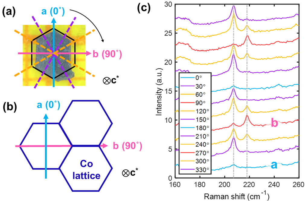
Single crystal growth. Single crystals of \chNa_3Co_2SbO_6 were synthesized with a flux method similar to the method for growing \chNa_2Co_2TeO_6 [37]. Highest-quality crystals were of a dark purple color and a flaky hexagonal shape [Fig. S7(a)]. The correspondence between sample shape and the cobalt honeycomb sub-lattice is shown in Fig. S7.
Single-crystal -ray diffraction. The measurements were performed with a custom-designed instrument equipped with a Xenocs Genix3D Mo Kα (17.48 keV) -ray source, which produced a beam-spot diameter of 150 m at the sample position with photons/sec. The samples were mounted on a Huber four-circle diffractometer. A highly sensitive PILATUS3 R 1M solid-state pixel array detector (with pixels, 172 m m per pixel) was used to collect the diffraction signals. Three-dimensional mapping of the reciprocal space was achieved by taking diffraction images in 0.1∘ sample-rotation increments. Due to a small non-monochromaticity of the -rays, each observed Bragg peak consisted of closely-spaced Kα1 and Kα2 components, and was further accompanied by a weak Kβ peak and a radial tail [red dashed lines in Fig. 1(c)].
Raman spectroscopy screening for twin-free samples. The measurements were performed under a microscope in a confocal backscattering geometry, using a Horiba Jobin Yvon LabRAM HR Evolution spectrometer equipped with 1800 gr/mm gratings and a liquid-nitrogen-cooled CCD detector. A He-Ne laser with nm was used for excitation (with 1.2 mW laser power). The measurements were performed at room temperature, in air, and on crystal surfaces parallel to the -plane. Linear polarizations of both incident and scattered photons were set parallel to each other and along twelve in-plane directions, as illustrated in Fig. S7(a).
Specific-heat measurement and magnetometry. Specific-heat measurements were performed with a Quantum Design PPMS, using a relaxation method. DC magnetometry was performed with a Quantum Design MPMS equipped with a sample rotator. The angle dependence of magnetization in Fig. 2(c) was measured in a sweep-field mode (+3 Oe/sec) after initially cooling the sample in zero field. The field was reset to zero between adjacent orientations without thermal cycling. The systematic results in Fig. 2(c) indicate a lack of dependence on the field history.
Neutron diffraction on a twin-free sample. The experiment was performed on the SIKA cold neutron triple-axis spectrometer at the Australian Nuclear Science and Technology Organization (ANSTO) [80]. A twin-free crystal was mounted with reciprocal vectors in the horizontal scattering plane, and the measurement was performed with Å-1 neutrons and a beryllium filter. In this experiment, only diffractions in the horizontal scattering plane could be accessed, due to the use of a cryomagnet which did not allow for sample tilting. The applied vertical fields were at 60∘ (anticlockwise) from the -axis [Fig. S11(c)]. This geometry made the studied sample equivalent to S60 discussed in main text, and allowed us to access the reflection in the AFM state, as well as the reflection in the AFM state.
Neutron diffraction on twinned samples. The neutron diffraction experiments displayed in Figs. 3-6 of main text were performed with twinned crystal arrays, which were co-aligned in the -plane on aluminum plates with a hydrogen-free adhesive (Cytop). 1/3 of the sample mass belonged to (see text), for which the vertical direction was parallel to the -axis, and the corresponding horizontal scattering plane was . The other 2/3 of the sample belonged to , for which the vertical direction was at 60∘ from the -axis and the horizontal plane was (or symmetry equivalent). As explained in main text and below, we use to denote wave vectors. is along the vertical direction, and the horizontal plane is . in Fig. 3(a), Fig. 4 and Fig. 5(c) of main text denotes 2D wave vectors in the vertical (real-space) -plane.
The diffraction experiment in magnetic fields (Fig. 4 of main text) was performed on the HYSPEC time-of-flight spectrometer at the SNS, Oak Ridge National Laboratory [81], using a helium-3 insert and a 14 T vertical-field cryomagnet as sample environment. Equipped with the incident beam-focusing optics, the narrow vertical opening angle of of the cryomagnet allowed us to detect the magnetic diffractions out of the horizontal scattering plane. We used a relatively high incident neutron energy meV to have the full out-of-plane access for acquiring the data in Figs. 4 and S16, and a relatively low incident neutron energy meV for accurate peak indexing Fig. S13. The sample was 0.5 gram in total mass, and had a full mosaic spread of about 2.3∘. Diffraction data were acquired by rotating the sample over a 78∘ range in 0.5∘ steps, resulting in a three-dimensional data set, which we further symmetrize with and mirror operations for plotting.
The diffraction experiment at variable temperatures without magnetic field. (Figs. 3, 5, 6 of main text) was performed on the 4SEASONS time-of-flight spectrometer at the MLF, J-PARC, Japan [82]. The sample was 0.7 gram in total mass with a full mosaic spread of about 2.7∘. The data in Figs. 3(a), 5(c) 6(c) and S12 were collected at fixed temperatures by rotating the sample over an 85∘ range in 1∘ steps. The data in Fig. 5(a-b) were acquired with a “sit-and-count” method, i.e., the sample was rotated to fixed orientations, and counting was continuously performed during a slow warm-up of the sample at a rate of about 0.022 K/min. Data were reduced and analyzed with the Utsusemi [83] and Horace [84] software packages.
The time-of-flight scattering experiments generated high-dimensional data sets, which were sliced into lower dimensions for plotting. Detailed measurement conditions and slicing restrictions can be found in Table S2.
Conversion between coordinate systems for the reciprocal space. A fully-twinned sample contains all -related counterparts of any chosen monoclinic domain, where the rotation is about the normal direction of the -plane, or . It is straightforward to show that, if a monoclinic domain has a physical wave vector of , the wave vector will be observed at a total of six positions in the coordinate system: , , , , , . Note that they share the same . It is straightforward to verify that all magnetic peaks in Fig. S12 in zero field are consistent with and symmetry equivalent, and that all magnetic peaks in Fig. S13 above are consistent with and symmetry equivalent, where is a reciprocal lattice vector. A reference table for the conversion between the coordinate systems can be found in Table S3.
Discussion of magnetic structure. Recent indications of triple- order [47, 48] in the sister compound \chNa_2Co_2TeO_6 motivate us to consider a multi- possibility here as well. In this regard, the two-step transitions of (e.g., in ) are intriguing. In zero field, the magnetic Hamiltonian has symmetry around the -axis. In the single- zigzag scenario, the symmetry of magnetic ground state is spontaneously lowered to , and -symmetry-related magnetic domains are formed, featuring FM zigzag chains running in directions related to each other by rotation around the -axis. However, in an external magnetic field applied not along the -axis (or the -axis), as is the case for , the magnetization energy of the -related magnetic domains will no longer be degenerate. In other words, considering the combination of the crystallographic structure and the field direction, the Hamiltonian’s symmetry is lowered to none () from the first place. This low symmetry is at the origin of the split between and for essentially all field directions between the and -axes.
At and , since the wave vectors switch only along the same -M line, the resultant AFM order would continue to have their FM ribbons/chains running in inequivalent directions, i.e., same as in the original zigzag domains. Then, by the same symmetry argument, it is natural to expect the two types of AFM domains to have their next transitions occur at somewhat different . However, only a single transition is experimentally observed. Under the zigzag scenario, this empirical “simplicity” can only be attributed to a coincidence not required by symmetry, yet it holds over a wide angular range – magnetometry always sees a single transition [Fig. 2(c-d)]. Moreover, the two-step transitions between AFM and AFM are consistently observed independent of field history [Fig. 2(c)], which means that the above two types of domains always reappear after the symmetry-lowering field is removed. Such lack of domain repopulation by the fields is somewhat difficult to understand if the zero-field magnetic ground state’s symmetry is indeed spontaneously lowered to (as for zigzag). Alternatively, these remaining puzzles for the zigzag scenario could imply that the “domains” are not macroscopically separated, but instead, the two sets of wave vectors arise from the same part of the sample, hinting at a multi- scenario for at least some of the magnetic orders. We therefore believe that the precise nature of the AFM phases in \chNa_3Co_2SbO_6 is still open for further research.
V Supplementary Figures


VI Supplementary Tables
| low-T | high- | ||||||
|---|---|---|---|---|---|---|---|
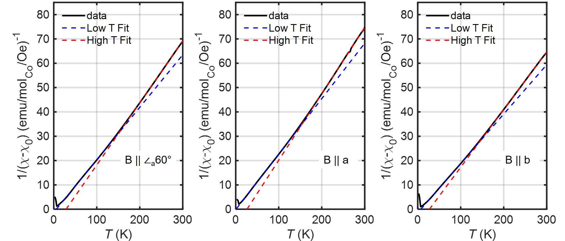
| 5.9 | 6.1 | 6.3 | 5.4 | 5.8 | 5.8 | 6.2 | |
| (K) | 1.0 | 5.7 | 6.8 | 26.2 | 22.4 | 27.6 | |
| 6.8 | 7.1 | 7.3 | 6.3 | 6.7 | 6.7 | 7.2 | |
| Data | range (r.l.u.) | range (r.l.u.) | range (r.l.u.) | (meV)111Incident neutron energy. | chopper frequency (Hz) | range (meV)222Neutron energy transfer. |
|---|---|---|---|---|---|---|
| Fig. 3(a) | - | - | [-1, 1] | 16.8 | 150 | [-0.1, 0.1] |
| Fig. 3(b) blue | [-0.1, 0.1] | [1.9, 2.1] | - | 16.8 | 150 | [-0.1, 0.1] |
| Fig. 3(b) orange | [0.4, 0.6] | [0.4, 0.6] | - | 16.8 | 150 | [-0.1, 0.1] |
| Fig. 3(c) | - | [0.4, 0.6] | [-5, 5] | 5.6 | 150 | [-0.1, 0.1] |
| Fig. 3(d) | [0.4, 0.6] | - | [-5, 5] | 5.6 | 150 | [-0.1, 0.1] |
| Fig. 4 left | - | - | [-0.2, 0.2] | 35 | 120 | [-0.2, 0.2] |
| Fig. 4 right | - | - | [0.3, 0.7] | 35 | 120 | [-0.2, 0.2] |
| Fig. 5(a) | - | [-0.2, 0.2] | [-2.2, 2.2] | 5.6 | 150 | [-0.2, 0.2] |
| Fig. 5(b) | [-0.2, 0.2] | - | [-2.2, 2.2] | 5.6 | 150 | [-0.2, 0.2] |
| Fig. 5(c) | - | - | [-2.2, 2.2] | 5.6 | 150 | [-0.2, 0.2] |
| Fig. 5(c) | - | - | [-2.2, 2.2] | 16.8 | 150 | [-0.1, 0.1] |
| Fig. 6(c) | - | - | [-1, 1] | 16.8 | 150 | [-0.1, 0.1] |
| Fig. S12(a) | - | - | [-1, 1] | 16.8 | 150 | [-0.1, 0.1] |
| Fig. S12(b) | [-0.1, 0.1] | - | - | 16.8 | 150 | [-0.1, 0.1] |
| Fig. S12(c) | [0.4, 0.6] | - | - | 16.8 | 150 | [-0.1, 0.1] |
| Fig. S12(d] | [-0.1, 0.1] | [0.9, 1.1] | - | 16.8 | 150 | [-0.1, 0.1] |
| Fig. S12(e) | [-0.1, 0.1] | [1.9, 2.1] | - | 16.8 | 150 | [-0.1, 0.1] |
| Fig. S13(b) | [-0.1, 0.1] | - | - | 5 | 360 | [-0.2, 0.2] |
| Fig. S13(c) | [-0.1, 0.1] | [0.567, 0.767] | - | 5 | 360 | [-0.2, 0.2] |
| Fig. S13(e) | [0.2, 0.285] | - | - | 15 | 360 | [-0.2, 0.2] |
| Fig. S13(f) | [0.2, 0.285] | [0.233, 0.433] | - | 15 | 360 | [-0.2, 0.2] |
| Fig. S14(a) | - | - | [-2.2, 2.2] | 5.6 | 150 | [-0.2, 0.2] |
| Fig. S14(b) | - | [0.6, 0.8] | - | 5.6 | 150 | [-0.2, 0.2] |
| Fig. S14(c) | - | [0.6, 0.8] | - | 5.6 | 150 | [-0.2, 0.2] |
|
|
from | from | from | from | from | |||||||||||||
|---|---|---|---|---|---|---|---|---|---|---|---|---|---|---|---|---|---|---|
|
![[Uncaptioned image]](/html/2204.04593/assets/2-1.jpg)
|
![[Uncaptioned image]](/html/2204.04593/assets/2-2.jpg)
|
![[Uncaptioned image]](/html/2204.04593/assets/2-3.jpg)
|
![[Uncaptioned image]](/html/2204.04593/assets/2-4.jpg)
|
![[Uncaptioned image]](/html/2204.04593/assets/2-5.jpg)
|
![[Uncaptioned image]](/html/2204.04593/assets/2-6.jpg)
|
||||||||||||
|
|
|
|
|
|
|
|||||||||||||
|
![[Uncaptioned image]](/html/2204.04593/assets/3-1.jpg)
|
![[Uncaptioned image]](/html/2204.04593/assets/3-2.jpg)
|
![[Uncaptioned image]](/html/2204.04593/assets/3-3.jpg)
|
![[Uncaptioned image]](/html/2204.04593/assets/3-4.jpg)
|
![[Uncaptioned image]](/html/2204.04593/assets/3-5.jpg)
|
![[Uncaptioned image]](/html/2204.04593/assets/3-6.jpg)
|
||||||||||||
|
|
|
|
|
|
|
VII Supplementary Animation
[height=15cm, label=Dmap, controls]2Dmap_rev2_0114