developed the algorithms, performed the analysis and finalized the manuscript
[orcid=0000-0002-6442-8343] \creditmanaged the data and designed the software
[orcid=0000-0002-2867-4838] \creditmanaged the data and designed the software
[orcid=0000-0002-6494-7514] \creditconceptualized, designed the project and finalized the manuscript
responsible for the clinical assessment of the images and script
[orcid=0000-0003-3529-3109] \creditmanaged the data and designed the software
[orcid=0000-0003-3602-4023] \cormark[1] \creditconceptualized, designed the project and finalized the manuscript
[cor1]Corresponding author
Which Generative Adversarial Network Yields High-Quality Synthetic Medical Images: Investigation Using AMD Image Datasets
Abstract
Deep learning has been proposed for the assessment and classification of medical images. However, many medical image databases with appropriately labeled and annotated images are small and imbalanced, and thus unsuitable to train and validate such models. The option is to generate synthetic images and one successful technique has been patented which limits its use for others. We have developed a free-access, alternate method for generating synthetic high-resolution images using Generative Adversarial Networks (GAN) for data augmentation and showed their effectiveness using eye-fundus images for Age-Related Macular Degeneration (AMD) identification. Ten different GAN architectures were compared to generate synthetic eye-fundus images with and without AMD. Data from three public databases were evaluated using the Fréchet Inception Distance (FID), two clinical experts and deep-learning classification. The results show that StyleGAN2 reached the lowest FID (166.17), and clinicians could not accurately differentiate between real and synthetic images. ResNet-18 architecture obtained the best performance with 85% accuracy and outperformed the two experts in detecting AMD fundus images, whose average accuracy was 77.5%. These results are similar to a recently patented method, and will provide an alternative to generating high-quality synthetic medical images. Free access has been provided to the entire method to facilitate the further development of this field.
keywords:
Medical images \sepAge-Related Macular Degeneration \sepData Augmentation \sepDeep Learning \sepGenerative Adversarial Networks \sepStyleGAN21 Introduction
Deep learning (DL) techniques are suitable for automated analysis of medical images and have been shown to outperform experts in several fields. Examples of applications of such approaches are to perform discriminative retinal image analysis, such as automated classification of fundus images for detection of referable diabetic retinopathy [21, 40, 17, 10], retinopathy of prematurity [4], exudates on the retina [31], and age-related macular degeneration (AMD) [7, 8, 5], and also for granular AMD severity classification [6, 19]. However the potential of this approach for medical images is limited because they require a large numbers of annotated images that are not currently available. It is also essential that the datasets should be balanced to avoid biased training. Usually, annotated medical image datasets are not sufficiently large nor balanced, and this is often due to issues of privacy of medical data [13].
Different approaches have been used to address this shortcoming such as transfer learning using previously trained models, and data augmentation. Pre-trained networks have limitations, for they are usually trained in a different learning domain. Additional images generated by spatial transformations [39] are acknowledged not to guarantee data variability. A promising solution to generate synthetic images lies in the Generative Adversarial Networks (GANs) [18]. Such an approach performs data augmentation by competitively creating new samples, i.e., a generator attempts to create synthetic images to fool the discriminator, which then tries to identify whether they are fake or real. GANs have provided exceptional results over a wide variety of medical applications, such as liver lesion classification [14], Barrett’s esophagus identification [44, 45], and chest pathology recognition [42]. However, these did not address problems with multi-lesion medical images in a class, when there is large class imbalance, or where there is a variable image quality in the dataset.
Bellemo et al. [2] described the possible advantages and limitations towards synthetic retina image generation using GANs. The authors highlighted the potential clinical applications of GANs concerning early- and late-stage AMD classification. Das et al. [9] proposed a quick and reliable super-resolution approach concerning Optical Coherence Tomography (OCT) imagery using GANs, achieving a classification accuracy of . Burlina et al. [7] trained a Progressive GAN [27] on color fundus images from age-related eye disease individuals to learn how to generate synthetic fundus images with and without AMD. Two retina specialists were asked to distinguish between images with and without AMD for original and synthetic images. Recognition rates varied from around for the first specialist to about for the second. The accuracies between synthetic and real images did vary slightly for both specialists. Although results are very promising, the authors have patented their methodology, and hence an alternate method is required.
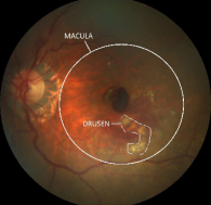
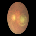
Age-related macular degeneration is a major cause of vision impairment and has affected approximately million people worldwide in 2020 [26]. With an aging population, such numbers are expected to rise to million by 2040 [46]. AMD is a progressive disorder of the macular region that causes central vision loss and is one of the most common causes of irreversible vision impairment in people over years-old [22]. Figure 1 depicts an example of a retina fundus affected by AMD.
This work has introduced an alternative approach for generating synthetic images for training deep networks and tested it for AMD identification. Retina images, positive and negative to AMD, from multiple databases were used to feed a GAN to provide a range of image qualities and lesions. Ten different GAN architectures were compared to generate synthetic eye-fundus images and trhe quality was assessed using the Fréchet Inception Distance (FID), two independent clinical experts who were label blinded and deep-learning classification.
The primary contributions of this work are fourfold:
-
•
To provide an open-source tool that can generate high-quality medical images of both, disease and healthy conditions;
-
•
Generation and validation of eye-fundus images suitable for training of deep-learning networks for computerized detection of AMD.
-
•
To introduce StyleGAN2-ADA [28] for eye fundus image generation; and
-
•
To present a comparison among several GAN architectures for synthetic image generation.
2 Theoretical Background
2.1 Generative Adversarial Networks
GANs are deep-learning based data generative networks. A significant improvement in more recent GANs is obtaining training stability to prevent the well-known “mode collapse", i.e., the generator is trapped in a local minimum, thus producing images that are very similar to each other. Gulrajani et al. [20] proposed the Wasserstein GAN with gradient penalty (WGAN-GP) to mitigate this problem, which implements a distinct loss function and eases the training convergence stability. Meanwhile, Karras et al. [27] proposed the Progressive GAN, showing that performance gains and learning capacity can be obtained by increasing the image resolution gradually during training.
2.2 Style-based Generative Adversarial Networks
The Style-based Generative Adversarial Network (StyleGAN) [29], depicted in Figure 2, is a state-of-the-art generative model that aims to create high-resolution images with greater fidelity. Additionally, such an architecture aims to enhance the diversity of outputs and simultaneously control image features. This gives more reliable control over the latent space which may allow users to edit image features, such as skin color, age, gender, and facial expression.
StyleGAN2 with Adaptive Discriminator Augmentation (StyleGAN2-ADA) has shown promising results in synthesizing high-quality images [28]. This variant uses an adaptive discriminator augmentation technique that stabilizes training in data-constrained environments. The method does not require modifications in the loss functions or architectures and can be used to train the network from scratch and fine-tuning.
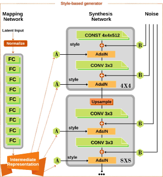
While traditional generators are designed as single neural networks, StyleGAN introduces a dual-network-based architecture, which is described below:
-
•
Mapping Network: it maps a latent input to an intermediate representation using eight fully connected layers, mitigating the entanglement issue (e.g., an -dimensional latent space is converted into an -dimensional intermediate space).
-
•
Synthesis Network: the outcome of the previous layer serves as an input to another network that uses different blocks, one for each image resolution. Each block comprises an upsampling operation (resolutions are increased progressively) followed by a convolutional layer with a kernel (CONV ). Further, Gaussian noise (B) is added to the output to feed an Adaptive Instance Normalization (AdaIn) layer, which also requires style input (high-level features) obtained by the affine transformation (A). The noise is vital to produce fine variations such as in hair of a facial image. The aforementioned process, i.e., convolution followed by an AdaIn layer, is executed once more, and the output is used to feed the next block. The only exception concerns the first block, which replaces the upsampling operation with a constant input (CONST ), and it has one convolutional layer.
2.3 Style-based Generative Adversarial Networks 2
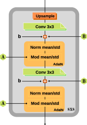
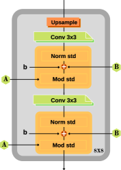
One recurring problem of StyleGAN lies in creating blob-like pixels (resembles water droplets), which are more visible when examining the intermediate feature maps of the generator. Such a problem initially appears in all feature maps at a low resolution such as of and is deepened when employing higher resolutions. Karras et al. [30] traced the problem and found out that AdaIn loses some crucial information when performing the instance normalization. They proposed the StyleGAN2, an alternative architecture that stabilizes the generation of high-resolution images. This has a simplified first processing step achieved by normalizing the features without their mean, and removing the noise addition. Figure 3 illustrates a comparison between StyleGAN and StyleGAN2 architectures.
 |
 |
| (a) | (b) |
3 Methodology
This section describes the datasets that were used in the study, the techniques employed to generate the synthetic images, and the methodology to evaluate the different neural architectures considered in the experimental section.
3.1 Dataset
This study used eye-fundus images from three public datasets reported in the literature to capture the typical differences due to equipment and demographics. Below, we summarized the datasets’ preliminary information:
-
•
iChallenge-AMD: Comprises of retinal fundus images that have been annotated for drusen and hemorrhage. The training set was made of 400 images (89 images of eyes with AMD and 311 from eyes without AMD), while the test sets contained the remaining images [15].
-
•
ODIR-2019: Contains colored fundus images from both left and right eyes of patients obtained from multiple hospitals/medical centers in China, with varying image resolutions and observations from several specialists. The dataset has was designed to address normal and six diseases: diabetes, glaucoma, cataract, AMD, hypertension, myopia, and other diseases/abnormalities111https://odir2019.grand-challenge.org/dataset/. The training set is made up of a structured dataset with images, of which images are labelled as having AMD. The testing set consists of colored fundus images, eliminating age and gender.
-
•
RIADD: contains fundus images recorded using 3 different cameras and multiple conditions. The images have been annotated through the consensus of two retina experts. The dataset has been sub-divided based on six diseases/abnormalities; diabetic retinopathy, AMD, media haze, drusen, myopia, and branch retinal vein occlusion [38]. The dataset was subdivided into three subsets: for training ( images), for validation ( images), and the remaining for testing purposes ( images).
We have used publicly available and online datasets, each of which have described their patient recruitment, clinical outcomes and human experiments ethics approval in their associated publications. Registration was necessary for access to the data from the three sources.
i-Challenge datset authors confirm in their publications they received ethics approval guidelines from Sun-Yat Sen University, China and Sixth People’s Hospital Affiliated to Shanghai Jiao Tong University, Shanghai, China. Details on https://amd.grand-challenge.org/Home/
ODIR dataset confirm that all data is from routine clinical examinations, have been de-identified and published after receiving clearance from Peiking University board. Details on https://odir2019.grand-challenge.org/dataset/
RIADD dataset confirm that data collection was conducted after ethics approval from Review Board of Shri Guru Gobind Singhji Institute of Engineering and Technology, Nanded, India. Details on https://riadd.grand-challenge.org/
The datasets mentioned above were designed to address a challenge and hence the labels of the test subset were not available. Therefore, we used only the training subset.
These datasets have an imbalanced class distribution, which is an inherent problem for most medical image datasets and using such a set generally leads to a bias towards the predominant class. Different research groups have developed these datasets with differences in the quality of the images and the demographics. Thus, these datasets offered both, data imbalance and wide range of image quality.
The ODIR-2019 and RIADD datasets were organized into two subsets, AMD and non-AMD images. Preprocessing methodology was same as proposed by Fu et al. [16], which comprises of a preprocessing step that detects the retinal mask using the Hough Circle Transform and then crops it to remove the impact of the black background. The cropped region was then scaled to a resolution. The resultant images were submitted to a three-level manual quality grading: “Good’, “Usable’ and “Reject’. Only those images graded with “Good" and “Usable" scores were used for further analysis. As a result, the number of images positive to AMD decreased from to , to , and to images in the iChallenge-AMD, ODIR-2019, and RIADD datasets, respectively while the number of non-AMD images decreased from to , to , and to images concerning the iChallenge-AMD, ODIR-2019, and RIADD datasets, respectively. Figure 4 illustrates the steps mentioned above for a sample image from the ODIR-2019 dataset.
 |
 |
 |
| (a) | (b) | (c) |
These images were then resampled to pixels, followed by a cropping procedure keeping the center of the image to pixels. Such a procedure is required to drive StyleGAN2-ADA generating images focused on the macula area (Figure 4-c). Ultimately, images were resized pixels and normalized within the range to be used as proper inputs to the deeper architectures considered in the manuscript.
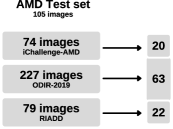
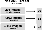
After quality assessment and selecting the images for the final dataset using the criterion described earlier, the resulting single dataset comprised totaL of images. In this, images were used to train the models ( with AMD and without AMD) and the remaining images ( with AMD) were used as a holdout test set. Figure 5 displays the number of images used per dataset to compose the final test set.
 |
 |
| (a) | (b) |
3.2 Evaluation Measures
We employed three evaluation measures, i.e., the Fréchet Inception Distance (FID), a well-known GAN evaluation score [24], the experts ability to identify the syntetic images and the classification accuracy. FID is often used to assess the quality and variety of the generated images and, even though it has been proposed to improve the standard Inception Score, it still uses the Inception-v3 architecture to extract features from both, synthetic and real images.
3.3 Experimental Setup
The experiments were divided into four rounds: (i) a FID comparison among StyleGAN2-ADA and nine different GAN architectures to assess the quality of the images generated, (ii) evaluation of the human experts to distinguish real and synthetic images, (iii) data perturbation with synthetic images generated by StyleGAN2-ADA and evaluated in three different deep networks and (iv) a comparison of the best-augmented model obtained in the previous step with human experts when identifying images that have AMD verses those that do not.
The first experiment employed FID to compare StyleGAN2-ADA and nine distinct GAN models. The following models were considered in the experiment: Deep Convolutional GAN (DCGAN) [41], Least Squares Generative Adversarial Networks (LSGAN) [35], Wasserstein GAN (WGAN) [1], Wasserstein GAN with Gradient Penalty (WGAN-GP) [20], Deep Regret Analytic Generative Adversarial Networks (DRAGAN) [33], Energy-based Generative Adversarial Network (EBGAN) [47], Boundary Equilibrium Generative Adversarial Networks (BEGAN) [3], Conditional GAN (CGAN) [36], and Auxiliary Classifier GAN (ACGAN) [37]. All models were trained with epochs, considering samples of size pixels and a batch size of . The training step used the ADAM[32] optimizer with a learning rate of and decay rates of and regarding the generator and the discriminator, respectively. The experiments were conducted using the training set with an Nvidia RTX 2060 GPU. Therefore, after training each GAN model, new images were generated and FID was computed.
The second experiment determines whether clinical experts, who are very experienced with the analysis of eye fundus images, can distinguish between synthetic and real images. Such a step is essential to evaluate the effectiveness of StyleGAN2-ADA for generating synthetic eye fundus images. The experts were provided with randomly generated image sets, one for AMD-diagnosed images and the other for non-AMD images, each consisting of ten synthetic images and ten real images. They were asked to identify the syntetic images in the mix.
In the next experiment, we considered three deep architectures pre-trained with ImageNet dataset [11] for performance comparison when the model is perturbed with synthetic images, i.e., SqueezeNet [25], AlexNet [34], and ResNet18 [23]. During training, synthetic and real images were mixed within each batch according to a pre-determined hyperparameter . For each image, a uniform distributed number was sampled and compared with : if the latter was greater than , the image was replaced by a synthetic one. We considered images generated by StyleGAN2-ADA, for it had obtained the best FID values in the first experiment (such outcomes are later described in Section 4). The deep networks were trained using a learning rate of , a decay rate of , batch size of , number of epochs equal to , and samples of size pixels.
To overcome the issue of unbalanced dataset, Weighted Random Sampler [12], was used which performs oversampling in the minority class. This assigns a higher weight to the minority classes, thus affecting the probability of drawing a point from the majority class by moving from a uniform distribution to a multinomial distribution. Further augmentation was performed using classical image transformations, such as resizing and color jittering, which changes the brightness, contrast, saturation, and horizontal flipping. The test set was kept intact.
The final round was aimed at comparing the best deep model obtained in the previous phase, ResNet-18, against human experts concerning the task of AMD identification. In this step, twenty real images (ten AMD images and ten non-AMD images) were randomly selected from the test set and provided to human experts for classification purposes. Following that, the same images were submitted to a ResNet-18 for comparison purposes. To allow a fair comparison between humans and deep models, we considered the following measures: standard accuracy (ACC), sensitivity, and specificity.
In this work, we used the StyleGAN2-ADA official source code, and the hyperparameters suggested by Karras et al. [28]. Concerning StyleGAN2-ADA hyperparameters, we used a batch size of , ADA target equal to (i.e., the probability of using ADA mechanism), the Adam algorithm with a learning rate of , decay values of and , and a convergence error of for the generator and discriminator. StyleGAN2-ADA framework enables different augmentations (rotation, geometric transformations, and color transformations) and class-conditional training. The output image resolution was set to pixels.
4 Results
The experimental results are presented in four sub-sections: (i) FID for synthetic image assessment, (ii) human experts detecting synthetic images, (iii) image perturbation assessment, and (iv) comparison between human experts and deep models for detecting AMD images.
4.1 Synthetic Image Assessment
Table 1 presents the FID values for each GAN-based architecture. StyleGAN2-ADA achieved the lowest FID value of , while EBGAN was placed in last and its FID value of was the highest. The smaller the FID value, the better is the quality of the generated image. Therefore, all further experiments only considered the StyleGAN2-ADA architecture for synthetic image generation.
| Type | Architecture | FID |
| Unconditional | EBGAN | |
| DCGAN | ||
| DRAGAN | ||
| WGAN | ||
| LSGAN | ||
| WGAN-GP | ||
| BEGAN | ||
| Conditional | CGAN | |
| ACGAN | ||
| StyleGAN2-ADA |
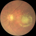
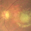
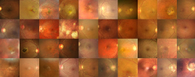
Figure 6 shows some examples of real and synthetic images produced by StyleGAN2-ADA. The trained model yields realistic-looking images for both, with and without AMD, conditioned by sampling from latent representations. Visual inspection shows that the generated images are similar to the real images. In the AMD images, macula degeneration is evident.
 |
 |
| (a) | (b) |
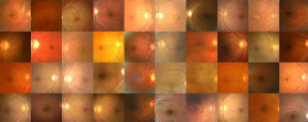
Figure 7 provides examples of real and synthetic images that are from eyes, positive and negative to AMD. One can observe the high-quality images that were generated for both, AMD and non-AMD images.
 |
 |
 |
 |
| (a) | (b) | (c) | (d) |
4.2 Distinguishing between Synthetic and Real Images
Table 2 presents the outcomes of each clinical expert. For AMD images, the accuracy was (standard deviation of ) for clinician #1 and (standard deviation of ) for clinician #2. For Non-AMD images, clinician #1 achieved an accuracy of (standard deviation of ), and clinician #2 obtained an accuracy of (standard deviation of ). These results highlight that both clinicians could not differentiate between real and synthetic images.
| ACC | Sensitivity | Specificity | ||
| AMD | Clinician #1 | |||
| Clinician #2 | ||||
| Non- AMD | Clinician #1 | |||
| Clinician #2 |
4.3 Image Perturbation Assessment
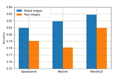
Figure 8 shows the accuracy over the test set concerning different percentages ( value) of real images that were replaced by synthetic images. Overall, the accuracy improved when combining synthetic and real images. While the accuracy lies between and when using only synthetic images, and between and when using only real images for training, the combination of both types of images gave the best results. However, this was network dependent: while SqueezeNet accuracy peaked at when using of synthetic images, AlexNet obtained its highest accuracy () when using only of synthetic images. ResNet18 achieved its best of with of synthetic images. In general, the networks performed poorly when the percentage of synthetic images exceeded .
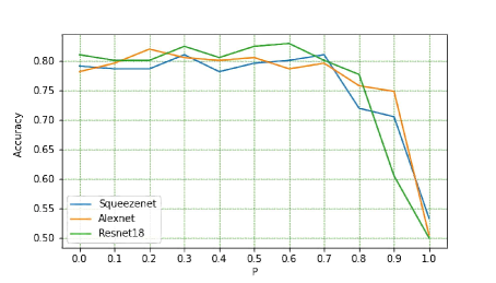
Figure 9 compares the improvement due to the mixing of the synthetic with real images on the three evaluated architectures. The most significant improvement was by AlexNet, its accuracy increased by approximately , while ResNet18 had highest accuracy (about ) using a mix of synthetic and real images.
Tables 3 and 4 present the confusion matrix and class-wise performance measures (precision, recall, and F1-score) when ResNet18 architecture was trained with (i) real data only and (ii) mixed data (), respectively. We can observe that ResNet18 trained with mixed data provided more reliable outcomes, where there was a minor difference between precision and recall values, attesting that the network can better distinguish between classes and not be prone to overfit on a specific class.
| AMD | Non-AMD | Precision | Recall | F-Score | |
| AMD | |||||
| Non-AMD |
| AMD | Non-AMD | Precision | Recall | F-Score | |
| AMD | |||||
| Non-AMD |
One of the main issues regarding deep learning in medicine is the difficulty to interpret the decision mechanisms. Techniques, such as heatmaps from the Gradient-weighted Class Activation Mapping (Grad-CAM) [43] can be used to identify and highlight regions of interest for visualization. Figure 10 depicts such heatmaps in images with AMD, which can help clinicians better interpret the decision-making process. The results also show that ResNet18 architecture trained with mixed data can highlight the location of the lesion better.


 (a)
(b)
(c)
(d)
(a)
(b)
(c)
(d)
4.4 Comparison between Human Experts and Deep Models
Table 5 presents the comparison of AMD detection by human experts and the best deep model, Resnet-18. Overall, the results of both clinicains and deep-learning were similar, with the best performance by deep-learning while the lowest specificity was by clinician #2. This shows that deep models can outperform clinicians for diagnosing AMD in eye fundus images.
| ACC | Sensitivity | Specificity | |
| Clinician #1 | |||
| Clinician #2 | |||
| Resnet-18 |
5 Discussion
DLN are suitable for automated and accurate analysis of medical images, and these are faster and often more accurate than the experts. These have been used for computerized classification of eye fundus images for detection of referable diabetic retinopathy [21, 40, 17, 10], retinopathy of prematurity [4], exudates on the retina [31], AMD [7, 8, 5], and AMD severity [6, 19]. However DLN require large numbers of annotated images, and the datasets should be balanced. Unbalanced datasets when used for training can lead to bias and erroneous outcomes. However, most suitably annotated medical image datasets are neither large nor balanced. There are number of diverse reasons why this happens, such as commercial interests, privacy of medical data, and issues with obtaining clearance from ethics boards of the hospitals [13]. While efforts are being made to develop bigger databases, the other alternate that has been proposed is to develop suitable synthetic images to overcome these shortcomings. Earlier efforts to develop synthetic images were by transforming the images, the use of deep-learning approach for this purpose has been found to be more effective [7, 8, 5].
Burlina et al have successfully developed a method for [7, 5] generating the synthetic images. Their method is based on GAN, and they tested their method by showing that experts were unable to identify the synthetic images. However, their method has been patented and hence not available for being used by others. We have developed an alternate method based on StyleGAN2, an extension of the progressive GAN, to generate synthetic medical images that human experts were unable to identify. We have also shown that these images were suitable for increase the performance of deep-learning networks. The results were similar to those reported in the literature using patented technology. While Burlina et al. trained a Progressive GAN over a large number of images positive to AMD, our technique has the potential to generate similar high synthetic images through a small number of images positive to AMD.
ResNet18 architecture trained over real and synthetic images provided the best results, marginally outperforming the human experts’ performance. Therefore, we can conclude that deep models appropriately trained can be as accurate as humans for specific medical image classification applications and datasets. We can extend such an argument to the problem of diagnosing AMD in eye fundus images. On the other hand, one limitation of this study is that these methods have handled the problem only as a binary classification task, i.e., AMD versus non-AMD images. However, medical images are typically We believe more classes will make the problem more challenging to cope with.
We have made available an online system with this trained network so that anyone can use it and test it, simply by uploading images. The software automatically labels the images as positive or negative to AMD. We have also provided the source code of the entire software and it is available publicly to facilitate researchers to use this as it is, or improve it. We are focused on fostering partnerships to facilitate and conduct research towards the usage of deep-learning to generate and recognize medical images.
6 Conclusion and Future Works
Deep-learning-based analysis of medical images has been found to be very useful, for it can learn from examples without requiring prior knowledge of features that matter. However, its applications are limited because labeled and/ or annotated medical image datasets of eye fundus are usually small and imbalanced.
This manuscript has developed a method to generate synthetic images that is not patented and the source code is publicly available. We have investigated the use of StyleGAN2-ADA to generate synthetic images using examples from publicly available databases to distinguish between AMD and healthy eyes. We found that experienced clinical experts were unable to differentiate between the synthetic and real images. We also demonstrated that synthetic images generated using StyleGAN2-ADA when mixed with real images improved the classification accuracy of deep learning networks, and these maginally outperformed clinical experts in separating the AMD and Non-AMD retinal images.
Future works regarding the application of this for AMD detection will need to broaden the scope to detect the severity of AMD, and for differentiating from other diseases. For generating medical images, there is the need to consider a broader range of deep architectures and applications. We would also need to investigate the effectiveness of heatmaps generated by Grad-CAM for helping the clinicians.
7 Data availability
The iChallenge-AMD dataset can be found in https://ai.baidu.com/broad/introduction?dataset=amd, while ODIR-2019 dataset is available on https://odir2019.grand-challenge.org/dataset/ and RIADD is available on https://riadd.grand-challenge.org/Home/.
8 Code availability
The official code for StyleGAN2-ADA is available at https://github.com/NVlabs/stylegan2-ada-pytorch. Our implementation of Deep Convolutional GAN (DCGAN), Least Squares Generative Adversarial Networks (LSGAN), Wasserstein GAN (WGAN), Wasserstein GAN with Gradient Penalty (WGAN-GP), Deep Regret Analytic Generative Adversarial Networks (DRAGAN), Energy-based Generative Adversarial Network (EBGAN), Boundary Equilibrium Generative Adversarial Networks (BEGAN), Conditional GAN (CGAN), and Auxiliary classifier GAN (ACGAN) are available for download at https://github.com/GuiCamargoX/gans_pytorch. All source code concerning image processing, Style-GAN2-ADA data generation, and the pre-trained networks are available at https://github.com/GuiCamargoX/amd-synthetic.
Acknowledgments
The authors thank Dr. Ione F. Alexim from BP Hospital - A Beneficência Portuguesa de São Paulo, who provided her eye fundus grading expertise to the project. We are grateful to São Paulo Research Foundation (FAPESP) under the grants #2013/07375-0, #2014/12236-1, #2018/15597-6, #2019/00585-5, #2019/07665-4, and #2019/02205-5, to the Brazilian National Council for Research and Development (CNPq) grants #307066/2017-7, #427968/2018-6, #309439/2020-5, and #606573/2021-0, as well as the Engineering and Physical Sciences Research Council (EPSRC) grant EP/T021063/1 and its principal investigator Ahsan Adeel. We also acknowledge the funding from Promobilia Foundation, Sweden, and SPARC grant (P-134, 2019), Department of Biotechnology (India) for financial their support.
Author Information
8.1 Contributions
G.O. developed the algorithms and performed the analysis. J.P. and D.K. conceptualized and designed the project. G.R., L.P., D.P. managed the data and designed the software. H.K. was responsible for the clinical assessment of the images and script. All authors contributed to writing the manuscript. D.K, G.O. and JP finalized the manuscript.
8.2 Corresponding authors
Correspondence to Guilherme C. Oliveira or Dinesh Kumar.
Conflict of Interest declarations
The authors declare no conflict of interest for this manuscript.
References
- Arjovsky et al. [2017] Arjovsky, M., Chintala, S., Bottou, L., 2017. Wasserstein generative adversarial networks, in: International conference on machine learning, PMLR. pp. 214–223.
- Bellemo et al. [2018] Bellemo, V., Burlina, P., Yong, L., Wong, T.Y., Ting, D.S.W., 2018. Generative adversarial networks (gans) for retinal fundus image synthesis, in: Asian Conference on Computer Vision, Springer. pp. 289–302.
- Berthelot et al. [2017] Berthelot, D., Schumm, T., Metz, L., 2017. Began: Boundary equilibrium generative adversarial networks. arXiv preprint arXiv:1703.10717 .
- Brown et al. [2018] Brown, J.M., Campbell, J.P., Beers, A., Chang, K., Ostmo, S., Chan, R.P., Dy, J., Erdogmus, D., Ioannidis, S., Kalpathy-Cramer, J., et al., 2018. Automated diagnosis of plus disease in retinopathy of prematurity using deep convolutional neural networks. JAMA ophthalmology 136, 803–810.
- Burlina et al. [2018a] Burlina, P., Joshi, N., Pacheco, K.D., Freund, D.E., Kong, J., Bressler, N.M., 2018a. Utility of deep learning methods for referability classification of age-related macular degeneration. JAMA ophthalmology 136, 1305–1307.
- Burlina et al. [2018b] Burlina, P.M., Joshi, N., Pacheco, K.D., Freund, D.E., Kong, J., Bressler, N.M., 2018b. Use of deep learning for detailed severity characterization and estimation of 5-year risk among patients with age-related macular degeneration. JAMA ophthalmology 136, 1359–1366.
- Burlina et al. [2019] Burlina, P.M., Joshi, N., Pacheco, K.D., Liu, T.A., Bressler, N.M., 2019. Assessment of deep generative models for high-resolution synthetic retinal image generation of age-related macular degeneration. JAMA ophthalmology 137, 258–264.
- Burlina et al. [2017] Burlina, P.M., Joshi, N., Pekala, M., Pacheco, K.D., Freund, D.E., Bressler, N.M., 2017. Automated grading of age-related macular degeneration from color fundus images using deep convolutional neural networks. JAMA ophthalmology 135, 1170–1176.
- Das et al. [2020] Das, V., Dandapat, S., Bora, P.K., 2020. Unsupervised super-resolution of oct images using generative adversarial network for improved age-related macular degeneration diagnosis. IEEE Sensors Journal 20, 8746–8756.
- De Fauw et al. [2018] De Fauw, J., Ledsam, J.R., Romera-Paredes, B., Nikolov, S., Tomasev, N., Blackwell, S., Askham, H., Glorot, X., O’Donoghue, B., Visentin, D., et al., 2018. Clinically applicable deep learning for diagnosis and referral in retinal disease. Nature medicine 24, 1342–1350.
- Deng et al. [2009] Deng, J., Dong, W., Socher, R., Li, L.J., Li, K., Fei-Fei, L., 2009. Imagenet: A large-scale hierarchical image database, in: 2009 IEEE conference on computer vision and pattern recognition, Ieee. pp. 248–255.
- Efraimidis and Spirakis [2008] Efraimidis, P., Spirakis, P., 2008. Weighted random sampling, in: Kao, M.Y. (Ed.), Encyclopedia of Algorithms, Springer US, Boston, MA. pp. 1024–1027. URL: https://doi.org/10.1007/978-0-387-30162-4_478, doi:10.1007/978-0-387-30162-4_478.
- Esteva et al. [2019] Esteva, A., Robicquet, A., Ramsundar, B., Kuleshov, V., DePristo, M., Chou, K., Cui, C., Corrado, G., Thrun, S., Dean, J., 2019. A guide to deep learning in healthcare. Nature medicine 25, 24–29.
- Frid-Adar et al. [2018] Frid-Adar, M., Diamant, I., Klang, E., Amitai, M., Goldberger, J., Greenspan, H., 2018. Gan-based synthetic medical image augmentation for increased cnn performance in liver lesion classification. Neurocomputing 321, 321–331.
- Fu et al. [2020] Fu, H., Li, F., Orlando, J., Bogunovic, H., Sun, X., Liao, J., Xu, Y., Zhang, S., Zhang, X., 2020. Adam: Automatic detection challenge on age-related macular degeneration. IEEE Dataport .
- Fu et al. [2019] Fu, H., Wang, B., Shen, J., Cui, S., Xu, Y., Liu, J., Shao, L., 2019. Evaluation of retinal image quality assessment networks in different color-spaces, in: International Conference on Medical Image Computing and Computer-Assisted Intervention, Springer. pp. 48–56.
- Gargeya and Leng [2017] Gargeya, R., Leng, T., 2017. Automated identification of diabetic retinopathy using deep learning. Ophthalmology 124, 962–969.
- Goodfellow et al. [2014] Goodfellow, I.J., Pouget-Abadie, J., Mirza, M., Xu, B., Warde-Farley, D., Ozair, S., Courville, A., Bengio, Y., 2014. Generative adversarial networks. arXiv preprint arXiv:1406.2661 .
- Grassmann et al. [2018] Grassmann, F., Mengelkamp, J., Brandl, C., Harsch, S., Zimmermann, M.E., Linkohr, B., Peters, A., Heid, I.M., Palm, C., Weber, B.H., 2018. A deep learning algorithm for prediction of age-related eye disease study severity scale for age-related macular degeneration from color fundus photography. Ophthalmology 125, 1410–1420.
- Gulrajani et al. [2017] Gulrajani, I., Ahmed, F., Arjovsky, M., Dumoulin, V., Courville, A., 2017. Improved training of wasserstein gans. arXiv preprint arXiv:1704.00028 .
- Gulshan et al. [2016] Gulshan, V., Peng, L., Coram, M., Stumpe, M.C., Wu, D., Narayanaswamy, A., Venugopalan, S., Widner, K., Madams, T., Cuadros, J., et al., 2016. Development and validation of a deep learning algorithm for detection of diabetic retinopathy in retinal fundus photographs. Jama 316, 2402–2410.
- Harvey [2003] Harvey, P.T., 2003. Common eye diseases of elderly people: identifying and treating causes of vision loss. Gerontology 49, 1–11.
- He et al. [2016] He, K., Zhang, X., Ren, S., Sun, J., 2016. Deep residual learning for image recognition, in: Proceedings of the IEEE conference on computer vision and pattern recognition, pp. 770–778.
- Heusel et al. [2017] Heusel, M., Ramsauer, H., Unterthiner, T., Nessler, B., Hochreiter, S., 2017. Gans trained by a two time-scale update rule converge to a local nash equilibrium. Advances in neural information processing systems 30.
- Iandola et al. [2016] Iandola, F.N., Han, S., Moskewicz, M.W., Ashraf, K., Dally, W.J., Keutzer, K., 2016. Squeezenet: Alexnet-level accuracy with 50x fewer parameters and< 0.5 mb model size. arXiv preprint arXiv:1602.07360 .
- Jonas [2014] Jonas, J.B., 2014. Global prevalence of age-related macular degeneration. The Lancet Global Health 2, e65–e66.
- Karras et al. [2017] Karras, T., Aila, T., Laine, S., Lehtinen, J., 2017. Progressive growing of gans for improved quality, stability, and variation. arXiv preprint arXiv:1710.10196 .
- Karras et al. [2020a] Karras, T., Aittala, M., Hellsten, J., Laine, S., Lehtinen, J., Aila, T., 2020a. Training generative adversarial networks with limited data, in: Proc. NeurIPS.
- Karras et al. [2019] Karras, T., Laine, S., Aila, T., 2019. A style-based generator architecture for generative adversarial networks, in: Proceedings of the IEEE/CVF Conference on Computer Vision and Pattern Recognition, pp. 4401–4410.
- Karras et al. [2020b] Karras, T., Laine, S., Aittala, M., Hellsten, J., Lehtinen, J., Aila, T., 2020b. Analyzing and improving the image quality of stylegan, in: Proceedings of the IEEE/CVF Conference on Computer Vision and Pattern Recognition, pp. 8110–8119.
- Khojasteh et al. [2019] Khojasteh, P., Passos, L.A., Carvalho, T., Rezende, E., Aliahmad, B., Papa, J.P., Kumar, D.K., 2019. Exudate detection in fundus images using deeply-learnable features. Computers in biology and medicine 104, 62–69.
- Kingma and Ba [2017] Kingma, D.P., Ba, J., 2017. Adam: A method for stochastic optimization. arXiv:1412.6980.
- Kodali et al. [2017] Kodali, N., Abernethy, J., Hays, J., Kira, Z., 2017. On convergence and stability of gans. arXiv preprint arXiv:1705.07215 .
- Krizhevsky et al. [2012] Krizhevsky, A., Sutskever, I., Hinton, G.E., 2012. Imagenet classification with deep convolutional neural networks. Advances in neural information processing systems 25, 1097--1105.
- Mao et al. [2017] Mao, X., Li, Q., Xie, H., Lau, R.Y., Wang, Z., Paul Smolley, S., 2017. Least squares generative adversarial networks, in: Proceedings of the IEEE international conference on computer vision, pp. 2794--2802.
- Mirza and Osindero [2014] Mirza, M., Osindero, S., 2014. Conditional generative adversarial nets. arXiv preprint arXiv:1411.1784 .
- Odena et al. [2017] Odena, A., Olah, C., Shlens, J., 2017. Conditional image synthesis with auxiliary classifier gans, in: International conference on machine learning, PMLR. pp. 2642--2651.
- Pachade et al. [2021] Pachade, S., Porwal, P., Thulkar, D., Kokare, M., Deshmukh, G., Sahasrabuddhe, V., Giancardo, L., Quellec, G., Mériaudeau, F., 2021. Retinal fundus multi-disease image dataset (rfmid): A dataset for multi-disease detection research. Data 6, 14.
- Perez and Wang [2017] Perez, L., Wang, J., 2017. The effectiveness of data augmentation in image classification using deep learning. arXiv preprint arXiv:1712.04621 .
- Quellec et al. [2017] Quellec, G., Charrière, K., Boudi, Y., Cochener, B., Lamard, M., 2017. Deep image mining for diabetic retinopathy screening. Medical image analysis 39, 178--193.
- Radford et al. [2015] Radford, A., Metz, L., Chintala, S., 2015. Unsupervised representation learning with deep convolutional generative adversarial networks. arXiv preprint arXiv:1511.06434 .
- Salehinejad et al. [2018] Salehinejad, H., Valaee, S., Dowdell, T., Colak, E., Barfett, J., 2018. Generalization of deep neural networks for chest pathology classification in x-rays using generative adversarial networks, in: 2018 IEEE International Conference on Acoustics, Speech and Signal Processing (ICASSP), IEEE. pp. 990--994.
- Selvaraju et al. [2017] Selvaraju, R.R., Cogswell, M., Das, A., Vedantam, R., Parikh, D., Batra, D., 2017. Grad-cam: Visual explanations from deep networks via gradient-based localization, in: Proceedings of the IEEE international conference on computer vision, pp. 618--626.
- Souza Jr et al. [2020] Souza Jr, L.A., Passos, L.A., Mendel, R., Ebigbo, A., Probst, A., Messmann, H., Palm, C., Papa, J.P., 2020. Assisting barrett’s esophagus identification using endoscopic data augmentation based on generative adversarial networks. Computers in Biology and Medicine , 104029.
- Souza Jr et al. [2021] Souza Jr, L.A., Passos, L.A., Mendel, R., Ebigbo, A., Probst, A., Messmann, H., Palm, C., Papa, J.P., 2021. Fine-tuning generative adversarial networks using metaheuristics: A case study on barrett’s esophagus identification, in: Bildverarbeitung für die Medizin 2021. Proceedings, German Workshop on Medical Image Computing, Regensburg, March 7-9, 2021, Springer Vieweg. pp. 205--210.
- Wong et al. [2014] Wong, W.L., Su, X., Li, X., Cheung, C.M.G., Klein, R., Cheng, C.Y., Wong, T.Y., 2014. Global prevalence of age-related macular degeneration and disease burden projection for 2020 and 2040: a systematic review and meta-analysis. The Lancet Global Health 2, e106--e116.
- Zhao et al. [2016] Zhao, J., Mathieu, M., LeCun, Y., 2016. Energy-based generative adversarial network. arXiv preprint arXiv:1609.03126 .