Multispectral time-resolved energy-momentum microscopy using high-harmonic extreme ultraviolet radiation
Abstract
A 790-nm-driven high-harmonic generation source with a repetition rate of 6 kHz is combined with a toroidal-grating monochromator and a high-detection-efficiency photoelectron time-of-flight momentum microscope to enable time- and momentum-resolved photoemission spectroscopy over a spectral range of – eV with sub-100-fs time resolution. Three-dimensional (3D) Fermi surface mapping is demonstrated on graphene-covered Ir(111) with energy and momentum resolutions of meV and Å-1, respectively. The table-top experiment sets the stage for measuring the -dependent ultrafast dynamics of 3D electronic structure, including band structure, Fermi surface, and carrier dynamics in 3D materials as well as 3D orbital dynamics in molecular layers.
I Introduction
Angle-resolved photoemission spectroscopy (ARPES) is the standard method to determine how electrons behave at surfaces of solid materials.Damascelli (2004); Lv, Qian, and Ding (2019); King et al. (2021); Sobota, He, and Shen (2021) Monochromatic photons having ultraviolet (UV) energies or higher eject electrons from a material’s surface, and the photocurrent is measured as a function of electron kinetic energy, emission direction, and photon energy. Direction is encoded in two emission angles or, equivalently, in the two components of the surface-parallel momentum . Since the four measurement parameters can be straightforwardly related to the energy relative to the Fermi level () and three-dimensional (3D) momentum of the electrons inside the material before photoexcitation,Chiang et al. (1980) the measured intensity distributions readily provide multidimensional images of the electronic structure in portions of four-dimensional (4D) energy-momentum space.Medjanik et al. (2017) Band structures and Fermi surfaces, but also momentum-dependent band renormalization and lifetime effects, can thus be accessed directly.Hüfner et al. (1999); Valla et al. (1999); Krasovskii et al. (2007); Watson et al. (2019) Another intriguing application is orbital tomography, which can provide reconstructed real-space tomograms of molecular orbitals on solid surfaces.Puschnig et al. (2009); Graus et al. (2019) Depending on whether emission angles or surface-parallel momentum components are imaged onto the detector,Wannberg (2009); Kotsugi et al. (2003); Schönhense, Medjanik, and Elmers (2015) the technique is referred to as ARPES or momentum microscopy, respectively.
In this energy-momentum imaging, the photon energy is an important parameter in at least three different ways. First, the photon energy determines the maximum detectable electron kinetic energy and 3D momentum, and thus the volume of the probed portion in energy-momentum space.Babenkov et al. (2019) Second, scanning the photon energy allows to retrieve the surface-perpendicular momentum component , hence to perform full 4D energy-momentum imaging.Rossnagel et al. (2001); Nielsen et al. (2003); Medjanik et al. (2017); Watson et al. (2019) And third, tunability in the photon energy can also be useful to enhance the contrast of specific features in ARPES data by exploiting excitation resonances, escape-depth variation, or matrix-element effects.Strocov et al. (2014); Moser (2017)
In principle, the same relevance of the probing photon energy and its tunability also applies to time-resolved ARPES (trARPES) or time-resolved momentum microscopy,Kutnyakhov et al. (2020); Maklar et al. (2020); Keunecke et al. (2020) in which time, or more precisely the time delay () between femtosecond-scale pump and probe pulses, is added as a fifth measurement parameter. Over the past fifteen years, trARPES has evolved into a powerful ARPES modality providing direct dynamical information on electronic structure at the fundamental time scales of electronic and atomic motion, particularly on photoinduced transient changes of electronic states and their population.Perfetti et al. (2006); Schmitt et al. (2008); Rohwer et al. (2011); Smallwood et al. (2012); Gierz et al. (2013); Wang et al. (2013); Mahmood et al. (2016); Nicholson et al. (2018); Na et al. (2019); Wallauer et al. (2021) trARPES is now routinely performed using table-top laser sources based on fourth and higher harmonic generation (HHG) in solidsGauthier et al. (2020); Bao et al. (2022) and gasesDakovski et al. (2010); Frietsch et al. (2013); Eich et al. (2014); Cilento et al. (2016); Rohde et al. (2016); Corder et al. (2018); Puppin et al. (2019); Buss et al. (2019); Sie et al. (2019); Mills et al. (2019); Liu et al. (2020); Cucini et al. (2020); Lee et al. (2020); Keunecke et al. (2020); Peli et al. (2020); Guo et al. (2022), respectively, as well as using free-electron lasers (FELs) based on the self-amplification of spontaneous emission (SASE) of free electrons in undulators.Kutnyakhov et al. (2020) The corresponding probe photon energies in trARPES range from the far UV to soft x-rays where a sweet spot currently is the intermediate extreme ultraviolet (XUV) regime.
In the XUV, HHG-based trARPES can optionally provide high time, energy, momentum, and spin resolution in conjunction with kHz-to-MHz repetition rates and a sufficiently wide detection window of the surface-parallel momentum to fully cover typical Brillouin-zone (BZ) dimensions.Dakovski et al. (2010); Frietsch et al. (2013); Eich et al. (2014); Cilento et al. (2016); Rohde et al. (2016); Plötzing et al. (2016); Eich et al. (2017); Corder et al. (2018); Gort et al. (2018); Puppin et al. (2019); Buss et al. (2019); Sie et al. (2019); Mills et al. (2019); Liu et al. (2020); Cucini et al. (2020); Lee et al. (2020); Keunecke et al. (2020); Peli et al. (2020); Bühlmann et al. (2020); Guo et al. (2022) Moreover, HHG sources combined with monochromators enable multispectral measurements as they can deliver an useable, discretely tunable photon-energy range of about 8–40 eV.Dakovski et al. (2010); Frietsch et al. (2013); Corder et al. (2018); Mills et al. (2019); Sie et al. (2019); Cucini et al. (2020) However, this multispectral capability has so far rarely been exploited in trARPES experiments, and recently several setups for HHG-based trARPES have even been optimized for operation at one specific photon energy only.Eich et al. (2014); Puppin et al. (2019); Lee et al. (2020); Keunecke et al. (2020) Here, contrary to this trend, we present the combination of a tunable, monochromatized, kHz-repetition-rate HHG source with a wide-momentum-acceptance time-of-flight (ToF) momentum microscope for efficient 4D energy-momentum mapping of ultrafast electronic structure dynamics. The overall system performance is demonstrated on bare and graphene-covered Ir(111).
II Experimental setup
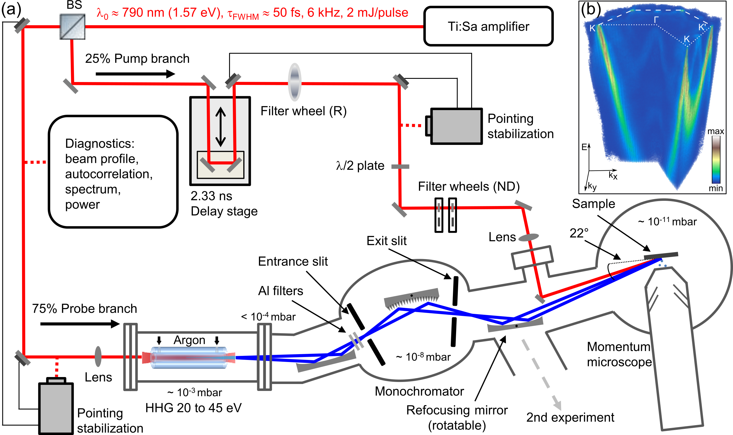
Our multispectral-HHG trARPES setup, as schematically illustrated in Fig. 1, measures a 5D photoemission data hypercube . The laser system gives the tuning of and , via adjustability of the probe photon energy and pump–probe delay, respectively, and the ToF momentum microscope provides the basic parallel 3D measurement of .
II.1 Multispectral photon source
The schematic layout of the laser system is shown in Fig. 1(a).Seo et al. (2016) A Ti:Sapphire laser amplifier (Wyvern 1000, KMLabs), operated at an output power of 12 W and a repetition rate of 6 kHz, delivers laser pulses at a center wavelength of 790 nm, pulse duration of 50 fs (FWHM, full width at half maximum), and pulse energy of 2 mJ. A quarter of the amplifier output is coupled into the pump branch, propagated through a delay stage, and focused onto the sample in the ultrahigh vacuum (UHV) photoemission chamber. The spot size of the pump beam on the sample is typically about m2. The available pump-pulse energy of 500 J can be used for frequency conversion or attenuated to the mid-nJ to low-J level for 790-nm excitation of the sample below the space-charge limit.Oloff et al. (2016); Schönhense et al. (2021)
The remaining 75% (50 fs, 1.5 mJ/pulse) of the amplifier output are coupled into the probe branch and focused with a lens (focal length mm) into an Ar-filled waveguide capillary (XUUS, KMLabs), where higher harmonics in the XUV are generated. During operation, the Ar pressure inside the capillary is 60 mbar, while the pressure outside stays below 5 mbar. A ZrO2-coated toroidal mirror ( mm) is used to focus the multispectral XUV light onto the entrance slit of the monochromator and to separate out much of the fundamental radiation. ZrO2 specifically provides XUV reflectivity comparable to Au for photon energies around 40 eV, and reduced reflectivity at eV.Wood and Nassau (1982); Polyanskiy ; Henke, Gullikson, and Davis (1993) The remaining 790-nm light is blocked using few-100-nm thick Al filters. Differential pumping is used to maintain a 5-orders-of-magnitude pressure difference between the HHG source and the monochromator chamber. A rotatable Au-coated toroidal grating ( mm) with 550 lines/mm (TGM300, Horiba Jobin Yvon SAS) images the entrance slit onto the exit slit with the selected higher harmonic light. For the 25th harmonic, the footprint on the monochromator grating is estimated to be mm2 translating into a nominal temporal probe-pulse broadening of 57 fs. A second toroidal mirror ( mm) focuses the selected and monochromatized XUV light onto the sample at an angle of 22∘ with respect to the surface. This mirror is rotatable and can alternatively direct the beam to a gas-phase experiment with combined ion- and electron-ToF spectroscopy.Gerken (2014); Gerken et al. (2014) Behind the refocusing mirror, the pump and probe beams propagate almost collinearly to the sample. At the sample, the XUV beam has a spot size of m2 and a flux of photons/s in the brightest harmonics, which are usually the 23 and 25 (see central panel of Fig. 4). The practically useable part of the harmonic spectrum contains all eight odd harmonics from the 15 to the 29 corresponding to a photon energy range of – eV. According to ray-tracing simulations, the temporal probe pulse broadening due to the monochromator lies in the range of 50–100 fs for all harmonics, independent of the slit size. The spectral resolution of the monochromator and the beamline is in the range of 100–240 meV under measurement conditions with slit sizes of 150 m. In particular, for the higher harmonics, the monochromator selects only a given harmonic rather than clipping the harmonic’s spectral bandwidth. The overall high stability of the HHG source enables pump-probe experiments over several days, with the temporal overlap (time zero) remaining within the experimental time resolution. Average drifts in HHG intensity stay within 30% over a week of continuous operation (with the possibility to retune and restore reduced intensity).
II.2 Electron momentum microscope
The ToF momentum microscope employed in our laboratory-based setup is the same instrument used for FEL-based photoemission spectroscopy at the PG2 beamline of FLASH (DESY, Hamburg).Kutnyakhov et al. (2020) The high photoelectron detection efficiency of the instrument, which compensates for the moderate repetition rate of the photon pulses, results from a combination of three separate capabilities: (i) direct 2D momentum imaging with a field of view of Å-1 in both surface-parallel momentum directions, (ii) simultaneous ToF energy recording in an energy window of 7 eV, and (iii) multi-hit detection of up to 3 electrons per pulse. The underlying principle of slit-less 3D photoelectron energy-momentum detection is implemented as follows:Schönhense, Medjanik, and Elmers (2015) The photocurrent emitted from the surface is imaged into an achromatic surface-parallel momentum image at the back-focal plane of the cathode objective lens; this hyperspectral image is subsequently magnified and high-pass-filtered by two lens systems, before it is spectrally dispersed in a field-free drift tube and finally captured on a delay-line detector (DLD). The 8-segment DLD (DLD6060-8s, Surface Concept) used in the current setup consists of two stacked 4-quadrant DLDs rotated by 45∘ with respect to each other. This novel detector provides improved multi-hit detection capability compared to a single 1- or 4-quadrant DLD, as well as improved resolution of hits occurring near segment boundaries. The length of the drift tube (800 mm) and temporal resolution of the detector (150 ps) translate into a nominal energy resolution of 40 meV for typical electron drift energies of 10–30 eV. The nominal momentum resolution is 0.01 Å-1, as given by the momentum field of view, active detector area (60 mm diameter), and the spatial resolution of the detector (80 m).
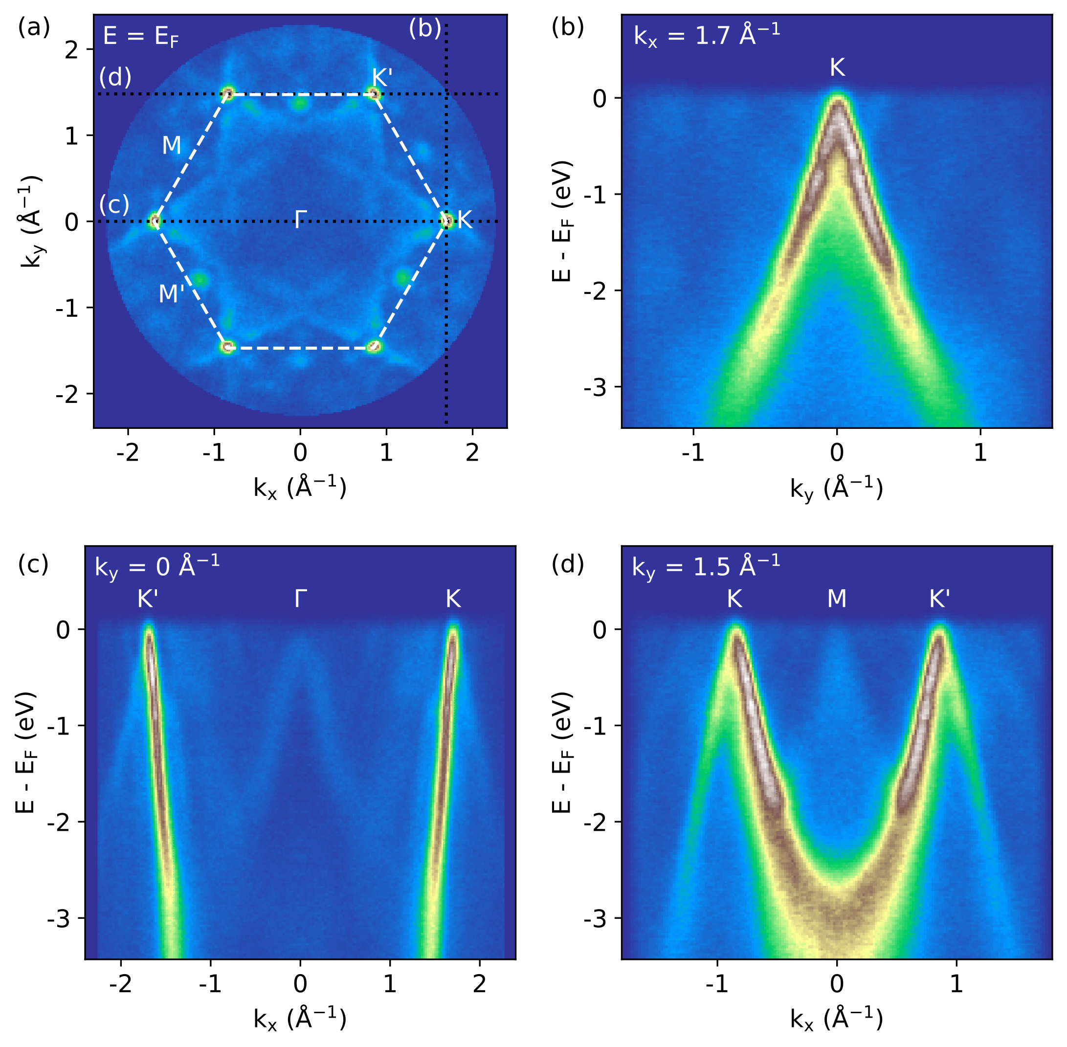
The 3D energy-momentum measurement system, based on single-event detection, fills in the photoemission data cube over energy and momentum intervals with a characteristic width of 7 eV and Å-1, respectively, at a repetition rate of 6 kHz. When delay-time scanning is added, the resulting size of a typical 4D data hypercube is 100 GB. An efficient data acquisition and data processing workflow is implemented using an open-source software package developed for high-throughput multidimensional photoemission spectroscopy experiments.Xian et al. (2020) Figure 1(b) shows a 3D representation of an exemplary data set taken from graphene/Ir(111). Four different 2D cuts through this data set are shown in Fig. 2, including the Fermi surface map [Fig. 2(a)] and selected band maps [Fig. 2(b)] and [Figs. 2(c) and 2(d)] for different constant values of corresponding to lines passing through high-symmetry points of the graphene BZ, as indicated in Fig. 2(a). In these maps, the strongest signal stems from the -band of graphene, with its linear dispersion toward and point-like Fermi surface at the and points. The Ir bands appear as much weaker features. Their interaction with the -band, however, leads to distinct kinks in the -band dispersion.Tusche et al. (2016) The presented data vividly illustrate the efficiency and completeness of the ToF momentum microscopy approach to photoelectron detection.
III Performance
We have characterized the performance of the experimental system by measuring the near- electronic structure and the above- carrier dynamics of graphene-covered and pristine Ir(111), respectively. Standard Ir(111) cleaning procedures and graphene growth recipes were applied.N’Diaye et al. (2008) Surface quality was checked by low-energy electron diffraction. All photoemission measurements were done at room temperature.
III.1 Experimental resolutions
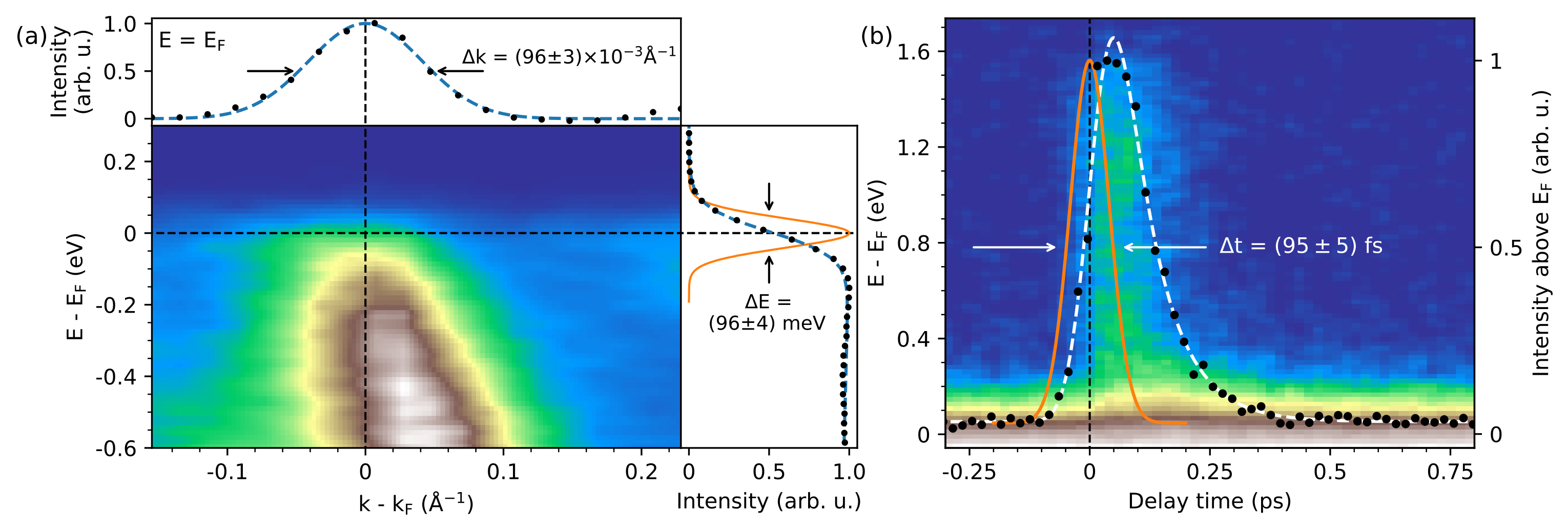
We estimated the effective experimental energy and momentum resolutions from the Fermi-level crossing of the graphene -band in the graphene/Ir(111) sample. Figure 3(a) shows a room-temperature – photoemission intensity map, which was measured in the vicinity of the point with a photon energy of 33 eV (21 harmonic) using probe-only photoemission. Also shown are the momentum distribution curve (MDC) extracted at (top panel) and an energy distribution curve (EDC) obtained by the MDC methodUlstrup et al. (2014) (right panel), representing the energy distribution of fitted MDC peak areas.
The EDC was fitted with a room-temperature Fermi-Dirac distribution function convoluted with a Gaussian resolution function [Fig. 3(a), right panel]. The resulting Gaussian FWHM, corresponding to the total energy resolution, is meV. This value includes a contribution of the photon source and monochromator of about 90 meV. The contribution of the electron spectrometer including space-charge broadening is estimated to be 30 meV. Over the entire usable spectral range of 24–46 eV, the effective energy resolution varied between 80 and 135 meV, where the photon-energy dependence of the grating resolution at fixed slit widths makes the dominant contribution. Note that these values are better than those given above for photon resolution because the field aperture of the momentum microscope typically acts as a virtual exit slit, selecting an effective field of view on the sample of 70 m.
The Gaussian FWHM determined from the momentum distribution curve at is Å-1 [Fig. 3(a), top panel]. After subtracting the intrinsic -band momentum width of Å-1,Kralj et al. (2011); Johannsen et al. (2013) the remaining effective momentum resolution is Å-1. We attribute the deterioration with respect to the nominal momentum resolution to less than optimal sample quality, electronic noise, and timing jitter in the position measurement on the detector. With varying probe photon energy, no noticeable changes of the momentum resolution were detected.
To estimate the temporal cross-correlation between pump and probe pulses, we performed pump–probe photoemission measurements on pristine Ir(111) using pump and probe photon energies of eV and eV (23 harmonic), respectively, and an incident pump fluence of mJ/cm2. Figure 3(b) shows a momentum-integrated – intensity map depicting the transient generation and relaxation of hot electrons above . A corresponding intensity transient (black data points), obtained by integrating over energies larger than , is overlaid. This signal was fitted with a step function multiplied by an exponential decay and convoluted with a Gaussian function. The resulting Gaussian FWHM is fs, giving an estimate of the temporal system response function. Based on this value and with a modeled pump-pulse duration of fs FWHM at the sample position (obtained by using an autocorrelation measurement in combination with a modeling of the additional optics), we estimate the duration of the probe pulse to fs FWHM, assuming uncorrelated Gaussian-shaped pulses.
III.2 Multispectral energy-momentum mapping
The key novel characteristic of our experimental setup is the combination of highly efficient 3D photoemission intensity imaging with a discrete tunability of the probe photon energy, thus making the energy-momentum mapping in trARPES -dependent and 4D. Figure 4 illustrates this experimental advance.
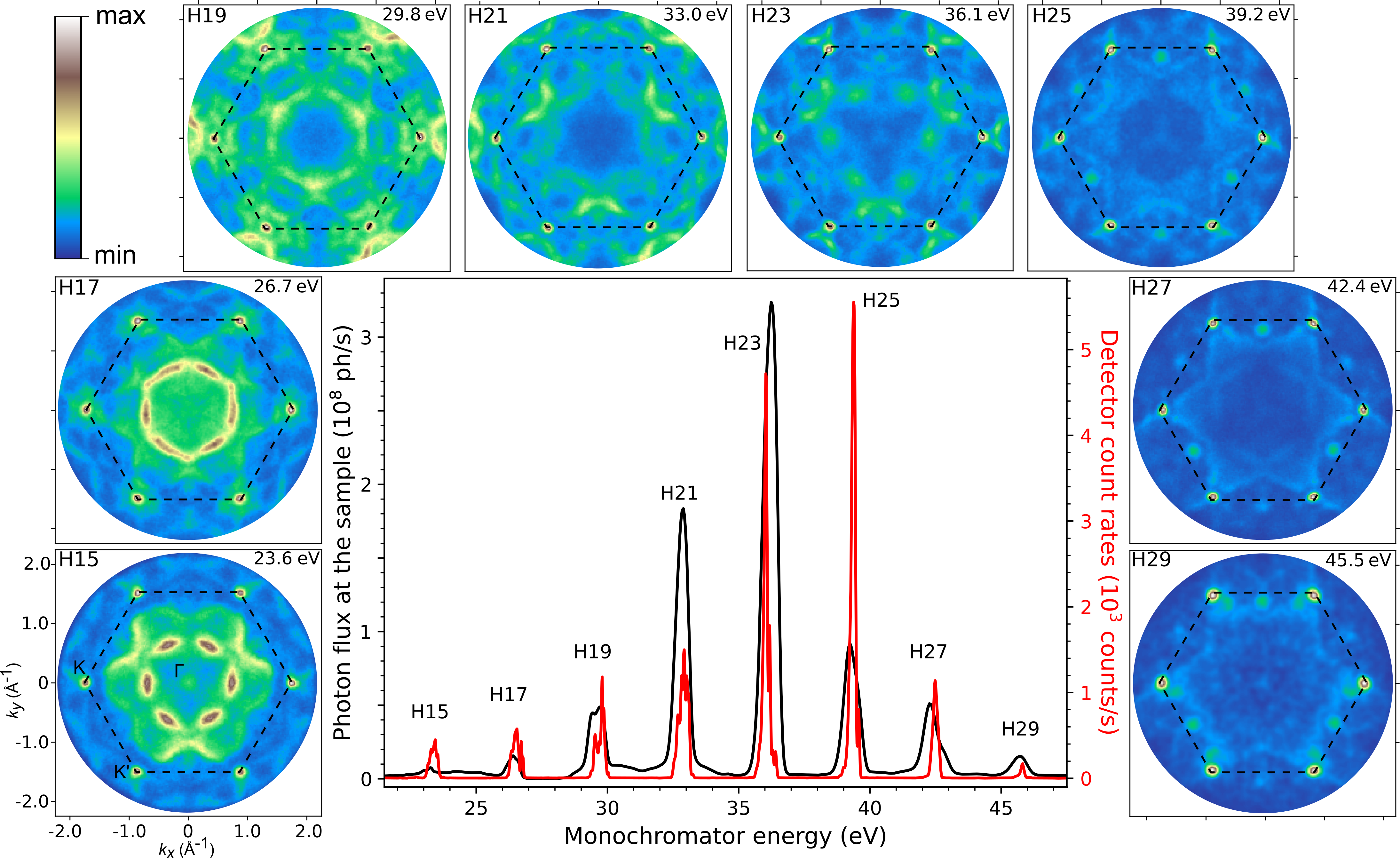
The central panel of Fig. 4 displays two typical XUV spectra as a function of the monochromator energy. The spectrum indicated by the black line gives the calculated photon flux at the sample position. This signal was measured by a calibrated XUV-sensitive Si photodiode behind the exit slit of the monochromator and corrected for beamline transmission, including Al-filter attenuation to prevent photocurrent saturation. The second spectrum (red line) represents the electron count rate at the detector, as measured from an electrically biased graphene/Ir(111) sample by the ToF momentum microscope under otherwise typical measurement settings. The practically useable photon-energy tuning range is – eV, at a spacing of eV, corresponding to the odd harmonic orders 15 to 29. The cutoff at 48 eV is typical for Ar gas as a generating medium.Krause, Schafer, and Kulander (1992)
The panels surrounding the XUV spectra in Fig. 4 show Fermi surface maps taken from graphene/Ir(111) with all eight harmonics. These 2D intensity maps are extracted from the full 3D data cubes that were originally measured. The data acquisition times varied from 2 to 12 h. The multispectral Fermi surface maps reflect a superposition of the point-like 2D Fermi surface of graphene (centered at the corners of the indicated hexagonal graphene BZ) and the complex multi-sheet 3D Fermi surface of Ir(111). There is no photon energy-dependent change in the shape of the graphene Fermi points, whereas the variation in the shape of the Ir intensity pattern, i.e., dispersion, is pronounced. Another observation is that the relative contribution of the Ir signal to the total photoemission intensity at is continuously suppressed upon increasing the photon energy from eV to eV. We attribute this effect to a decrease in the electron escape depth upon approaching the minimum of the universal curve of the inelastic mean free path around a kinetic energy of 50 eV:Tanuma, Powell, and Penn (2011) With increasing surface sensitivity, less photoemission signal is obtained from Ir(111), which is covered by a graphene monolayer.
Under the assumption of free-electron-like final states within the direct-transition model of ARPES, constant-energy maps, such as the ones displayed in Fig. 4, map onto a spherical surface in 3D momentum space. The kinematic equation relating the surface-perpendicular momentum component to the other measurement parameters is:Chiang et al. (1980)
| (1) |
where is the photon energy and and are the effective electron mass and inner potential (referenced to ) of the nearly-free-electron final-state parabola, respectively. The empirical parameters for Ir(111) are and eV.Elmers et al. (2017) Using equation (1) and exploiting point symmetry about the center of the BZ ( point), a tomogram of the 3D Fermi surface can be reconstructed from the stack of Fermi surface maps shown in Fig. 4.
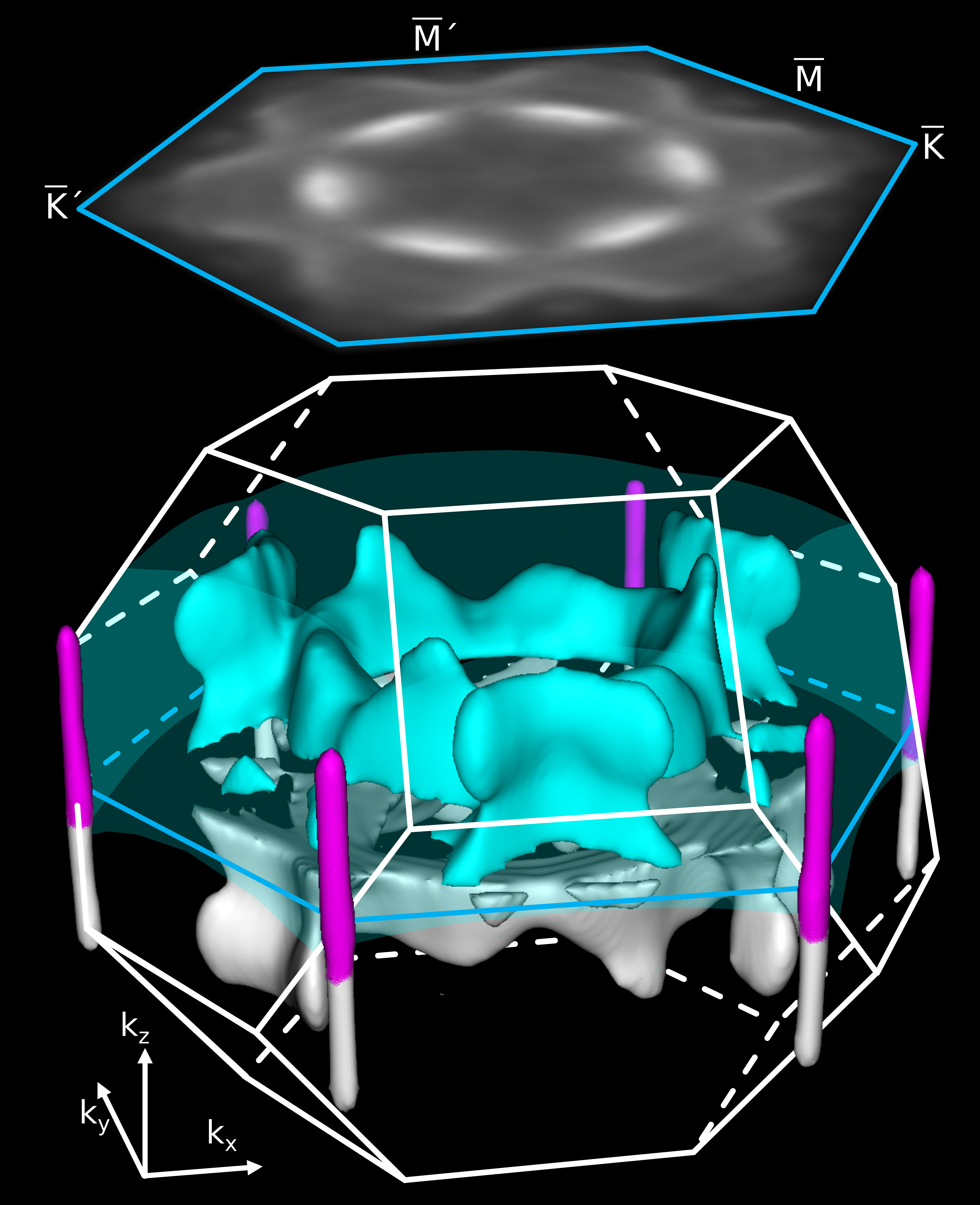
Figure 5 displays the reconstructed portion of the 3D Fermi surface for graphene/Ir(111). Three sets of isosurfaces can be identified: the non--dispersive graphene Fermi rods centered at the corners of the hexagonal graphene BZ as well as an inner hexagon-shaped Fermi surface sheet and an outer star-shaped Fermi surface sheet derived from Ir bulk states. The reconstructed Ir Fermi surface sheets are in good agreement with a 3D Fermi surface tomogram obtained from pristine Ir(111) by soft x-ray momentum microscopy.Elmers et al. (2017) The available photon-energy range translates into a finite probing interval of Å-1, smaller than the characteristic dimension ( Å-1) of the fcc BZ of Ir(111). Thus, our tomographic data cover 39% of the BZ volume. For crystalline materials with smaller dimensions, particularly layered 3D electron materials,Rossnagel et al. (2001); Krasovskii et al. (2007); Watson et al. (2019); Bao et al. (2022) larger portions of the bulk BZ or entire bulk BZs can be scanned. Similarly, for layers of 3D molecules,Haag et al. (2020) 3D orbital momentum tomograms can be recorded.
IV Conclusions
In conclusion, by combining a 790-nm-driven kHz-repetition-rate HHG source with a toroidal-grating monochromator and a high-detection-efficiency ToF momentum microscope, we have realized an experimental setup for probe photon energy-dependent time-resolved XUV-ARPES with good data collection efficiency and sub-100-fs time resolution. The photon energy tuning range is 24–46 eV, sufficient to map band structures and Fermi surfaces as well as molecular orbital densities over an interval of 1 Å-1. The system thus specifically enables -selective probing of ultrafast electronic structure dynamics in 3D materials as well as ultrafast 3D orbital tomography of molecular layers.Wallauer et al. (2021); Baumgärtner et al. (2022) Moreover, the setup complements time-resolved momentum microscopy in the XUV to soft x-ray regime at the FEL FLASH, for which the same ToF momentum microscope is used.Kutnyakhov et al. (2020) The two complementary probe photon sources particularly enable a unique merging of trARPES with time-resolved x-ray photoelectron spectroscopyHellmann et al. (2010); Dendzik et al. (2020); Pressacco et al. (2021) and diffractionCurcio et al. (2021) for combined investigations of ultrafast electronic, chemical, and geometric structure dynamics.
Acknowledgements.
This work is dedicated to the late Wilfried Wurth. The work was supported by the Deutsche Forschungsgemeinschaft (DFG, German Research Foundation)—Project ID 491245950 and via the Collaborative Research Center (CRC) 925—Project ID 170620586 (project B2). We thank Holger Meyer and Sven Gieschen for support with the instrumentation. We also thank former members of Wolfgang Eberhardt’s group, particularly Daniel Ramm, for help with the design and initial installation of the HHG source.Data Availability Statement
The data that support the findings of this study are available from the corresponding author upon reasonable request.
References
- Damascelli (2004) A. Damascelli, “Probing the electronic structure of complex systems by ARPES,” Physica Scripta T109, 61 (2004).
- Lv, Qian, and Ding (2019) B. Lv, T. Qian, and H. Ding, “Angle-resolved photoemission spectroscopy and its application to topological materials,” Nature Reviews Physics 1, 609–626 (2019).
- King et al. (2021) P. D. C. King, S. Picozzi, R. G. Egdell, and G. Panaccione, “Angle, spin, and depth resolved photoelectron spectroscopy on quantum materials,” Chemical Reviews 121, 2816–2856 (2021), pMID: 33346644, https://doi.org/10.1021/acs.chemrev.0c00616 .
- Sobota, He, and Shen (2021) J. A. Sobota, Y. He, and Z.-X. Shen, “Angle-resolved photoemission studies of quantum materials,” Rev. Mod. Phys. 93, 025006 (2021).
- Chiang et al. (1980) T. C. Chiang, J. A. Knapp, M. Aono, and D. E. Eastman, “Angle-resolved photoemission, valence-band dispersions , and electron and hole lifetimes for gaas,” Phys. Rev. B 21, 3513–3522 (1980).
- Medjanik et al. (2017) K. Medjanik, O. Fedchenko, S. Chernov, D. Kutnyakhov, M. Ellguth, A. Oelsner, B. Schönhense, T. R. F. Peixoto, P. Lutz, C.-H. Min, F. Reinert, S. Däster, Y. Acremann, J. Viefhaus, W. Wurth, H. J. Elmers, and G. Schönhense, “Direct 3d mapping of the fermi surface and fermi velocity,” Nature Materials 16, 615–621 (2017).
- Hüfner et al. (1999) S. Hüfner, R. Claessen, F. Reinert, T. Straub, V. Strocov, and P. Steiner, “Photoemission spectroscopy in metals:: band structure-fermi surface–spectral function,” Journal of Electron Spectroscopy and Related Phenomena 100, 191–213 (1999).
- Valla et al. (1999) T. Valla, A. V. Fedorov, P. D. Johnson, and S. L. Hulbert, “Many-body effects in angle-resolved photoemission: Quasiparticle energy and lifetime of a mo(110) surface state,” Phys. Rev. Lett. 83, 2085–2088 (1999).
- Krasovskii et al. (2007) E. E. Krasovskii, K. Rossnagel, A. Fedorov, W. Schattke, and L. Kipp, “Determination of the hole lifetime from photoemission: Ti states in ,” Phys. Rev. Lett. 98, 217604 (2007).
- Watson et al. (2019) M. D. Watson, O. J. Clark, F. Mazzola, I. Marković, V. Sunko, T. K. Kim, K. Rossnagel, and P. D. C. King, “Orbital- and -selective hybridization of se and ti states in the charge density wave phase of ,” Phys. Rev. Lett. 122, 076404 (2019).
- Puschnig et al. (2009) P. Puschnig, S. Berkebile, A. J. Fleming, G. Koller, K. Emtsev, T. Seyller, J. D. Riley, C. Ambrosch-Draxl, F. P. Netzer, and M. G. Ramsey, “Reconstruction of molecular orbital densities from photoemission data,” Science 326, 702–706 (2009), https://www.science.org/doi/pdf/10.1126/science.1176105 .
- Graus et al. (2019) M. Graus, C. Metzger, M. Grimm, P. Nigge, V. Feyer, A. Schöll, and F. Reinert, “Three-dimensional tomographic imaging of molecular orbitals by photoelectron momentum microscopy,” The European Physical Journal B 92, 80 (2019).
- Wannberg (2009) B. Wannberg, “Electron optics development for photo-electron spectrometers,” Nuclear Instruments and Methods in Physics Research Section A: Accelerators, Spectrometers, Detectors and Associated Equipment 601, 182–194 (2009), special issue in honour of Prof. Kai Siegbahn.
- Kotsugi et al. (2003) M. Kotsugi, W. Kuch, F. Offi, L. I. Chelaru, and J. Kirschner, “Microspectroscopic two-dimensional fermi surface mapping using a photoelectron emission microscope,” Review of Scientific Instruments 74, 2754–2758 (2003), https://doi.org/10.1063/1.1569404 .
- Schönhense, Medjanik, and Elmers (2015) G. Schönhense, K. Medjanik, and H.-J. Elmers, “Space-, time- and spin-resolved photoemission,” Journal of Electron Spectroscopy and Related Phenomena 200, 94 – 118 (2015), special Anniversary Issue: Volume 200.
- Babenkov et al. (2019) S. Babenkov, K. Medjanik, D. Vasilyev, S. Chernov, C. Schlueter, A. Gloskovskii, Y. Matveyev, W. Drube, B. Schönhense, K. Rossnagel, H.-J. Elmers, and G. Schönhense, “High-accuracy bulk electronic bandmapping with eliminated diffraction effects using hard x-ray photoelectron momentum microscopy,” Communications Physics 2, 107 (2019).
- Rossnagel et al. (2001) K. Rossnagel, L. Kipp, M. Skibowski, C. Solterbeck, T. Strasser, W. Schattke, D. Voß, P. Krüger, A. Mazur, and J. Pollmann, “Three-dimensional fermi surface determination by angle-resolved photoelectron spectroscopy,” Phys. Rev. B 63, 125104 (2001).
- Nielsen et al. (2003) M. B. Nielsen, Z. Li, S. Lizzit, A. Goldoni, and P. Hofmann, “Bulk fermi surface mapping with high-energy angle-resolved photoemission,” Journal of Physics: Condensed Matter 15, 6919–6930 (2003).
- Strocov et al. (2014) V. N. Strocov, M. Kobayashi, X. Wang, L. L. Lev, J. Krempasky, V. V. Rogalev, T. Schmitt, C. Cancellieri, and M. L. Reinle-Schmitt, “Soft-x-ray arpes at the swiss light source: From 3d materials to buried interfaces and impurities,” Synchrotron Radiation News 27, 31–40 (2014), https://doi.org/10.1080/08940886.2014.889550 .
- Moser (2017) S. Moser, “An experimentalist’s guide to the matrix element in angle resolved photoemission,” Journal of Electron Spectroscopy and Related Phenomena 214, 29–52 (2017).
- Kutnyakhov et al. (2020) D. Kutnyakhov, R. P. Xian, M. Dendzik, M. Heber, F. Pressacco, S. Y. Agustsson, L. Wenthaus, H. Meyer, S. Gieschen, G. Mercurio, A. Benz, K. Bühlman, S. Däster, R. Gort, D. Curcio, K. Volckaert, M. Bianchi, C. Sanders, J. A. Miwa, S. Ulstrup, A. Oelsner, C. Tusche, Y.-J. Chen, D. Vasilyev, K. Medjanik, G. Brenner, S. Dziarzhytski, H. Redlin, B. Manschwetus, S. Dong, J. Hauer, L. Rettig, F. Diekmann, K. Rossnagel, J. Demsar, H.-J. Elmers, P. Hofmann, R. Ernstorfer, G. Schönhense, Y. Acremann, and W. Wurth, “Time- and momentum-resolved photoemission studies using time-of-flight momentum microscopy at a free-electron laser,” Review of Scientific Instruments 91, 013109 (2020), https://doi.org/10.1063/1.5118777 .
- Maklar et al. (2020) J. Maklar, S. Dong, S. Beaulieu, T. Pincelli, M. Dendzik, Y. W. Windsor, R. P. Xian, M. Wolf, R. Ernstorfer, and L. Rettig, “A quantitative comparison of time-of-flight momentum microscopes and hemispherical analyzers for time- and angle-resolved photoemission spectroscopy experiments,” Review of Scientific Instruments 91, 123112 (2020), https://doi.org/10.1063/5.0024493 .
- Keunecke et al. (2020) M. Keunecke, C. Möller, D. Schmitt, H. Nolte, G. S. M. Jansen, M. Reutzel, M. Gutberlet, G. Halasi, D. Steil, S. Steil, and S. Mathias, “Time-resolved momentum microscopy with a 1 mhz high-harmonic extreme ultraviolet beamline,” Review of Scientific Instruments 91, 063905 (2020), https://doi.org/10.1063/5.0006531 .
- Perfetti et al. (2006) L. Perfetti, P. A. Loukakos, M. Lisowski, U. Bovensiepen, H. Berger, S. Biermann, P. S. Cornaglia, A. Georges, and M. Wolf, “Time evolution of the electronic structure of through the insulator-metal transition,” Phys. Rev. Lett. 97, 067402 (2006).
- Schmitt et al. (2008) F. Schmitt, P. S. Kirchmann, U. Bovensiepen, R. G. Moore, L. Rettig, M. Krenz, J.-H. Chu, N. Ru, L. Perfetti, D. H. Lu, M. Wolf, I. R. Fisher, and Z.-X. Shen, “Transient electronic structure and melting of a charge density wave in tbte<sub>3</sub>,” Science 321, 1649–1652 (2008), https://www.science.org/doi/pdf/10.1126/science.1160778 .
- Rohwer et al. (2011) T. Rohwer, S. Hellmann, M. Wiesenmayer, C. Sohrt, A. Stange, B. Slomski, A. Carr, Y. Liu, L. M. Avila, M. Kalläne, S. Mathias, L. Kipp, K. Rossnagel, and M. Bauer, “Collapse of long-range charge order tracked by time-resolved photoemission at high momenta,” Nature 471, 490–493 (2011).
- Smallwood et al. (2012) C. L. Smallwood, J. P. Hinton, C. Jozwiak, W. Zhang, J. D. Koralek, H. Eisaki, D.-H. Lee, J. Orenstein, and A. Lanzara, “Tracking cooper pairs in a cuprate superconductor by ultrafast angle-resolved photoemission,” Science 336, 1137–1139 (2012), https://www.science.org/doi/pdf/10.1126/science.1217423 .
- Gierz et al. (2013) I. Gierz, J. C. Petersen, M. Mitrano, C. Cacho, I. C. E. Turcu, E. Springate, A. Stöhr, A. Köhler, U. Starke, and A. Cavalleri, “Snapshots of non-equilibrium dirac carrier distributions in graphene,” Nature Materials 12, 1119–1124 (2013).
- Wang et al. (2013) Y. H. Wang, H. Steinberg, P. Jarillo-Herrero, and N. Gedik, “Observation of floquet-bloch states on the surface of a topological insulator,” Science 342, 453–457 (2013), https://www.science.org/doi/pdf/10.1126/science.1239834 .
- Mahmood et al. (2016) F. Mahmood, C.-K. Chan, Z. Alpichshev, D. Gardner, Y. Lee, P. A. Lee, and N. Gedik, “Selective scattering between floquet–bloch and volkov states in a topological insulator,” Nature Physics 12, 306–310 (2016).
- Nicholson et al. (2018) C. W. Nicholson, A. Lücke, W. G. Schmidt, M. Puppin, L. Rettig, R. Ernstorfer, and M. Wolf, “Beyond the molecular movie: Dynamics of bands and bonds during a photoinduced phase transition,” Science 362, 821–825 (2018), https://www.science.org/doi/pdf/10.1126/science.aar4183 .
- Na et al. (2019) M. X. Na, A. K. Mills, F. Boschini, M. Michiardi, B. Nosarzewski, R. P. Day, E. Razzoli, A. Sheyerman, M. Schneider, G. Levy, S. Zhdanovich, T. P. Devereaux, A. F. Kemper, D. J. Jones, and A. Damascelli, “Direct determination of mode-projected electron-phonon coupling in the time domain,” Science 366, 1231–1236 (2019), https://www.science.org/doi/pdf/10.1126/science.aaw1662 .
- Wallauer et al. (2021) R. Wallauer, M. Raths, K. Stallberg, L. Münster, D. Brandstetter, X. Yang, J. Güdde, P. Puschnig, S. Soubatch, C. Kumpf, F. C. Bocquet, F. S. Tautz, and U. Höfer, “Tracing orbital images on ultrafast time scales,” Science (2021), 10.1126/science.abf3286.
- Gauthier et al. (2020) A. Gauthier, J. A. Sobota, N. Gauthier, K.-J. Xu, H. Pfau, C. R. Rotundu, Z.-X. Shen, and P. S. Kirchmann, “Tuning time and energy resolution in time-resolved photoemission spectroscopy with nonlinear crystals,” Journal of Applied Physics 128, 093101 (2020), https://doi.org/10.1063/5.0018834 .
- Bao et al. (2022) C. Bao, H. Zhong, S. Zhou, R. Feng, Y. Wang, and S. Zhou, “Ultrafast time- and angle-resolved photoemission spectroscopy with widely tunable probe photon energy of 5.3–7.0 ev for investigating dynamics of three-dimensional materials,” Review of Scientific Instruments 93, 013902 (2022), https://doi.org/10.1063/5.0070004 .
- Dakovski et al. (2010) G. L. Dakovski, Y. Li, T. Durakiewicz, and G. Rodriguez, “Tunable ultrafast extreme ultraviolet source for time- and angle-resolved photoemission spectroscopy,” Review of Scientific Instruments 81, 073108 (2010), https://doi.org/10.1063/1.3460267 .
- Frietsch et al. (2013) B. Frietsch, R. Carley, K. Döbrich, C. Gahl, M. Teichmann, O. Schwarzkopf, P. Wernet, and M. Weinelt, “A high-order harmonic generation apparatus for time- and angle-resolved photoelectron spectroscopy,” Review of Scientific Instruments 84, 075106 (2013), https://doi.org/10.1063/1.4812992 .
- Eich et al. (2014) S. Eich, A. Stange, A. Carr, J. Urbancic, T. Popmintchev, M. Wiesenmayer, K. Jansen, A. Ruffing, S. Jakobs, T. Rohwer, S. Hellmann, C. Chen, P. Matyba, L. Kipp, K. Rossnagel, M. Bauer, M. Murnane, H. Kapteyn, S. Mathias, and M. Aeschlimann, “Time- and angle-resolved photoemission spectroscopy with optimized high-harmonic pulses using frequency-doubled ti:sapphire lasers,” Journal of Electron Spectroscopy and Related Phenomena 195, 231–236 (2014).
- Cilento et al. (2016) F. Cilento, A. Crepaldi, G. Manzoni, A. Sterzi, M. Zacchigna, P. Bugnon, H. Berger, and F. Parmigiani, “Advancing non-equilibrium arpes experiments by a 9.3ev coherent ultrafast photon source,” Journal of Electron Spectroscopy and Related Phenomena 207, 7–13 (2016).
- Rohde et al. (2016) G. Rohde, A. Hendel, A. Stange, K. Hanff, L.-P. Oloff, L. X. Yang, K. Rossnagel, and M. Bauer, “Time-resolved arpes with sub-15 fs temporal and near fourier-limited spectral resolution,” Review of Scientific Instruments 87, 103102 (2016), https://doi.org/10.1063/1.4963668 .
- Corder et al. (2018) C. Corder, P. Zhao, J. Bakalis, X. Li, M. D. Kershis, A. R. Muraca, M. G. White, and T. K. Allison, “Ultrafast extreme ultraviolet photoemission without space charge,” Structural Dynamics 5, 054301 (2018), https://doi.org/10.1063/1.5045578 .
- Puppin et al. (2019) M. Puppin, Y. Deng, C. W. Nicholson, J. Feldl, N. B. M. Schröter, H. Vita, P. S. Kirchmann, C. Monney, L. Rettig, M. Wolf, and R. Ernstorfer, “Time- and angle-resolved photoemission spectroscopy of solids in the extreme ultraviolet at 500 khz repetition rate,” Review of Scientific Instruments 90, 023104 (2019), https://doi.org/10.1063/1.5081938 .
- Buss et al. (2019) J. H. Buss, H. Wang, Y. Xu, J. Maklar, F. Joucken, L. Zeng, S. Stoll, C. Jozwiak, J. Pepper, Y.-D. Chuang, J. D. Denlinger, Z. Hussain, A. Lanzara, and R. A. Kaindl, “A setup for extreme-ultraviolet ultrafast angle-resolved photoelectron spectroscopy at 50-khz repetition rate,” Review of Scientific Instruments 90, 023105 (2019), https://doi.org/10.1063/1.5079677 .
- Sie et al. (2019) E. J. Sie, T. Rohwer, C. Lee, and N. Gedik, “Time-resolved XUV arpes with tunable 24-33 ev laser pulses at 30 mev resolution,” Nature Communications 10, 3535– (2019).
- Mills et al. (2019) A. K. Mills, S. Zhdanovich, M. X. Na, F. Boschini, E. Razzoli, M. Michiardi, A. Sheyerman, M. Schneider, T. J. Hammond, V. Süss, C. Felser, A. Damascelli, and D. J. Jones, “Cavity-enhanced high harmonic generation for extreme ultraviolet time- and angle-resolved photoemission spectroscopy,” Review of Scientific Instruments 90, 083001 (2019), https://doi.org/10.1063/1.5090507 .
- Liu et al. (2020) Y. Liu, J. E. Beetar, M. M. Hosen, G. Dhakal, C. Sims, F. Kabir, M. B. Etienne, K. Dimitri, S. Regmi, Y. Liu, A. K. Pathak, D. Kaczorowski, M. Neupane, and M. Chini, “Extreme ultraviolet time- and angle-resolved photoemission setup with 21.5 mev resolution using high-order harmonic generation from a turn-key yb:kgw amplifier,” Review of Scientific Instruments 91, 013102 (2020), https://doi.org/10.1063/1.5121425 .
- Cucini et al. (2020) R. Cucini, T. Pincelli, G. Panaccione, D. Kopic, F. Frassetto, P. Miotti, G. M. Pierantozzi, S. Peli, A. Fondacaro, A. De Luisa, A. De Vita, P. Carrara, D. Krizmancic, D. T. Payne, F. Salvador, A. Sterzi, L. Poletto, F. Parmigiani, G. Rossi, and F. Cilento, “Coherent narrowband light source for ultrafast photoelectron spectroscopy in the 17–31 ev photon energy range,” Structural Dynamics 7, 014303 (2020), https://doi.org/10.1063/1.5131216 .
- Lee et al. (2020) C. Lee, T. Rohwer, E. J. Sie, A. Zong, E. Baldini, J. Straquadine, P. Walmsley, D. Gardner, Y. S. Lee, I. R. Fisher, and N. Gedik, “High resolution time- and angle-resolved photoemission spectroscopy with 11 ev laser pulses,” Review of Scientific Instruments 91, 043102 (2020), https://doi.org/10.1063/1.5139556 .
- Peli et al. (2020) S. Peli, D. Puntel, D. Kopic, B. Sockol, F. Parmigiani, and F. Cilento, “Time-resolved vuv arpes at 10.8 ev photon energy and mhz repetition rate,” Journal of Electron Spectroscopy and Related Phenomena 243, 146978 (2020).
- Guo et al. (2022) Q. Guo, M. Dendzik, A. Grubišić-Čabo, M. H. Berntsen, C. Li, W. Chen, B. Matta, U. Starke, B. Hessmo, J. Weissenrieder, and O. Tjernberg, “A narrow bandwidth extreme ultra-violet light source for time- and angle-resolved photoemission spectroscopy,” (2022), arXiv:2201.12456 [cond-mat.str-el] .
- Plötzing et al. (2016) M. Plötzing, R. Adam, C. Weier, L. Plucinski, S. Eich, S. Emmerich, M. Rollinger, M. Aeschlimann, S. Mathias, and C. M. Schneider, “Spin-resolved photoelectron spectroscopy using femtosecond extreme ultraviolet light pulses from high-order harmonic generation,” Review of Scientific Instruments 87, 043903 (2016), https://doi.org/10.1063/1.4946782 .
- Eich et al. (2017) S. Eich, M. Plötzing, M. Rollinger, S. Emmerich, R. Adam, C. Chen, H. C. Kapteyn, M. M. Murnane, L. Plucinski, D. Steil, B. Stadtmüller, M. Cinchetti, M. Aeschlimann, C. M. Schneider, and S. Mathias, “Band structure evolution during the ultrafast ferromagnetic-paramagnetic phase transition in cobalt,” Science Advances 3, e1602094 (2017), https://www.science.org/doi/pdf/10.1126/sciadv.1602094 .
- Gort et al. (2018) R. Gort, K. Bühlmann, S. Däster, G. Salvatella, N. Hartmann, Y. Zemp, S. Holenstein, C. Stieger, A. Fognini, T. U. Michlmayr, T. Bähler, A. Vaterlaus, and Y. Acremann, “Early stages of ultrafast spin dynamics in a ferromagnet,” Phys. Rev. Lett. 121, 087206 (2018).
- Bühlmann et al. (2020) K. Bühlmann, R. Gort, A. Fognini, S. Däster, S. Holenstein, N. Hartmann, Y. Zemp, G. Salvatella, T. U. Michlmayr, T. Bähler, D. Kutnyakhov, K. Medjanik, G. Schönhense, A. Vaterlaus, and Y. Acremann, “Compact setup for spin-, time-, and angle-resolved photoemission spectroscopy,” Review of Scientific Instruments 91, 063001 (2020), https://doi.org/10.1063/5.0004861 .
- Seo et al. (2016) H. O. Seo, T. Arion, F. Roth, D. Ramm, C. Lupulescu, and W. Eberhardt, “Improving the efficiency of high harmonic generation (hhg) by ne-admixing into a pure ar gas medium,” Applied Physics B 122, 70 (2016).
- Oloff et al. (2016) L.-P. Oloff, K. Hanff, A. Stange, G. Rohde, F. Diekmann, M. Bauer, and K. Rossnagel, “Pump laser-induced space-charge effects in hhg-driven time- and angle-resolved photoelectron spectroscopy,” Journal of Applied Physics 119, 225106 (2016), https://doi.org/10.1063/1.4953643 .
- Schönhense et al. (2021) G. Schönhense, D. Kutnyakhov, F. Pressacco, M. Heber, N. Wind, S. Y. Agustsson, S. Babenkov, D. Vasilyev, O. Fedchenko, S. Chernov, L. Rettig, B. Schönhense, L. Wenthaus, G. Brenner, S. Dziarzhytski, S. Palutke, S. K. Mahatha, N. Schirmel, H. Redlin, B. Manschwetus, I. Hartl, Y. Matveyev, A. Gloskovskii, C. Schlueter, V. Shokeen, H. Duerr, T. K. Allison, M. Beye, K. Rossnagel, H. J. Elmers, and K. Medjanik, “Suppression of the vacuum space-charge effect in fs-photoemission by a retarding electrostatic front lens,” Review of Scientific Instruments 92, 053703 (2021), https://doi.org/10.1063/5.0046567 .
- Wood and Nassau (1982) D. L. Wood and K. Nassau, “Refractive index of cubic zirconia stabilized with yttria,” Appl. Opt. 21, 2978–2981 (1982).
- (59) M. N. Polyanskiy, “Refractive index database,” https://refractiveindex.info/?shelf=main&book=ZrO2&page=Wood, accessed on 2022-05-24.
- Henke, Gullikson, and Davis (1993) B. Henke, E. Gullikson, and J. Davis, “X-ray interactions: Photoabsorption, scattering, transmission, and reflection at e = 50-30,000 ev, z = 1-92,” Atomic Data and Nuclear Data Tables 54, 181–342 (1993).
- Gerken (2014) N. C. Gerken, XUV Multiphoton Excitation and Charged State Dynamics in Small Quantum Systems (Verlag Dr. Hut, 2014).
- Gerken et al. (2014) N. Gerken, S. Klumpp, A. A. Sorokin, K. Tiedtke, M. Richter, V. Bürk, K. Mertens, P. Juranić, and M. Martins, “Time-dependent multiphoton ionization of xenon in the soft-x-ray regime,” Phys. Rev. Lett. 112, 213002 (2014).
- Xian et al. (2020) R. P. Xian, Y. Acremann, S. Y. Agustsson, M. Dendzik, K. Bühlmann, D. Curcio, D. Kutnyakhov, F. Pressacco, M. Heber, S. Dong, T. Pincelli, J. Demsar, W. Wurth, P. Hofmann, M. Wolf, M. Scheidgen, L. Rettig, and R. Ernstorfer, “An open-source, end-to-end workflow for multidimensional photoemission spectroscopy,” Scientific Data 7, 442 (2020).
- Tusche et al. (2016) C. Tusche, P. Goslawski, D. Kutnyakhov, M. Ellguth, K. Medjanik, H. J. Elmers, S. Chernov, R. Wallauer, D. Engel, A. Jankowiak, and G. Schönhense, “Multi-mhz time-of-flight electronic bandstructure imaging of graphene on ir(111),” Applied Physics Letters 108, 261602 (2016), https://doi.org/10.1063/1.4955015 .
- N’Diaye et al. (2008) A. T. N’Diaye, J. Coraux, T. N. Plasa, C. Busse, and T. Michely, “Structure of epitaxial graphene on ir(111),” New Journal of Physics 10, 043033 (2008).
- Ulstrup et al. (2014) S. Ulstrup, J. C. Johannsen, M. Grioni, and P. Hofmann, “Extracting the temperature of hot carriers in time- and angle-resolved photoemission,” Review of Scientific Instruments 85, 013907 (2014), https://doi.org/10.1063/1.4863322 .
- Kralj et al. (2011) M. Kralj, I. Pletikosić, M. Petrović, P. Pervan, M. Milun, A. T. N’Diaye, C. Busse, T. Michely, J. Fujii, and I. Vobornik, “Graphene on ir(111) characterized by angle-resolved photoemission,” Phys. Rev. B 84, 075427 (2011).
- Johannsen et al. (2013) J. C. Johannsen, S. Ulstrup, M. Bianchi, R. Hatch, D. Guan, F. Mazzola, L. Hornekær, F. Fromm, C. Raidel, T. Seyller, and P. Hofmann, “Electron–phonon coupling in quasi-free-standing graphene,” Journal of Physics: Condensed Matter 25, 094001 (2013).
- Krause, Schafer, and Kulander (1992) J. L. Krause, K. J. Schafer, and K. C. Kulander, “High-order harmonic generation from atoms and ions in the high intensity regime,” Phys. Rev. Lett. 68, 3535–3538 (1992).
- Tanuma, Powell, and Penn (2011) S. Tanuma, C. J. Powell, and D. R. Penn, “Calculations of electron inelastic mean free paths. ix. data for 41 elemental solids over the 50 ev to 30 kev range,” Surface and Interface Analysis 43, 689–713 (2011), https://onlinelibrary.wiley.com/doi/pdf/10.1002/sia.3522 .
- Elmers et al. (2017) H. J. Elmers, D. Kutnyakhov, S. V. Chernov, K. Medjanik, O. Fedchenko, A. Zaporozhchenko-Zymakova, M. Ellguth, C. Tusche, J. Viefhaus, and G. Schönhense, “Hosting of surface states in spin–orbit induced projected bulk band gaps of w(1 1 0) and ir(1 1 1),” Journal of Physics: Condensed Matter 29, 255001 (2017).
- Haag et al. (2020) N. Haag, D. Lüftner, F. Haag, J. Seidel, L. L. Kelly, G. Zamborlini, M. Jugovac, V. Feyer, M. Aeschlimann, P. Puschnig, M. Cinchetti, and B. Stadtmüller, “Signatures of an atomic crystal in the band structure of a thin film,” Phys. Rev. B 101, 165422 (2020).
- Baumgärtner et al. (2022) K. Baumgärtner, M. Reuner, C. Metzger, D. Kutnyakhov, M. Heber, F. Pressacco, C.-H. Min, T. R. F. Peixoto, M. Reiser, C. Kim, W. Lu, R. Shayduk, M. Izquierdo, G. Brenner, F. Roth, A. Schöll, S. Molodtsov, W. Wurth, F. Reinert, A. Madsen, D. Popova-Gorelova, and M. Scholz, “Ultrafast orbital tomography of a pentacene film using time-resolved momentum microscopy at a fel,” Nature Communications 13, 2741 (2022).
- Hellmann et al. (2010) S. Hellmann, M. Beye, C. Sohrt, T. Rohwer, F. Sorgenfrei, H. Redlin, M. Kalläne, M. Marczynski-Bühlow, F. Hennies, M. Bauer, A. Föhlisch, L. Kipp, W. Wurth, and K. Rossnagel, “Ultrafast melting of a charge-density wave in the mott insulator ,” Phys. Rev. Lett. 105, 187401 (2010).
- Dendzik et al. (2020) M. Dendzik, R. P. Xian, E. Perfetto, D. Sangalli, D. Kutnyakhov, S. Dong, S. Beaulieu, T. Pincelli, F. Pressacco, D. Curcio, S. Y. Agustsson, M. Heber, J. Hauer, W. Wurth, G. Brenner, Y. Acremann, P. Hofmann, M. Wolf, A. Marini, G. Stefanucci, L. Rettig, and R. Ernstorfer, “Observation of an excitonic mott transition through ultrafast core-cum-conduction photoemission spectroscopy,” Phys. Rev. Lett. 125, 096401 (2020).
- Pressacco et al. (2021) F. Pressacco, D. Sangalli, V. Uhlíř, D. Kutnyakhov, J. A. Arregi, S. Y. Agustsson, G. Brenner, H. Redlin, M. Heber, D. Vasilyev, J. Demsar, G. Schönhense, M. Gatti, A. Marini, W. Wurth, and F. Sirotti, “Subpicosecond metamagnetic phase transition in FeRh driven by non-equilibrium electron dynamics,” Nature Communications 12, 5088 (2021).
- Curcio et al. (2021) D. Curcio, S. Pakdel, K. Volckaert, J. A. Miwa, S. Ulstrup, N. Lanatà, M. Bianchi, D. Kutnyakhov, F. Pressacco, G. Brenner, S. Dziarzhytski, H. Redlin, S. Y. Agustsson, K. Medjanik, D. Vasilyev, H.-J. Elmers, G. Schönhense, C. Tusche, Y.-J. Chen, F. Speck, T. Seyller, K. Bühlmann, R. Gort, F. Diekmann, K. Rossnagel, Y. Acremann, J. Demsar, W. Wurth, D. Lizzit, L. Bignardi, P. Lacovig, S. Lizzit, C. E. Sanders, and P. Hofmann, “Ultrafast electronic linewidth broadening in the c core level of graphene,” Phys. Rev. B 104, L161104 (2021).