Non-monotonic dc Stark shifts in the rapidly ionizing orbitals of the water molecule
Abstract
We extend a previously developed model for the Stark resonances of the water molecule. The method employs a partial-wave expansion of the single-particle orbitals using spherical harmonics. To find the resonance positions and decay rates, we use the exterior complex scaling approach which involves the analytic continuation of the radial variable into the complex plane and yields a non-hermitian Hamiltonian matrix. The real part of the eigenvalues provides the resonance positions (and thus the Stark shifts), while the imaginary parts are related to the decay rates , i.e., the full-widths at half-maximum of the Breit-Wigner resonances. We focus on the three outermost (valence) orbitals, as they are dominating the ionization process. We find that for forces directed in the three Cartesian co-ordinates, the fastest ionizing orbital always displays a non-monotonic Stark shift. For the case of fields along the molecular axis we also compare results as a function of the number of spherical harmonics included (). We also compare our results to the total molecular Stark shifts for the Hartree-Fock and coupled cluster methods.
I Introduction
Ionization of the water molecule by a dc electric field been dealt with in the past by effective potential formulations, in which a local potential is formed to solve a single-electron Schrödinger equation Pirkola and Horbatsch (2022); Laso and Horbatsch (2017). Another recent approach includes calculations using the Hartree-Fock (HF), and the correlated coupled-cluster singles and doubles, CCSD(T) methods Jagau (2018). Molecules without external field are often treated using density functional theory Heßelmann et al. (2007). Both the HF and CCSD methods are approximations to the multi-electron Schrödinger equation Jagau (2018). In density functional theory one minimizes the total energy of the system, where one deals with the total electron density rather than the multi-electron wavefunction.
To solve the resonance problem one employs an analytic continuation method, such as exterior complex scaling (ECS) or the inclusion of a complex absorbing potential (CAP). This is applied to avoid the use of outgoing waves describing the accelerating ionized electrons. Analytic continuation allows the use of square-normalizable wave functions to describe an exponentially decaying state. This can be done at the level of the -electron wave function, such as in Ref. Jagau (2018), or at the level of molecular orbitals (MOs), as in Ref. Pirkola and Horbatsch (2022). One might criticize the orbital-based method as being too restrictive by not including electron correlation, but it should be noted that electron spectroscopy can be used to determine different first ionization energies Nixon et al. (2021); Milne-Brownlie et al. (2004), and therefore the orbital picture offers interesting insights despite its shortcomings. The removal of inner-shell electrons is possible by exposing molecules to X-rays and such experiments have been carried out Jahnke et al. (2021). Tunnel ionization for aligned molecular orbitals has been shown to provide an understanding of harmonic generation in attosecond pulses Mairesse et al. (2008).
We have recently carried out work Pirkola and Horbatsch (2022) to follow up on the single-center expansion HF method of Moccia Moccia (1964) for the field-free problem.
In the present work we apply the partial-wave methodology to solve for the resonance parameters for the three valence orbitals, using a finite-element implementation for the radial basis functions, which results in a matrix problem. The angular parts of the wave function are represented by complex spherical harmonics. The spherical harmonic basis is truncated at for much of the work, except for fields oriented with the molecular axis where those results results Pirkola and Horbatsch (2022) will be compared with the case of . We use an effective potential borrowed from the literature (Errea et al., 2015), and expand the hydrogenic parts in spherical harmonics, which allows for an efficient implementation. The main new aspect of the present paper is the additional inclusion of the dc field along the and directions in order to observe how the Stark shifts behave as a function of field stength. This work, therefore, complements the findings of Jagau for the overall Stark shift of the molecule where these directions were also investigated Jagau (2018).
The paper is organized as follows. In Sect. II we discuss the potential and wavefunction models employed in the calculation. In Sect. III.1 we present density plots for the orbitals under the Cartesian force directions, and in Sect. III.2 the corresponding resonance parameters. Here we make our point about the non-monotonic behavior of the dc Stark shift for the MO which ionizes most rapidly for a given field direction. Sect. III.3 provides a comparison with the net ionization parameters of Ref. Jagau (2018). We offer some conclusions and an outlook in Sect. IV. Throughout the paper, atomic units (a.u.), characterized by , are used.
II Model
We use an effective potential for the water molecule that has been developed previously for various applications (Refs. Lüdde et al. (2020); Errea et al. (2015); Illescas et al. (2011); Jorge et al. (2019, 2020)). The model combines three spherically symmetric potentials for the atoms which make up the water molecule. Each part contains a screening contribution, and the parameters are adjusted such that the overall potential falls of as at large distances, as is expected to avoid contributions from electronic self-interaction.
The potential is defined as follows,
| (1) |
| (2) | ||||
where , and . These determine how the screening changes as a function of the radial distance. The variables (with ) represent the electron-proton separations. The parameters defining the effective nuclear charges are given by and . The opening angle is chosen as 105 degrees, with an O-H bond length of 1.8 a.u.. These were chosen in accord with Ref. Errea et al. (2015). The molecular plane is chosen as , and the geometric arrangement follows the HF calculation of Moccia Moccia (1964).
The wavefunction is given as,
| (3) |
where the are complex-valued spherical harmonics. The functions are local basis functions on interval of the radial box. The index labels the polynomial basis functions Scrinzi and Elander (1993). As outlined in Ref. Pirkola and Horbatsch (2022), we expand the hydrogen potentials using spherical harmonics. For a basis of spherical harmonics describing the wavefunction expanded up to order , these potentials are expanded up to a level . This is validated by the selection rules imposed by the Gaunt integrals, which are expressed in terms of Wigner coefficients.
We employ the exterior complex scaling (ECS) method, which is outlined in Refs. Scrinzi (2010); Scrinzi and Elander (1993). The radial variable is scaled as follows:
| (4) |
This scaling is applied to the radial variable wherever it appears in the Hamiltonian (cf. Ref Pirkola and Horbatsch (2022)).
To extend the previous work to fields in more directions than along the molecular axis () we can write the Schrödinger equation, e.g., for a water molecule with a force experienced by the electron, with behavior as
| (5) |
The radial variable (which appears also in the expanded hydrogenic -dependent parts for ) is scaled in accord with Eq. (4). The screening functions are obtained from Eqs. (2) by multiplication with the appropriate radial coordinate. A description of the ECS methodology can be found in Refs. Pirkola and Horbatsch (2022); Scrinzi (2010); Scrinzi and Elander (1993).
In addition to fields oriented along the -axis and the -axis, we also implemented fields oriented in the plane (the results for which are given in the Supplementary Material [link to be provided]). Their representation in terms of spherical harmonics is straightforward as indicated in the Supplementary Material ([link to be provided]). We focus in this paper on the field orientations along the Cartesian axes, since they result in an interesting behavior of the dc Stark shift for the MO which is most effectively ionized for a given field direction. We present the data as a function of the force as experienced by the electron, rather than the field.
For the scaling radius we have chosen a large value, 16.4 a.u. (compared to the molecular size) so that we can scale the potential of the molecule when it is approximated well by . The scaling angle is approximately 1.4 radians, and the radial functions extend to a.u.. The FEM implements Neumann conditions at , and we have enforced a boundary condition of at .
III Results
III.1 Visualization of molecular orbitals for -, -, and -directed forces
We have calculated the orbitals from the ECS-FEM eigenvectors, rather than the CAP-FEM solutions from Mathematica® presented in Ref. Pirkola and Horbatsch (2022). The shapes are generally consistent between these methods to the point that visual inspection alone cannot identify differences (the color schemes are similar, but not identical). For the same force orientation in Ref. Pirkola and Horbatsch (2022) we now add a comparison of 3 and 4.
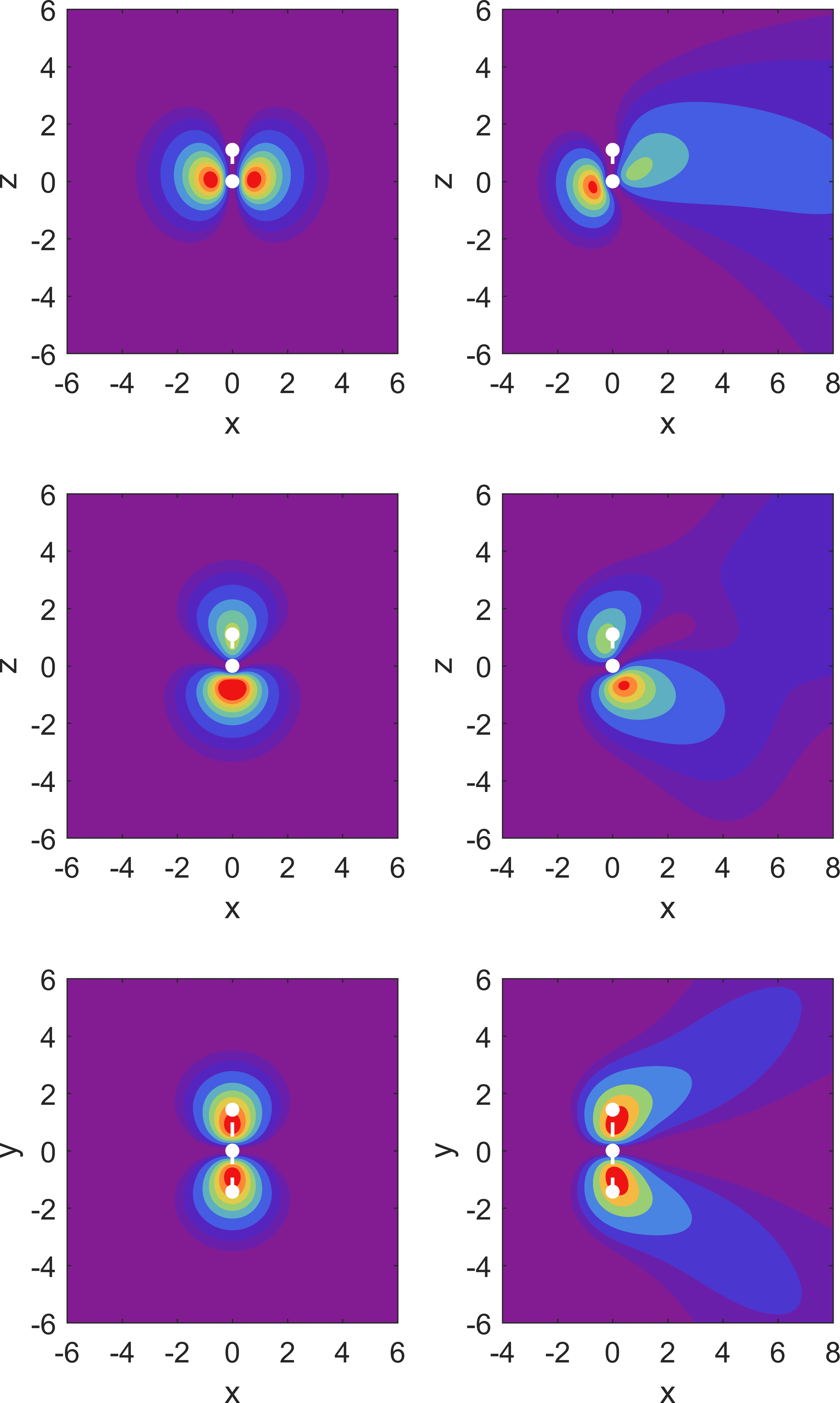
All density plots show some peculiarities. For the force applied in the direction, the MO in Fig. 1 shows a structure in the orbital shape, where the left lobe of the orbital bends down, along with the right lobe of the orbital. In other words, the electron density is not purely directed along the direction of the force. For the MO we observe that it is pushed toward the top right of the box, which implies that the directed force leads to part of the density being pushed in the positive direction. For the MO there is a run-away of the density to both positive and negative along with a movement towards positive .
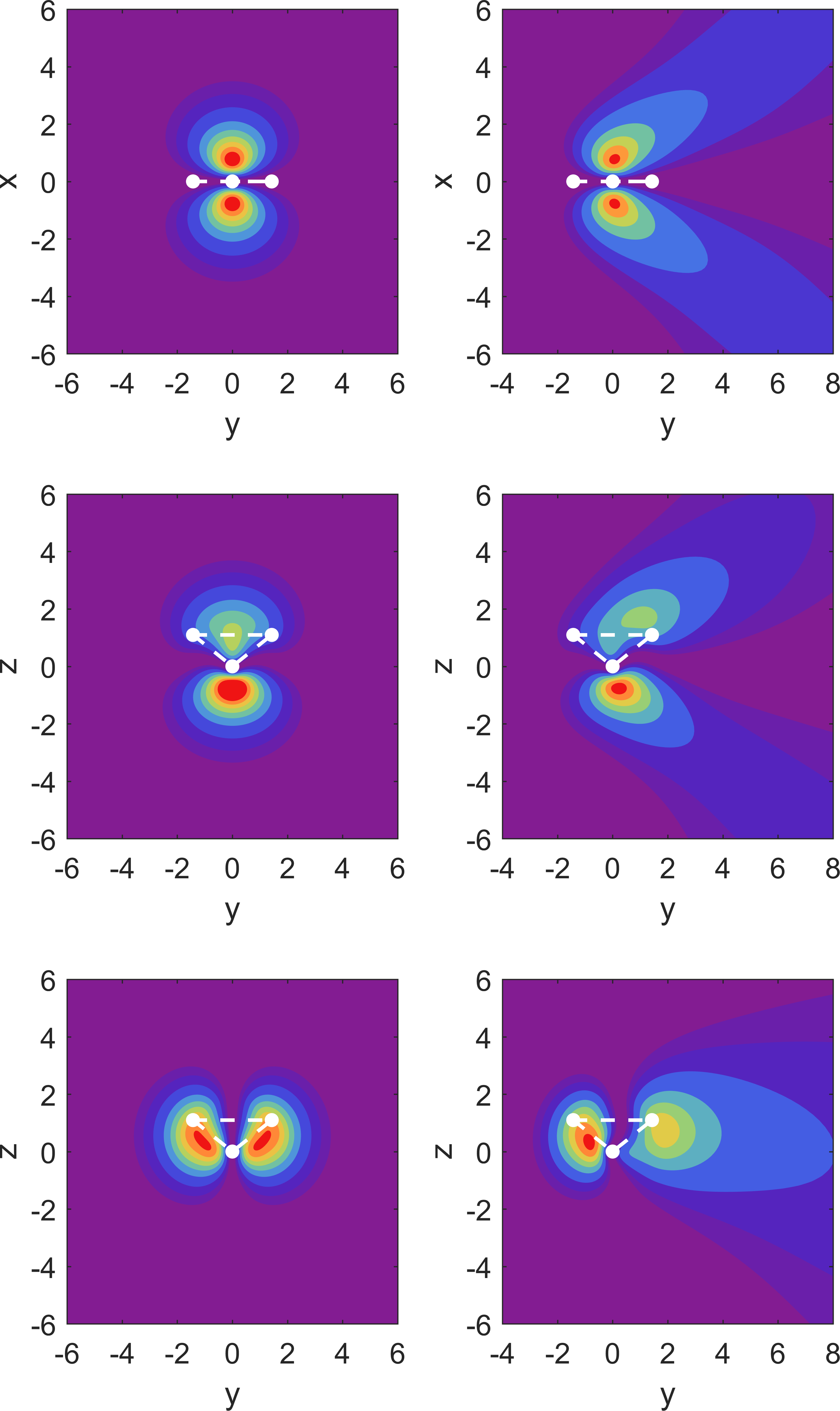
While looking at results for the -directed force in Fig. 2 one notices the continuation of a trend as the MOs and also have a run-away movement of the density away from the direction of the force. Finally, for we also observe a structure in the right lobe downwards, away from the direction of the force. All these deformations of the MO probability densities reflect the combined influence of molecular potential and external dc electric field potential.


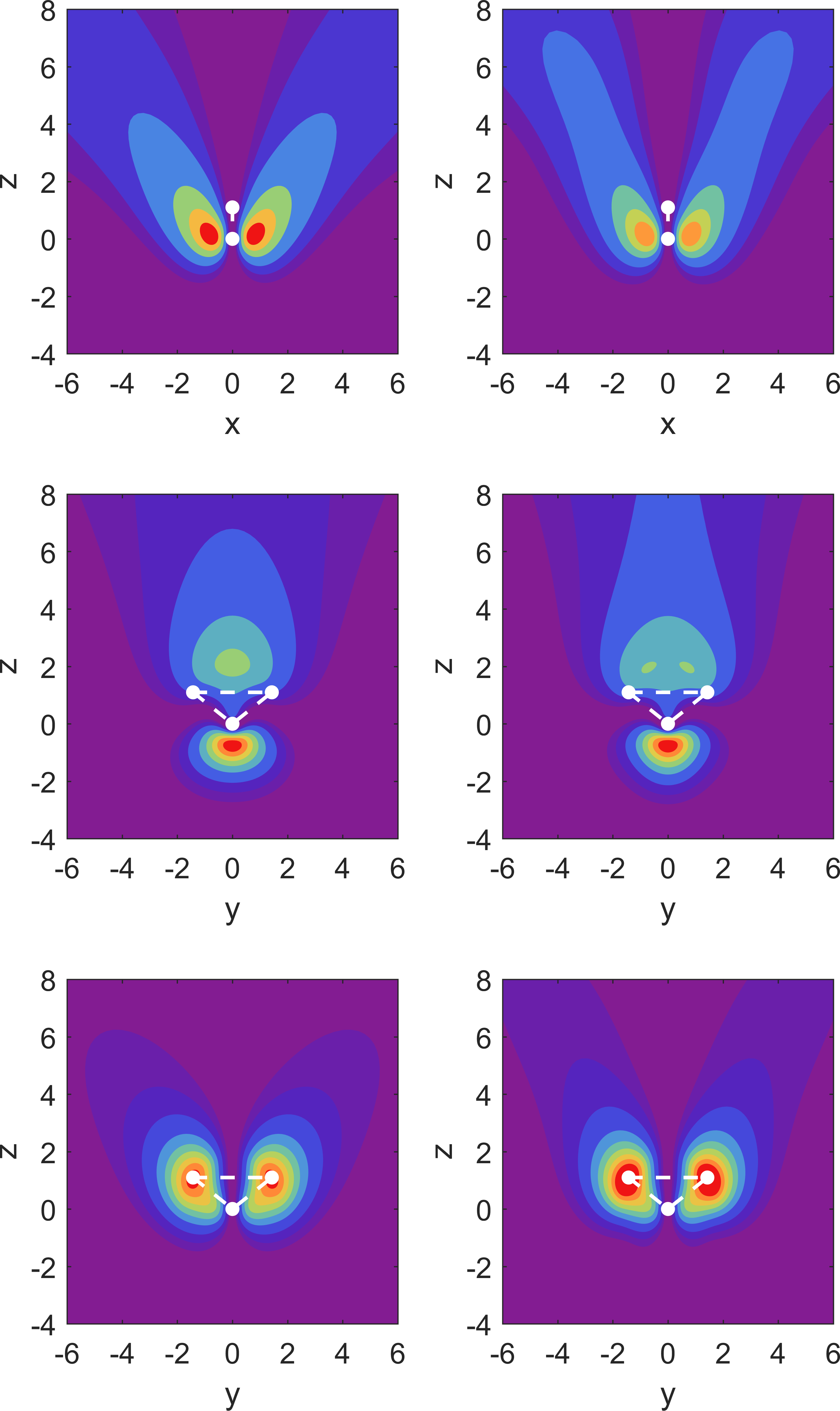
In Fig. 3 we display the MO densities for the case when the force is directed in the direction. Here we carry out a comparison between 3 and 4. When increasing we observe: (i) a narrowing and re-direction of the MO density of towards the direction of the force; (ii) the same occurs for , although there are also two bumps in density which form at the base of the uppermost lobe; and (iii) we find that the MO slightly changes in shape, and the lobes lengthen, in part towards the direction of the force. This is caused by the added flexibility in the partial-wave expansion.
To summarize the visualization of our results, which required added computational effort compared to the resonance parameter calculations, we note that the spatial distribution of ionized electrons even under dc field conditions is far from trivial, and would present a fertile ground for comparison with experiment. We note that in the context of ac laser field ionization R-matrix theory was used recently to predict interesting ionization patterns for the water molecule Benda et al. (2020).
III.2 Dominant field direction for ionization of MOs and their dc Stark shift
We now proceed with a presentation of the resonance parameters for the MOs, and focus on the behavior of the dc Stark shift for the MO with the largest ionization rate for a given field direction. Our main observation in this work is that the orbital with the strongest ionization rate for a given external field direction displays a non-monotonic behavior in the dc Stark shift.

Graphs of the resonance position for fields along the molecular axis as a function of field strength were shown in Ref. Pirkola and Horbatsch (2022) to display such behavior for the orbital for the orientation where electrons are pushed out opposite to the hydrogen atoms. The shift was initially positive, then reached a maximum, became zero at about a.u., in order to continue to negative values for stronger fields.
For the orbital non-monotonic dependence of the the dc shift was observed in both directions with minima occurring at force strength of the order of a.u. and a.u. respectively. The MO is the orbital with the strongest ionization rate in that case, and the minimum in the shift at a.u. corresponds to an almost vanishing dc Stark shift. The bonding MO on the other hand showed monotonic behavior for fields along . The purpose of this section is to show that these features can be generalized to other field directions.
In Fig. 4 the three valence orbitals are shown for the case of an external field perpendicular to the molecular plane. The results are obviously symmetric with respect to reversing the field orientation. The half-widths are showing clearly that the MO oriented with lobes in this direction () is the most easily ionized valence orbital with rates exceeding the other MOs by one or more orders of magnitude before saturation sets in.
While the MO shows non-monotonicity in the dc Stark shift with a minimum at a.u., and a vanishing shift at a.u. the other MOs simply acquire an increasingly negative dc shift.
We can now ask whether this behavior is more than coincidental: is it true that for a field oriented along the direction the bonding orbital might be affected similarly to the MO’s behavior in the case of a field along ?
The answer is provided in Fig. 5 below: Indeed, despite its deep binding energy for substantial field strengths, such as a.u. this orbital is clearly the most easily ionized of the three valence orbitals. Note that this is not the case for weak fields, i.e., in the pure tunneling regime.
The reason for the dominance as far as ionization rate is concerned, geometric considerations are, of course, an important reason: in a simplistic representation of the three orbitals, i.e., , , and it is obvious that they respond strongly to fields aligned with respectively due to the occurrence of substantial dipole matrix elements from the external field.
Interestingly, the non-monotonic behavior in the dc Stark shift for the orbital occurs only for field strength a.u., with a minimum at a.u., and a vanishing shift at a.u.. Thus the overall trend is best comparable to the orbital which displays very similar behaviour when under a force in the direction. The trends of both these orbitals under their respective forces outside of a.u. are very similar to the trend in when the force is in the negative direction.


For the case of fields along the molecular axis which was discussed in Ref. Pirkola and Horbatsch (2022), we report some additional results here, having extended the calculations to the level of . Compared to the field orientations along and we have the complication of asymmetry of the molecule along the molecular axis, and, thus, we separate the presentation for fields aligned such that the force pushes electrons out along this axis (Fig. 6), and in the opposite direction (Fig. 7).
We begin with electrons being pushed out towards the oxygen atom. When one considers field strengths beyond the tunneling regime (where the width turns over towards saturation) the MO emerges as the one with the highest ionization rate. Due to the asymmetry the dc Stark shift has a non-zero slope at zero field. The resonance position decreases with increasing field and reaches a minimum at a.u.. The higher-convergence results run parallel to those for for the dc shift. For the resonance width the two results are not well distinguishable on a logarithmic scale except for the weakest field strengths shown.
Concerning the convergence with we can state that the weakest bound MO () is showing the smallest discrepancy, while the bonding orbital () is affected most, because the partial-wave expansion is not yet sensitive to the full potential from the hydrogenic parts. We observe, however, that the convergence (or lack thereof) does not have a strong impact on the features reported in this work, i.e., the non-monotonicity of the shifts.
For fields in the opposite direction, i.e., ionization into the half-space on the hydrogenic side, we again notice that the dominant ionization contribution is from the MO , and that this MO displays complicated non-monotonic behavior in the dc Stark shift.
To summarize this section we observe that the conclusions are consistent for the four possible field orientations (the direction has two possible orientations due to asymmetry of the molecule) associated with symmetry axes of the orbitals. To phrase it simply: the non-monotonic behavior of the dc Stark shift goes hand in hand with the relatively strong ionization rate for the orbital of that particular symmetry.
While our results for the widths of the orbital show an asymmetry when changing the force direction from positive to negative, this behavior is different from what was observed for the net molecular width in HF theory Jagau (2018), as shown in Fig. 7 of Ref. Pirkola and Horbatsch (2022).

Our own net widths are dominated by the orbital, and so a natural question to ask is whether there is a convergence issue in our partial-wave approach. The results do not deviate, however, substantially from those for . Thus, one will have to investigate further whether this difference in behavior is related to the determination of the net decay width, or whether it is the model potential approach that fails to account for self-consistent field effects in the presence of the external field.
III.3 Comparison with HF and CCSD(T) calculations for net ionization

In Fig. 8 we compare with the HF and CCSD(T) total molecular Stark shifts and resonance widths given in Ref. Jagau (2018). As explained in Ref. Pirkola and Horbatsch (2022) for our model calculation a meaningful comparison is to consider the direct sum of orbital energies. Such an analysis corresponds to total, (or net) ionization from the molecule, and the five MOs are counted twice to account for the spin degeneracy.
The most direct comparison for the present results should be with the mean-field single-particle approach, i.e., the HF method, as our model potential is designed to match HF orbital energies. The comparison of the dc Stark shifts for field directions along and shows that the present model calculation yields stronger shifts as the field strength increases, but that the overall trend agrees with the HF results of Ref. Jagau (2018).
We observe a similar trend in the decay widths: they agree at the factor-of-two level for the cases involving fields along and . This is an improvement compared to the results for forces along the molecular axis in the positive direction, while comparable to the case when the force is in the negative direction Pirkola and Horbatsch (2022). We note that the additional corrections due to electronic correlation, i.e., the CCSD(T) over the HF results is also on a similar scale, i.e., a discrepancy at the level of factors of two-three when the ionization rate is strong.
The blue mark the DS (2) values, i.e., the direct sum method, in which we calculate the total molecular Stark shift and width by adding the resonance parameters for every MO (as reported in Sect. III.2) assuming double occupancy due to spin degeneracy Ref. Pirkola and Horbatsch (2022). The agreement between DS (2) and the HF results of Ref. Jagau (2018) is generally good for the Stark shifts but less so for the widths. At higher field strengths, the agreement weakens, and more so for the force in the direction. For the widths, DS (2) in begins with improving in agreement with HF in relative to the the force strength. We note that the correlated CCSD(T) calculations of Ref. Jagau (2018) generally do not deviate much from the HF data, and that the present model calculations in some cases, perhaps fortuitously, agree with them.
The Stark shifts for the -directed force, agree very well with the CCSD(T) results of Ref. Jagau (2018) with single-digit percentage deviations. It would be of interest to compare the MO resonance parameters from HF calculations with the present model potential results in order to complement the comparison of net quantities which follow from the total energy. For the purposes of comparison with experiment, and understanding the theory more clearly, more work must be done to bridge the gap between the multi-electron solutions, HF and CCSD(T), and the present single-electron, local potential approach. For future work it is planned to extend the current work to a potential model from density functional theory, such as the local HF potential method Della Sala and Görling (2002); Sala (2007).
IV Conclusions
In this work we have extended our previous model calculations for dc field ionization of the water molecule Pirkola and Horbatsch (2022) mostly in two respects:
(i) we have included two orientations for the external field to complement the previous work which was restricted to fields along the molecular axis;
(ii) for fields oriented with the molecular axis we have validated the conclusions based on the limited calculations in the angular momentum basis by comparing results for with those for .
Extension (i) allowed us to gain some understanding concerning the non-monotonic behaviour of the dc Stark shift, as being associated with the MO that is most easily ionized by a given field orientation. Extension (ii), while not a complete convergence analysis, nevertheless provides strong evidence that the major conclusions concerning dc Stark shifts and resonance widths will not be overturned by the inclusion of more partial waves. These higher- contributions are likely to play a significant role when one analyzes the spatial emission properties of the ionized electrons.
It would be of great interest to explore in experiments with infrared laser fields and oriented molecules the predictions made for the non-monotonic dc Stark shifts. Experiments with oriented nitrogen and carbon dioxide molecules have been performed Mairesse et al. (2008), and water vapor does represent a challenge. Ionization from particular MOs would require some method of vacancy detection, so this is definitely a challenge compared to ionization from particular MOs by electron Song et al. (2021) or X ray Benda et al. (2020) impact where one has some control through the incident particle energy, or even the secondary electron energy in an (e,2e) process Nixon et al. (2021).
Acknowledgements.
Discussions with Tom Kirchner and Michael Haslam are gratefully acknowledged. We would also like to thank Steven Chen for support with the high performance computing server used for our calculations. Financial support from the Natural Sciences and Engineering Research Council of Canada is gratefully acknowledged.References
- Pirkola and Horbatsch (2022) P. Pirkola and M. Horbatsch, Phys. Rev. A 105, 032814 (2022).
- Laso and Horbatsch (2017) S. A. Laso and M. Horbatsch, Journal of Physics B: Atomic, Molecular and Optical Physics 50, 225001 (2017).
- Jagau (2018) T.-C. Jagau, The Journal of Chemical Physics 148, 204102 (2018), https://doi.org/10.1063/1.5028179 .
- Heßelmann et al. (2007) A. Heßelmann, A. W. Götz, F. Della Sala, and A. Görling, The Journal of Chemical Physics 127, 054102 (2007), https://doi.org/10.1063/1.2751159 .
- Nixon et al. (2021) K. L. Nixon, C. Kaiser, and A. J. Murray, “Ionization of water using the (e, 2e) technique from the homo state and from the n-homo state,” http://es1.ph.man.ac.uk/AJM2/Water.html (2021), accessed: 2021-12-08.
- Milne-Brownlie et al. (2004) D. S. Milne-Brownlie, S. J. Cavanagh, B. Lohmann, C. Champion, P. A. Hervieux, and J. Hanssen, Phys. Rev. A 69, 032701 (2004).
- Jahnke et al. (2021) T. Jahnke, R. Guillemin, L. Inhester, S.-K. Son, G. Kastirke, M. Ilchen, J. Rist, D. Trabert, N. Melzer, N. Anders, T. Mazza, R. Boll, A. De Fanis, V. Music, T. Weber, M. Weller, S. Eckart, K. Fehre, S. Grundmann, A. Hartung, M. Hofmann, C. Janke, M. Kircher, G. Nalin, A. Pier, J. Siebert, N. Strenger, I. Vela-Perez, T. M. Baumann, P. Grychtol, J. Montano, Y. Ovcharenko, N. Rennhack, D. E. Rivas, R. Wagner, P. Ziolkowski, P. Schmidt, T. Marchenko, O. Travnikova, L. Journel, I. Ismail, E. Kukk, J. Niskanen, F. Trinter, C. Vozzi, M. Devetta, S. Stagira, M. Gisselbrecht, A. L. Jäger, X. Li, Y. Malakar, M. Martins, R. Feifel, L. P. H. Schmidt, A. Czasch, G. Sansone, D. Rolles, A. Rudenko, R. Moshammer, R. Dörner, M. Meyer, T. Pfeifer, M. S. Schöffler, R. Santra, M. Simon, and M. N. Piancastelli, Phys. Rev. X 11, 041044 (2021).
- Mairesse et al. (2008) Y. Mairesse, N. Dudovich, J. Levesque, M. Y. Ivanov, P. B. Corkum, and D. M. Villeneuve, New Journal of Physics 10, 025015 (2008).
- Moccia (1964) R. Moccia, The Journal of Chemical Physics 40, 2186 (1964), https://doi.org/10.1063/1.1725491 .
- Errea et al. (2015) L. Errea, C. Illescas, L. Méndez, I. Rabadán, and J. Suárez, Chemical Physics 462, 17 (2015), inelastic Processes in Atomic, Molecular and Chemical Physics.
- Lüdde et al. (2020) H. J. Lüdde, A. Jorge, M. Horbatsch, and T. Kirchner, Atoms 8 (2020), 10.3390/atoms8030059.
- Illescas et al. (2011) C. Illescas, L. F. Errea, L. Méndez, B. Pons, I. Rabadán, and A. Riera, Phys. Rev. A 83, 052704 (2011).
- Jorge et al. (2019) A. Jorge, M. Horbatsch, C. Illescas, and T. Kirchner, Phys. Rev. A 99, 062701 (2019).
- Jorge et al. (2020) A. Jorge, M. Horbatsch, and T. Kirchner, Phys. Rev. A 102, 012808 (2020).
- Scrinzi and Elander (1993) A. Scrinzi and N. Elander, The Journal of Chemical Physics 98, 3866 (1993).
- Scrinzi (2010) A. Scrinzi, Phys. Rev. A 81, 053845 (2010).
- Benda et al. (2020) J. Benda, J. D. Gorfinkiel, Z. Mašín, G. S. J. Armstrong, A. C. Brown, D. D. A. Clarke, H. W. van der Hart, and J. Wragg, Phys. Rev. A 102, 052826 (2020).
- Della Sala and Görling (2002) F. Della Sala and A. Görling, The Journal of Chemical Physics 116, 5374 (2002), https://doi.org/10.1063/1.1453958 .
- Sala (2007) F. D. Sala, Theoretical Chemistry Accounts 117, 981 (2007).
- Song et al. (2021) M.-Y. Song, H. Cho, G. P. Karwasz, V. Kokoouline, Y. Nakamura, J. Tennyson, A. Faure, N. J. Mason, and Y. Itikawa, Journal of Physical and Chemical Reference Data 50, 023103 (2021), https://doi.org/10.1063/5.0035315 .