2022
These authors contributed equally to this work. \equalcontThese authors contributed equally to this work. [2,3]\fnmJoseba \surAlonso
1]\orgnameTesoro Imaging S.L., \orgaddress\cityValencia, \postcode46022, \countrySpain 2]\orgdivInstitute for Molecular Imaging and Instrumentation, \orgnameSpanish National Research Council, \orgaddress\cityValencia, \postcode46022, \countrySpain 3]\orgdivInstitute for Molecular Imaging and Instrumentation, \orgnameUniversitat Politècnica de València, \orgaddress\cityValencia, \postcode46022, \countrySpain 4]\orgnamePhysioMRI Tech S.L., \orgaddress\cityValencia, \postcode46022, \countrySpain 5]\orgnameAsociación de investigación MPC, \orgaddress\citySan Sebastián, \postcode20018, \countrySpain 6]\orgdivDepartment of Electronic Engineering, \orgnameUniversitat de València, \orgaddress\cityBurjassot, \postcode46100, \countrySpain 7]\orgnameHelios School, \orgaddress\cityLa Eliana, \postcode46183, \countrySpain 8]\orgnameOxford Ionics, \orgaddress\cityOxford, \postcodeOX5 1PF, \countryUnited Kingdom 9]\orgdivMedical Imaging Department, \orgnameHospital Universitari i Politècnic La Fe, \orgaddress\cityValencia, \postcode46026, \countrySpain
Portable magnetic resonance imaging of patients indoors, outdoors and at home
Abstract
Mobile medical imaging devices are invaluable for clinical diagnostic purposes both in and outside healthcare institutions1, 2, 3, 4. Among the various imaging modalities, only a few are readily portable. Magnetic resonance imaging (MRI), the gold standard for numerous healthcare conditions5, 6, 7, 8, 9, 10, does not traditionally belong to this group. Recently, low-field MRI start-up companies have demonstrated the first decisive steps towards portability within medical facilities11, 12, 13, but these are so far incompatible with more demanding use cases such as in remote and developing regions, sports facilities and events, medical and military camps, or home healthcare. Here we present in vivo images taken with a light, home-made, low-field extremity MRI scanner outside the controlled environment provided by medical facilities. To demonstrate the true portability of the system and benchmark its performance in various relevant scenarios, we have acquired images of a volunteer’s knee in: i) an MRI physics laboratory; ii) an office room; iii) outside a campus building, connected to a nearby power outlet; iv) in open air, powered from a small fuel-based generator; and v) at the volunteer’s home. All images have been acquired within clinically viable times, and signal-to-noise ratios (SNR) and tissue contrast suffice for 2D and 3D reconstructions with diagnostic value, with comparable overall image quality across all five situations. Furthermore, the volunteer carries a fixation metallic implant screwed to the femur, which leads to strong artifacts in standard clinical systems14, 15, 16 but appears sharp in our low-field acquisitions. Altogether, this work opens a path towards highly accessible MRI under circumstances previously unrealistic.
keywords:
mobile medical imaging, home healthcare, point of care imaging, outdoors imaging, portable MRI, low field MRIThe average adult human body contains over four thousand trillion trillion hydrogen nuclei, each a minuscule quantum compass needle susceptive to the presence of external magnetic fields. Standard clinical MRI scanners make use of powerful superconducting magnets that interact strongly with these protons and enable the high SNR and image contrast and resolution typical for magnetic resonance images17. Regrettably, these magnets also require cryogenic refrigeration, they are bulky, heavy, environmentally unfriendly, expensive to build, site, operate and maintain, and they ultimately constitute a formidable barrier to the accessibility and democratization of MRI18, 19, 20. Besides, high-field scanners are constrained by patient safety considerations, due to increased specific absorption rates (SAR) of electromagnetic energy in tissues at the corresponding higher excitation radio-frequencies (RF)21; they generate undesirable acoustic noise due to strong magnetic interactions during imaging sequences22; and they induce severe image artifacts around metallic implants due to magnetic susceptibility effects14, 15, 16. Low-field systems ( T) can overcome all of the above and are nowadays gaining traction as affordable complements to standard MRI scanners. Recent achievements with low-field scanners include in vivo brain and extremity imaging23, 24, hard-tissue imaging25, 26, 27 and even quantitative MRI and fingerprinting28, 29. The main penalty to pay for operating in this regime is a significant loss in SNR and spatial resolution. However, the diagnostic value of the resulting reconstructions is not necessarily compromised, due to a number of reasons: i) contrast-to-noise ratio (CNR), a more relevant metric for diagnosis than SNR, does not depend as strongly on field strength30; ii) multiple health conditions and diseases may be diagnosed without the exquisite detail provided by high-field images18; iii) SAR effects can be safely disregarded at low fields, allowing for efficient pulse sequences which increase the duty cycle to partly compensate the SNR loss18; and iv) machine learning and deep convolutional neural networks can be trained to reconstruct high quality images from low-field acquisitions by e.g. transfer learning31, 32.
The scope of conceivable applications for MRI technologies widens extraordinarily once the need for large superconducting magnets is removed. For instance, a vehicle has been equipped with a 0.2 T system large enough for a human arm33, and point-of-care and bedside neuroimaging have been demonstrated with a 64 mT FDA-cleared scanner portable within a clinical facility11, 12. Low-cost devices with improved mobility would open the door to expanding MRI applications to home and hospice care, small clinics, rural areas or sports clubs and school facilities. Autonomously powered scanners could even be operated outdoors, e.g. in sports events, campaign hospitals, NGO and military camps34 or medicalized vehicles33, making MRI available to a large fraction of the world population with no or insufficient access18, 19, 20.
In this article we present a 70 mT extremity MRI scanner mounted on a wheeled structure, with an overall weight kg and component cost k€. After checking that the system performs as expected for in vivo images under controlled ambient conditions in an MRI physics laboratory, we took images of the right knee of a volunteer in different indoor and outdoor environments, including the living room at the volunteer’s apartment, and in open air connected to a portable gasoline generator. All knee images were acquired with identical 3-dimensional turbo spin echo (3D-TSE) sequences, in about 12 min each. The electromagnetic interference (EMI) spectrum was different at the various locations, which results in slightly different noise patterns in the reconstructed images. Nevertheless, they all yield valuable anatomical information in clinically acceptable times. The volunteer had undergone a femoral shaft osteotomy and carried a fixation metallic implant screwed to the femur. This hardware is sharply defined in our low-field acquisitions, where previous high-field images suffered from strong susceptibility-induced image distortions.
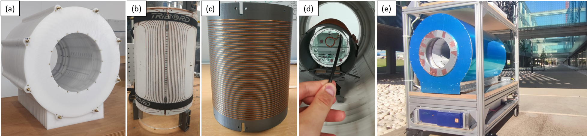
The system (Fig. 1) is built around a permanent magnet array in a Halbach configuration 24, for a field strength of mT homogeneous down to ppm (parts-per-million) over a diameter of spherical volume (DSV) of 20 cm. The gradient coil assembly can generate roughly linear fields of up to 40 mT/m in the DSV. All the below images are taken with a single RF coil used for both transmission and reception (Tx/Rx), tuned and impedance-matched to MHz, the proton resonance (Larmor) frequency at our field strength. The control electronics are based on MaRCoS, an open-source, high-performance Magnetic Resonance Control System35, 36. The diameter and length of the scanner are around 53 and 51 cm respectively, excluding electronics and the mobile structure, with a bore opening cm and a weight of kg. Once on the mobile, open structure and equipped with all the required electronics and the control computer, the overall system dimensions are cm3, the weight is kg, and the power consumption is kW. Further details on the scanner can be found in the Methods.
The scanner is usually in the controlled environment of an MRI physics laboratory, where the temperature is stabilized at C and the air relative humidity at %. In these conditions, the Larmor frequency is stable down to the kilo-hertz at 3.076 MHz over weeks, and the different couplings of the subjects to the RF circuitry can be compensated for with minor corrections to the impedance matching electronics. The abundant surrounding electronic equipment and scanners generate substantial EMI in the laboratory at frequencies within our detection bandwidth. For this reason, we conceal the resonant RF coil behind three grounded shields: one is the outermost scanner housing (blue in Fig. 1(e)); another is inside the magnet, between the RF coil and the gradient assembly; and last is an electrically conducting cloth that can be wrapped around the subject at both scanner ends, to avoid antenna effects that otherwise couple EMI to the coil from the inside, despite the other two shields.
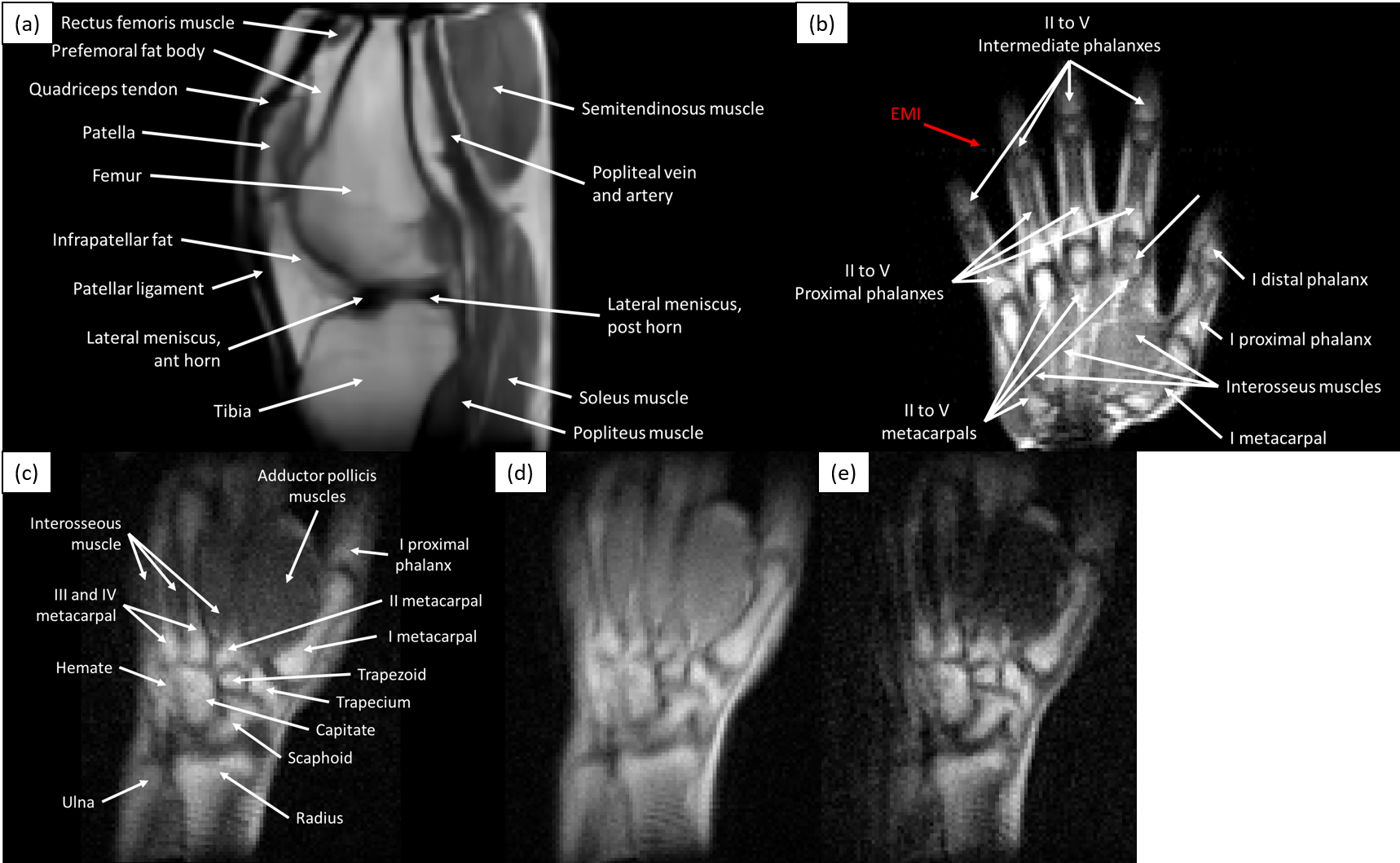
The images in Fig. 2 show the scanner performance in the laboratory. They correspond to in vivo 3D-TSE acquisitions of different healthy subjects on different days, showing selected slices of a left knee, a right hand and a right wrist. Acquisition times ranged from 10 to 19 min (Methods). All images show sufficient tissue contrast and spatial resolution to identify relevant anatomical features, including muscles, fat, cortical bone, bone marrow, tendons, ligaments, veins, arteries and fascia. In these images we show different contrast mechanisms17, with weightings on , and proton density (). The knee image in Fig. 2(a) is BM4D-filtered37 and rescaled to increase the number of pixels by a factor of , whereas the hand in Fig. 2(b) is unprocessed after Fourier reconstruction and weak EMI effects result in a faint line along the horizontal (phase-encoded) direction.
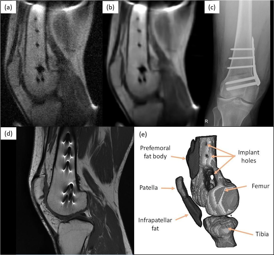
In a second set of experiments, we demonstrate in vivo MR images in the presence of metallic implants without the strong susceptibility-induced artifacts typical of high-field acquisitions14, 16, which often hamper post-operative assessment of orthopedic procedures15. The volunteer for these tests had been diagnosed with lateral gonarthrosis due to cartilage damage in their right knee and had a femoral shaft osteotomy to remove pressure from the damaged tissue. The fixation metallic implant screwed to the femur is cleanly visible in a lateral X-ray computed radiographic image (Fig. 3(c)), but leads to high intensity fringes around the metallic hardware in high-field MR images due to incorrect spin mapping (see Fig. 3(d), taken at 3 T). These effects depend supralinearly on the magnetic field strength and are barely perceptible38 at fields T. The field dependence is notorious in the images: the SNR and resolution are much higher in the 3 T system, but the metallic implant geometry is accurately defined in our 70 mT 2D and 3D reconstructions, and can be readily segmented with standard data post-processing. The low-field images were taken in 12 min (Fig. 3(a) and (b)) and 20 min (Fig. 3(e)) with -weighted 3D-TSE acquisitions (Methods).
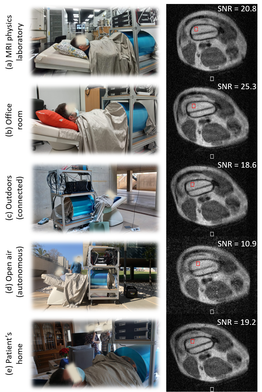
The goal of the last set of experiments is to evaluate the portability of the scanner and its performance under various environments and conditions. To establish comparisons as unbiased as possible, the acquisitions across all five scenarios are of the intervened knee of the same volunteer as in Fig. 3, and all with the same sequence parameters: -weighted 3D-TSE with a total scan time min (Methods). The slices in Fig. 4 have been selected to show the third screw from the top (Fig. 3) as it runs through the bone from the top of the image, where the prosthetic plate is implanted. As a general indicator of the image quality, we measure the SNR in a region of interest (ROI) in the femur bone marrow (red boxes in the unprocessed reconstructions in Fig. 4). To this end, we estimate the signal strength as the average voxel brightness in the ROI, and the noise as the average voxel brightness in the background (white boxes). Prior to each acquisition, we measured the spectral noise density as picked up by the detection RF coil with the subject inside the scanner. The average signal strength in these spectra speaks of the white noise amplitude in the Rx chain, which is ideally close to thermal (Johnson) noise in the coil (Methods). Besides, we often encounter stronger peaks, indicating EMI at discrete frequencies. These can be suppressed by covering the subject meticulously with the shielding cloth.
The first acquisition (Fig. 4(a)) took place in the same laboratory as above and serves as reference under controlled ambient conditions. For the acquisition in the MRI physics laboratory, the Larmor frequency was MHz, the measured noise level (50 nV/Hz1/2) was compatible with Johnson noise (Methods), there is no visible EMI and the femur SNR is .
The second scan took place in an office room (Fig. 4(b)) around 20 m away from the laboratory, in the same building and floor. The Larmor frequency here decreased to MHz due to a higher temperature. The noise amplitude is still consistent with Johnson noise levels and EMI is not visible in the reconstruction. The SNR in the marrow ROI is and the overall image quality is comparable to the reference image, perhaps even slightly sharper.
The third image was acquired outdoors, at basement level, just outside the laboratory building (Fig. 4(c)). The system was powered through a 30 m cable running down three floors from the laboratory. The conducting cloth wrapped around the subject purposely connected the scanner shielding to the concrete floor to improve the otherwise resistive connection between the laboratory ground and earth. During this acquisition the volunteer reported sensing the presence and conversations of bypassers, a light breeze on the grounded cloth, and weak tremors due to vehicles driving through the underground parking. The resulting image quality seems not to be strongly influenced by any of these, with an SNR of in the ROI, and a noise spectrum of comparable amplitude to indoor acquisitions. The Larmor frequency was MHz.
The fourth scan was also taken outdoors, in this case in open air in a university campus (Fig. 4(d), Larmor frequency MHz), far from power outlets and operating autonomously with a portable electricity generator. The latter is based on a low-consumption gasoline engine, weighs kg, costs € and has an autonomy hours with the scanner at continuous operation (Methods). We grounded the system electrically as before, with the conducting cloth offering low-resistance paths between the scanner shielding, the floor concrete and the ground terminal in the generator. The spectrum was significantly more populated in this case, presumably due to noise originating at the motor. Consequently, the quality of the resulting image is somewhat lower than in the previous acquisitions (), and an EMI line is visible along the vertical (phase-encoding) direction. Nevertheless, the main anatomic features, different tissues and metallic implants are all still clearly identifiable.
The last image was acquired at the volunteer’s apartment. This is located in a low-density town in the province of Valencia, Spain. The system was transported in a small truck from the university campus to a parking lot approximately 300 m away from the entrance to the apartment block, and pushed along the sidewalk into the building, the elevator, the apartment and ultimately, the living room. Throughout the way, the only wheelchair-adapted elements were the lowered-sidewalks at pedestrian crossings. After transport, the system required re-connecting some of the RF electronics modules we had packed in a separate box, and tightening some screwed connectors that had become loose during transport through the rugged, tiled sidewalks. Other than that, the system was plugged to a wall power outlet, tuned to MHz and ready to use. The noise spectrum in the apartment was clean, again compatible with Johnson noise levels. The SNR in the ROI for this acquisition is .
In conclusion, we have demonstrated the viability of a portable, low-cost system for magnetic resonance imaging indoors, outdoors and at home. In this work, we have focused on healthy volunteers and subjects carrying metallic implants. Nevertheless, the acquired images contain sufficient anatomical information to diagnose a large variety of articular diseases, including effusion, synovial engorgement, tendon disruption or bone fractures. Upon completion of this manuscript, we have learned about an independent effort towards portable MRI with a commercial 64 mT scanner mounted on a modified cargo van39.
Looking ahead, our 70 mT scanner can be still upgraded in various ways. Machine learning algorithms have been shown to boost the performance in other low-field systems and can be readily incorporated to ours. These can be used, via transfer learning, to increase the spatial resolution of scans a posteriori based on multiple acquisitions, prior knowledge about the sample40, or with networks trained with paired datasets of low and high-field images, to recover from the former features visible otherwise only with the latter32, 41. Deep learning and convolutional neural networks can also be employed to increase reconstruction quality through image denoising, artifact detection and active noise cancellation31, 42, 43. Quantitative MRI, radiomics and fingerprinting28, 29, 44 show promising potential in situations where subtle texture changes contain potentially valuable information for the patients. Also special-purpose pulse sequences and reconstruction methods can enhance the efficiency of low-field MRI29, 25, and hardware developments and contrast agents which are mainstays in clinical high-field MRI (e.g. parallel imaging, optimization of RF detection coils for different body parts, gadolinium contrast enhancement), are seldom used in the still mostly experimental low-field systems available18, 45. Finally, for our particular scanner, the GUI and overall system usability can be improved to facilitate operation by non-experts.
All in all, the scanner performance demonstrated in this work, especially if upgraded with the above capabilities, sets a path towards accessible MRI, democratizing its use and benefits, and qualitatively expanding the circumstances where it can provide value.
Acknowledgments
We thank the anonymous volunteers for their participation, Andrew Webb and Thomas O’Reilly for discussions on MRI hardware, and Benjamin Menküc for contributions to MaRCoS.
Funding
This work was supported by the Ministerio de Ciencia e Innovación of Spain through research grant PID2019-111436RB-C21. Action co-financed by the European Union through the Programa Operativo del Fondo Europeo de Desarrollo Regional (FEDER) of the Comunitat Valenciana 2014-2020 (IDIFEDER/2018/022 and IDIFEDER/2021/004) and Future and Emerging Technologies (FET, grant 101034644). JMG and JB acknowledge support from the Innodocto program of the Agencia Valenciana de la Innovación (INNTA3/2020/22 and INNTA3/2021/17).
Conflict of interest
PhysioMRI Tech S.L. is a for-profit organization spun off the Institute for Molecular Imaging and Instrumentation and proprietor of the low-field scanner presented in this work. JMA, FG, JB, JMB and JA have patents pending that are licensed to PhysioMRI Tech S.L. JMA, FG, AR, JMB and JA are co-founders of PhysioMRI Tech S.L. All other authors declare no competing interests.
Ethics approval
All experiments were carried out following Spanish regulations and under the research agreement from La Fe Hospital in Valencia (IIS-F-PG-22-02, agreement number 2019-139-1).
Consent to participate
Informed consent was obtained from all volunteers prior to study commencement.
Consent for publication
Informed consent for publication was obtained from all participants prior to study commencement.
Availability of data and materials
All anonymized datasets, reconstruction and postprocessing methods generated and/or used during the present study are available from the corresponding author upon reasonable request.
Code availability
MaRCoS and the GUI are publicly available from open-source repositories at https://github.com/vnegnev/marcos_server and https://github.com/yvives/PhysioMRI_GUI, respectively.
Authors’ contributions
Low-field images taken by TGN and JMA with help from RB, PM, FJL, JPR and JA. Portable system built by RPG, TGN, JMA, FG, RB, EP, JMG and JA with help from JPR, PM and FJL. Data analysis and evaluation by JMA, TGN, FG, LMB and JA. Control electronics and software developed by VN, YVG, JMA, TGN and JB. Portability experiments conceived by JA, JMA, TGN and AMP. Project conceived and supervised by AR, JMB and JA. Paper written by JA and LMB, with input from all authors.
References
- \bibcommenthead
- 1 B.P. Nelson, E.R. Melnick, J. Li, Portable Ultrasound for Remote Environments, Part I: Feasibility of Field Deployment. The Journal of Emergency Medicine 40(2), 190–197 (2011). 10.1016/J.JEMERMED.2009.09.006
- 2 P. Pittayapat, C. Oliveira-Santos, P. Thevissen, K. Michielsen, N. Bergans, G. Willems, D. Debruyckere, R. Jacobs, Image quality assessment and medical physics evaluation of different portable dental X-ray units. Forensic Science International 201(1-3), 112–117 (2010). 10.1016/J.FORSCIINT.2010.04.041
- 3 N. Kosaka, M. Ogawa, P.L. Choyke, H. Kobayashi, Clinical implications of near-infrared fluorescence imaging in cancer. Future Oncology 5(9), 1501–1511 (2009). 10.2217/FON.09.109
- 4 A.S. Zubair, A. Crawford, A.M. Prabhat, K.N. Sheth, Use of Portable Imaging Modalities in Patients With Neurologic Disorders: A Case-Based Discussion. Cureus 13(6) (2021). 10.7759/CUREUS.15841
- 5 N.B. Smith, A. Webb, Introduction to Medical Imaging (Cambridge University Press, 2010). 10.1017/cbo9780511760976
- 6 N. Gordillo, E. Montseny, P. Sobrevilla, State of the art survey on MRI brain tumor segmentation. Magnetic Resonance Imaging 31(8), 1426–1438 (2013). 10.1016/J.MRI.2013.05.002
- 7 C.R. Jack, M.A. Bernstein, N.C. Fox, P. Thompson, G. Alexander, D. Harvey, B. Borowski, P.J. Britson, J.L. Whitwell, C. Ward, A.M. Dale, J.P. Felmlee, J.L. Gunter, D.L. Hill, R. Killiany, N. Schuff, S. Fox-Bosetti, C. Lin, C. Studholme, C.S. DeCarli, G. Krueger, H.A. Ward, G.J. Metzger, K.T. Scott, R. Mallozzi, D. Blezek, J. Levy, J.P. Debbins, A.S. Fleisher, M. Albert, R. Green, G. Bartzokis, G. Glover, J. Mugler, M.W. Weiner, The Alzheimer’s disease neuroimaging initiative (ADNI): MRI methods. Journal of Magnetic Resonance Imaging 27(4), 685–691 (2008). 10.1002/JMRI.21049
- 8 M. Carlsson, H. Arheden, C.B. Higgins, M. Saeed, Magnetic resonance imaging as a potential gold standard for infarct quantification. Journal of Electrocardiology 41(6), 614–620 (2008). 10.1016/J.JELECTROCARD.2008.06.010
- 9 C. Anthoulakis, N. Nikoloudis, Pelvic MRI as the “gold standard” in the subsequent evaluation of ultrasound-indeterminate adnexal lesions: A systematic review. Gynecologic Oncology 132(3), 661–668 (2014). 10.1016/J.YGYNO.2013.10.022
- 10 C. Pawlyn, L. Fowkes, S. Otero, J.R. Jones, K.D. Boyd, F.E. Davies, G.J. Morgan, D.J. Collins, B. Sharma, A. Riddell, M.F. Kaiser, C. Messiou, Whole-body diffusion-weighted MRI: a new gold standard for assessing disease burden in patients with multiple myeloma? Leukemia 2016 30:6 30(6), 1446–1448 (2015). 10.1038/leu.2015.338
- 11 K.N. Sheth, M.H. Mazurek, M.M. Yuen, B.A. Cahn, J.T. Shah, A. Ward, J.A. Kim, E.J. Gilmore, G.J. Falcone, N. Petersen, K.T. Gobeske, F. Kaddouh, D.Y. Hwang, J. Schindler, L. Sansing, C. Matouk, J. Rothberg, G. Sze, J. Siner, M.S. Rosen, S. Spudich, W.T. Kimberly, Assessment of Brain Injury Using Portable, Low-Field Magnetic Resonance Imaging at the Bedside of Critically Ill Patients. JAMA Neurology 78(1), 41–47 (2021). 10.1001/JAMANEUROL.2020.3263
- 12 M.H. Mazurek, M.M. Yuen, B.A. Cahn, M.S. Rosen, K.T. Gobeske, E.J. Gilmore, D. Hwang, F. Kaddouh, J.A. Kim, G. Falcone, N. Petersen, J. Siner, S. Spudich, G. Sze, W.T. Kimberly, K.N. Sheth, Low-Field, Portable Magnetic Resonance Imaging at the Bedside to Assess Brain Injury in Patients with Severe COVID-19 (1349). Neurology 96(15 Supplement) (2021). 10.7759/CUREUS.15841
- 13 J. Nasri, V.G. Wagaskar, S. Parekha, J.D. Adams, D. Kumar, S.S. Venkataraman, A.K. Tewari, Office-Based, Point-of-Care, Low-Field MRI System to Guide Prostate Interventions: Recent Developments. EMJ Urology 9(1), 83–90 (2021)
- 14 K.M. Lüdeke, P. Röschmann, R. Tischler, Susceptibility artefacts in NMR imaging. Magnetic Resonance Imaging 3(4), 329–343 (1985). 10.1016/0730-725X(85)90397-2
- 15 C.A. Harris, L.M. White, Metal Artifact Reduction in Musculoskeletal Magnetic Resonance Imaging. Orthopedic Clinics of North America 37(3), 349–359 (2006). 10.1016/J.OCL.2006.04.001
- 16 P. Stradiotti, A. Curti, G. Castellazzi, A. Zerbi, Metal-related artifacts in instrumented spine. Techniques for reducing artifacts in CT and MRI: State of the art. European Spine Journal 18(SUPPL. 1), 102–108 (2009). 10.1007/S00586-009-0998-5
- 17 E.M. Haacke, R.W. Brown, M.R. Thompson, R. Venkatesan, et al., Magnetic resonance imaging: physical principles and sequence design, vol. 82 (Wiley-liss New York:, 1999)
- 18 J.P. Marques, F.F. Simonis, A.G. Webb, Low-field MRI: An MR physics perspective. Journal of Magnetic Resonance Imaging 49(6), 1528–1542 (2019). 10.1002/jmri.26637
- 19 M. Sarracanie, N. Salameh, Low-Field MRI: How Low Can We Go? A Fresh View on an Old Debate. Frontiers in Physics 8, 172 (2020). 10.3389/fphy.2020.00172
- 20 L.L. Wald, P.C. McDaniel, T. Witzel, J.P. Stockmann, C.Z. Cooley, Low-cost and portable MRI. Journal of Magnetic Resonance Imaging 52(3), 686–696 (2020). 10.1002/JMRI.26942
- 21 L.P. Panych, B. Madore, The physics of MRI safety. Journal of Magnetic Resonance Imaging 47(1), 28–43 (2018). 10.1002/JMRI.25761
- 22 D.L. Price, J.P. De Wilde, A.M. Papadaki, J.S. Curran, R.I. Kitney, Investigation of Acoustic Noise on 15 MRI Scanners from 0.2 T to 3 T (2001). 10.1002/1522-2586
- 23 C.Z. Cooley, P.C. McDaniel, J.P. Stockmann, S.A. Srinivas, S.F. Cauley, M. Śliwiak, C.R. Sappo, C.F. Vaughn, B. Guerin, M.S. Rosen, M.H. Lev, L.L. Wald, A portable scanner for magnetic resonance imaging of the brain. Nature Biomedical Engineering 2020 5:3 5(3), 229–239 (2020). 10.1038/s41551-020-00641-5
- 24 T. O’Reilly, W.M. Teeuwisse, D. Gans, K. Koolstra, A.G. Webb, In vivo 3D brain and extremity MRI at 50 mT using a permanent magnet Halbach array. Magnetic Resonance in Medicine p. mrm.28396 (2020). 10.1002/mrm.28396
- 25 J.M. Algarín, E. Díaz-Caballero, J. Borreguero, F. Galve, D. Grau-Ruiz, J.P. Rigla, R. Bosch, J.M. González, E. Pallás, M. Corberán, C. Gramage, S. Aja-Fernández, A. Ríos, J.M. Benlloch, J. Alonso, Simultaneous imaging of hard and soft biological tissues in a low-field dental MRI scanner. Scientific Reports 10(1), 21,470 (2020). 10.1038/s41598-020-78456-2
- 26 J.M. González, J. Borreguero, E. Pallás, J.P. Rigla, J.M. Algarín, R. Bosch, F. Galve, D. Grau-Ruiz, R. Pellicer, A. Ríos, J.M. Benlloch, J. Alonso, Prepolarized MRI of Hard Tissues and Solid-State Matter. arXiv preprint (2021). arXiv:2110.03417v1
- 27 J. Borreguero, F. Galve, J.M. Algarín, J.M. Benlloch, J. Alonso, Slice-selective Zero Echo Time imaging of ultra-short T2 tissues based on spin-locking. arXiv preprint p. 2201.06305 (2022). arXiv:2201.06305v1
- 28 T. O’Reilly, A.G. Webb, In vivo T1 and T2 relaxation time maps of brain tissue, skeletal muscle, and lipid measured in healthy volunteers at 50 mT. Magnetic Resonance in Medicine 87(2), 884–895 (2021). 10.1002/MRM.29009
- 29 M. Sarracanie, Fast Quantitative Low-Field Magnetic Resonance Imaging With OPTIMUM - Optimized Magnetic Resonance Fingerprinting Using a Stationary Steady-State Cartesian Approach and Accelerated Acquisition Schedules. Investigative Radiology (2021). 10.1097/RLI.0000000000000836
- 30 S. Ghazinoor, J.V. Crues, C. Crowley, Low-field musculoskeletal MRI. Journal of Magnetic Resonance Imaging 25(2), 234–244 (2007). 10.1002/jmri.20854
- 31 N. Koonjoo, B. Zhu, G.C. Bagnall, D. Bhutto, M.S. Rosen, Boosting the signal-to-noise of low-field MRI with deep learning image reconstruction. Scientific Reports 2021 11:1 11(1), 1–16 (2021). 10.1038/s41598-021-87482-7
- 32 A. Garcia Hernandez, P. Fau, S. Rapacchi, J. Wojak, H. Mailleux, M. Benkreira, M. Adel, Improving Image Quality In Low-Field MRI With Deep Learning pp. 260–263 (2021). 10.1109/ICIP42928.2021.9506659
- 33 M. Nakagomi, M. Kajiwara, J. Matsuzaki, K. Tanabe, S. Hoshiai, Y. Okamoto, Y. Terada, Development of a small car-mounted magnetic resonance imaging system for human elbows using a 0.2 T permanent magnet. Journal of Magnetic Resonance 304, 1–6 (2019). 10.1016/j.jmr.2019.04.017
- 34 M. Sarracanie, C.D. LaPierre, N. Salameh, D.E.J. Waddington, T. Witzel, M.S. Rosen, Low-Cost High-Performance MRI. Scientific Reports 5(1), 15,177 (2015). 10.1038/srep15177
- 35 V. Negnevitsky, T. O’Reilly, R. Pellicer-Guridi, Y. Vives-Gilabert, L. Craven-Brightman, D. Schote, J.M. Algarín, M. Prier, J. Stockmann, T. Witzel, B. Menküc, J. Alonso, A. Webb, in Book of Abstracts ESMRMB 2021 38th Annual Scientific Meeting, vol. 34 (Springer, 2021), p. 172. 10.1007/s10334-021-00947-8
- 36 L. Craven-Brightman, T. O’Reilly, B. Menküc, M. Prier, R. Pellicer-Guridi, J. Alonso, L.L. Wald, M. Zaitsev, J. Stockmann, T. Witzel, A. Webb, V. Negnevitsky, in Proceedings of the 2021 ISMRM & SMRT annual meeting and exhibition (ISMRM, 2021), p. Abstract 0748. URL https://cds.ismrm.org/protected/21MPresentations/abstracts/0748.html
- 37 M. Maggioni, V. Katkovnik, K. Egiazarian, A. Foi, Nonlocal transform-domain filter for volumetric data denoising and reconstruction. IEEE Transactions on Image Processing 22(1), 119–133 (2013). 10.1109/TIP.2012.2210725
- 38 C. Van Speybroeck, T. O’Reilly, W. Teeuwisse, P. Arnold, A. Webb, Characterization of displacement forces and image artifacts in the presence of passive medical implants in low-field (<100 mT) permanent magnet-based MRI systems, and comparisons with clinical MRI systems. Physica Medica 84, 116–124 (2021). 10.1016/j.ejmp.2021.04.003
- 39 S.C. Deoni, A.T. Deoni, P. Burton, J. Beauchemin, V. D’Sa, E. Boskamp, B. Samantha, C. McNulty, W. Mileski, B.E. Welch, M. Huentelman, Residential MRI: Development of A Mobile Anywhere-Everywhere MRI Lab. Research Square preprint pp. rs–1121,934 (2021). 10.21203/RS.3.RS-1121934/V1
- 40 E. Van Reeth, I.W.K. Tham, C.H. Tan, C.L. Poh, Super-resolution in magnetic resonance imaging: A review. Concepts in Magnetic Resonance Part A 40A(6), 306–325 (2012). 10.1002/cmr.a.21249
- 41 J.E. Iglesias, R. Schleicher, S. Laguna, B. Billot, P. Schaefer, B. McKaig, J.N. Goldstein, K.N. Sheth, M.S. Rosen, W.T. Kimberly, Accurate super-resolution low-field brain mri. arXiv preprint arXiv:2202.03564 (2022)
- 42 T. Küstner, A. Liebgott, L. Mauch, P. Martirosian, F. Bamberg, K. Nikolaou, B. Yang, F. Schick, S. Gatidis, Automated reference-free detection of motion artifacts in magnetic resonance images. Magnetic Resonance Materials in Physics, Biology and Medicine 31(2), 243–256 (2018). 10.1007/S10334-017-0650-Z/FIGURES/10
- 43 Y. Liu, A.T.L. Leong, Y. Zhao, L. Xiao, H.K.F. Mak, A. Chun, O. Tsang, G.K.K. Lau, G.K.K. Leung, E.X. Wu, X.. Linfang, A low-cost and shielding-free ultra-low-field brain MRI scanner. Nature Communications 2021 12:1 12(1), 1–14 (2021). 10.1038/s41467-021-27317-1
- 44 G. Simpson, B. Spieler, N. Dogan, L. Portelance, E.A. Mellon, D. Kwon, J.C. Ford, F. Yang, Predictive value of 0.35 T magnetic resonance imaging radiomic features in stereotactic ablative body radiotherapy of pancreatic cancer: A pilot study. Medical physics 47(8), 3682–3690 (2020). 10.1002/MP.14200
- 45 D.E. Waddington, T. Boele, R. Maschmeyer, Z. Kuncic, M.S. Rosen, High-sensitivity in vivo contrast for ultra-low field magnetic resonance imaging using superparamagnetic iron oxide nanoparticles. Science Advances 6(29), 998–1015 (2020). 10.1126/sciadv.abb0998
Methods
Scanner
The scanner is based on a Halbach magnet including almost 4,600 NdFeB cubes of side 12 mm to generate mT at the field of view, and another smaller cuboids (64 mm3) to shim the inhomogeneity from down to ppm over a spherical volume of 20 cm in diameter. The gradient coil geometry is optimized with conventional target-field methods for efficiencies between 0.52 and 0.91 mT/m/A. These coils are wound on and glued to curved 3D-printed Nylon molds, and the whole assembly is supported by a methacrylate cylinder. Gradient waveforms are generated with an OCRA1 board connected to the Red Pitaya in the MaRCoS system via Serial Peripheral Interface (SPI), and amplified by AE Techron 7224 power amplifiers (Indiana, USA). The single Tx/Rx RF antenna is a solenoid coil tuned and impedance-matched to the proton Larmor frequency ( MHz). The RF coil holder was 3D-printed in polylactic acid (PLA), and the wire was fixed with cyanoacrylate adhesive. The coil is inside a grounded faraday cage for noise immunity and to prevent interference between the gradients and the RF system, and a conductive cloth covers the subject during in vivo acquisitions. The RF low-noise and power amplifiers, as well as the Tx/Rx switch, were purchased from Barthel HF-Technik GmbH (Aachen, Germany).
Pulse sequences
The knee image in Fig. 2(a) was acquired with a -weighted 3D-TSE sequence, with mm3, a pixel resolution of mm3, , ms, ms, kHz, and 4 averages for a total scan time of 19.2 min. The duration of resonant and -pulses in all images are and , respectively, and dephasing gradient pulses after the RF pulses are pre-emphasized by a factor to place the echoes at the center of the data acquisition windows.
The hand image in Fig. 2(b) was acquired with a -weighted 3D-TSE sequence, with mm3, a pixel resolution of mm3, , ms, ms, kHz, and 13 averages for a total scan time of 10.4 min.
The wrist image in Fig. 2(c) was acquired with a -weighted 3D-TSE sequence, with mm3, a pixel resolution of mm3, , ms, ms, kHz, and 30 averages for a total scan time of 12 min.
The wrist image in Fig. 2(d) was acquired with a -weighted 3D-TSE sequence, with mm3, a pixel resolution of mm3, , ms, ms, kHz, and 5 averages for a total scan time of 12 min.
The wrist image in Fig. 2(e) was acquired with a -weighted 3D-TSE sequence, with mm3, a pixel resolution of mm3, , echo spacing of 20 ms, effective ms, ms, kHz, and 5 averages for a total scan time of 12 min.
The knee images in Fig. 3(a) and (b) were acquired with a -weighted 3D-TSE sequence, with mm3, a pixel resolution of mm3, , ms, ms, kHz, and 9 averages for a total scan time of 12 min.
The knee image in Fig. 3(e) was acquired with a -weighted 3D-TSE sequence, with mm3, a pixel resolution of mm3, , ms, ms, kHz, and 4 averages for a total scan time of 20 min.
The knee images in Fig. 4 were acquired with a -weighted 3D-TSE sequence, with mm3, a pixel resolution of mm3, , ms, ms, kHz, and 9 averages for a total scan time of 12 min.
Data acquisition
The receive chain consists of an analog stage (RF coil, passive Tx/Rx switch, low-noise amplifier and low-pass filter) followed by a digital stage. The digitization is performed at 122.88 Ms/s by an analog-to-digital converter in the Red Pitaya Stemlab board of MaRCoS. The digital signal is mixed down by complex multiplication with a numerically-controlled oscillator set to the Larmor frequency. The real and imaginary data components pass first a cascaded integrator-comb filter and finally a finite impulse response filter. The resulting data conform the sought in-phase and quadrature components of the magnetic resonance signal. These are sent to the control computer and can be Fourier-transformed for image reconstruction and post-processing.
Noise
The spectral noise density of the MR data is bounded from below by Johnson noise due to thermal fluctuations of electrons in the resistive elements in the detector. These are dominated by the coil, with quality factor (88) and in the unloaded (loaded) case. For a given acquisition bandwidth, the integrated noise amplitude is expected to be , with the Boltzmann constant. In the controlled environment of the MRI physics laboratory, we measure nV/Hz1/2 after a 45 dB low-noise pre-amplifier, in agreement with the estimated Johnson level. We use this as a reference to evaluate the signal quality and the shielding efficiency of the conductive cloth, both in the laboratory and in the rest of locations.
We have found situations where suppressing noise down to Johnson levels is not trivial, and indeed did not achieve it when the system was powered by the portable generator. The control computer is another significant source of 50 Hz noise and needs to be as far as possible in the rack to reconstruct clean images. We also find it often necessary to ensure the subject is sufficiently covered by the conductive cloth, and extending some of it on the floor helps.
Generator
For the autonomous experiments outdoors we powered the system from a “Limited 2000i” gasoline-fueled generator from Genergy (Calahorra, Spain). This motor delivers up to 2 kW at 230 V and 50 Hz (single phase). It costs €, weighs 19 kg and has a fuel tank capacity of 4 l and an autonomy of 10.8 hours at 25 % load (500 W), which is more than required for continuous operation of the scanner.