[table]capposition=top
Resolving molecular diffusion and aggregation of antibody proteins with megahertz X-ray free-electron laser pulses
Abstract
X-ray free-electron lasers (XFELs) with megahertz repetition rate can provide novel insights into structural dynamics of biological macromolecule solutions. However, very high dose rates can lead to beam-induced dynamics and structural changes due to radiation damage. Here, we probe the dynamics of dense antibody protein (Ig-PEG) solutions using megahertz X-ray photon correlation spectroscopy (MHz-XPCS) at the European XFEL. By varying the total dose and dose rate, we identify a regime for measuring the motion of proteins in their first coordination shell, quantify XFEL-induced effects such as driven motion, and map out the extent of agglomeration dynamics. The results indicate that for average dose rates below in a time window up to , it is possible to capture the protein dynamics before the onset of beam induced aggregation. We refer to this approach as correlation before aggregation and demonstrate that MHz-XPCS bridges an important spatio-temporal gap in measurement techniques for biological samples.
Introduction
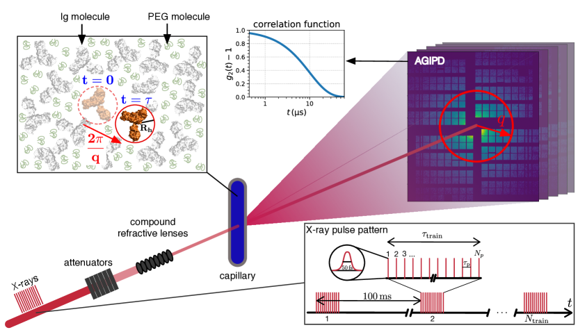
The European X-ray Free-Electron Laser Facility (EuXFEL) is the first X-ray free electron laser (XFEL) generating ultrashort hard X-ray pulses with megahertz repetition rate. Megahertz X-ray photon correlation spectroscopy (MHz-XPCS) [1, 2, 3] makes use of this high repetition rate and the high degree of transverse coherence to measure diffusive dynamics with (sub-) microsecond temporal resolution. In biological systems, typical diffusion coefficients in dense cellular environments range from [4, 5, 6, 7, 8, 9] which requires to resolve time scales from (Fig. 1) in order to trace the complex many-body interactions between proteins and the solvent on molecular length scales. This window of length and time scales is not accessible by optical techniques such as dynamic light scattering, which measures longer length scales (micrometers), or neutron spectroscopy techniques such as neutron spin echo or inelastic neutron scattering, which typically measure on faster time scales of nanoseconds and below. Clearly, experimental techniques are needed to close this gap and measure collective dynamics on microsecond time scales and nanometer length scales. By analyzing fluctuating X-ray speckle patterns, MHz-XPCS is potentially capable of closing this gap, as we demonstrate here, and enables us to gain information on equilibrium and out-of-equilibrium collective dynamics of protein solutions.
Protein dynamics in crowded environments are particularly relevant in the context of intracellular transport in the cytoplasm of eukaryotic cells [10], phase transitions in biomolecular condensates [11, 12, 13], aggregation phenomena [14, 15] and drug production [16]. In highly concentrated environments, the dynamics differ significantly from that of a dilute system, whereas the exact mechanisms that influence the dynamics on different time scales are not yet fully understood [7, 6, 17]. It was found that in vivo dynamics in cells exhibit tremendously reduced diffusion compared to in vitro measurements of diluted proteins in buffer solutions [18, 19, 20, 21, 22, 23, 24, 25]. It is believed that the level of slowing-down depends on the particular system and possibly additional crowding agents [26, 18, 27, 28, 6]. In addition to excluded volume effects [29, 30], there can be contributions from the local water dynamics of the hydration layer [31], quinary interactions of proteins with other cytoplasmic constituents [32, 33, 34, 18, 19, 35], and transient cluster formation [36, 37, 38, 36, 39, 40] that influence intracellular protein diffusion. Also, the dynamics often exhibit anomalous behavior–i.e., non-Brownian and in particular subdiffusive dynamics [26, 41, 42]–and making it difficult to extrapolate the dynamics from the dilute regime. Clearly, new methods are needed to directly probe diffusive dynamics in crowded biological solutions on (sub-) microsecond time scales and nanometer length scales to study these phenomena.
Radiation damage constitutes a major challenge for X-ray scattering experiments with protein solutions. Radiolysis of water and the fast distribution of the free radicals formed rapidly degrade the protein molecules. Hence, a typical upper limit of tolerable absorbed doses is estimated on the order of a few kilo Gray in these experiments with the exact value depending on the chemical composition of the system [43, 44, 45, 46]. Protein aggregation is a signature of beam-induced damage in protein solutions visible via changes in the X-ray scattering form factor. Aggregation processes and the spread of free radicals are both driven by diffusive dynamics and act on nano- and microsecond time scales [14, 47, 48, 49]. The study of such time-dependent dynamic processes in aqueous solutions of bio-molecules when illuminated with X-rays is of considerable relevance for understanding biological aspects of ionizing radiation. In addition, MHz-XFEL experiments deliver extremely high dose rates to the sample. Utilizing MHz repetition rates and high attenuation, the X-ray pulses are delivered on (sub-) microsecond time scales such that an average dose rate on the order of several kilo Gray per microsecond can be reached. The effects of such high dose rates on structure and dynamics of protein solutions are still unknown.
Here, we report a MHz-XPCS experiment with radiation sensitive protein solutions at the Materials Imaging and Dynamics (MID) instrument [50] at EuXFEL. We investigate the dynamics in a concentrated bovine immunoglobulin (Ig) solution where of the Ig is constituted by IgG [51, 52]. Immunoglobulin is an abundant antibody protein that can be found, for instance, in the blood of animals and humans. Polyethylene glycol (PEG) is added to the solution as a depletant and induces attractive protein-protein interactions that–depending on concentration and temperature–can result in liquid-liquid phase separation (LLPS) [51, 52]. This combination renders the Ig-PEG system an interesting candidate for the MHz-XPCS measurements in the context of both crowding dynamics in concentrated protein solutions and the formation of biomolecular condensates.
Results
Measurement Scheme And Data Collection
We employed X-ray pulses with delays between successive pulses corresponding roughly to repetition rates of , respectively. The X-ray pulses were delivered in trains of up to pulses with a train frequency of (Fig. 1). This time structure makes it possible to conduct MHz-XPCS measurements within a single train, while the time between subsequent trains is sufficiently long to refresh the sample via translation.
The data presented here were acquired at the MID instrument in small-angle X-ray scattering (SAXS) geometry with a pink beam, i.e., using self-amplified spontaneous emission (SASE) without a monochromator, and a photon energy of [50]. A sketch of the experimental setup is shown in Fig. 1. The Adaptive Gain Integrating Pixel Detector (AGIPD) [53] was placed behind the sample with most of the sample-detector flight path being evacuated. The Ig-PEG solutions were filled into quartz capillaries with an outer diameter of and a wall thickness of . A Linkam scientific instruments stage was used to control and stabilize the sample temperature at , which is above the binodal in the single phase regime of the Ig-PEG system [52, 51]. The X-ray beam was focused to a diameter of (FWHM) using compound refractive lenses to increase the measured speckle contrast and the signal-to-noise (SNR) of the XPCS measurements [54].
Competing interests contains a summary of the measurement parameters. The intensity of the X-rays was reduced by chemically vapour deposited (CVD) diamond attenuators of various thickness and adjusted such that the samples were exposed to the lowest possible dose while keeping the scattered intensity high enough to reach a sufficient SNR. For example, with an average pulse energy of and CVD attenuator thickness per X-ray pulse illuminate the sample. The incoming flux results in an average scattering signal of less than per pixel per image. In addition to the absolute dose also the average dose rate was varied, i.e., the absorbed dose per time, measured in . The actual dose rate value is calculated as an average over the first ten X-ray pulses and all trains of a measurement (see Methods).
Megahertz small angle X-ray scattering (MHz-SAXS)
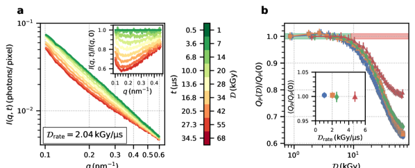
The evolution of the time-resolved SAXS signal as a function of dose and dose rate is analyzed by computing the azimuthally integrated intensity as a function of absolute momentum transfer, , where is the X-ray wavelength and is the scattering angle, and measurement time or dose (Fig. 2a). The absorbed dose is proportional to the measurement time and is calculated with Eq. 5 (see Methods). The data displayed were recorded with a dose rate of , but with absolute doses varying between (green) and (red). With increasing dose, we observe significant changes in , with the largest decrease of intensity visible at momentum transfers of accompanied by an increasing scattering signal at small momentum transfers. The inset shows the data normalized by . The Ig-PEG system exhibits a structure factor peak close to that was studied in a previous work by [51]. Additionally, the phase behavior of the Ig-PEG systems is characterized by an upper critical solution temperature of around . The overall decrease of intensity in Fig. 2 indicates that the system is moving away from the LLPS binodal in the phase diagram, presumably due to beam-induced local heating. Deeper in the single phase regime, increasingly repulsive protein-protein interactions lead to a reduced SAXS intensity. On the other hand, at -values below , the visible increase of indicates the formation of X-ray induced aggregation of the proteins.
We quantify the evolution of structural changes by calculating the Porod invariant
| (1) |
in the accessible -range (, ) as a function of dose (Fig. 2b). displays an initial plateau up to a maximum dose of after which it starts to decrease more than two percent from its initial value. At doses below , the protein structure seems unaffected by the X-ray illumination–at least on the length scales probed here. In this low-dose regime, we also extract the dose rate dependence of the Porod invariant by averaging the data for . The results are displayed in the inset in Fig. 2b demonstrating the absence of a dose rate dependence in the SAXS signal. This is in agreement with previous work reporting that the absolute absorbed dose is the main driver for radiation damage and dose rate effects are only weak [46].
Megahertz X-ray photon correlation spectroscopy (MHz-XPCS)
The disordered protein solutions give rise to a speckle pattern in the far-field when illuminated by coherent radiation. The dynamics can be studied by analyzing the speckle intensity fluctuations that are related to the microscopic motion of the protein molecules. The intensity is measured at time by pixel within a concentric region of interest (ROI) of constant absolute momentum transfer. We utilized an XPCS adapted acquisition scheme in which the sample is continuously moving through the X-ray beam with . The sample movement is negligible during an X-ray train ensuring illumination of the same sample spot on microsecond time scales. In between two trains the sample position offset is large enough to completely renew the sample volume, and thus to avoid accumulated damage. The low intensity scattering signal requires averaging correlation functions from many trains (between and Competing interests) to increase the SNR. Approximately of the acquired trains are used for the XPCS analysis while the rest are discarded after applying filters based on diagnostics such as extremely low intensity due to the SASE fluctuations.
We compare measurements with average dose rates, , from . The influence of dose and dose rate on the protein dynamics can be quantified with the help of two-time correlation functions (TTCs) [55, 56] which essentially represent the correlation coefficient between speckle images taken at times and at momentum transfer :
| (2) |
Here, denotes an average over all pixels with the same absolute momentum transfer, , and denotes an average over all trains where . The data calibration and analysis workflow for MHz-XPCS with AGIPD is described in detail in [3].
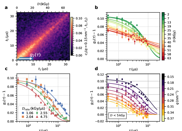
Fig. 3a displays a TTC measured with an average dose rate of at . The abscissa and ordinate of the TTC show the measurement times and , respectively, within an X-ray pulse train while the additional label at the top indicates the corresponding absorbed dose. The TTC decays with increasing distance from the diagonal describing the temporal decorrelation of the speckle fluctuations due to the sample dynamics. The fact that the diagonal does not exhibit a constant width indicates that the dynamics change throughout the measurement.
Time-resolved intra-train intensity auto-correlation functions, , are calculated by averaging sections of the TTCs as indicated by the white arrow in Fig. 3a:
| (3) |
To obtain the correlation function for a particular initial dose, , during the measurement, the time is chosen on the diagonal of the TTC in Fig. 3a, while noting that the dose increases further with each point of the correlation function. The average over can be seen as rebinning along to increase the statistics. This approach yields a set of per dose and dose rate.
The correlation functions are modelled by a Kohlrausch-Williams-Watts (KWW) function:
| (4) |
where is the -dependent speckle contrast [57] () and is the KWW exponent. Brownian diffusion is characterized by a quadratic -dependence of the relaxation rates , where is the diffusion coefficient, and simple exponential behavior (). KWW exponents smaller than one are typically observed in supercooled liquids, and gels and can indicate heterogeneous dynamics with a distribution of relaxation times [58]. A quadratic -dependence and a -independent KWW exponent are used to model the data. Fig. 3d shows that within the experimental accuracy the KWW function describes the data well.
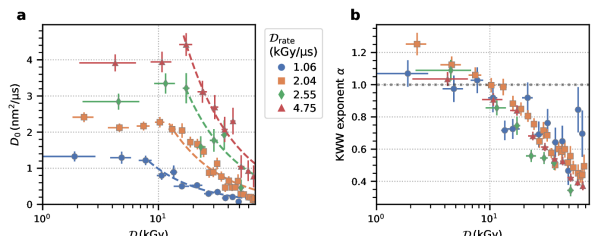
Fig. 3c shows correlation functions for different dose rates for absolute doses below . Fig. 3c indicates that the dynamics become faster with dose rate as the correlation functions shift to shorter time scales while the overall lineshape appears to change only slightly. This is different from the behavior observed with increasing total dose in Fig. 3b where the shape of the correlation functions drastically changes from a simple exponential decay at low doses to a highly stretched () and almost logarithmic decay at higher dose values. We account for these changing KWW exponents by computing the average relaxation rate [59, 60] , where is the -function. Using these average relaxation rates one pair of parameters is calculated per initial dose and dose rate, where . The results are displayed in Fig. 4.
The diffusion coefficients in Fig. 4a reveal a pronounced dependence on the initial dose and dose rate as already indicated by the correlation functions in Fig. 3 and are higher than expected for the base temperature of . Therefore, we denote reported here is as an effective diffusion coefficient discussed in more detail in the following section. The numbers obtained for are on the order of a few , which is the typical range of diffusion coefficients found for dense protein systems [6, 26, 7]. For a given dose rate, all diffusion coefficients follow a similar pattern as a function of initially absorbed dose: is nearly independent of the initial dose up to a threshold value, above which the steadily decreases. The threshold initial dose is slightly below for a dose rate of and increases above for . The average values of below these thresholds increase linearly by about a factor of four from with increasing dose rate. The corresponding KWW exponents do not show any pronounced dose rate dependence, but a clear dependence on the initial dose (Fig. 4b). The KWW exponents further reveal that the correlation functions exhibit a simple exponential shape (=1) for low doses while they are increasingly stretched above , which approximately coincides with the dose value where a decrease in becomes apparent (Fig. 4a). The simultaneous decrease of and the KWW exponent for high doses points towards beam-induced aggregation of the proteins (cf. Fig. 2), which results in slower diffusion and increasingly stretched exponential behavior.
Discussion
Our results indicate that static and dynamic properties are influenced in different ways by the intense X-ray pulses of the European XFEL. MHz-SAXS reveals that the static scattering signal–within the accessible -window–is preserved below an absorbed dose of . This threshold value is independent of the applied dose rate (Fig. 2b inset) within the limited range of dose rates. It is noteworthy that the extremely high dose rates and microsecond time scales probed with an XFEL yield similar threshold values () as the orders of magnitude lower dose rates used at a synchrotron ( [52]).
Understanding the dose rate dependence of radiation-induced effects is crucial for comparing and optimizing experiments at different radiation sources (rotating anodes, synchrotrons, XFELs). At comparably moderate dose rates of tens of Gray per second at synchrotrons, the aggregation rate of proteins was found to exhibit a dose rate dependence [46] favoring measurements with low dose rates. On the other hand, high dose rates seem to be preferable in room temperature protein crystallography measurements [61, 62, 63].
Generally, radiation damage in aqueous protein solutions is mainly attributed to the diffusion and successive reaction of proteins with radicals produced by radiolysis, such as \ceOH-. Radiolysis itself involves a variety of different time and length scales where the radicals are not uniformly generated in the solvent, but distributed initially in nanoscale traces which broaden and diffuse into the bulk on timescales of hundreds of nanoseconds to microseconds during the chemical stage [64]. The primary yield of \ceOH- radicals is high, with \ceOH- per absorbed after one microsecond [65], leading to an average of about \ceOH- radicals per Ig protein molecule needed to induce measurable changes to the SAXS signal (Fig. 2). The observed threshold dose of represents a typical time window of when using a dose rate of . The absence of a measurable dose rate effect on this static threshold value indicates that diffusion rates of radicals, recombination and quenching effects do not affect the overall agglomeration probability.
Additional insight can be obtained from the MHz-XPCS data, which allows to trace time-resolved non-equilibrium dynamics via the TTCs. With regard to protein diffusion the typical mean square distances probed here can be estimated via
yielding values of at to at at decay of the correlation functions. Thus, in the present configuration, MHz-XPCS is sensitive to the motion inside the first coordination shell of the protein molecules in the dense solution.
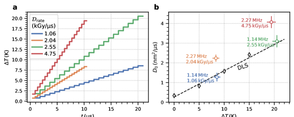
Furthermore, it is interesting to examine the dynamics at dose values below the static damage threshold of obtained from the MHz-SAXS analysis. The diffusion constants are almost independent of the initial dose for a given dose rate. However, displays a pronounced rate dependence and increases by almost a factor of four between the lowest and the highest dose rate (Fig. 4b). Illuminating a sample with highly intense X-ray pulses can lead to a temperature increase. Based on the X-ray beam size of , we estimate that the generated heat dissipates with a time constant of (see Methods section below), which is much longer than the measurement window covered by a -function here (). Thus, the illuminated sample volume does not cool down noticeably during a measurement and the maximum accumulated heat only depends on the fluence per pulse and the number of pulses illuminating the same sample volume which is equivalent to the accumulated dose.
The increase of temperature for the different XFEL parameters is estimated following the model used by [1] using a weighted average heat capacity of the constituents of . With a maximum number of photons per X-ray pulse (Competing interests) the energy density is , which is an order of magnitude smaller than in the work of [1]. Fig. 5a displays the corresponding temperature rise during a pulse train with a temperature increase of after the low intensity pulse trains (blue and orange) and after the high intensity pulse trains (red and green). For comparison, we measured the temperature dependence of the equilibrium diffusion constant with dynamic light scattering (DLS), where the sample was equilibrated at each temperature before a measurements, and display the results in Fig. 5b. The values of measured with MHz-XPCS and X-ray pulse repetition rates of are close to their equilibrium values obtained from DLS, when taking the XFEL-induced temperature increase into account. In contrast, employing higher XFEL frequencies of yields consistently higher diffusion coefficients which cannot be explained by a temperature increase alone. We hypothesize that the intense MHz XFEL pulses create a non-equilibrium state triggering processes on the sub-microsecond time scale. One example of such processes is the spatial homogenization of the aforementioned radiolysis products. The typical rates of secondary products such as \ceOH- radicals are on the order of microseconds [66, 67]. Thus, on sub-microsecond time scales, the XFEL pulses simultaneously produce and probe a spatially inhomogeneous local distribution of the radiolysis products. The resulting chemical gradients, molecular repulsion due to dose rate dependent protein charging, and possibly changes of the ionic strength of the solution, as well as damage to the PEG molecules, could contribute to the observed enhanced diffusive motion. Clearly, more systematic data and additional work by theory and simulation is needed to understand this XFEL driven motion.
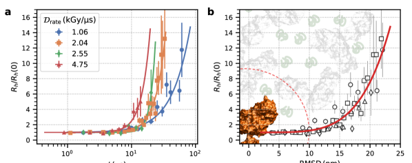
The question arises why the faster dynamics at higher dose rates do not lead to a dose rate dependent aggregation visible in the SAXS signal. We address this question by employing the Stokes-Einstein relation and estimating the temporal evolution of the relative changes to the apparent hydrodynamic radii via , where and represent the respective values at the minimum dose in Fig. 4. The increase of this ratio serves as an indicator for protein aggregation. Fig. 6 shows that aggregation sets in earlier and develops faster for higher dose rates. For a dose rate of , has approximately doubled after and after for (Supplementary Fig. 4). Using the measured diffusion coefficients , we further calculate the time dependent root mean square displacement (RMSD) of the proteins and plot as a function of RMSD (Fig. 6b). The data for the different dose rates collapse onto a single master curve (red line) indicating that the onset of aggregation mainly depends on the RMSD of the protein molecules. Higher dose rates induce faster movement of the proteins, and thus the RMSD necessary for aggregation is reached earlier. Fig. 6b also reveals that aggregation sets in after a RMSD of about and the space a single protein can explore in the crowded solution before that happens is indicated by a the red dashed circle. This area is quite large considering that the sample is a densely packed solution of , where the mean free path between two molecules is typically smaller than their radius. We estimate from the number density and the molecular radius of an Ig molecule which in turn implies an average number of contacts between proteins on the order of before aggregation sets in.
Our analysis indicates that aggregation is not strictly translational diffusion limited, but multiple contacts are necessary to attach two protein molecules to each other and form aggregates. This may hint towards the importance of specific interaction sites driving the aggregation process [68]. In addition, we note that unfolding processes which increase the protein propensity to aggregate do also occur on time scales of microseconds [69]. Thus, the observed initial period of constant points towards a minimum incubation time on the order of before the proteins locally unfold or a time needed for rotational motion of molecules in order to allow activated sites to form local bonds. This incubation time and the minimum RMSD of define a window of opportunity where dynamics can be measured in a correlation before aggregation scheme. Analogously to diffraction before destruction on femtosecond time scales [70, 71, 72, 73], correlation before aggregation will allow to obtain experimental information about the structural sample dynamics on (sub-)microsecond time scales before X-ray induced changes will become apparent in the XPCS signal. Additional data with more dose rates could allow developing novel methods to estimate the diffusion coefficients at zero dose rate [74]. However, the dose rate dependence might be highly dependent on the sample. On the other hand, developing the correlation before aggregation approach further would provide a window of opportunity, where with moderate doses and dose rates, the SNR is increased and the overall measurement time and sample consumption could be reduced. Reducing the sample consumption is crucial for measuring particularly precious solutions or systems that exhibit phase transitions on microsecond time scales, e.g., biomolecular condensates, which is hard to repeat thousands of times [52, 58, 75]. Improving the experiments could be achieved for instance by making use of the self-seeding schemes which provide a much larger longitudinal coherence length. A larger longitudinal coherence length would allow for a larger beam size with similar measured speckle contrast yielding a lower photon density on the sample. This reduces the radiation damage to the sample and the amount of sample needed. It also increases the scattering volume and scattering intensity and thus strongly increases the signal-to-noise ratio [76]. Further technical improvements such as MHz detectors with smaller pixel size are needed to improve the SNR even further which allows extending the accessible -range and lowering the dose rate needed.
Summarizing, we demonstrated that MHz-XPCS bears the potential to become a useful tool for measuring dynamics of biological macromolecules in solution on molecular length scales and on the time scales relevant for diffusive motion in cells. Importantly, our results indicate that taking the temperature rise of the solution into account allows for studying equilibrium dynamics within the first coordination shell of the molecules. Higher XFEL frequencies drive the dynamics and lead to increasing diffusion coefficients and aggregation which sets in after a time window of . We refer to this approach as correlation before aggregation which allows to capture protein dynamics in solution before the manifestation of X-ray induced effects. Additional experiments and simulations are needed to fully understand the underlying physics of the involved processes. Understanding the observed dose rate dependence of the diffusion process involves accurate knowledge of a number of yet unknown factors, such as the role of the interaction potentials, concentration, solvent chemical composition, and size and masses of the proteins. Resolving these properties and the role of radiolysis processes and their products in this context will determine the best data acquisition strategies for measuring the unperturbed dynamic properties.
Methods
Sample Preparation
The sample preparation followed a procedure provided by the literature [51]. Polyclonal bovine immunoglobulin (purity , Sigma-Aldrich, SRE0011), PEG 1000 (Sigma-Aldrich, 81188), NaCl (Merck 106404), HEPES (Roth, HN78) and NaN3 (Sigma-Aldrich, S8032) were used as received. All solutions were prepared in a buffer of composition 20 mM HEPES pH = 7.0, 2 mM NaN3, using degassed Milli-Q water (Merck Millipore 18.2 Mcm). The concentration of the immunoglobulin stock solutions was assessed by UV absorption at 280nm, using an extinction coefficient of with a Cary 50 UV-Vis spectrophotometer. The experimental phase diagram of this system has been established in our previous work [51]. The “parent solution” was equilibrated for about 24 h at and then briefly centrifuged, resulting in a clear dense and a dilute phase, separated by a sharp meniscus. The parent solution composition was immunoglobulin 200 mg/mL, PEG 12% w/v and NaCl 150 mM. The dense liquid phase was used for XPCS measurements with a concentration of roughly .
Measurement Protocol
The sample was moved continuously through the X-ray beam to refresh the sample volume between trains. The translation motor speed was leading to an absolute sample translation of between successive trains (a train arrives every ). X-ray generated heat diffuses with a thermal diffusion constant of . Assuming homogeneous heat diffusion in radial direction perpendicular from the beam, after the heat diffused in a cylinder with radius . The illuminated volume of the next train is then of that volume. Therefore, beam-induced heating effects generated by the previous train can be neglected for the following illumination.
Calculation of the Absorbed Dose and Dose Rate
On the time scale of an individual X-ray pulse ( [50]) and with maximum flux, the peak dose rate can reach several hundred mega Gray per pulse [77]. Utilizing MHz repetition rates and high attenuation, the X-ray pulses are delivered on (sub-) microsecond time scales such that an average dose rate on the order of several kilo Gray per microsecond can be reached. Correspondingly, synchrotron sources produce typical average dose rates of kilo Gray per second.
In order to quantify the amount of energy absorbed by a certain sample mass, we calculate the dose, , absorbed by the sample after pulses:
| (5) |
where denotes the sample absorption where is the sample thickness and is the effective attenuation length of the solution calculated as the weighted harmonic mean of the individual components [78, 52], the photon energy, the number of photons in pulse in train , is the sample density, and is the beam size. denotes an average over trains. The dose is measured in Gray ().
To account for SASE related intensity fluctuations, the average dose rate is calculated as the average dose absorbed by the sample after ten pulses divided by the delay between two successive pulses, :
| (6) |
Error Bar Calculation
The error bars of the correlation functions in Fig. 3 are calculated as the standard deviation of the fluctuations of the contrast values within a -bin. This standard deviation is used to calculate the weighted average over trains and times in Eqs. 2 and 3. These error bars are used in the fits for the parameters estimation. The error bars in the following plots, which display the fit results of the correlation functions, indicate the parameter uncertainty obtained from the fits using least-squares minimization.
X-ray Induced Heating
The relaxation time of heat diffusion used in Fig. 5 to estimate the X-ray induced heating, is calculated as , where is the heat capacity of the solution and is the thermal conductivity. The heat capacity is calculated by an average of the heat capacities of water, PEG, and IgG weighted with their volume fractions. The temperature increase induced by a pulse with energy is . This formalism leads to values from depending on and the number of X-ray pulses (Fig. 5). It should be noted that and less of the maximum possible at MID has been used to reduce beam-induced effects.
Data availability
The data are available from the authors upon request.
Code availability
Analysis scripts are available from the authors upon request.
References
- [1] Felix Lehmkühler et al. “Emergence of Anomalous Dynamics in Soft Matter Probed at the European XFEL” In Proc. Natl. Acad. Sci. U.S.A. 117.39, 2020, pp. 24110–24116 DOI: 10.1073/pnas.2003337117
- [2] Francesco Dallari et al. “Microsecond Hydrodynamic Interactions in Dense Colloidal Dispersions Probed at the European XFEL” In IUCrJ 8.5, 2021, pp. 775–783 DOI: 10.1107/S2052252521006333
- [3] Francesco Dallari et al. “Analysis Strategies for MHz XPCS at the European XFEL” In Appl. Sci. 11.17, 2021, pp. 8037 DOI: 10.3390/app11178037
- [4] José García de la Torre, María L. Huertas and Beatriz Carrasco “Calculation of Hydrodynamic Properties of Globular Proteins from Their Atomic-Level Structure” In Biophys. J. 78.2, 2000, pp. 719–730 DOI: 10.1016/S0006-3495(00)76630-6
- [5] Douglas Ridgway et al. “Coarse-Grained Molecular Simulation of Diffusion and Reaction Kinetics in a Crowded Virtual Cytoplasm” In Biophys. J. 94.10, 2008, pp. 3748–3759 DOI: 10.1529/biophysj.107.116053
- [6] Marco Grimaldo et al. “Dynamics of Proteins in Solution” In Q. Rev. Biophys. 52, 2019, pp. E7 DOI: 10.1017/S0033583519000027
- [7] F. Roosen-Runge et al. “Protein Self-Diffusion in Crowded Solutions” In Proc. Natl. Acad. Sci. U.S.A. 108.29, 2011, pp. 11815–11820 DOI: 10.1073/pnas.1107287108
- [8] Sören Bülow, Marc Siggel, Max Linke and Gerhard Hummer “Dynamic Cluster Formation Determines Viscosity and Diffusion in Dense Protein Solutions” In Proc. Natl. Acad. Sci. U.S.A. 116.20, 2019, pp. 9843–9852 DOI: 10.1073/pnas.1817564116
- [9] Marco Grimaldo et al. “Protein Short-Time Diffusion in a Naturally Crowded Environment” In J. Phys. Chem. Lett. 10.8, 2019, pp. 1709–1715 DOI: 10.1021/acs.jpclett.9b00345
- [10] Mats Leeman et al. “Proteins and Antibodies in Serum, Plasma, and Whole Blood—Size Characterization Using Asymmetrical Flow Field-Flow Fractionation (AF4)” In Anal. Bioanal. Chem. 410.20, 2018, pp. 4867–4873 DOI: 10.1007/s00216-018-1127-2
- [11] Jin Suk Myung et al. “Weak Shape Anisotropy Leads to a Nonmonotonic Contribution to Crowding, Impacting Protein Dynamics under Physiologically Relevant Conditions” In J. Phys. Chem. B 122.51, 2018, pp. 12396–12402 DOI: 10.1021/acs.jpcb.8b07901
- [12] Saskia Bucciarelli et al. “Dramatic Influence of Patchy Attractions on Short-Time Protein Diffusion under Crowded Conditions” In Sci. Adv. 2.12, 2016, pp. e1601432 DOI: 10.1126/sciadv.1601432
- [13] Lei Tang “Protein Translation inside Synthetic Membraneless Organelles” In Nat. Methods 16.6, 2019, pp. 456–456 DOI: 10.1038/s41592-019-0439-2
- [14] Leonard F. Pease et al. “Determination of Protein Aggregation with Differential Mobility Analysis: Application to IgG Antibody” In Biotechnol. Bioeng. 101.6, 2008, pp. 1214–1222 DOI: 10.1002/bit.22017
- [15] Nicolas Martin et al. “Prevention of Thermally Induced Aggregation of IgG Antibodies by Noncovalent Interaction with Poly(Acrylate) Derivatives” In Biomacromolecules 15.8, 2014, pp. 2952–2962 DOI: 10.1021/bm5005756
- [16] Nicholas Skar-Gislinge et al. “A Colloid Approach to Self-Assembling Antibodies” In Mol. Pharm. 16.6, 2019, pp. 2394–2404 DOI: 10.1021/acs.molpharmaceut.9b00019
- [17] Anita Girelli et al. “Molecular Flexibility of Antibodies Preserved Even in the Dense Phase after Macroscopic Phase Separation” In Mol. Pharm. 18.11, 2021, pp. 4162–4169 DOI: 10.1021/acs.molpharmaceut.1c00555
- [18] Yaqiang Wang, Conggang Li and Gary J. Pielak “Effects of Proteins on Protein Diffusion” In J. Am. Chem. Soc. 132.27, 2010, pp. 9392–9397 DOI: 10.1021/ja102296k
- [19] Conggang Li, Yaqiang Wang and Gary J. Pielak “Translational and Rotational Diffusion of a Small Globular Protein under Crowded Conditions” In J. Phys. Chem. B 113.40, 2009, pp. 13390–13392 DOI: 10.1021/jp907744m
- [20] R. London, C. Gregg and N. Matwiyoff “Nuclear Magnetic Resonance of Rotational Mobility of Mouse Hemoglobin Labeled with (2-13C)Histidine” In Science 188.4185, 1975, pp. 266–268 DOI: 10.1126/science.1118727
- [21] S.P. Williams, P.M. Haggie and K.M. Brindle “19F NMR Measurements of the Rotational Mobility of Proteins in Vivo” In Biophys. J. 72.1, 1997, pp. 490–498 DOI: 10.1016/S0006-3495(97)78690-9
- [22] T. Ando and J. Skolnick “Crowding and Hydrodynamic Interactions Likely Dominate in Vivo Macromolecular Motion” In Proc. Natl. Acad. Sci. U.S.A. 107.43, 2010, pp. 18457–18462 DOI: 10.1073/pnas.1011354107
- [23] J.. Wojcieszyn, R.. Schlegel, E.. Wu and K.. Jacobson “Diffusion of Injected Macromolecules within the Cytoplasm of Living Cells.” In Proc. Natl. Acad. Sci. U.S.A. 78.7, 1981, pp. 4407–4410 DOI: 10.1073/pnas.78.7.4407
- [24] Martine Arrio-Dupont et al. “Translational Diffusion of Globular Proteins in the Cytoplasm of Cultured Muscle Cells” In Biophys. J. 78.2, 2000, pp. 901–907 DOI: 10.1016/S0006-3495(00)76647-1
- [25] Alan S Verkman “Solute and Macromolecule Diffusion in Cellular Aqueous Compartments” In Trends Biochem. Sci. 27.1, 2002, pp. 27–33 DOI: 10.1016/S0968-0004(01)02003-5
- [26] Daniel S. Banks and Cécile Fradin “Anomalous Diffusion of Proteins Due to Molecular Crowding” In Biophys. J. 89.5, 2005, pp. 2960–2971 DOI: 10.1529/biophysj.104.051078
- [27] N. Muramatsu and A.. Minton “Tracer Diffusion of Globular Proteins in Concentrated Protein Solutions.” In Proc. Natl. Acad. Sci. U.S.A. 85.9, 1988, pp. 2984–2988 DOI: 10.1073/pnas.85.9.2984
- [28] James A. Dix and A.S. Verkman “Crowding Effects on Diffusion in Solutions and Cells” In Annu. Rev. Biophys. 37.1, 2008, pp. 247–263 DOI: 10.1146/annurev.biophys.37.032807.125824
- [29] S B Zimmerman and A P Minton “Macromolecular Crowding: Biochemical, Biophysical, and Physiological Consequences” In Annu. Rev. Biophys. Biomol. Struct. 22.1, 1993, pp. 27–65 DOI: 10.1146/annurev.bb.22.060193.000331
- [30] Sanjib K. Mukherjee et al. “Do Macromolecular Crowding Agents Exert Only an Excluded Volume Effect? A Protein Solvation Study” In J. Phys. Chem. B 119.44, 2015, pp. 14145–14156 DOI: 10.1021/acs.jpcb.5b09446
- [31] Ryuhei Harada, Yuji Sugita and Michael Feig “Protein Crowding Affects Hydration Structure and Dynamics” In J. Am. Chem. Soc. 134.10, 2012, pp. 4842–4849 DOI: 10.1021/ja211115q
- [32] Rachel D. Cohen and Gary J. Pielak “A Cell Is More than the Sum of Its (Dilute) Parts: A Brief History of Quinary Structure” In Protein Sci. 26.3, 2017, pp. 403–413 DOI: 10.1002/pro.3092
- [33] E.. McConkey “Molecular Evolution, Intracellular Organization, and the Quinary Structure of Proteins.” In Proc. Natl. Acad. Sci. U.S.A. 79.10, 1982, pp. 3236–3240 DOI: 10.1073/pnas.79.10.3236
- [34] Isseki Yu et al. “Biomolecular Interactions Modulate Macromolecular Structure and Dynamics in Atomistic Model of a Bacterial Cytoplasm” In eLife 5, 2016, pp. e19274 DOI: 10.7554/eLife.19274
- [35] Michael Feig and Yuji Sugita “Variable Interactions between Protein Crowders and Biomolecular Solutes Are Important in Understanding Cellular Crowding” In J. Phys. Chem. B 116.1, 2012, pp. 599–605 DOI: 10.1021/jp209302e
- [36] Frédéric Cardinaux et al. “Cluster-Driven Dynamical Arrest in Concentrated Lysozyme Solutions” In J. Phys. Chem. B 115.22, 2011, pp. 7227–7237 DOI: 10.1021/jp112180p
- [37] P. Kowalczyk, A. Ciach, P.A. Gauden and A.P. Terzyk “Equilibrium Clusters in Concentrated Lysozyme Protein Solutions” In J. Colloid Interface Sci. 363.2, 2011, pp. 579–584 DOI: 10.1016/j.jcis.2011.07.043
- [38] Lionel Porcar et al. “Formation of the Dynamic Clusters in Concentrated Lysozyme Protein Solutions” In J. Phys. Chem. Lett. 1.1, 2010, pp. 126–129 DOI: 10.1021/jz900127c
- [39] Yun Liu et al. “Lysozyme Protein Solution with an Intermediate Range Order Structure” In J. Phys. Chem. B 115.22, 2011, pp. 7238–7247 DOI: 10.1021/jp109333c
- [40] Grzegorz Nawrocki et al. “Slow-Down in Diffusion in Crowded Protein Solutions Correlates with Transient Cluster Formation” In J. Phys. Chem. B 121.49, 2017, pp. 11072–11084 DOI: 10.1021/acs.jpcb.7b08785
- [41] Matthias Weiss, Markus Elsner, Fredrik Kartberg and Tommy Nilsson “Anomalous Subdiffusion Is a Measure for Cytoplasmic Crowding in Living Cells” In Biophys. J. 87.5, 2004, pp. 3518–3524 DOI: 10.1529/biophysj.104.044263
- [42] Gernot Guigas and Matthias Weiss “Sampling the Cell with Anomalous Diffusion—The Discovery of Slowness” In Biophys. J. 94.1, 2008, pp. 90–94 DOI: 10.1529/biophysj.107.117044
- [43] Steve P. Meisburger et al. “Breaking the Radiation Damage Limit with Cryo-SAXS” In Biophys. J. 104.1, 2013, pp. 227–236 DOI: 10.1016/j.bpj.2012.11.3817
- [44] Jesse B. Hopkins and Robert E. Thorne “Quantifying Radiation Damage in Biomolecular Small-Angle X-ray Scattering” In J. Appl. Crystallogr. 49.3, 2016, pp. 880–890 DOI: 10.1107/S1600576716005136
- [45] Elspeth F. Garman and Martin Weik “X-Ray Radiation Damage to Biological Macromolecules: Further Insights” In J. Synchrotron Radiat. 24.1, 2017, pp. 1–6 DOI: 10.1107/S160057751602018X
- [46] Shigeo Kuwamoto, Shuji Akiyama and Tetsuro Fujisawa “Radiation Damage to a Protein Solution, Detected by Synchrotron X-ray Small-Angle Scattering: Dose-Related Considerations and Suppression by Cryoprotectants” In J. Synchrotron Radiat. 11.6, 2004, pp. 462–468 DOI: 10.1107/S0909049504019272
- [47] Linda Young et al. “Photon-In/Photon-Out X-ray Free-Electron Laser Studies of Radiolysis” In Appl. Sci. 11.2, 2021, pp. 701 DOI: 10.3390/app11020701
- [48] Clare L. Hawkins and Michael J. Davies “Generation and Propagation of Radical Reactions on Proteins” In Biochim. Biophys. Acta Bioenerg. 1504.2-3, 2001, pp. 196–219 DOI: 10.1016/S0005-2728(00)00252-8
- [49] Julia M. Khalack and Alexander P. Lyubartsev “Solvation Structure of Hydroxyl Radical by Car-Parrinello Molecular Dynamics” In J. Phys. Chem. A 109.2, 2005, pp. 378–386 DOI: 10.1021/jp0461807
- [50] A. Madsen et al. “Materials Imaging and Dynamics (MID) Instrument at the European X-ray Free-Electron Laser Facility” In J. Synchrotron Radiat. 28.2, 2021, pp. 637–649 DOI: 10.1107/S1600577521001302
- [51] Stefano Da Vela et al. “Effective Interactions and Colloidal Stability of Bovine -Globulin in Solution” In J. Phys. Chem. B 121.23, 2017, pp. 5759–5769 DOI: 10.1021/acs.jpcb.7b03510
- [52] Anita Girelli et al. “Microscopic Dynamics of Liquid-Liquid Phase Separation and Domain Coarsening in a Protein Solution Revealed by X-Ray Photon Correlation Spectroscopy” In Phys. Rev. Lett. 126.13, 2021, pp. 138004 DOI: 10.1103/PhysRevLett.126.138004
- [53] Aschkan Allahgholi et al. “The Adaptive Gain Integrating Pixel Detector at the European XFEL” In J. Synchrotron Radiat. 26.1, 2019, pp. 74–82 DOI: 10.1107/S1600577518016077
- [54] P. Falus, L.. Lurio and S… Mochrie “Optimizing the Signal-to-Noise Ratio for X-ray Photon Correlation Spectroscopy” In J. Synchrotron Radiat. 13.3, 2006, pp. 253–259 DOI: 10.1107/S0909049506006789
- [55] Mark Sutton, Khalid Laaziri, F. Livet and F. Bley “Using Coherence to Measure Two-Time Correlation Functions” In Opt. Express 11.19, 2003, pp. 2268 DOI: 10.1364/OE.11.002268
- [56] Luca Cipelletti et al. “Time-Resolved Correlation: A New Tool for Studying Temporally Heterogeneous Dynamics” In J. Phys. Condens. Matter 15.1, 2003, pp. S257–S262 DOI: 10.1088/0953-8984/15/1/334
- [57] S.. Hruszkewycz et al. “High Contrast X-ray Speckle from Atomic-Scale Order in Liquids and Glasses” In Phys. Rev. Lett. 109.18, 2012, pp. 185502 DOI: 10.1103/PhysRevLett.109.185502
- [58] Nafisa Begam et al. “Kinetics of Network Formation and Heterogeneous Dynamics of an Egg White Gel Revealed by Coherent X-Ray Scattering” In Phys. Rev. Lett. 126.9, 2021, pp. 098001 DOI: 10.1103/PhysRevLett.126.098001
- [59] B. Ruta et al. “Wave-Vector Dependence of the Dynamics in Supercooled Metallic Liquids” In Phys. Rev. Lett. 125.5, 2020, pp. 055701 DOI: 10.1103/PhysRevLett.125.055701
- [60] Hongyu Guo et al. “Entanglement-Controlled Subdiffusion of Nanoparticles within Concentrated Polymer Solutions” In Phys. Rev. Lett. 109.5, 2012, pp. 055901 DOI: 10.1103/PhysRevLett.109.055901
- [61] Eugenio Mora et al. “Radiation Damage and Dose Limits in Serial Synchrotron Crystallography at Cryo- and Room Temperatures” In Proc. Natl. Acad. Sci. U.S.A. 117.8, 2020, pp. 4142–4151 DOI: 10.1073/pnas.1821522117
- [62] Robert J. Southworth-Davies, Melissa A. Medina, Ian Carmichael and Elspeth F. Garman “Observation of Decreased Radiation Damage at Higher Dose Rates in Room Temperature Protein Crystallography” In Structure 15.12, 2007, pp. 1531–1541 DOI: 10.1016/j.str.2007.10.013
- [63] Matthew A. Warkentin et al. “Lifetimes and Spatio-Temporal Response of Protein Crystals in Intense X-ray Microbeams” In IUCrJ 4.6, 2017, pp. 785–794 DOI: 10.1107/S2052252517013495
- [64] Harold A. Schwarz “Applications of the Spur Diffusion Model to the Radiation Chemistry of Aqueous Solutions” In J. Phys. Chem. 73.6, 1969, pp. 1928–1937 DOI: 10.1021/j100726a047
- [65] GV Buxton “Radiation chemistry: principles and applications” In Verlag Chemie Publishers, Weinheim, 1987
- [66] Pankaj Attri et al. “Generation Mechanism of Hydroxyl Radical Species and Its Lifetime Prediction during the Plasma-Initiated Ultraviolet (UV) Photolysis” In Sci. Rep. 5.1, 2015, pp. 9332 DOI: 10.1038/srep09332
- [67] Kentaro Baba et al. “Quantitative Estimation of Track Segment Yields of Water Radiolysis Species under Heavy Ions around Bragg Peak Energies Using Geant4-DNA” In Sci. Rep. 11.1, 2021, pp. 1524 DOI: 10.1038/s41598-021-81215-6
- [68] S.. Northrup and H.. Erickson “Kinetics of Protein-Protein Association Explained by Brownian Dynamics Computer Simulation.” In Proc. Natl. Acad. Sci. U.S.A. 89.8, 1992, pp. 3338–3342 DOI: 10.1073/pnas.89.8.3338
- [69] Jan Kubelka, James Hofrichter and William A Eaton “The Protein Folding ‘Speed Limit”’ In Curr. Opin. Struct. Biol. 14.1, 2004, pp. 76–88 DOI: 10.1016/j.sbi.2004.01.013
- [70] Karol Nass “Radiation Damage in Protein Crystallography at X-ray Free-Electron Lasers” In Acta Crystallogr D Struct Biol 75.2, 2019, pp. 211–218 DOI: 10.1107/S2059798319000317
- [71] Karol Nass et al. “Indications of Radiation Damage in Ferredoxin Microcrystals Using High-Intensity X-FEL Beams” In J Synchrotron Rad 22.2, 2015, pp. 225–238 DOI: 10.1107/S1600577515002349
- [72] Ilme Schlichting “Serial Femtosecond Crystallography: The First Five Years” In IUCrJ 2.2, 2015, pp. 246–255 DOI: 10.1107/S205225251402702X
- [73] Richard Neutze et al. “Potential for Biomolecular Imaging with Femtosecond X-ray Pulses” In Nature 406.6797, 2000, pp. 752–757 DOI: 10.1038/35021099
- [74] Yuriy Chushkin “Deciphering the Intrinsic Dynamics from the Beam-Induced Atomic Motions in Oxide Glasses” In J. Synchrotron Radiat. 27.5, 2020, pp. 1247–1252 DOI: 10.1107/S1600577520009753
- [75] Anastasia Ragulskaya et al. “Interplay between Kinetics and Dynamics of Liquid–Liquid Phase Separation in a Protein Solution Revealed by Coherent X-ray Spectroscopy” In J. Phys. Chem. Lett. 12.30, 2021, pp. 7085–7090 DOI: 10.1021/acs.jpclett.1c01940
- [76] J. Möller, M. Sprung, A. Madsen and C. Gutt “X-Ray Photon Correlation Spectroscopy of Protein Dynamics at Nearly Diffraction-Limited Storage Rings” In IUCrJ 6.5, 2019, pp. 794–803 DOI: 10.1107/S2052252519008273
- [77] Henry N. Chapman et al. “Femtosecond X-ray Protein Nanocrystallography” In Nature 470.7332, 2011, pp. 73–77 DOI: 10.1038/nature09750
- [78] Stephen Seltzer “Tables of X-Ray Mass Attenuation Coefficients and Mass Energy-Absorption Coefficients, NIST Standard Reference Database 126” National Institute of Standards and Technology, 1995 DOI: 10.18434/T4D01F
Acknowledgements
We acknowledge the European XFEL in Schenefeld, Germany, for provision of X-ray free electron laser beamtime at Scientific Instrument MID (Materials Imaging and Dynamics) and would like to thank the staff for their assistance. We acknowledge DESY (Hamburg, Germany), a member of the Helmholtz Association HGF, for the provision of experimental facilities. This research is supported by Center of Molecular Water Science (CMWS) of DESY in an Early Science Project, the MaxWater initiative of the Max-Planck-Gesellschaft and the Wenner-Gren Foundations. This work is supported by the Swedish National Research Council Vetenskapsrådet ( 2019-05542, FP), the Röntgen-Ångström Cluster ( 2019-06075 FP), BMBF (05K19PS1 and 05K20PSA, CG ; 05K19VTB, FS and FZ), DFG-ANR (SCHR700/28-1, SCHR700/42-1, ANR-16-CE92-0009, FS and FZ). AR acknowledges the Studienstiftung des deutschen Volkes for a personal fellowship. NB acknowledges the Alexander von Humboldt-Stiftung for postdoctoral research fellowship.
Author contributions
FP, FZ, FS, and CG conceived the experiment, which was designed and coordinated by MR, FP, FZ and CG. AG and AR prepared and handled the samples. UB, JH, JM, ARF, MS, RS, AZ, AM operated MID and collected data together with MR, AG and AR. MR, AG, AR, FP, ST, SD, MSA and MB performed online data processing and analysis, whereas SB, HFP, MLP, MF, and LR were in addition responsible for the elog. MR performed offline data processing and analysis. MR, FP, FS, FZ, and CG discussed the XPCS data analysis with input from all authors. R.R and R.S supported the development of analysis routines and the integration of experimental equipment. All authors jointly performed the experiment and discussed the final results, to a large extent remotely due to travel restrictions imposed by the COVID-19 pandemic. The manuscript was written by MR, FP and CG with input from all authors.
Competing interests
The authors declare no competing interests.