∎
11email: lichen201096@hotmail.com 22institutetext: Microscopic Image and Medical Image Analysis Group, College of Medicine and Biological Information Engineering, Northeastern University, China School of Control Engineering, Chengdu University of Information Technology, China College of Software Engineering, Northeastern University, China Shengjing Hospital of China Medical University, China Institute for Medical Informatics, University of Luebeck, Germany
A State-of-the-art Survey of U-Net in Microscopic Image Analysis: from Simple Usage to Structure Mortification
Abstract
Image analysis technology is used to solve the inadvertences of artificial traditional methods in disease, wastewater treatment, environmental change monitoring analysis and convolutional neural networks (CNN) play an important role in microscopic image analysis. An important step in detection, tracking, monitoring, feature extraction, modeling and analysis is image segmentation, in which U-Net has increasingly applied in microscopic image segmentation. This paper comprehensively reviews the development history of U-Net, and analyzes various research results of various segmentation methods since the emergence of U-Net and conducts a comprehensive review of related papers. First, This paper has summarizes the improved methods of U-Net and then listed the existing significances of image segmentation techniques and their improvements that has introduced over the years. Finally, focusing on the different improvement strategies of U-Net in different papers, the related work of each application target is reviewed according to detailed technical categories to facilitate future research. Researchers can clearly see the dynamics of transmission of technological development and keep up with future trends in this interdisciplinary field.
Keywords:
Microscopic image analysisU-NetImage segmentation Deep learning Convolutional Neural Network1 Introduction
1.1 Background Knowledge of Microscopic Images
Microscopic image refers to the image use to see in a microscope Wu-2010-MIP . There are many types of microscopes: The general magnification of the optical microscope is generally 1500-2000 times Zenhausern-1994-ANO ; Toledo-1992-NDS ; Inouye-1994-NSO , which has never exceeded 2000 times. However, the maximum magnification of the electron microscope exceeds three million times, such as transmission electron microscope (TEM) Williams-1996-TTE , scanning electron microscopev Seiler-1983-SEE . Scanning tunneling microscope magnification up to 300 million times Tersoff-1985-TOT . Furthermore, there are other types of microscopes, such as atomic force microscope Binnig-1986-AFM , Raman microscope and cryo-electron microscopy Duncan-1982-SCA ; Adrian-1984-CMO .
Microscopic image analysis has a wide range of application scenarios, such as microorganism image analysis Li-2020-ASM ; Li-2019-ASF ; zhang-2021l-LAN , histopathological image analysis Li-2021-ACR ; Zhou-2020-ACR , cytopathological image analysis Rahaman-2020-ASF ; Hore-2015-FCO , metal structure analysis Oien-2014-DSA , rock structure analysis Clelland-1991-ARC , soil structure analysis Pagliai-2002-IAA , material structure analysis and image analysis in plant pathology Abell-1999-MIP ; Nilsson-1995-RSA .
1.2 Background Knowledge of Intelligent Microscopic Image Analysis
Manual operations of microscopic image analysis has some limitations, including:
-
•
In the case of big data, it takes a long time.
-
•
Operator has a heavy workload.
-
•
Analysis of experimenters is easy to be subjective.
-
•
Poor quantification.
Therefore, upcoming technologies should be introduced to this field to improve the level of image analysis.
As mentioned above, efficient artificial intelligence technology is introduced to the field of microscopic image analysis, which can effectively solve the above problems. First of all, computer efficiency is high and big data problems are easy to solve. Secondly, computer assistance can efficiently reduce the amount of hardwork. The third point, the computer is more objective. Finally, computer algorithms can quantitatively output the numerical results Nilsson-2014-POA . Especially, deep learning is the game changer and effective machine learning technology in the field of artificial intelligence in recent years, which has a high accuracy (ACC) rate. With the increasing amount of training data, the ACC rate is higher Shen-2017-DLI . Deep learning litjens-2017-ASO has a strong learning ability, wide coverage, adaptability and good portability. In different deep learning methods, CNN has the characteristics of non-contact and high precision for image recognition, classification and other operations. CNN is extremely applicable to this non-contact method in the process of image segmentation, detection, identification and classification, which can directly take image data as input Tajbakhsh-2016-CNN . Among all well-known CNN methods, U-Net Ronneberger-2015-UCN is clearly the most successful model for microscopic image segmentation, which does not require too many training data to obtain better segmentation results. Furthermore, the training and test time of U-Net is shot, which supports a practical possibility for some real tasks, such as histopathology image analysis, microorganism image analysis and cell image analysis.
1.3 Typical U-Net Architecture
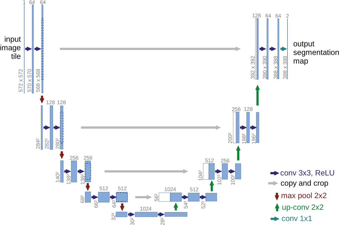
U-Net is originated from Fully Convolutional Networks(FCN), which is a semantic segmentation network Long-2015-FCN ; Ronneberger-2015-UCN . This network is named U-Net because of the U-shaped network structure, which is very suitable for medical image segmentation. The network structure is shown in Fig. 1. Structure of U-Net is symmetrical. The left side is the encoder which can extract the input features, the right side is the decoder, which can output the encoded features as a picture. Red square represents down-sampling, green square represents up-sampling and conv represents the convolution operation with as the core network. The networks on the left are the traditional convolutional layer and pooling layer. The convolution and rectified linear unit (ReLU) activation function are performed twice and there is a maximum pooling. The process is repeated four times and the filter is doubled at each stage. The right part uses the transposed convolution to upsampling. After performing the 3 convolution and ReLU activation function twice, it is also executed four times in a loop and the filter is reduced by half after each upsampling. Finally, add a convolution plus SIGMOID activation function to get the result. In addition, every time downsampling is performed when preparing for pooling, it will be fused to the feature map after transposed convolution, so that the output on the encoder can be directly connected to the decoder to continue propagation. It can be seen that the network is not fully connected. This is also an end-to-end image, that is, the input is an image and the output is also an image.
1.4 Motivation of This Survey Paper
Because of the efficiency and popularity of U-Net in microscopic image segmentation, many researchers choose to use it. Currently, there are some review articles involving U-Net, but there is no survey that focuses on U-Net in microscopic image analysis. Hence, we decide to prepare this suvery paper to organize and summarize lated works for future work as a reference.
Taghanaki-2020-DSS introduces the development of segmentation neural network from the perspective of deep semantic segmentation of natural and medical images, including the development of U-Net and the variant of U-Net. There are 163 references in this paper, but only 16 are about microscopic image analysis. In the work of Du-2020-MIS , a review of U-Net is given from the use of U-Net in different application scenarios. There are 63 references in this work, but only eight references about microscopic image analysis are discussed. Compared with the above published reviews, we summarize 158 related works from 2015 to 2021, involving microorganism images, histopathological images, cytopathological images, rock microscopic images, metal microscopic images, plant microscopic images and material microscopic images. Our survey paper can provide services for the following two groups of people: (1) People who focus on microscopic image analysis; (2) People who do related to deep learning research and development, such as U-Net.
1.5 Structure of This Paper
The general structure of this paper is as follows: The first section summarizes the background of microscopic images, development status, motivation for this work and the structure of the paper. Section two describes the application scenarios of the unmodified U-Net. The third section writes the deformation method of U-Net under the primary changes and the dataset and application scenarios that it is good at segmenting after deformation. The fourth section is about some advanced and complex changes with Residuals in U-Net. This section contains many different types of changes.
2 Undeformed Classic U-Net
This section discusses about the unmodified U-Net in microscopic image segmentation and summarize the literature.
2.1 Applications in Cytology
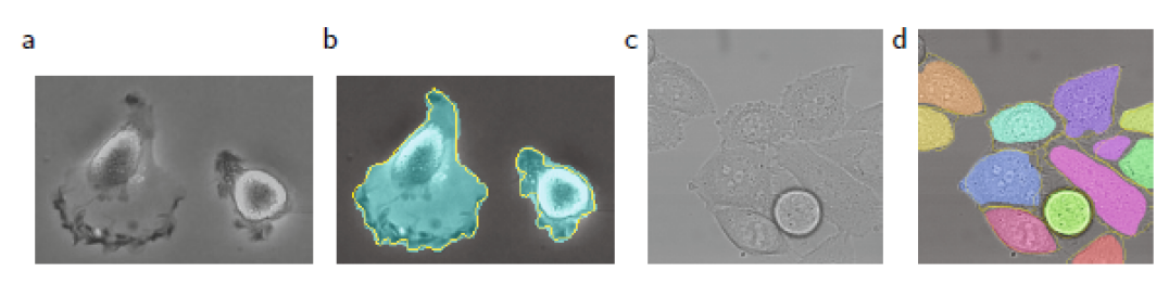
In Ronneberger-2015-UCN , U-Net is proposed to solve a problem of microscopic image segmentation. In this work, original images ( pixels) are resized into pixels to fit the input scale of U-Net. The method is applied to three datasets. Neuron structure has 30 pieces of training data. First, “PhC-U373” dataset has 35 training images with a small number of annotations. Second, “DIC-HeLa” dataset has 20 training images, some of them are annotated. At last, “PhC-U373” and “DIC-HeLa” obtain intersection over union (IOU) of and , respectively. Segmentation results are show in Fig. 2. In Sec. 3.1.1, we will discuss about 3D U-Net.
In Colonna-2018-SOC , undeformed U-Net can accurately segment corneal nerves. A dataset contains 30 people is tested, of which 10 are healthy and 20 are sick. This dataset is adjusted appropriately: first, outermost periphery (10 pixels) of the image to be analyzed is cropped. In addition each image is reduced to 0.7 times of the original size. Experimental result shows that SE reaches .
In Seong-2019-AIO , an original U-Net is used to automatically segment nerve cells. In this paper, a classification task of excitatory, inhibitory neurons and glia cells is also performed. The original U-Net is trained on 126 images containing 5000 cells and an experimental result of an ACC of is obtained.
Since the change of density of corneal endothelial cells (CEC) can monitor corneal diseases. In Daniel-2019-ASO , a baseline U-Net is used to automatically segment corneal endothelium in a large set of “real-world” specular microscopy images. Compared with the baseline U-Net Ronneberger-2015-UCN , neural network structure used for segmentation that has no structural changes. A mirror microscope database containing 16000 images (from corneal consultation service) is selected to evaluate this method and 158 training images are randomly selected as the training set. Finally, the experimental results with a CEC recall (RE) of and an ACC of are obtained.
2.2 Applications on Microorganisms
In Nunez-2020-ASS , Tuberculosis (TB) cords are segmented by U-Net, taking into account the ACC of U-Net segmentation of TB cords. This method is also easy to operate when small reserve of expertise. Datasets are obtained through image reconstruction, a total number 300 images, of which 120 sub-images form the training set and 30 sub-images form the test set. Then, U-Net processes and segments sub-images. Finally, this method connects each sub-image to TB cords for the full images and the segmentation is completed. Finally, this work achieves an IoU of and an ACC of .
In Ojeda-2020-CNN , in order to reduce the time and physical waste of the operator, U-Net is used to segment the parasites in the blood to implement an automated system. This method is tested on a private dataset composed of 974 images. In the dataset, 600 images form the training set and 200 images form the testing set. The result shows that this method obtains an ACC of and a Dice similariy coefficient (DICE) score of .
2.3 Other Applications
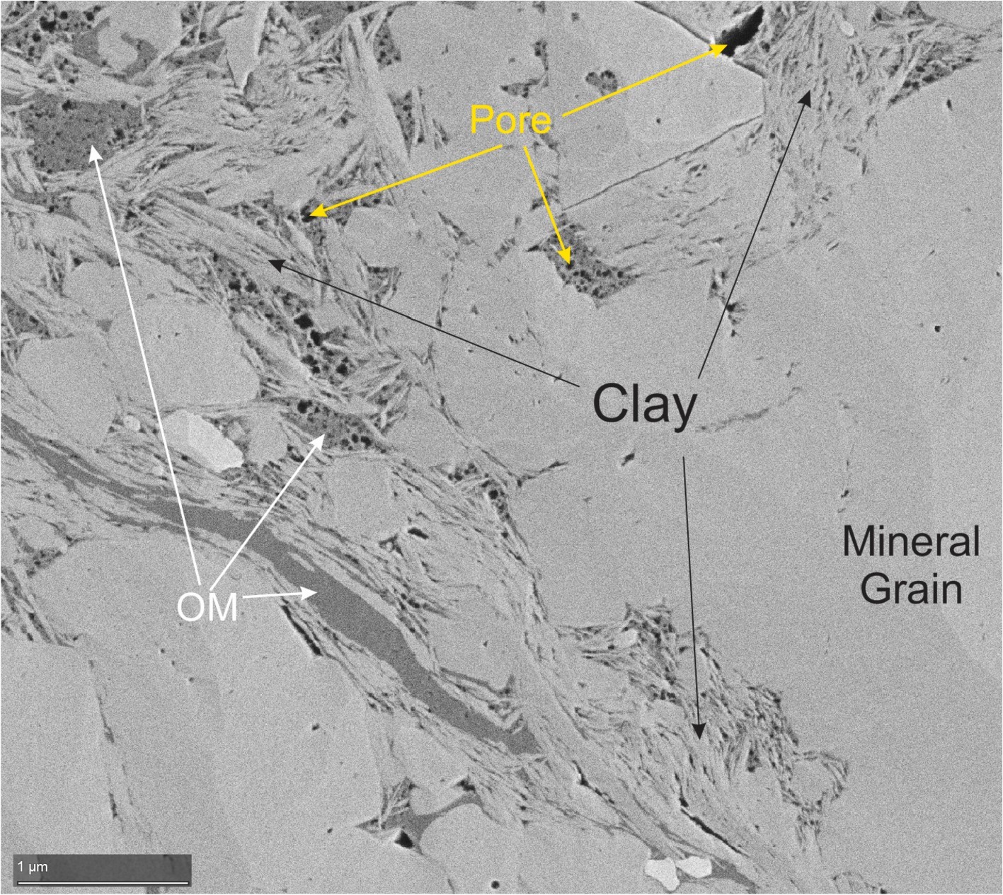
In Chen-2020-DLB , in order to evaluate the geological characteristics of the rock samples, U-Net is used to segment the scanning electron microscope (SEM) images of it (as shown in Fig. 3). The dataset contains 8000 rock slice images, of which are used for training and for testing. The experiment result shows that the highest IOU reaches . It can be seen that when processing texture features, U-Net has a good segmentation performance.
In Oktay-2019-ADL , a new method of detecting nanoparticles is proposed, in which U-Net is used to segment nanostructures. The dataset has eight TEM images synthesized by Fe3O4 nano-particles and nine images of Fe3O4 nano-particles smeared with silica, a total of 17 images. The experiment obtains an Acc higher than .
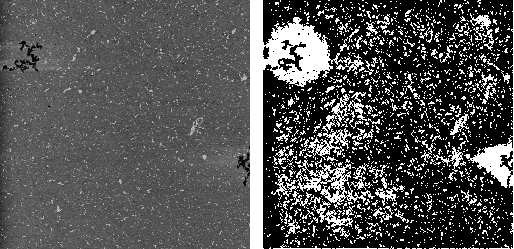
In Farley-2020-ITS , because manual segmentation of nanostructured surface images is time-consuming and requires high professionalism. Therefore, it is necessary to develop an automated method for segmenting nanoparticles based on U-Net. A method based on U-Net is better than the traditional automation method. The experimental dataset contains 728 images of gold nanoparticles generated by the atomic force microscope (AFM) in the absence of moisture, of which are used as the training set and are used as the test set. When banding, the average rate of U-Net pixel change is . The result of segmentation with U-Net is shown in Fig. 4.
Skin healthy condition is evaluated through detection and analysis of blood vessel structure. In Jaworek-2018-ADL , a fully automatic method using a baseline U-Net is developed to segment the blood vessel structure in dermoscopy color images. This work utilizes a dataset from the University of Naples, Italy and the University of Graz, Austria, which contains 74 images of different types of blood vessel patterns in total. In the dataset, of the samples are used for training and the remaining are used for testing. The final experimental results are: a sensitivity (SE) of and a specificity (SP) of .
Many diseases can be diagnosed by retinal blood vessels, in Meyer-2017-ADN , the original U-Net is used to segment Scanning Laser Ophthalmoscopy (SLO) (introduced by Webb-1980-FST ) images. SLO assists blood vessels to be extracted features better. This proposed method is evaluated by IOSTAR Zhang-2016-RRV . In IOSTAR, 20 images form the training set and 10 images form the test set. The proposed method obtains an experimental result of an SE of , a SP of , an ACC of and an area under curve (AUC) of , respectively.
Since a method of manually obtaining tissue sections can easily introduce errors (such as deformation, tissue fracture). Therefore, the baseline U-Net Swiderska-2018-DLF is developed to segment the undesired areas in whole slide images (WSIs) to reduce errors. This proposed method can be used in the preprocessing step of WSIs automated analysis. The method may prevent the damaged area from being used. Meanwhile, the classification task is carried out by the baseline U-Net. Original WSIs are classified as Damaged and non-Damaged classes. This work utilizes a dataset from the archives of the Department of Pathology at the Military Institute of Medicine in Warsaw, Poland. The dataset contains 34 brain tissue cohorts corresponding to brain tumor areas (meningiomas and oligodendrogliomas) in total. However among the dataset, 10 WSIs for training and 24 WSIs be used in testing. Regarding Ki-67 brain tumor specimens segmentation, an experimental result is as follows: SE = 0.83, SP = 0.92, precision (PR) = 0.80, ACC = 0.90 and IoU = 0.69.
2.4 Summary
From the above analysis and review we can reach to a conclusion that the significant segmentation performance of U-Net on microscopic images, the original U-Net without any changes is often used to perform segmentation tasks and achieve good results. It shows that U-Net has strong versatility. U-Net also has wealthy application scenarios, such as cytology, histopathology, microorganism and nanoparticle image analysis. From birth of U-Net in 2015 to 2021, the original U-Net is used in a total number of papers that are listed above. With the development over the time, more and more variant network structures based on U-Net appears. In the subsequent analysis of the deformed structure section, the specific U-Net based on deformed network structure introduced. Tab. 1 is a summary of the evaluation indicators and results in the paper using the original U-Net.
| Aim | Detial | Year | Reference | Team | Data Information | CNN type | Evaluation | ||||||
| Segmentation |
|
2015 | Ronneberger-2015-UCN | O-Ronneberger | 35 annotated images | U-Net | IoU = 92.03% | ||||||
| corneal nerves | 2018 | Colonna-2018-SOC | A.Colonna |
|
U-Net | SE = 97.2% | |||||||
| nuclei | 2019 | Seong-2019-AIO | S.Seong |
|
U-Net | ACC = 93.2% | |||||||
| corneal endothelium | 2019 | Daniel-2019-ASO | M.C.Daniel | 158 training images | U-Net | RE = 34%, PR = 84% | |||||||
| TB cords | 2020 | Nunez-2020-ASS | L.Ballan |
|
U-Net | IoU = 88%, ACC = 92.01% | |||||||
| the T. cruzi parasite | 2020 | Ojeda-2020-CNN | A.Ojeda-Pat |
|
U-Net | ACC = 63.04%, DICE = 68.25% | |||||||
| rock slice SEM images | 2020 | Chen-2020-DLB | Z.Chen |
|
U-Net | IoU = 91.7% | |||||||
| nano-particles | 2019 | Oktay-2019-ADL | A.B.Oktay | 17 images | U-Net | SE = 78.59%, ACC = 96.59% | |||||||
| nanostructured surfaces | 2020 | Farley-2020-ITS | S.Farley |
|
U-Net | average proportion of pixels changed pixels changed = 12.2% | |||||||
|
2018 | Jaworek-2018-ADL | J.Jaworek-Korjakowska |
|
U-Net | DICE = 0.84, SE = 0.85, SP = 0.81 | |||||||
| retinal blood vessels | 2017 | Meyer-2017-ADN | M.I.Meyer |
|
U-Net |
|
|||||||
|
2018 | Swiderska-2018-DLF | Z.Swiderska-Chadaj |
|
U-Net |
|
3 Simple and Low-level Deformable U-Net
From the above section we can see: in most cases, the original U-Net segmentation without any changes performed well. However, firstly, U-Net published in 2015. In recent years, a large number of new network structure changes appear, such as: many new network modules are proposed and new ideas in skip connection are proposed. Secondly, U-Net can not fit all datasets perfectly. In many cases, the segmentation performance is not ideal, therefore, it is appropriate and simple to change the U-Net for better performance over the segmentation tasks. This section introduces some simple and conventional methods to change original implementation of U-Net structure.
3.1 Redesigned Convolution
3.1.1 3D Convolution
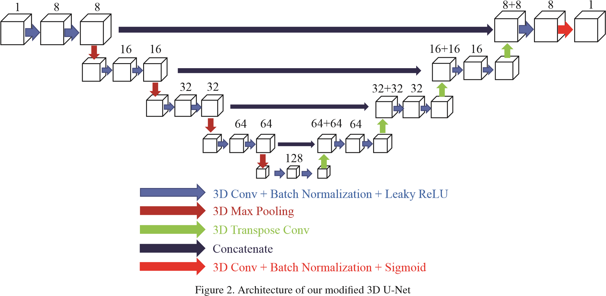
In Cciccek-2016-3UN , a 3D U-Net that replaces 2D convolution with 3D convolution is creatively proposed to achieve accurate semi-automatic segmentation of 3D datasets. Compared with baseline U-Net, 3D U-Net replaces all 2D operations in the paper Ronneberger-2015-UCN with 3D operations and adds Batch Normalization (BN) before each ReLU (similar addition BN’s approach is also reflected in the paper Fang-2019-NSB ). A dataset with 77 manually annotated Xenopus kidney slices under confocal microscopic is used in the experiment. Under the dataset, the proposed 3D U-Net obtains an IoU of 0.863 in the semi-automated test experiments of three-fold cross validation.
In Fu-2018-TDF , a 3D U-Net composed of 3D convolution combined with spatially constrained cycle-consistent adversarial networks is proposed. It solves the problem of high learning rate for manually labeling 3D datasets. The structure of this 3D U-Net is shown in Fig. 5. First, a subvolume of the original image volumes train spatially constrained CycleGAN (SpCycleGAN). Then, the above operations generate 3D synthetic data to evaluate the 3D U-Net. Finally, the proposed method obtains an ACC of .
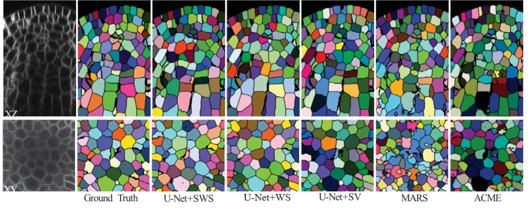
In Eschweiler-2019-CBP , a 3D U-Net combined with a seeded watershed approach (SWS) is proposed. It solves a problem of the large amount of data in the 3D microscopic image of the cell membrane, which is not easy to segment. This method is tested on a training set (the training set comes from Willis-2016-CSA ) composed of 109296 Arabidopsis thaliana cells and a validation set composed of 972 single cells, which manually annotate 3D images. At the same time, 3D U-Net watershed algorithm (WS), 3D U-Net supervoxel merging approach (SV), multi-angle image acquisition, three-dimensional reconstruction and cell segmentation (MARS) Fernandez-2010-IPG , automated cell morphology extractor for comprehensive reconstruction of cell membranes (ACME) Mosaliganti-2012-AAC are also tested by the dataset. The experimental results show that among these methods, U-Net SWS performs best and it obtains a JI of 0.870 and a DICE of 0.931. Fig. 6 is the segmentation result of the method mentioned in Eschweiler-2019-CBP .
In Heinrich-2018-SCS , a 3D U-Net is proposed to fit the non-isotropic nature of serial section Transmission Electron Microscopy (ssTEM) data, the sparsity of synapses improves the performance of segmentation and detection of insect nervous system. Challenge on Circuit Reconstruction from Electron Microscopy Images (CREMI) datasets from Medical Image Computing and Computer Assisted Intervention Society (MICCAI) is used in the experiment. It contains 6 volumes of nerve tissue under an electron microscope, of which are used in the training set and are used in the validation set. The proposed 3D U-Net obtains the experimental results of CREMI score with an average of .
In Wang-2019-SNS , because of the complex structure of neurons and poor imaging quality in some cases, a teacher-student learning framework based on 3D U-Net Cciccek-2016-3UN is proposed to segment neurons to obtain higher ACC and efficiency. Like Cciccek-2016-3UN ; Fu-2018-TDF ; Eschweiler-2019-CBP ; Heinrich-2018-SCS , the teacher-student learning framework uses 3D convolution as the basic unit. Unlike Cciccek-2016-3UN ; Fu-2018-TDF ; Eschweiler-2019-CBP ; Heinrich-2018-SCS , first, the teacher-student network is divided into 2 parts: Teacher network and student network. Second, Residual modules (the specific usage of Residual modules is detailed in Sec. 4) are added to the teacher-student network. A data is obtained from the Janelia dataset from the BigNeuron project (contains 42 images of adult Drosophila nervous system, 38 images for training and 4 images for testing). A PR-RE curves in Wang-2019-SNS shows that the teacher-student network can obtain more accurate segmentation performance.
3.1.2 Other Convolution
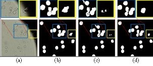
In Zhang-2017-ISA , deformable U-Net with variable convolution is used to segment and classify sickle cell disease (SCD) cells. Similarly, Zhang-2018-RSS is the study of SCD cell segmentation from the same team. Compared with U-Net, in addition to variable convolution, deformable U-Net has only three deformable convolution blocks in the encoding and decoding parts respectively. A dataset with 128 SCD cells obtained from University of Pittsburgh Medical Center (UPMC) is used in the experiment. The deformable U-Net is tested under the dataset. It obtains a segmentation ACC of and a classification ACC of . The segmentation results of proposed method in Fig. 7.
In Zhang-2018-RSS , in order to solve the inaccurate segmentation and classification of SCD cells are caused by the change of cell shape and the image blur caused by noise and artifacts, deformable U-Net with variability convolution is proposed. The structure of the deformable U-Net is shown in Fig. 8, which is consistent with the network structure of Zhang-2017-ISA . The ordinary convolution becomes the deformable convolution and the convolution block is reduced. In contrast to Zhang-2017-ISA , the dataset in this paper is a public dataset of red blood cells (RBCs) of SCD patients, from Xu-2017-ADC , there are four different types of 266 original images. Under the dataset, they proposed new network is tested to achieve a segmentation ACC of and an IoU of .

In Qin-2020-MFU , a Match Feature U-Net used in the field of medical image dynamic reception is proposed to perform cell segmentation. Like Zhang-2018-RSS ; Zhang-2017-ISA ; Cciccek-2016-3UN ; Fu-2018-TDF ; Heinrich-2018-SCS ; Wang-2019-SNS , Match Feature U-Net improves the ability to segment-specific or public datasets by changing basic convolution units. Unlike the above works that plain convolution becomes 3D convolution and deformable convolution, an adaptive receptive field mechanism is embedded in the Match Feature U-Net. The mechanism is introduced by adding a large number of Dynamic Convolution Units with Adaptive Receptive Field (ARF) convolution. Match Feature U-Net is evaluated by 670 cell nuclei (each original image is augmented to 16 times) from Data Science Bowl 2018, of which is used for 5-fold cross-validation and is used for testing. Finally, the Match Feature U-Net with Match operator obtains a MIoU of experimental results. Similar technical methods are not only used for cell segmentation. In Rad-2018-BCC , the U-Net-based method of changing convolution is applied to cell counting and centroid localization.
In Rad-2020-TSI , a variant U-Net composed of Inception modules (introduced in Szegedy-2015-GDW ) with dilated convolution is proposed. The variant U-Net is proposed to obtain accurate segmentation of trophectoderm (TE) and to achieve an automatic evaluation of the quality of human embryos. It is different from deformed U-Nets formed by Inception modules, dilated convolutions with different dilation rates in the variant U-Net replaces plain convolutions. Datasets come from Saeedi-2017-AIO (contains 235 human blastocyst images) and a private dataset (including 592 human blastocyst images), of which are used as the train set and are used as the test set.
3.2 Add convolution block, reduce convolution block
In Matuszewski-2018-MAT , a deformed U-Net is used to segment images with minimal annotation. In addition to the reduction in the number of feature maps of the convolutional layer in the encoding and decoding part, the other parts of the deformed U-Net are consistent with the classical U-Net. One of the advantages of the deformed U-Net: the training parameters of the structure are reduced, which prevents overfitting. The deformed U-Net is tested by the Rift Valley virus dataset (there are 143 TEM images) Kylberg-2012-SOV and obtains a DICE of 0.900 and an IoU of 0.831.
In Mocan-2018-ADO , in order to identify whether cells are normal or circulating tumor cells (CTCs), a modified U-Net is used to automatically segment the cells. The modified U-Net has three more layers in the encoding and decoding parts than the original U-Net structure. In addition, an additional convolution and ReLU are added to each layer in the decoding part. Like Xu-2019-UFR ; Li-2017-NDL , the modified U-Net from Mocan-2018-ADO has a change in the number of convolutional layers compared with the baseline U-Net. Unlike Xu-2019-UFR ; Li-2017-NDL , the modified U-Net from Mocan-2018-ADO segmentation of cell images in the blood instead of histopathological images. In terms of data, tumor cell datasets from the Oncology Institute of Cluj-Napoca are used. 120 image data expand into 56000 images by generating small patches, furthermore, of it is used as the training set and the rest is the test set. The final result shows that under their dataset, an ACC of is obtained.
By observing corneal endothelial cells, information about corneal health can be obtained in time. Because of the size of endothelial cells in the specular microscope image needs to be analyzed, a U-Net-based CNN is developed to segment endothelial cells Fabijanska-2018-SOC . Improvements compared with the baseline U-Net are described as follows: First, convolution blocks are reduced and the downsampling is reduced twice (the same operation is in Zhang-2017-ISA ). Second, the number of feature vectors in each layer is from the original. A dataset employed in the experiment contains 30 images of the corneal endothelium. In the dataset, of the samples are used for training and the remaining are used for testing. A result of the experiment is that a DICE reaches .
In Xu-2019-UFR , a new variant U-Net (named US-Net) is proposed for robust nuclei instance segmentation in histopathology images. Like Li-2017-NDL , US-Net has a post-processing part after the output layer. Unlike Li-2017-NDL , US-Net combines a single shot multibox detector (SSD) to form a post-processing sub networks, rather than simply adding a post-processing layer. A training dataset curated by the Segmentation of Nuclei in Images Contest (SNIC) and the Medical Image Computing and MICCAI. 32 patches from SNIC and 30 patches from MoNuSeg are pre-processed to obtain 878 patches, of which 650 patches are used for training and 228 patches are used for evaluation. an experimental result shows that the proposed US-Net performs better than many advanced nuclear detection and segmentation networks.
In Li-2017-NDL , noise-tolerant U-Net is proposed to fully automate the segmentation of histopathological images. Two differences between the noise-tolerant U-Net and the baseline U-Net are: noise-tolerant has two less convolutional blocks. However, it adds a noise-tolerant layer after a softmax output layer. Like Xu-2019-UFR ; Mocan-2018-ADO , noise-tolerant U-Net changes the number of layers of convolution. Unlike Xu-2019-UFR ; Mocan-2018-ADO , noise-tolerant U-Net reduces the convolutional layer. A dataset includes five groups of histopathological images of Duchenne Muscular Dystrophy (DMD). The first group contains 110 images as a training set and the remaining four groups (100 images in each group) are used for validation and comparison.
However the original network requires more annotated images. Throughout the study of kumar-2020-CSB , a modified U-Net that only needs a small amount of annotated images and has a more appropriate amount of calculation is proposed. Compared with the classical U-Net, this modified U-Net has three main improvements: reducing the number of filters, adding a BN layer after the convolutional layer and adding rectified-Adaptive Moment Estimation (Adam). From ISBI cell tracking Challenge, 120 images (data augmentation from 30 images to 120 images by cutting and flipping) of the Drosophila first instar larva ventral nerve cord (VNC) are used to evaluate the modified U-Net. Experimental results show that an IoU of is obtained.
3.3 Composite Structure Appearance Multiple U-Net Chains
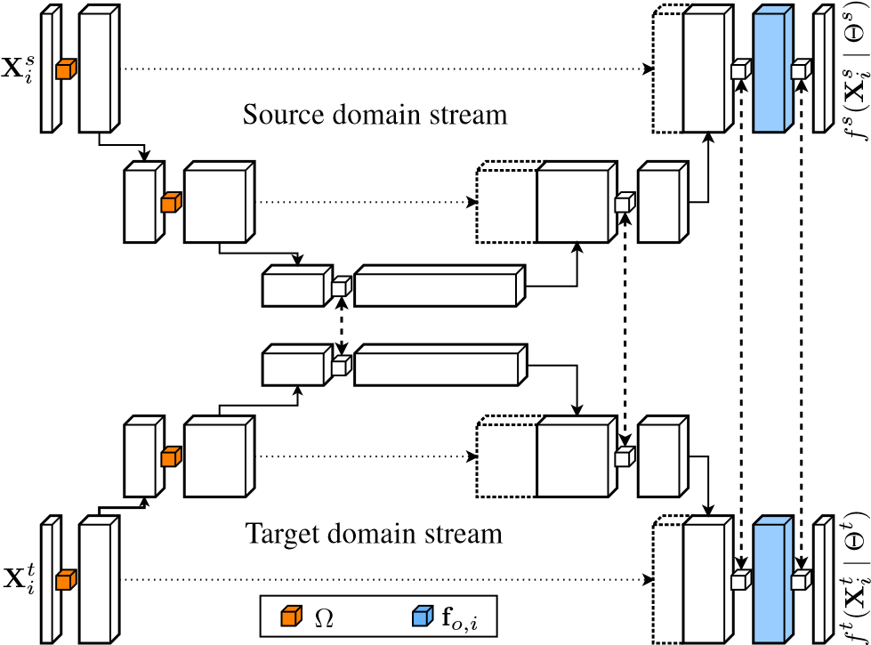
In Bermudez-2018-ADT , since the mitochondria and synapses in the mouse brain under the electron microscope are not easily segmented, a two-stream U-Net with two coupled U-Nets is proposed. Two-stream U-Net is different from the classic U-Net Ronneberger-2015-UCN . First of all, it consists of two symmetrically distributed U-Nets. Furthermore, one U-Net acts on the source domain and the another one acts on the target. Finally, the weight is shared by two U-Nets. Like Jha-2020-DAD ; Zhuang-2018-LMN , two-stream U-Net consists of two similar U-Nets. Unlike Jha-2020-DAD ; Zhuang-2018-LMN , two U-Nets of two-stream U-Net are trained by the source domain and the target domain, respectively. However, in Jha-2020-DAD and Zhuang-2018-LMN , the input of U-Net at the back is related to the output of U-Net at the front. TEM volumes of mouse somatosensory cortex and cerebellum are used to test the two-stream U-Net and a JI of 0.7230 is obtained. The architecture of two-stream U-Net is shown in Fig. 9.
In many segmentation tasks, the traditional encoder, decoder and skip connection structure cannot complete the task perfectly and there are few channels for information flow to circulate. In Zhuang-2018-LMN , LadderNet is proposed to solve the problem. The same point as Bermudez-2018-ADT ; Jha-2020-DAD , the weights are shared between the two U-Nets. The difference from Bermudez-2018-ADT ; Jha-2020-DAD , LadderNet shares the weights in the decoding part of the first U-Net and the encoding part of the second U-Net. But, two-stream U-Net shares weights in the decoding part of the two U-Nets. A dataset is obtained from one source: the CHASE DB1 dataset (contains 28 retinal images, of which is used for training and is used for testing). In the dataset, 0.8031 F1-score, 0.7978 SE, 0.9818 SP, 0.9656 ACC and 0.9839 AUC are obtained.
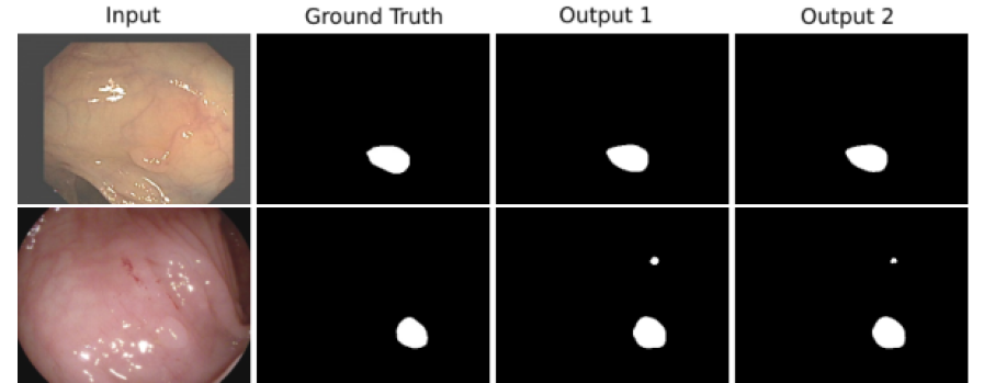
In Jha-2020-DAD , a stacked double U-Net is proposed to obtain better segmentation ACC and is named DoubleU-Net. Like Zhuang-2018-LMN ; Bermudez-2018-ADT , encoding and decoding ideas of U-Net are not changed. Unlike Zhuang-2018-LMN ; Bermudez-2018-ADT , first, the encoding part of a U-Net in DoubleU-Net is replaced with VGG-19 Simonyan-2014-VDC . Second, the information flows from the previous U-Net encoding part to the next decoding part. Third, DoubleU-Net is evaluated by the alike-microscopic dataset. DoubleU-Net is tested on the CVC-ClinicDB dataset Bernal-2015-WMF to obtain a DICE of 0.9239, a mean Intersection over Union (mIoU) of 0.8611, a 0.8457 RE and a 0.9592 PR. The segmentation results of DoubleU-Net are shown in Fig. 10.
The variant structure based on U-Net is not easy to segment adjacent cells, in Torr-2020-DSO DeepSplit is proposed to segment the cell contact areas. Like Bermudez-2018-ADT ; Zhuang-2018-LMN ; Jha-2020-DAD , DeepSplit has multiple U-Net chains with a composite structure appearance. Unlike Bermudez-2018-ADT ; Zhuang-2018-LMN ; Jha-2020-DAD , DeepSplit only has one encoder branch and two decoder branches. The second decoder branch is a separate branch, its main purpose is to segment the cell contact areas. This method is evaluated on an MCF-10a epithelial breast cells dataset (contains 50 manually annotated images, of which is used for training, of which is used for verification and of which is used for testing). Finally, a cell detection score (CDS) of 0.903 is obtained.
In Bozkurt-2018-AMC , a MUnet nested with three U-Nets is proposed, which is used to segment the image of morphological patterns of human skin under reflectance confocal microscopy (RCM). Because the traditional method is time-consuming, MUNet is used to assist in the diagnosis of this type of skin cancer. 56 RCM mosaics (46 mosaics are used for training and 10 mosaics are used for testing) annotated by experts with 6 types of labels are used to evaluate MUnet. Because three nested U-Net structures are designed, the segmentation operation of MUet can be performed at different resolutions. Finally, for the background segmentation under the dataset, MUNet obtains a SE, a SP, a DICE and a PR.
In Zhao-2020-TUH , a Triple U-Net is proposed for nuclear segmentation to avoid blurry tumor nucleus boundaries and overlapping tumor cells as much as possible. Like Bozkurt-2018-AMC , the Triple U-Net is composed of three U-Net branches: a red-green-blue (RGB) branch, a Hematoxylin branch and a Segmentation branch. Unlike Bozkurt-2018-AMC , two branches of the Triple U-Net are composed of Progressive Dense Feature Aggregation Module (PDFA) based on the densely connected block (be introduced by Huang-2017-DCC ). This work utilizes the MoNuSeg dataset from 7 organs which contain 30 images in total, 21000 nuclear boundaries are annotated. In the total dataset, 16 images are used for training and 14 images are used for testing. Comparison of Triple U-Net and existing nuclei segmentation models, the final Triple U-Net result is the best (0.837 DICE).
3.4 Attention U-Net
An attention mechanism is introduced into U-Net in Oktay-2018-AUL and applied to a CT dataset to highlight features. In Lian-2018-AGU , an ATTention U-Net (ATT-UNet) is proposed to segment an iris to solve a problem that a segmentation network is susceptible to irrelevant noise pixels outside the iris area. ATT-UNet enables a network to focus on a region of interest (ROI), avoid wasting time and over-computing features of irrelevant regions. The purpose of the attention mechanism is introduced to guide ATT-UNet to learn more features to separate the iris and non-iris pixels. An attention mask is generated to evaluate the most likely areas of the iris and a bounding box regression module is used to evaluate the coordinates. Furthermore, the attention mask guides ATT-UNet to segment the specific area. A dataset employed in the experiments comes from UBIRIS.v2 Proencca-2009-TUA , of which 500 images are used as the training set and 500 images are used as the test set. In the end, an experiment result shows: an IoU of is obtained.
In Lv-2020-AGU , an attention guided U-Net with atrous convolution (AA-UNet) is proposed to segment retinal blood vessels. A precise segmentation of retinal blood vessels has an important auxiliary role in the diagnosis of diabetes, hypertension and other diseases. Like Lian-2018-AGU , an attention module is used to force the network to pay attention to ROI. Unlike Lian-2018-AGU , atrous convolution replaces ordinary convolution in the feature blocks, which is beneficial to increase the receptive field. AA-UNet is tested on three retinal vessels segmentation datasets (DRIVE Bansal-2013-RVD , STARE Guo-2018-ARV and CHASEDB1 Thangaraj-2018-RVS ). The DRIVE (Digital Retinal Images for Vessel Extraction) dataset contains 40 fundus images, of which 20 images are used for training and 20 images are used for testing. Under the DRVIE dataset, AA-UNet obtains ACC, F1-scores, Jaccard similarity (JS) and AUC.
In Mou-2019-CCA , in order to segment curved structures (such as blood vessels), CS-Net is proposed to assist experts in diagnosing diseases. Channel attention block (CAB) and spatial attention block (SAB) with attention ideas are integrated into the baseline U-Net. Attention idea appears in the form of a module after the encoder. STARE is a fundus dataset used to evaluate this proposed method. An experimental result is obtained by CS-Net ( ACC, SE, SP and AUC).
In Li-1903-CSA , a connection sensitive attention U-Net (CSAU) is proposed to segment retinal blood vessels. Like Mou-2019-CCA ; Lv-2020-AGU ; Jiang-2020-multi , CSAU is proposed to segment retinal blood vessels. Unlike Mou-2019-CCA ; Lv-2020-AGU ; Jiang-2020-multi , a connection sensitive loss is proposed and combines with attention gates. CSAU is trained on the DRIVE, STARE and HRF datasets, respectively. STARE contains 20 fundus images, of which are used for training and are used for testing. Under the test data of STARE, an experimental result of CSAU is as follows: an F1-score of 0.8435, a SE of 0.8465 and an ACC of 0.9673.
In Jiang-2020-multi , a novel multi-path recurrent U-Net with attention gate (MPAR) is developed to segment retinal fundus images. An innovative idea of many variants of U-Net is that attention is integrated into the baseline U-Net Lian-2018-AGU ; Lv-2020-AGU ; Mou-2019-CCA ; Li-1903-CSA ; Zhang-2020-PCS ; Zhu-2020-SWR ). However, in MPAR, Attention Recurrent Unit (formed by combining recurrent neural network and attention) can further improve target features. Two datasets (Drishti-GS1 dataset Sivaswamy-2015-ACR and DRIVE dataset Bansal-2013-RVD ) are used to evaluate MPAR. The Drishti-GS1 dataset has 101 retinal fundus vascular images. For the Drishti-GS1 dataset, in the training phase, 50 images are used as the training set. In the testing phase, 51 images are used as the test set. Experimental results of testing MPAR under the DRISHTI-GS1 dataset are as follows. For optic disc segmentation, an ACC of and a DICE of are obtained. For optic cup segmentation, an ACC of and a DICE of are obtained.
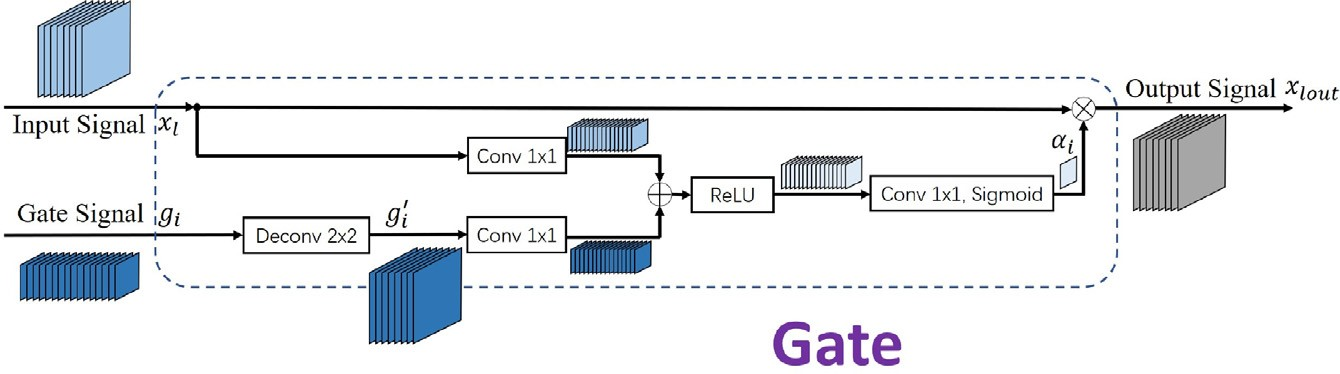
In Zhang-2020-PCS , a method based on ATT-UNet and graph-based Random Walk (RW) is proposed to extract nucleus and cytoplasm from overlapping cervical cells. This method proposed is mainly the following four steps: (1) ATT-UNet is used to separate the nuclei; (2) images are acquired by polar coordinate sampling; (3) ATT-UNet predicts the cytoplasm boundary; (4) RW is used to refine the cytoplasm boundary. Because of the repeated operation of the encoding part, some spatial detail information is lost. Attention Gates (AG) Oktay-2018-AUL is used to obtain the missing information. The architecture of Attention Gate mentioned in Fig. 11. The experimental data are training images of the ISBI 2014 Challenge Dataset. 135 synthetic cervical cytology images from eight training depth of field (EDF) images are splited into 45 images (train set) and 90 images (test set). Under the test set, a 0.93 DICE is obtained.
In Zhu-2020-SWR , irregular cell boundaries are often difficult to segment. Residual Attention U-Net (ResAttU-Net) is proposed to segment the cells in fluorescence widefield microscopy images. Like Zhang-2020-PCS , AG is applied to ResAttU-Net. Unlike Zhang-2020-PCS , Residual blocks He-2016-DRL are integrated into ResAttU-Net. This work utilizes a baby hamster kidney (BHK) cell dataset from Columbia University in the Department of Biomedical Engineering. After data enhancement such as random horizontal and vertical flipping, 4600 cell images constitute the training set, 1000 images constitute the training set and 500 images constitute the test set. This proposed method obtains good experimental results (SE=0.97, SP=0.93, F1-score=0.95, JS=0.91 and DICE=0.95).
In Xiancheng-2018-RBV , in order to assist ophthalmologists in the diagnosis of eye diseases, a method that relies on data enhancement and combined with U-Net is proposed to segment retinal blood vessel images. Like Fabijanska-2018-SOC , an identical structural deformation is applied to the baseline U-Net. Unlike Fabijanska-2018-SOC , this method is to segment the retinal blood vessels images. 20 DRIVE training images generate 19,000 patches to form a dataset. From the training set, of samples are used for training and the remaining are used for validation. This experiment obtains satisfactory results.
In Leng-2018-CUF , in order to fully consider contextual information, a context-aware U-Net is proposed to conduct the segmentation tasks of the Drosophila first instar larva VNC. Like Chidester-2019-ERU , the context-aware U-Net improves the long connection by placing a model on the connection. Unlike Chidester-2019-ERU , the context-aware U-Net places a context transfer module instead of a Residual module. A dataset used for the Drosophila first instar larva ventral nerve cord images segmentation in experiments from EM segmentation challenge (ISBI 2012). A training data is 30 serial section transmission electron microscopy images. The context-aware U-Net obtains a segmentation result: A warping error of 0.000121, a rand error of 0.0212 and a pixel error of 0.0346.
3.5 Dense U-Net
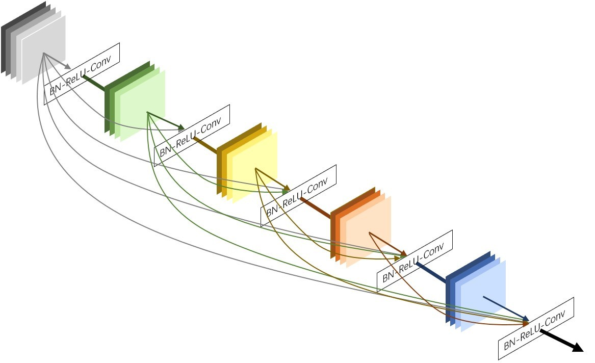
A densely connected convolutional network (DenseNet) is proposed Huang-2017-DCC,Huang-2017-MDC. The feature of a dense U-Net is that in the dense block, each layer is directly connected to the previous layer. The structure of the dense block is shown in Fig. 12. The advantage of this structure is that the vanishing gradient problem can be alleviated. In Jegou-2017-TOH , DenseNet is modified and integrated into U-Net to propose a new neural network and used to segment some trial scenes.
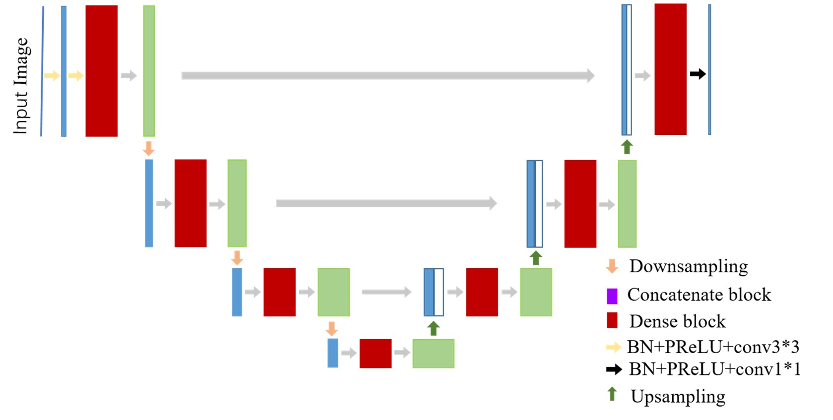
In Cheng-2020-RBV , a new dense block-based U-Net is proposed to perform segmentation tasks of blood vessels. The structure of the new block-based dense U-Net is shown in Fig. 13. It differs from the baseline U-Net in: (1) dense block replaces ordinary convolutional blocks (in Zhao-2020-TUH , a PDFA module improves densely connected block); (2) Parametric Rectified Linear Unit (PReLU) He-2015-DDI replaces ReLU; (3) encoder and decoder part is missing a convolution block. two datasets (CHASEDB1 dataset Owen-2009-MRV and DRIVE dataset Bansal-2013-RVD ) are used to evaluate the new dense block-based U-Net. The DRIVE dataset has 40 retinal fundus vascular images. For the DRIVE dataset from a diabetic fundus lesion screening organization, in the training phase, 20 images are used as the training set. In the testing phase, 20 images are used as the test set. Experimental results of testing the new block-based dense U-Net under the DRIVE dataset are as follows: 0.9834 SP, 0.7672 SE, 0.9559 ACC and 0.9793 AUC.
In Wang-2019-DUB , an improved U-Net combining patch-based learning strategy and dense idea is proposed to segment retinal vessels. Like Cheng-2020-RBV , compared with the baseline U-Net, dense block replaces ordinary convolutional blocks and the number of convolutional blocks is reduced. Unlike Cheng-2020-RBV , the Residual idea is applied before the dense block and the convolution block is reduced by two blocks in the encoder and decoder parts. This work utilizes two public datasets (DRIVE and STARE). For the DRIVE dataset, during the training process, 40000 image patches are extracted from 20 source images, of which are used for cross-validation. Finally, 0.7986 SE, 0.9736 SP, 0.9511 ACC and 0.9740 AUC are obtained.
In order to use deep learning models to assist pathologists in achieving precise treatment, in Samanta-2021-CAN , a new improved U-Net (HistNet) is proposed for segmentation of colorectal histopathology. Like Cheng-2020-RBV ; Wang-2019-DUB , dense blocks are applied to U-Net. Unlike Cheng-2020-RBV ; Wang-2019-DUB , dense blocks are improved (modified dense block uses dilated convolution). To evaluate the proposed model thoroughly, the DigestPath 2019 dataset Li-2019-SRC and the Gland Segmentation (GlaS) dataset (in Histology Image Challenge held at MICCAI 2015) Sirinukunwattana-2017-GSI are applied. For DigestPath 2019 dataset, it contains 660 tissue images from 324 WSI, the tissue images are randomly divided into training, validation and test sets at a ratio of . Finally, DICE and IoU are obtained by HistNet.
Since, biomedical image segmentation plays an important role in diagnosing diseases, therefore, a novel Multi-scale Dense U-Net (MDU-Net) is proposed to segment biomedical images Zhang-2018-MMD . Like Cheng-2020-RBV ; Wang-2019-DUB , MDU-Net uses a dense idea. Unlike Cheng-2020-RBV ; Wang-2019-DUB , MDU-Net is a multi-scale densely idea, more precisely, Cross Dense connections, Up Dense connections, Down Dense connections are widely used in the MDU-Net, but MDU-Net does not have dense blocks. A dataset containing 165 biomedical images (originate from Histology Image Challenge held at MICCAI 2015) is used. The dataset is divided into two subsets, the first subset (containing 85 images) is used for training and the second subset (containing 85 images) is used for testing. Finally, experiment reveals that a DICE obtained by this proposed method is higher than baseline U-Net.
In Liu-2018-DCS , a Densely Connected Stacked U-Network (DCSU) is used to segment confocal microscopy images of filament. Unlike Cheng-2020-RBV ; Wang-2019-DUB ; Zhang-2018-MMD , DCSU is a cascaded U-Net (combination of multiple U-Nets, the output of previous level U-Net is related to the input of the next U-Net Wu-2019-ABS ) and dense connections occur between convolutional blocks of different U-Nets. A microtubule dataset containing 5032407 training patches is proposed to evaluate DCSU. Under the microtubule dataset, this proposed method obtains an IoU of 0.9439 and a Skeletonized IoU (SKIoU) of 0.9775.
3.6 U-Net redesigned Skip Connections
In Zhou-2018-UAN , a simple and effective modified U-Net that redesigns the original U-Net skip connection is creatively proposed. It reduces the loss of information in the copy aggregation from the encoder to the decoder. For different datasets, the importance of each convolutional layer is different. The information carried by the third layer may be effective for the segmentation of the dataset or it may be the second layer. The advantage of this modified U-Net is that all copy aggregation operations contain all the depth feature information of the previous convolutional layer. A data from Data Science Bowl 2018 containing 670 nuclear images are used. Under the dataset, an IoU of core segmentation result is obtained. Fig. 14 shows the proposed deformable U-Net and the analysis of the variant thinking.
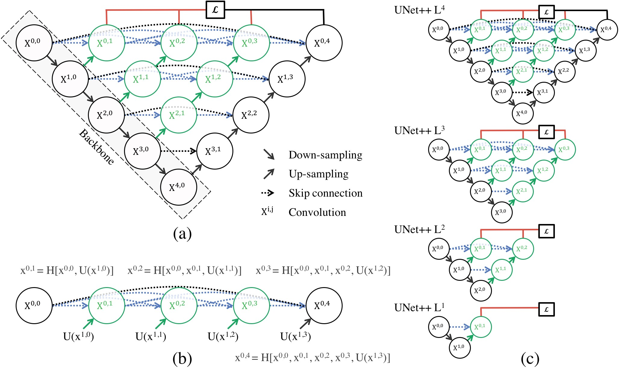
In Zhou-2019-URS , a U-Net that changes skip connection is explored in more depth (similarly, Zhou-2018-UAN is the study of skip connection from the same team). A purpose of the U-Net that changes the skip connection is to reduce the loss of information when convolutional layers of different depths are aggregated and connected. Like Zhou-2018-UAN , the deformed U-Net is deleted some mandatory aggregations (or different depth features are extracted), different performances are obtained. Unlike Zhou-2018-UAN , the deformed U-Net is evaluated by more datasets.
In Wang-2020-AIB , a powerful improvement U-Net++ is proposed to segment tiny breast cancer nuclei. The difference between the improved U-Net++ and the baseline U-Net++ Zhou-2018-UAN is that an Inception-Resnet-V2 network is integrated into U-Net++ and the network improves U-Net++ segmentation capabilities. This work utilizes a dataset from Janowczyk-2016-DLF , which contains 141 RGB HE stained estrogen receptor positive (ER+) breast cancer images in total. Finally, the dataset has a total of 3366 sub-images after being resized and cropped. The proportions of the training set, validation set and test set are , and , respectively. The improved U-Net++ is evaluated by this dataset and obtains 0.9505 ACC, 0.5581 PR, 0.6035 RE and 0.5207 DICE.
Doppler optical coherence tomography (OCT) vessels images can observe the vascular structure and blood flow to facilitate surgery. So in Wu-2019-ABS , a cascaded U-Net (CU-net) is proposed to segment the vascular intensity images, the boundary image of the outer vessel wall and the inner blood flow lumen (this cascaded U-Net idea is also reflected in Liu-2018-DCS ). In Cu-Net, the first U-Net segment the intensity image, then the segmentation result is used as the input of the second U-Net (as a mask, it is better to select the region of interest and remove the non-target region). The above operations can reduce training time. This method is examined on a traceable dataset of Doppler OCT images of mouse arteries. The dataset used in the experiments is consists of 190 images, of which 150 images are used for training and 40 images are used for testing. These experimental results show that for the segmentation of the outer vessel wall boundary, CU-net obtains an ACC of , furthermore, for the segmentation of the contour of the inner blood flowing lumen area, CU-net obtains an ACC of .
In Tsunomura-2020-SOM , a dyed particle usually needs to be divided manually by an expert and the reproducibility of the operation is low. Therefore, a simple and effective deformed U-Net is proposed to solve the problem and achieve precise segmentation. A dataset is composed of art papers printed by mixing carbon black pigments and inks, with a total of 60 images. First, improved U-Net is obtained by changing the number of channels. Then, improved U-Net outputs high-precision large particle segmentation results. Finally, the segmentation results are applied to improved U-Net (delete some skip connections) to obtain the best segmentation results. Two different variants of U-Net combined with the proposed method flow obtain a root mean squares error (RMSE) of 45.2187 and a standard deviation of 7.8371.
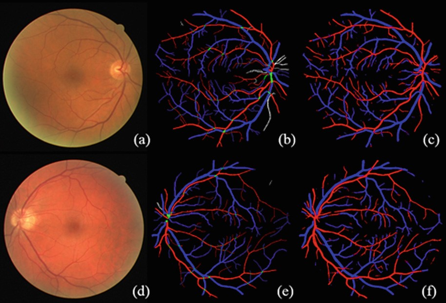
Hence, the segmentation of retinal arterioles and venules has an important auxiliary role in the diagnosis of eye diseases. Therefore, in Xu-2018-AIU , an improved U-Net is proposed to segment arterioles and venules. Like improved U-Net from Tsunomura-2020-SOM and improved U-Net from Xu-2018-AIU deletes the connections. On the contrary, improved U-Net from Tsunomura-2020-SOM and improved U-Net from Xu-2018-AIU only deletes the fourth connection between the encoder 512-channel feature map. The segmentation results of the proposed method are shown in Fig. 15. This experiment uses a DRIVE dataset curated by a diabetic retinopathy screening program. DRIVE (contains 40 color fundus photographs) is used to evaluate six different segmentation methods. Finally, the improved U-Net obtains an optimal experimental result (0.870 SE and 0.980 SP).
In Liang-2020-WPM , in order to better realize target spore identification and spore count, an effective variant U-Net is proposed to segment wheat powdery mildew spore images. The variant U-Net has two differences from the baseline U-Net as follows: The variant U-Net deletes the three skip connections in the baseline U-Net and adds the pyramid pool module after the encoding is completed. Like improved U-Net from Tsunomura-2020-SOM and improved U-Net from Xu-2018-AIU , the variant U-Net deletes some connections. Unlike improved U-Net from Tsunomura-2020-SOM and improved U-Net from Xu-2018-AIU , the variant U-Net from Liang-2020-WPM only 1 connection between encoder 512-channel feature map is reserved. 835 wheat powdery mildew spore images are divided into a training set and a test set, of which 550 images are used as training set and 285 images are used as the test set. The variant U-Net obtains an MIoU of experimental results.
3.7 Summary
From the survey above, since 2016, U-Net has improved, but after 2018, deep neural networks based on U-Net become more and more widely used in the field of biomedical image segmentation. The main reasons for this development trend are as follows: (a) More advanced computers are developed, which can handle more complex networks. (b) The improved U-Net architecture that can avoid problems such as over-fitting and enhanced computing time. Table 2 summarizes the work done by different teams in using the improved U-Net to analyze microscopic images.
| Aim | Detial | Year | Reference | Team | Data Information | CNN type: points of improvement | Evaluation | |||||||||||
| semi-automatic segmentation | Xenopus kidney slices | 2016 | Cciccek-2016-3UN | A. Abdulkadir, et al. | 77 images | 3D U-Net | IoU = 86.3% | |||||||||||
| segmentation | 3D synthetic data | 2018 | Fu-2018-TDF | C. Fu, et al. | 3D synthetic data | 3D U-Net | ACC = 95.56% | |||||||||||
| segmentation | cell membrane | 2019 | Eschweiler-2019-CBP | D. Eschweiler, et al. |
|
3D U-Net combined with SWS | JI = 87%, DICE = 93.1% | |||||||||||
| segmentation | nerve tissue | 2018 | Heinrich-2018-SCS | L. Heinrich, et al. |
|
3D U-Net | CREMI score = 50 | |||||||||||
| segmentation | neurons | 2019 | Wang-2019-SNS | H. Wang, et al. |
|
|
\ | |||||||||||
| segmentation and classification | SCD cells | 2017 | Zhang-2017-ISA | M. Zhang, et al. | 128 images | variable convolution is applied |
|
|||||||||||
| 2018 | Zhang-2018-RSS | 266 original images |
|
|||||||||||||||
| segmentation | cell nuclei | 2020 | Qin-2020-MFU | X. F. Qin, et al. |
|
Match Feature U-Net | IoU = 91.83% | |||||||||||
| segmentation | trophectoderm | 2020 | Rad-2020-TSI 58 | R. M. Rad, et al. |
|
|
\ | |||||||||||
| segmentation | Rift V alley virus | 2018 | Matuszewski-2018-MAT | D. J. Matuszewski, et al. | 143 TEM images |
|
|
|||||||||||
| segmentation | circulating tumor cells | 2018 | Mocan-2018-ADO | I. Mocan, et al. |
|
Convolution block increased | ACC = 99.81% | |||||||||||
| segmentation | endothelial cells | 2018 | Fabijanska-2018-SOC | A. Fabijanska, et al. |
|
Convolution blocks are reduced | DICE = 86% | |||||||||||
| segmentation | nuclei | 2019 | Xu-2019-UFR | Z. Y. Xu, et al. |
|
|
\ | |||||||||||
| automate segmentation | histopathological | 2017 | Li-2017-NDL | W. Z. Li, et al. |
|
|
\ | |||||||||||
| segmentation | ventral nerve cord | 2020 | kumar-2020-CSB 67 | C. A. Kumar, et al. | 120 images |
|
IoU = 92.54% | |||||||||||
| segmentation | mitochondria and synapses | 2018 | Bermudez-2018-ADT | R. Bermudez, et al. | \ | 2 distributed U-Nets | JI = 72.3% | |||||||||||
| segmentation | retinal images | 2018 | Zhuang-2018-LMN | J. T. Zhuang, et al. |
|
|
|
|||||||||||
| segmentation | Colorectal polyps | 2020 | Jha-2020-DAD | D. Jha, et al. | CVC-ClinicDB dataset | DoubleU-Net: the information flows from the previous to the next decoding part |
|
|||||||||||
| segmentation | adjacent cells | 2020 | Torr-2020-DSO | A. Torr, et al. |
|
|
|
|||||||||||
| segmentation | human skin | 2018 | Bozkurt-2018-AMC | A. Bozkurt, et al. |
|
Munet: nested with three U-Nets |
|
|||||||||||
| segmentation | nuclear | 2020 | Zhao-2020-TUH 75 | B. Zhao, et al. |
|
|
|
|||||||||||
| segmentation | iris | 2018 | Lian-2018-AGU | S. Lian, et al. |
|
ATT-Unet: an attention mask is generated | IoU of 91.37% | |||||||||||
| segmentation | retinal blood vessels | 2020 | Lv-2020-AGU | Y. Lv, et al. |
|
|
|
|||||||||||
| segmentation | retinal blood vessels | 2019 | Mou-2019-CCA | L. Mou, et al. | STARE dataset | CS-Net: CAB and SAB is added |
|
|||||||||||
| segmentation | retinal blood vessels | 2019 | Li-1903-CSA | R. Li, et al. |
|
CSAU: attention gates is added |
|
|||||||||||
| segmentation | retinal fundus | 2020 | Jiang-2020-multi | Y. Jiang, et al. |
|
AG is added |
|
|||||||||||
| segmentation | cells | 2020 | Zhang-2020-PCS | H. Zhang, et al. | 135 images | AG is added |
|
|||||||||||
| segmentation | cell boundaries | 2020 | Zhu-2020-SWR | N. Y. Zhu, et al. |
|
|
|
|||||||||||
| segmentation | retinal blood vessel | 2018 | Xiancheng-2018-RBV | W. Xiancheng, et al. |
|
|
\ | |||||||||||
| segmentation | l nerve | 2018 | Leng-2018-CUF 90 | J. X. Leng, et al. | 30 images for training |
|
|
|||||||||||
| segmentation | retinal fundus vascular | 2020 | Cheng-2020-RBV | Y. L. Cheng, et al. |
|
|
|
|||||||||||
| segmentation | retinal vessels | 2019 | Wang-2019-DUB | C. Wang, et al. | 40,000 image patches |
|
|
|||||||||||
| segmentation | colorectal histopathology | 2021 | Samanta-2021-CAN | P. Samanta, et al. |
|
|
|
|||||||||||
| segmentation | biomedical images | 2018 | Zhang-2018-MMD | J. W. Zhang, et al. |
|
|
\ | |||||||||||
| segmentation | filament | 2018 | Liu-2018-DCS 104 | Y. Liu, et al. | 5032407 training patches |
|
IoU = 97.75%, | |||||||||||
| segmentation | nuclear | 2018 | Zhou-2018-UAN | Z. W. Zhou, | 670 images |
|
IoU of 92.63%, | |||||||||||
| segmentation | cell, nuclei | 2019 | Zhou-2019-URS | Z. W. Zhou, | a new skip connection is applied |
|
||||||||||||
| segmentation | nucle | 2020 | Wang-2020-AIB | H. Wang, |
|
|
|
|||||||||||
| segmentation | OCT vessels | 2019 | Wu-2019-ABS | C. C. Wu, |
|
|
ACC = 94.8% ± 0.2% | |||||||||||
| segmentation | pigments and inks | 2020 | Tsunomura-2020-SOM | M. Tsunomura, | 60 images |
|
|
|||||||||||
| segmentation | color fundus photographs | 2018 | Xu-2018-AIU | X. Y. Xu, | DRIVE: 40 images |
|
|
|||||||||||
| segmentation | mildew spore | 2020 | Liang-2020-WPM 114 | X. S. Liang |
|
|
MIoU = 91.477% |
4 Modified U-Net Related to Residual Block and Residual Idea
4.1 Only Residual Block
A depth of the network affects the extraction of features, but, as the network becomes deeper, the problem of overfitting and training errors in the network increases significantly. A Residual block is proposed to reuse the features of the previous layer to train a deeper network He-2016-DRL . In Patel-2019-CSO , an improved U-Net based on Residual blocks has introduced. It is used to segment the regions of human-induced pluripotent Retinal Pigment Epithelial stem cells (iRPE) under Bright-field microscopy. This improved U-Net is tested on 1032 absorbance images of IRPE cells from Age-related Macular Degeneration (AMD) patients. In the dataset, the training set is composed of 800 images and the verification set is composed of 232 images. The experiment shows that this method is superior to the prior art method and the proposed deformed U-Net obtains the experimental result of a DICE of 0.8366.
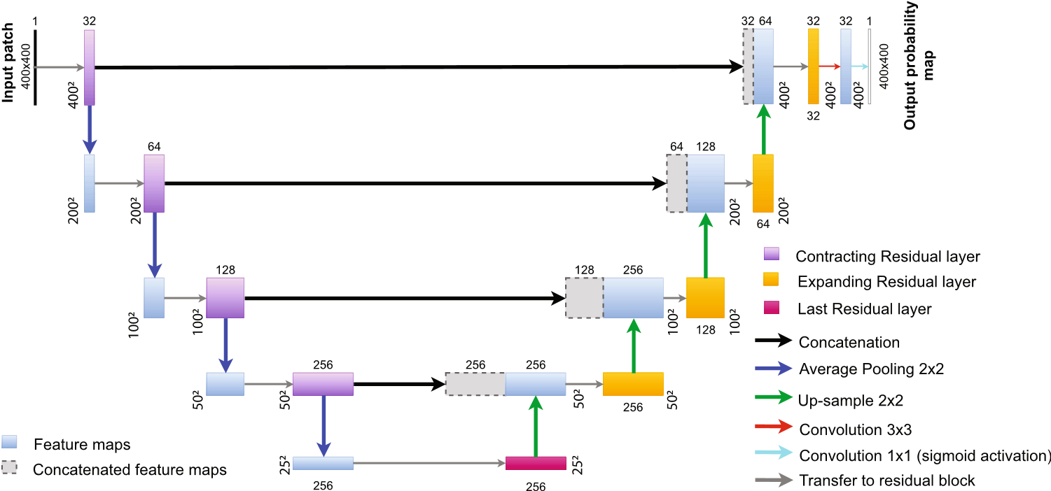
In Gomez-2019-DLB , a variant of U-Net with 9 Residual layers is proposed. It is used for Small extracellular vesicles (sEVs) collected by TEM to achieve good detection and segmentation performance. The architecture of variant U-Net is shown in Fig. 16. 688 sEVs of Mouse fibroblasts (L-cells), mouse embryonic fibroblast (MEF), human embryonic kidney 293 (HEK-293) and the ovarian cancer cell are used as a dataset. Under the dataset, the proposed method obtains a JS of 0.88.
In Quan-2016-FAF , FusionNet with Residual block based on U-Net is proposed to automatically segment neuronal cells. The biggest difference between FusionNet and the original U-Net is that a Residual block containing 3 convolutions is added to each block. Like Patel-2019-CSO ; Gomez-2019-DLB , 3 variable U-Nets proposed in the three papers are all based on U-Net and add Residual blocks. Unlike Patel-2019-CSO ; Gomez-2019-DLB ; Patel-2019-CSO ; Gomez-2019-DLB transformed the original convolution block into a Residual block, FusionNet retains the original convolution block and inserts Residual blocks with three convolutional. FusionNet is trained on 30 sections of Drosophila Electron Microscopic images (from Arganda-2015-CTC ) and tested on a private dataset.
In Mehta-2018-YJS , a simple concept network named Y-Net is used to segment different tissues in breast biopsy images to generate segmentation masks to assist breast cancer diagnosis. Y-Net differs from the baseline U-Net in that: Pyramid spatial pooling (PSP) and Residual convolutional blocks (RCB) are integrated into Y-Net, Y-Net adds two new hyperparameters and new fully connected layers.
The breast biopsy dataset containing 58 regions of interest (ROIs) is divided into a training set containing 29 images and a test set containing 29 images. Y-Net obtains a mIoU of experimental results.
However, a large number of manual annotations are time-consuming and labor-intensive, a Multi-Tasking U-Net is proposed to solve this problem Ke-2019-AMU . The Multi-Tasking U-Net is trained by coarse data labels to combine with a few pixel-wise annotations images. A Residual Multi-Tasking Block is proposed, each Multi-Tasking Block has three paths. The Multi-Tasking Block is composed of several sub-blocks with three tasks: task one is detection, task two is separation and task three is segmentation. Like Torr-2020-DSO , the Multi-Tasking U-Net is also a Multi-Tasking network. Unlike Torr-2020-DSO , the multi-tasking process of Multi-Tasking U-Net is embodied in a module instead of a decoder and Multi-Tasking U-Net has 1 more detection task. A training dataset contains 20 ice-cream Scanning Electron Microscopy (ESM) images and a test dataset contains 12 ice-cream SEM images. This Multi-Tasking U-Net obtains a DICE of 0.94 experimental results.
4.2 Residual Block Skip Connection
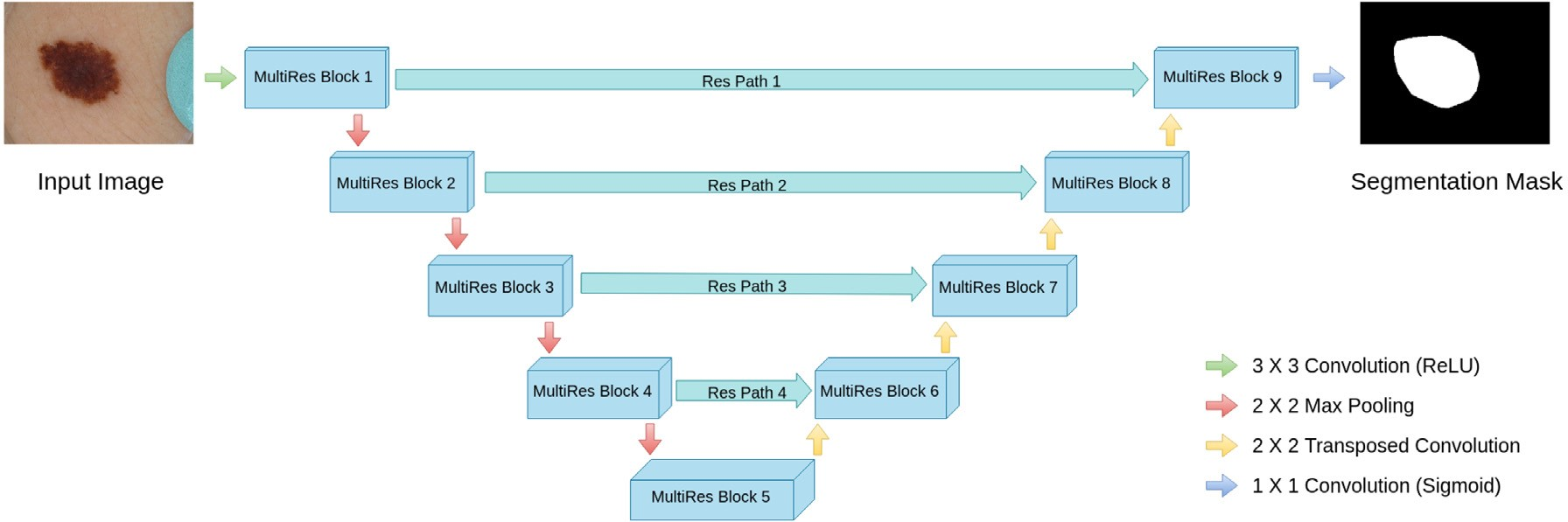
In Ibtehaz-2020-MRT , in order to solve a problem that the classical U-Net does not perform well in the segmentation of some challenging datasets, MultiResUNet is proposed. Compared with the classical U-Net, the improved performance of MultiResUNet lies in the following two points. First, the MultiRes block is proposed and replaces all 2D convolution blocks in the classical U-Net. Second, a Res path with four convolution and four short connections of Residual properties are creatively proposed to replace skip connections. MultiResUNet is tested and evaluated on five datasets, one of them obtains a JI of experimental results (the dataset containing 97 fluorescence microscopy images from Coelho-2009-NSI ). This architecture of MultiResUNet mentioned is shown in Fig. 17.
In Lou-2021-DRT , to minimize the limitations of the classical U-Net in some aspects and obtain more accurate segmentation results, DC-UNet is proposed. Like Ibtehaz-2020-MRT , the network structure of both uses the same Res path instead of skip connection. Unlike Ibtehaz-2020-MRT , DC-UNet creates a Dual-Channel block (consisting of six Residual convolutions in two rows) to replace all convolution blocks of the original U-Net. The dataset from the ISBI 2012 challenge (a training set of 30 images and a test set of 30 images) is used to evaluate DC-UNet. An experiment is carried out 5-fold cross-validation and the experimental result of an average ACC of is obtained.
In Gadosey-2020-SSD , in order to segment biomedical images quickly, effectively and with high PR, SD-UNet is proposed. It has three advantages: small model size (23 times smaller than U-Net), fewer parameters (eight times less than U-Net) and fast computing time. Compared with U-Net, its change is that a SD-UNet block is designed and replaces the original convolution block. From International Symposium on Biomedical Imaging (ISBI) challenge dataset, 30 fruit fly images under ssTEM are used to test SD-UNet to obtain an IoU of and a DICE of .
In Arbelle-2019-MCS , a Convolutional Long Short Term Memory (C-LSTM) block is integrated into U-Net and LSTM U-Net is proposed. The limitation of time information is incorporated into LSTM U-Net. Like Gadosey-2020-SSD , a new module is designed to replace the original convolution module. Unlike Gadosey-2020-SSD , the C-LSTM blocks of LSTM U-Net only exist in the encoder part. The structure facilitates the segmentation of single touch cells and partially visible cells. Furthermore, similar to an application of the LSTM module is discussed in this paper Abdallah-2020-RAR , which an extended LSTM block is designed based on the LSTM module and a Res block is designed based on the baseline U-Net and Residual. The LSTM U-Net is tested by the fluorescent simulated dataset (Fluo-N2DH-SIM+) in the Cell Tracking Challenge and a quantitative result of 0.811 is obtained.
In Chidester-2019-ERU , since U-Net still has room for improvement in the task of nuclear segmentation, Rotation-Equivariant U-Net (REU-Net) is proposed to be used for histopathological images of seven different organs. Like Ibtehaz-2020-MRT ; Gadosey-2020-SSD ; Lou-2021-DRT , the REU-Net adds Residual blocks and modifies long connection. Unlike Ibtehaz-2020-MRT ; Gadosey-2020-SSD ; Lou-2021-DRT , in REU-Net, a long connection is between the second encoder and the third encoder. The dataset contains of 30 pathological images curated by Kumar-2017-ADA . In the dataset, 4 images are used as the training set and 7 images are used for validation and testing set respectively. The proposed method obtains an experimental result of an aggregated JS of 0.6291, an F1-score of 0.8469 and a DICE of 0.7980.
4.3 Residual Block Recurrent
Models such as CNN can not accurately simulate high-level dependencies between object boundary points. Furthermore, in order to prevent overfitting and reduce the computational time, Recurrent Active Contour Evolution Network (RACE-Net) is proposed to segment Optic disc (OD) and Optic Cup (OC) in fundus images Chakravarty-2018-RAR . Feedforward neural network (FFNN) architecture is added to RACE-Net to simulate every step of the curve evolution. A generalized the level set based deformable models (LDM) evolving is simulated by RACE-net. RACE-Net utilizes the DRISHTI-GS1 dataset from Sivaswamy-2015-ACR , which contains 101 images in total (50 for training and 51 for testing). This RACE-Net-based method has the following experimental results. For OD segmentation, a DICE of 0.97 and a Boundary Localization Error (BLE) of 6.06 are obtained. For OC segmentation, a DICE of 0.87 and a BLE of 7.63 are obtained.
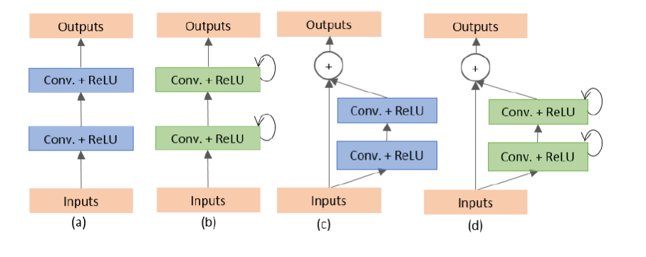
In Alom-2018-RRC ; Alom-2019-RRU , Recurrent U-Net (RU-Net) and Recurrent Residual U-Net (R2U-Net) are developed to accurately segment the blood vessel images on the retina and skin cancer images. The biggest difference between RU-Net, R2U-Net and the baseline U-Net Ronneberger-2015-UCN is that the convolution units are different. RU-Net uses recurrent convolutional layers (RCL) and R2U-Net uses recurrent Residual convolutional layers (RRCL). Different variants of convolutional and recurrent convolutional units are shown in Fig. 18. The dataset contains 25000 patches of 20 images from Structured Analysis of the Retina (STARE) and CHASEDB1 (of which is train set and is validation set) is used to train and verify RU-Net, R2U-Net. RU-Net obtains experimental results of an F1-score of 0.8396, a SE of 0.8108, an SP of 0.9871, an ACC of 97.06% and an AUC of 0.9909, respectively. R2U-Net obtains the experimental results of an F1-score of 0.8475, a SE of 0.8298, an SP of 0.9862, an ACC of 97.12% and an AUC of 0.9914, respectively.
In Alom-2018-NSW (from the same team as Alom-2018-RRC ), R2U-Net is applied to the segmentation task of a nuclear cell. An unfolded version of the recurrent convolutional units is proposed. Like Alom-2018-RRC , R2U-Net from Alom-2018-NSW has the same structure. Unlike Alom-2018-RRC , R2U-Net from Alom-2018-NSW is applied to nuclear segmentation for the first time. This R2U-Net is evaluated on the Data Science Bowl Grand Challenge in 2018 (a total of 735 images, of which 536 are used for training, 134 are used for validation and 65 are used for testing). Finally, an experimental result shows that a DICE of is obtained on the test set.
In Zahangir-2018-MNC , an R2U-Net model is developed to segment the nucleus. R2U-Net is introduced in Alom-2018-RRC , compared with R2U-Net from Alom-2018-RRC , this R2U-Net has not changed at all. A dataset used for image segmentation in experiments is composed of 735 cell images from the 2018 Data Science Bowl Grand Challenge. The database is divided into two groups: training and testing. The training dataset includes 650 images (in the training set, is used for training and is used for validation), while the testing dataset includes 65 images. Finally, this nuclei segmentation task obtains testing ACC.
In Yang-2020-ESA , a baseline U-Net is introduced to segment label-free multiphoton microscopy (MPM) images of epithelial cells in prostate tissue. Segmentation results (segmented by the baseline U-Net) combined with the input of the baseline U-Net to obtain a merged image to train AlexNet Krizhevsky-2012-ICW for classification. Nine tissue slides and 70 tissue microarray (TMA) cores of prostate cancer (PCa) tissues from 79 patients are used as a dataset. The dataset is curated by the First Affiliated Hospital of Fujian Medical University. Segmentation results are obtained: A mean F1-score of 0.839.
4.4 Inception-ResNet Block
Inception Szegedy-2017-IIA and ResNet He-2016-DRL receive widespread attention since they are creatively proposed. Different receptive fields are the characteristics of Inception structures and ResNet has a unique connection method to avoid vanishing gradients. In Szegedy-2017-IIA , Inception and ResNet are combined to form an Inception-ResNet block that combines the advantages of the above two structures.
In order to solve the problem of spatial information loss caused by continuous pooling and convolution in U-Net, a context encoder network (CE-Net) is proposed to segment medical images Gu-2019-CCE . By combining Inception-ResNet-V2 block and atrous convolution, dense atrous convolution (DAC) blocks are proposed. The structural improvements are: (1) in the coding part, pre-trained ResNet-34 He-2016-DRL replaces original ordinary blocks; (2) DAC blocks and Residual multi-kernel pooling (RMP) blocks are inserted into the context extractor section. This work utilizes an ORIGA dataset from Zhang-2010-OAO , which contains 650 optic disc images in total. The proportions of the training set and test set are and , respectively. Under the dataset, CE-Net obtains an experimental result of an overlapping error of 0.058.
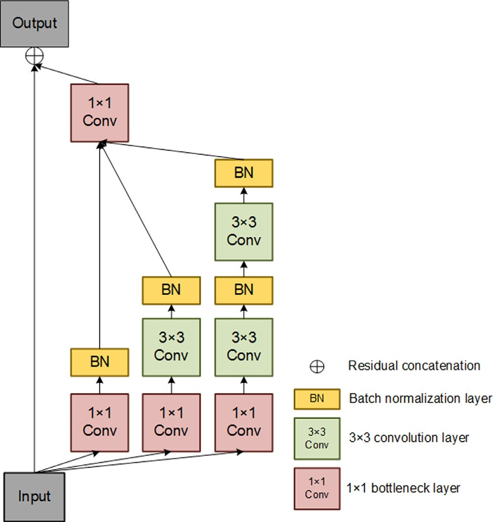
In order to solve the problem of vanishing gradient and excessive computation, in Zhang-2020-DUF , a Dense-Inception U-net (DIU-Net) with an Inception-res block is proposed to segment blood vessels. Like Gu-2019-CCE , Residual (short connection) and Inception are combined to form an Inception-Res block (The structure of the Inception-Res block is shown in Fig. 19). Unlike Gu-2019-CCE , the Dense-Inception block is applied in DIU-Net and the baseline Inception-Res block from Szegedy-2017-IIA is replaced by a modified Residual Inception module. Similar to Lv-2020-AGU , three datasets (DRIVE Bansal-2013-RVD , STARE Guo-2018-ARV and CHASHDB1 Thangaraj-2018-RVS ) are used to evaluate the improved U-Net. The dataset has 136 samples of RGB images, of which is used for training and is used for testing. five-fold cross-validation is used in this experiment. Finally, a good experimental result is obtained (0.9582 DICE, 0.9338 JS, 0.9657 ACC, 0.7967 SE, 0.9863 SP, F0.8003 1-score and 0.9802 AUC).
In order to better distinguish tiny features between different categories, in Huang-2020-MFM , a Mini-Inception-Residual-Dense network (MIRD-Net) is proposed to segment cervical cancer cells, blood vessels, and nuclei. Like Gu-2019-CCE ; Zhang-2020-DUF , MIRD-Net uses Inception blocks and Residual blocks. Unlike Gu-2019-CCE ; Zhang-2020-DUF , MIRD-Net integrates the Inception block, Residual block and dense block into a Mini-Inception-Residual-Dense Block, instead of being used independently for the network. A dataset of 30 images of nuclei from Data Science bowl 2018 is used. Under 5-fold cross-validation, an experimental result of a DICE of 0.954 is obtained.
In Trimeche-2020-FAC , an improved U-Net is proposed to automatically segment retinal vessel branches and bifurcations. In Trimeche-2020-FAC , the baseline convolution block from Ronneberger-2015-UCN is replaced by Residual Inception blocks from Szegedy-2017-IIA . Different from Gu-2019-CCE ; Zhang-2020-DUF ; Huang-2020-MFM , Firesqueeze blocks are inserted into U-Net. The improved U-Net is evaluated on a private dataset containing 65 Adaptive Optics Ophthalmoscopy (AOO) images of the eye fundus. In the dataset, 30 images are selected as the training set, 5 images are selected as the validation set and 30 images are selected as the test set. Finally, experiments reveal that the improved U-Net obtains a PR of 0.97, a RE of 0.96 and an F1-score of 0.96.
4.5 Summary
According to the review above, we can see that, since 2018, modified U-Nets related to Residual block and Residual ideas are widely used. The variant U-Net without the Residual idea has some limitations, such as easy over-fitting and slack training speed, while the Residual U-Net has better encode and decode information. The combination of Residual ideas and Inception, recurrent, skip connection changes can perform their respective advantages and the image segmentation efficiency is better. Table 3 summarizes the work done by different teams in using modified U-Net related to Residual block and Residual idea U-Net to analyze microscopic images.
| Aim | Detial | Year | Reference | Team | Data Information | CNN type: points of improvement | Evaluation | ||||||||||||
| segmentation | Irpe stem cells | 2019 | Patel-2019-CSO | G. Patel, et al. |
|
Residual blocks instead of ordinary blocks | DICE = 83.66% | ||||||||||||
| segmentation | sEVs | 2019 | Gomez-2019-DLB | E. Gomez de Mariscal, et al. | 688 images | 9 Residual layers instead of ordinary blocks | JI = 88% | ||||||||||||
| segmentation | neuronal cells | 2016 | Quan-2016-FAF | T. M. Quan, et al. | 30 for test |
|
|
||||||||||||
| segmentation | tissues in breast biopsy | 2018 | Mehta-2018-YJS | S. Mehta, et al. |
|
|
mIoU = 44.19% | ||||||||||||
| segmentation | ice cream | 2019 | Ke-2019-AMU 120 | R. Ke, et al. |
|
Multi-Task U-Net: each Multi-Task Block has three paths | DICE = 0.94% | ||||||||||||
| segmentation | fluorescence microscopy images | 2020 | Ibtehaz-2020-MRT | N. Ibtehaz, et al. | 97 fluorescence microscopy images |
|
JI = 91.6537% | ||||||||||||
| segmentation | cells | 2021 | Lou-2021-DRT | A. Lou, et al. |
|
|
ACC = 92.62% | ||||||||||||
| segmentation | neuronal cells | 2020 | Gadosey-2020-SSD | P. K. Gadosey, et al. |
|
|
|
||||||||||||
| segmentation | fluorescence microscopy images | 2019 | Arbelle-2019-MCS | A. Arbelle, et al. |
|
|
Quantitative results = 81.1% | ||||||||||||
| segmentation | nuclear | 2019 | Chidester-2019-ERU 91 | B. Chidester, et al. |
|
|
|
||||||||||||
| segmentation | OD and OC | 2018 | Chakravarty-2018-RAR | A. Chakravarty |
|
|
|
||||||||||||
| segmentation | blood vessel | 2018 | Alom-2018-RRC , Alom-2019-RRU | M. Z. Alom, et al. |
|
RU-Net and R2U-Net (use RCL and RRCL) |
|
||||||||||||
| segmentation | nuclear cell | 2018 | Alom-2018-NSW |
|
R2U-Net | DICE = 92.15% | |||||||||||||
| segmentation | nucleus | 2018 | Zahangir-2018-MNC | A. M. Zahangir, et al. |
|
R2U-Net | ACC = 92.15% | ||||||||||||
| segmentation | TMA cores of PCA tissues | 2020 | Yang-2020-ESA 133 | Q. Q. Yang, et al. | 70 TMA cores | baseline U-Net | F1-score = 83.9% | ||||||||||||
| segmentation | optic disc | 2019 | Gu-2019-CCE | Z. W. Gu, et al. |
|
|
Overlapping error = 5.8% | ||||||||||||
| segmentation | blood vessels | 2020 | Zhang-2020-DUF | Z. Zhang, et al. |
|
|
|
||||||||||||
| segmentation | cervical cancer cells | 2020 | Huang-2020-MFM | Y. F. Huang, et al. | 30 images |
|
DICE = 95.4% | ||||||||||||
| segmentation | retinal vessel branches and bifurcations | 2020 | Trimeche-2020-FAC 140 | I. Trimeche, et al. |
|
|
|
5 Analysis of Methodology
5.1 Analysis of Several Typical Improvement Methods for U-Net
According to the survey of improved U-Net: Residual, attention and Inception are used more frequently in the improvement of U-Net. Because different datasets are used, each method cannot be compared longitudinally, so this paper analyzes from the perspective of the neural network itself.
The essence of Residual is a short connection, which means that it adds a skip connection that passes by the nonlinear transformation. A pre-made Reset-34 in the feature encoder module is used to replace the convolutional layer and a maximum pooling layer. Some previous features can be reused, it solves the overfitting problem. Furthermore, because it adds a shortcut mechanism, it avoids the disappearance of the gradient and accelerates the network convergence. The papers involved in this article are Patel-2019-CSO ; Gomez-2019-DLB ; Quan-2016-FAF ; Mehta-2018-YJS ; Ke-2019-AMU .
Inception layers are a method of decomposing several convolutions from 1 convolution. The Inception layer connects convolutions of different filter sizes (, , ) and the mixing pool layer. Because of the difficulty in choosing the type of convolutional layer, Inception is proposed to automate the selection of various layers. The papers involved in this article are Gu-2019-CCE ; Zhang-2020-DUF ; Huang-2020-MFM ; Trimeche-2020-FAC . In deep learning, Inception and Residual are two representative architectures. Inception uses different acceptance fields to expand the architecture. On the contrary, Residual uses a shortcut connection mechanism to avoid explosions and disappear gradients. Because of its existence, the neural network can have thousands of layers. In Szegedy-2017-IIA , the Inception-Reset block combines the advantages of Inception and Residual to form a new module.
Attention is also an important way to improve U-Net, a bounding box of the potential region is regressed, an attention mask is generated and the attention mask is used as a weighted function. It merges discriminative feature maps into the model. Attention gates make the model pay more attention to vascular regions. The attention gate further improves the ACC on detailed vessels by additionally concatenating attention weights to features before output. The papers involved in this article are Lian-2018-AGU ; Lv-2020-AGU ; Mou-2019-CCA ; Li-1903-CSA ; Jiang-2020-multi ; Zhang-2020-PCS ; Zhu-2020-SWR ; Xiancheng-2018-RBV ; Leng-2018-CUF .
5.2 Analysis of the Outstanding Methods in Each Review Task
In the same dataset, some excellent improved U-Nets are proposed. For example, in the STARE dataset task, the best result is obtained by Mou-2019-CCA , where a CS-Net based on the self-attention mechanism is designed, so local features and their global dependencies can be effectively integrated. In addition, the spatial attention module is introduced, the context information is encoded into local features to increase representative capability.
For the Drishti-GS1 dataset segmentation task, the best result is obtained by multi-path recurrent U-Net with attention gate Jiang-2020-multi , which introduces the attention gating unit to obtain global information of the current input. In addition, maximum pooling and average pooling merge operations are introduced to the left and right branches respectively to reduce the error.
In the DRIVE dataset segmentation task, AA-UNet obtains the best segmentation results Lv-2020-AGU which generates an attention mask and multiplies it with the feature map in the model. The main advantage of this model is that it can pay more attention to the blood vessel area. Furthermore, ordinary convolution is replaced by atrous convolution, which increases the receiving range and reduces the amount of calculation.
5.3 The Potential of the Mentioned Methods in this Survey in Other Research Fields
In addition, this review discussed the U-Net method not only can be applied to microscopic images, but also in other image analysis fields. Because of artifacts such as noise, weak edges and uneven intensity in Magnetic Resonance Imaging (MRI) images Piantadosi-2018-BSI ; Wang-2019-3UB ; Lee-2020-ASO ; Rundo-2019-UIS ; Kermi-2018-DCN ; Chen-2018-AMU , many classic segmentation methods perform poorly. Deep learning neural networks based on U-Net can solve this problem.
However, there is no effective way to segment Computed Tomography (CT) images of gallstones. In Song-2019-UAN , researchers proposed a new deep learning segmentation model based on U-Net with attentional upsampling blocks and spatial pyramid pools, which can give the importance of different shapes of target structures in the image. Therefore it is very suitable for segmentation of CT images Roth-2018-AAO ; Roth-2018-DLA ; Chen-2019-CAS ; Saeedizadeh-2021-CTS ; Weng-2019-NNA .
Due to the existence of various ultrasound artifacts, accurate tumor segmentation is still a challenge. Therefore, a U-Net segmentation framework based on a deep learning structure is often used to segment ultrasound images. A following lists ultrasound image analysis Almajalid-2018-DOA ; Wang-2018-AMS ; Li-2019-ASO ; Yang-2019-RSO ; Amiri-2020-TUI ; Amiri-2019-FTU .
No matter from the selection of datasets, image collection, preprocessing and from the design and proposal of an improved U-Net model, the deep artificial neural network summarized in this survey paper can provide support for research in other fields as well.
6 Conclusion and future work
In this survey paper, U-Net-based microscopic image analysis methods are comprehensively summarized. In addition, when summarizing the deep neural network method, the related work is grouped according to the type of improvement. In each type of improvement, performances with similar improvements idea has categorized similarly.
From classical review works in Sec. 2, U-Net without modification is used in cytology, microorganisms, nano-particles, histopathology. Deep learning techniques, especially deep convolutional neural networks, have achieved excellent results in the segmentation of microscopic images. Pathological images can help patients detect and treat diseases early. A sampling of contaminated water sources can separate the microorganisms in it, which is beneficial to protect the environment. According to this review Sec. 2, the original U-Net is a very common method, but from the review works in Sec. 3 and Sec. 4, the deep learning method based on the improved U-Net is the most commonly used. This review works in Sec. 3 and Sec. 4, improved and novel network frameworks of U-Net tend to perform well under different datasets.
In the future, there is still room for improvement. First, researchers can combine various new microscopic images to develop new U-Net-based models to make them more suitable for new microscopic images, such as COVID-19. Secondly, there is no large-scale, full-label, open microscopic image dataset with many types and a very large dataset should be established to have a positive significance for the exploration of the field of microscopic image segmentation. Thirdly, for different medical image microscopy datasets, we should find an improved U-Net that is more suitable for them. Since a hospital processes a large amount of data every day, more accurate and faster output is also a goal with more potential and value in the future. Finally, only a few researchers study the improved U-Net composed of multiple U-Nets, which can better realize the flow of information. In the future, this improved U-Net composed of multiple U-Nets will be a more potential and valuable research direction, which is a key direction of our future work.
Acknowledgements
This work is supported by the “National Natural Science Foundation of China” (No. 61806047). We also thank Miss. Zixian Li and Mr. Guoxian Li for their important discussion in this work.
Declaration of Competing Interest
The authors declare that they have no conflict of interest in this paper.
References
- [1] Q. Wu, F. Merchant, and K. Castleman. Microscope image processing. Elsevier, 2010.
- [2] F. Zenhausern, M. Boyle, and H. Wickramasinghe. Apertureless near-field optical microscope. Applied Physics Letters, 65(13):1623–1625, 1994.
- [3] R. Toledo-Crow, P. Yang, Y. Chen, and M. Vaez-Iravani. Near-field differential scanning optical microscope with atomic force regulation. Applied Physics Letters, 60(24):2957–2959, 1992.
- [4] Y. Inouye and S. Kawata. Near-field scanning optical microscope with a metallic probe tip. Optics Letters, 19(3):159–161, 1994.
- [5] D. Williams and C. Carter. The transmission electron microscope. In Transmission Electron Microscopy, pages 3–17. 1996.
- [6] H. Seiler. Secondary electron emission in the scanning electron microscope. Journal of Applied Physics, 54(11):R1–R18, 1983.
- [7] J. Tersoff and D. Hamann. Theory of the scanning tunneling microscope. Physical Review B, 31(2):805, 1985.
- [8] G. Binnig, C. Quate, and C. Gerber. Atomic force microscope. Physical Review Letters, 56(9):930, 1986.
- [9] M. Duncan, J. Reintjes, and T. Manuccia. Scanning coherent anti-Stokes Raman microscope. Optics Letters, 7(8):350–352, 1982.
- [10] M. Adrian, J. Dubochet, J. Lepault, and A. McDowall. Cryo-electron microscopy of viruses. Nature, 308(5954):32–36, 1984.
- [11] C. Li, J. Zhang, F. Kulwa, S. Qi, and Z. Qi. A SARS-CoV-2 Microscopic Image Dataset with Ground Truth Images and Visual Features. In Proc. of PRCV 2020, pages 244–255, 2020.
- [12] C. Li, K. Wang, and N. Xu. A survey for the applications of content-based microscopic image analysis in microorganism classification domains. Artificial Intelligence Review, 51(4):577–646, 2019.
- [13] J. Zhang, C. Li, S. Kosov, M. Grzegorzek, K. Shirahama, T. Jiang, C. Sun, Z. Li, and H. Li. LCU-Net: A novel low-cost U-Net for environmental microorganism image segmentation. Pattern Recognition, 115:107885, 2021.
- [14] X. Li, C. Li, M. Rahaman, X. Li, H. Sun, H. Zhang, Y. Zhang, X. Li, J. Wu, and Y. Yao. A Comprehensive Review of Computer-aided Whole-slide Image Analysis: from Datasets to Feature Extraction, Segmentation, Classification, and Detection Approaches. Artificial Intelligence Review, Online first:1–70, 2022.
- [15] X. Zhouand C. Li, M. Rahaman, Y. Yao, S. Ai, C. Sun, Q. Wang, Y. Zhang, M. Li, and X. Li. A comprehensive review for breast histopathology image analysis using classical and deep neural networks. IEEE Access, 8:90931–90956, 2020.
- [16] M. Rahaman, C. Li, X. Wu, Y. Yao, Z. Hu, T. Jiang, X. Li, and S. Qi. A survey for cervical cytopathology image analysis using deep learning. IEEE Access, 8:61687–61710, 2020.
- [17] S. Hore, S. Chakroborty, A. Ashour, N. Dey, A. Ashour, D. Sifaki-Pistolla, T. Bhattacharya, and S. Chaudhuri. Finding contours of hippocampus brain cell using microscopic image analysis. Journal of Advanced Microscopy Research, 10(2):93–103, 2015.
- [18] S.Øien, D. Wragg, H. Reinsch, S. Svelle, S. Bordiga, C. Lamberti, and K. Lillerud. Detailed structure analysis of atomic positions and defects in zirconium metal–organic frameworks. Crystal Growth & Design, 14(11):5370–5372, 2014.
- [19] W. Clelland and T. Fens. Automated rock characterization with SEM/image-analysis techniques. SPE Formation Evaluation, 6(04):437–443, 1991.
- [20] M. Pagliai and N. Vignozzi. Image analysis and microscopic techniques to characterize soil pore system. In Physical Methods in agriculture, pages 13–38. 2002.
- [21] A. Abell, K. Willis, and D. Lange. Mercury intrusion porosimetry and image analysis of cement-based materials. Journal of Colloid and Interface science, 211(1):39–44, 1999.
- [22] H. Nilsson. Remote sensing and image analysis in plant pathology. Annual Review of Phytopathology, 33(1):489–528, 1995.
- [23] N. Nilsson. Principles of artificial intelligence. Morgan Kaufmann, 2014.
- [24] D. Shen, G. Wu, and H. Suk. Deep learning in medical image analysis. Annual Review of Biomedical Engineering, 19:221–248, 2017.
- [25] G. Litjens, T. Kooi, B. Bejnordi, A. Setio, F. Ciompi, M. Ghafoorian, J. Laak, B. Ginneken, and C. Sánchez. A survey on deep learning in medical image analysis. Medical Image Analysis, 42:60–88, 2017.
- [26] N. Tajbakhsh, J. Shin, S. Gurudu, R. Hurst, C. Kendall, M. Gotway, and J. Liang. Convolutional neural networks for medical image analysis: Full training or fine tuning? IEEE Transactions on Medical Imaging, 35(5):1299–1312, 2016.
- [27] O. Ronneberger, P. Fischer, and T. Brox. U-net: Convolutional networks for biomedical image segmentation. In Proc. of ICMICCA 2015, pages 234–241, 2015.
- [28] J. Long, E. Shelhamer, and T. Darrell. Fully convolutional networks for semantic segmentation. In Proc. of CVPR 2015, pages 3431–3440, 2015.
- [29] S. Taghanaki, K. Abhishek, J. Cohen, J. Cohen-Adad, and G. Hamarneh. Deep semantic segmentation of natural and medical images: a review. Artificial Intelligence Review, pages 1–42, 2020.
- [30] G. Du, X. Cao, J. Liang, X. Chen, and Y. Zhan. Medical image segmentation based on u-net: A review. Journal of Imaging Science and Technology, 64(2):20508–1, 2020.
- [31] A. Colonna, F. Scarpa, and A. Ruggeri. Segmentation of corneal nerves using a u-net-based convolutional neural network. In Computational Pathology and Ophthalmic Medical Image Analysis, pages 185–192. 2018.
- [32] S. Seong and H. Park. Automated identification of neural cells in the multi-photon images using deep-neural networks. arXiv: 1909.11269, 2019.
- [33] M. Daniel, L. Atzrodt, F. Bucher, K. Wacker, S. Böhringer, T. Reinhard, and D. Böhringer. Automated segmentation of the corneal endothelium in a large set of ‘real-world’specular microscopy images using the U-Net architecture. Scientific Reports, 9(1):1–7, 2019.
- [34] D. Núñez-Fernández, L. Ballan, G. Jiménez-Avalos, J. Coronel, and M. Zimic. Automatic semantic segmentation for prediction of tuberculosis using lens-free microscopy images. arXiv: 2007.02482, 2020.
- [35] A. Ojeda-Pat, A. Martin-Gonzalez, and R. Soberanis-Mukul. Convolutional Neural Network U-Net for Trypanosoma cruzi Segmentation. In Proc. of ISICS 2020, pages 118–131, 2020.
- [36] Z. Chen, X. Liu, J. Yang, E. Little, and Y. Zhou. Deep learning-based method for SEM image segmentation in mineral characterization, an example from Duvernay Shale samples in Western Canada Sedimentary Basin. Computers & Geosciences, 138:104450, 2020.
- [37] A. Oktay and A. Gurses. Automatic detection, localization and segmentation of nano-particles with deep learning in microscopy images. Micron, 120:113–119, 2019.
- [38] S. Farley, J. Hodgkinson, O. Gordon, J. Turner, A. Soltoggio, P. Moriarty, and E. Hunsicker. Improving the segmentation of scanning probe microscope images using convolutional neural networks. Machine Learning: Science and Technology, 2(1):015015, 2020.
- [39] J. Jaworek-Korjakowska. A deep learning approach to vascular structure segmentation in dermoscopy colour images. BioMed Research International, 2018, 2018.
- [40] M. Meyer, P. Costa, A. Galdran, A. Mendonça, and A. Campilho. A deep neural network for vessel segmentation of scanning laser ophthalmoscopy images. In Proc. of ICIAR 2017, pages 507–515, 2017.
- [41] R. Webb, G. Hughes, and O. Pomerantzeff. Flying spot TV ophthalmoscope. Applied Optics, 19(17):2991–2997, 1980.
- [42] J. Zhang, B. Dashtbozorg, E. Bekkers, J. Pluim, R. Duits, and B. Romeny. Robust retinal vessel segmentation via locally adaptive derivative frames in orientation scores. IEEE Transactions on Medical Imaging, 35(12):2631–2644, 2016.
- [43] Z. Swiderska-Chadaj, T. Markiewicz, J. Gallego, G. Bueno, B. Grala, and M. Lorent. Deep learning for damaged tissue detection and segmentation in Ki-67 brain tumor specimens based on the U-net model. Bulletin of the Polish Academy of Sciences. Technical Sciences, 66(6), 2018.
- [44] Özgün Çiçek, A. Abdulkadir, S. Lienkamp, T. Brox, and O. Ronneberger. 3D U-Net: learning dense volumetric segmentation from sparse annotation. In Proc. of ICMICCAI 2016, pages 424–432, 2016.
- [45] Z. Fang, W. Yue, X. Zhitao, G. Lei, W. Jun, L. Yanbei, and W. Wen. Nanoparticle segmentation based on U-Net convolutional neural network. Laser & Optoelectronics Progress, 56(6):061005, 2019.
- [46] C. Fu, S. Lee, D. Ho, S. Han, P. Salama, K. Dunn, and E. Delp. Three dimensional fluorescence microscopy image synthesis and segmentation. In Proc. of CVPR 2018, pages 2221–2229, 2018.
- [47] D. Eschweiler, T. Spina, R. Choudhury, E. Meyerowitz, A. Cunha, and J. Stegmaier. CNN-based preprocessing to optimize watershed-based cell segmentation in 3D confocal microscopy images. In Proc. of ISBI 2019, pages 223–227, 2019.
- [48] L. Willis, Y. Refahi, R. Wightman, B. Landrein, José Teles, K. Huang, E. Meyerowitz, and Henrik Jönsson. Cell size and growth regulation in the Arabidopsis thaliana apical stem cell niche. Proceedings of the National Academy of Sciences, 113(51):E8238–E8246, 2016.
- [49] R. Fernandez, P. Das, V. Mirabet, E. Moscardi, J. Traas, J. Verdeil, G. Malandain, and C. Godin. Imaging plant growth in 4D: robust tissue reconstruction and lineaging at cell resolution. Nature Methods, 7(7):547, 2010.
- [50] K. Mosaliganti, R. Noche, F. Xiong, I. Swinburne, and S. Megason. ACME: automated cell morphology extractor for comprehensive reconstruction of cell membranes. PLoS Comput Biol, 8(12):e1002780, 2012.
- [51] L. Heinrich, J. Funke, C. Pape, J. Nunez-Iglesias, and S. Saalfeld. Synaptic cleft segmentation in non-isotropic volume electron microscopy of the complete drosophila brain. In Proc. of ICMICCAI 2018, pages 317–325, 2018.
- [52] H. Wang, D. Zhang, Y. Song, S. Liu, Y. Wang, D. Feng, H. Peng, and W. Cai. Segmenting neuronal structure in 3D optical microscope images via knowledge distillation with teacher-student network. In Proc. of ISBI 2019, pages 228–231, 2019.
- [53] M. Zhang, X. Li, M. Xu, and Q. Li. Image segmentation and classification for sickle cell disease using deformable u-net. arXiv: 1710.08149, 2017.
- [54] M. Zhang, X. Li, X. Xu, and Q. Li. RBC semantic segmentation for sickle cell disease based on deformable U-Net. In Proc. of ICMICCAI 2018, pages 695–702, 2018.
- [55] X. Xu, D. Papageorgiou, S. Abidi, M. Dao, H. Zhao, and G. Karniadakis. A deep convolutional neural network for classification of red blood cells in sickle cell anemia. PLoS Computational Biology, 13(10):e1005746, 2017.
- [56] X. Qin, C. Wu, H. Chang, H. Lu, and X. Zhang. Match Feature U-Net: Dynamic Receptive Field Networks for Biomedical Image Segmentation. Symmetry, 12(8):1230, 2020.
- [57] R. Rad, P. Saeedi, J. Au, and J. Havelock. Blastomere cell counting and centroid localization in microscopic images of human embryo. In Proc. of MMSP 2018, pages 1–6, 2018.
- [58] R. Rad, P. Saeedi, J. Au, and J. Havelock. Trophectoderm segmentation in human embryo images via inceptioned U-Net. Medical Image Analysis, 62:101612, 2020.
- [59] C. Szegedy, W. Liu, Y. Jia, P. Sermanet, S. Reed, D. Anguelov, D. Erhan, V. Vanhoucke, and A. Rabinovich. Going deeper with convolutions. In Proc. of CVPR 2015, pages 1–9, 2015.
- [60] P. Saeedi, D. Yee, J. Au, and J. Havelock. Automatic identification of human blastocyst components via texture. IEEE Transactions on Biomedical Engineering, 64(12):2968–2978, 2017.
- [61] D. Matuszewski and I. Sintorn. Minimal annotation training for segmentation of microscopy images. In Proc. of ISBI 2018, pages 387–390, 2018.
- [62] G. Kylberg, M. Uppström, K. HEDLUND, G. Borgefors, and I. SINTORN. Segmentation of virus particle candidates in transmission electron microscopy images. Journal of Microscopy, 245(2):140–147, 2012.
- [63] I. Mocan, R. Itu, A. Ciurte, R. Danescu, and R. Buiga. Automatic Detection of Tumor Cells in Microscopic Images of Unstained Blood using Convolutional Neural Networks. In Proc. of ICCP 2018, pages 319–324, 2018.
- [64] Z. Xu, F. Sobhani, C. Moro, and Q. Zhang. Us-net for robust and efficient nuclei instance segmentation. In Proc. of ISBI 2019, pages 44–47, 2019.
- [65] W. Li, X. Qian, and J. Ji. Noise-tolerant deep learning for histopathological image segmentation. In Proc. of ICIP 2017, pages 3075–3079, 2017.
- [66] A. Fabijańska. Segmentation of corneal endothelium images using a U-Net-based convolutional neural network. Artificial Intelligence in Medicine, 88:1–13, 2018.
- [67] C. Kumar, M. TN, and A. Narasimhadhan. Cell Segmentation by Modified U-Net Architecture for Biomedical Images. In Proc. of CONECCT 2020, pages 1–6, 2020.
- [68] R. Bermúdez-Chacón, P. Márquez-Neila, M. Salzmann, and P. Fua. A domain-adaptive two-stream U-Net for electron microscopy image segmentation. In Proc. of ISBI 2018, pages 400–404, 2018.
- [69] D. Jha, M. Riegler, D. Johansen, P. Halvorsen, and H. Johansen. Doubleu-net: A deep convolutional neural network for medical image segmentation. In Proc. of CBMS 2020, pages 558–564, 2020.
- [70] J. Zhuang. Laddernet: Multi-path networks based on u-net for medical image segmentation. arXiv: 1810.07810, 2018.
- [71] K. Simonyan and A. Zisserman. Very deep convolutional networks for large-scale image recognition. arXiv: 1409.1556, 2014.
- [72] J. Bernal, F. Sánchez, G. Fernández-Esparrach, D. Gil, C. Rodríguez, and F. Vilariño. WM-DOVA maps for accurate polyp highlighting in colonoscopy: Validation vs. saliency maps from physicians. Computerized Medical Imaging and Graphics, 43:99–111, 2015.
- [73] A. Torr, D. Basaran, J. Sero, J. Rittscher, and H. Sailem. DeepSplit: Segmentation of Microscopy Images Using Multi-Task Convolutional Networks. In Annual Conference on Medical Image Understanding and Analysis, pages 155–167, 2020.
- [74] A. Bozkurt, K. Kose, C. Alessi-Fox, M. Gill, J. Dy, D. Brooks, and M. Rajadhyaksha. A multiresolution convolutional neural network with partial label training for annotating reflectance confocal microscopy images of skin. In Proc. of ICMICCAI 2018, pages 292–299, 2018.
- [75] B. Zhao, X. Chen, Z. Li, Z. Yu, S. Yao, L. Yan, Y. Wang, Z. Liu, C. Liang, and C. Han. Triple U-net: Hematoxylin-aware nuclei segmentation with progressive dense feature aggregation. Medical Image Analysis, 65:101786, 2020.
- [76] G. Huang, Z. Liu, L. Maaten, and K. Weinberger. Densely connected convolutional networks. In Proc. of CVPR 2017, pages 4700–4708, 2017.
- [77] O. Oktay, J. Schlemper, L. Folgoc, M. Lee, M. Heinrich, K. Misawa, K. Mori, S. McDonagh, N. Hammerla, and B. Kainz. Attention u-net: Learning where to look for the pancreas. arXiv: 1804.03999, 2018.
- [78] S. Lian, Z. Luo, Z. Zhong, X. Lin, S. Su, and S. Li. Attention guided U-Net for accurate iris segmentation. Journal of Visual Communication and Image Representation, 56:296–304, 2018.
- [79] H. Proença, S. Filipe, R. Santos, J. Oliveira, and L. Alexandre. The UBIRIS. v2: A database of visible wavelength iris images captured on-the-move and at-a-distance. IEEE Transactions on Pattern Analysis and Machine Intelligence, 32(8):1529–1535, 2009.
- [80] Y. Lv, H. Ma, J. Li, and S. Liu. Attention guided U-Net with atrous convolution for accurate retinal vessels segmentation. IEEE Access, 8:32826–32839, 2020.
- [81] N. Bansal and M. Dutta. Retina vessels detection algorithm for biomedical symptoms diagnosis. International Journal of Computer Applications, 71(20), 2013.
- [82] Y. Guo, Ü. Budak, L. Vespa, E. Khorasani, and A. Şengür. A retinal vessel detection approach using convolution neural network with reinforcement sample learning strategy. Measurement, 125:586–591, 2018.
- [83] S. Thangaraj, V. Periyasamy, and R. Balaji. Retinal vessel segmentation using neural network. IET Image Processing, 12(5):669–678, 2018.
- [84] L. Mou, Y. Zhao, L. Chen, J. Cheng, Z. Gu, H. Hao, H. Qi, Y. Zheng, A. Frangi, and J. Liu. CS-Net: channel and spatial attention network for curvilinear structure segmentation. In Proc. of ICMICCAI 2019, pages 721–730, 2019.
- [85] R. Li, M. Li, J. Li, and Y. Zhou. Connection sensitive attention u-net for accurate retinal vessel segmentation. arXiv 2019. arXiv: 1903.05558.
- [86] Y. Jiang, F. Wang, J. Gao, and S. Cao. Multi-path recurrent u-net segmentation of retinal fundus image. Applied Sciences, 10(11):3777, 2020.
- [87] H. Zhang, H. Zhu, and X. Ling. Polar coordinate sampling-based segmentation of overlapping cervical cells using attention U-Net and random walk. Neurocomputing, 383:212–223, 2020.
- [88] N. Zhu, C. Liu, Z. Singer, T. Danino, A. Laine, and J. Guo. Segmentation with Residual Attention U-Net and an Edge-Enhancement Approach Preserves Cell Shape Features. arXiv: 2001.05548, 2020.
- [89] J. Sivaswamy, S. Krishnadas, A. Chakravarty, G. Joshi, and A. Tabish. A comprehensive retinal image dataset for the assessment of glaucoma from the optic nerve head analysis. JSM Biomedical Imaging Data Papers, 2(1):1004, 2015.
- [90] K. He, X. Zhang, S. Ren, and J. Sun. Deep residual learning for image recognition. In Proc. CVPR 2016, pages 770–778, 2016.
- [91] W. Xiancheng, L. Wei, M. Bingyi, J. He, Z. Jiang, W. Xu, Z. Ji, G. Hong, and S. Zhaomeng. Retina blood vessel segmentation using a U-net based Convolutional neural network. Procedia Comput Sci, pages 1–11, 2018.
- [92] J. Leng, Y. Liu, T. Zhang, P. Quan, and Z. Cui. Context-aware u-net for biomedical image segmentation. In Proc. of BIBM 2018, pages 2535–2538, 2018.
- [93] B. Chidester, T. Ton, M. Tran, J. Ma, and M. Do. Enhanced rotation-equivariant u-net for nuclear segmentation. In Proc. of CVPRW 2019, page Onine, 2019.
- [94] S. Jégou, M. Drozdzal, D. Vazquez, A. Romero, and Y. Bengio. The one hundred layers tiramisu: Fully convolutional densenets for semantic segmentation. In Proc. of CVPR 2017, pages 11–19, 2017.
- [95] Y. Cheng, M. Ma, L. Zhang, C. Jin, L. Ma, and Y. Zhou. Retinal blood vessel segmentation based on Densely Connected U-Net. Math. Biosci. Eng, 17:3088–3108, 2020.
- [96] K. He, X. Zhang, S. Ren, and J. Sun. Delving deep into rectifiers: Surpassing human-level performance on imagenet classification. In Proc. of ICCV 2015, pages 1026–1034, 2015.
- [97] C. Owen, A. Rudnicka, R. Mullen, S. Barman, D. Monekosso, P. Whincup, J. Ng, and C. Paterson. Measuring retinal vessel tortuosity in 10-year-old children: validation of the computer-assisted image analysis of the retina (CAIAR) program. Investigative Ophthalmology & Visual Science, 50(5):2004–2010, 2009.
- [98] C. Wang, Z. Zhao, Q. Ren, Y. Xu, and Y. Yu. Dense U-net based on patch-based learning for retinal vessel segmentation. Entropy, 21(2):168, 2019.
- [99] P. Samanta, G. Raipuria, and N. Singhal. Context Aggregation Network For Semantic Labeling In Histopathology Images. In Proc. of ISBI 2021, pages 673–676, 2021.
- [100] J. Li, S. Yang, X. Huang, Q. Da, X. Yang, Z. Hu, Q. Duan, C. Wang, and H. Li. Signet ring cell detection with a semi-supervised learning framework. In Proc. of ICIPMI 2019, pages 842–854, 2019.
- [101] K. Sirinukunwattana, J. Pluim, H. Chen, X. Qi, P. Heng, Y. Guo, L. Wang, B. Matuszewski, E. Bruni, and U. Sanchez. Gland segmentation in colon histology images: The glas challenge contest. Medical Image Analysis, 35:489–502, 2017.
- [102] J. Zhang, Y. Jin, J. Xu, X. Xu, and Y. Zhang. Mdu-net: Multi-scale densely connected u-net for biomedical image segmentation. arXiv: 1812.00352, 2018.
- [103] Y. Liu, W. Treible, A. Kolagunda, A. Nedo, P. Saponaro, J. Caplan, and C. Kambhamettu. Densely connected stacked u-network for filament segmentation in microscopy images. In Proc. of ECCV 2018, page Online, 2018.
- [104] C. Wu, Y. Xie, L. Shao, J. Yang, D. Ai, H. Song, Y. Wang, and Y. Huang. Automatic boundary segmentation of vascular Doppler optical coherence tomography images based on cascaded U-net architecture. OSA Continuum, 2(3):677–689, 2019.
- [105] Z. Zhou, M. RSiddiquee, N. Tajbakhsh, and J. Liang. Unet++: A nested u-net architecture for medical image segmentation. In Deep Learning in Medical Image Analysis and Multimodal Learning for Clinical Decision Support, pages 3–11. 2018.
- [106] Z. Zhou, M. Siddiquee, N. Tajbakhsh, and J. Liang. Unet++: Redesigning skip connections to exploit multiscale features in image segmentation. IEEE Transactions on Medical Imaging, 39(6):1856–1867, 2019.
- [107] H. Wang, Y. Li, and Z. Luo. An Improved Breast Cancer Nuclei Segmentation Method Based on UNet++. In Proc. of ICCAI 2020, pages 193–197, 2020.
- [108] A. Janowczyk and A. Madabhushi. Deep learning for digital pathology image analysis: A comprehensive tutorial with selected use cases. Journal of Pathology Informatics, 7, 2016.
- [109] M. Tsunomura, M. Shishikura, T. Ishii, R. Takahashi, and N. Tsumura. Segmentation of Microscopic Image of Colorants Using U-Net Based Deep Convolutional Networks for Material Appearance Design. In International Conference on Image and Signal Processing, pages 197–204, 2020.
- [110] X. Xu, T. Tan, and F. Xu. An improved U-net architecture for simultaneous arteriole and venule segmentation in fundus image. In Proc. of ACMIUA 2018, pages 333–340, 2018.
- [111] X. Liang and B. Wang. Wheat Powdery Mildew Spore Images Segmentation Based on U-Net. In Journal of Physics: Conference Series, volume 1631, page 012074, 2020.
- [112] G. Patel, H. Tekchandani, and S. Verma. Cellular segmentation of bright-field absorbance images using residual u-net. In Proc. of ICAC 2019, pages 1–5, 2019.
- [113] E.Gómez de Mariscal, M. Maška, A. Kotrbová, V. Pospíchalová, P. Matula, and A. Muñoz-Barrutia. Deep-learning-based segmentation of small extracellular vesicles in transmission electron microscopy images. Scientific Reports, 9(1):1–10, 2019.
- [114] T. Quan, D. Hildebrand, and W. Jeong. Fusionnet: A deep fully residual convolutional neural network for image segmentation in connectomics. arXiv: 1612.05360, 2016.
- [115] I. Arganda-Carreras, S. Turaga, D. Berger, D. Cireşan, A. Giusti, L. Gambardella, J. Schmidhuber, D. Laptev, S. Dwivedi, and J. Buhmann. Crowdsourcing the creation of image segmentation algorithms for connectomics. Frontiers in Neuroanatomy, 9:142, 2015.
- [116] S. Mehta, E. Mercan, J. Bartlett, D. Weaver, J. G. Elmore, and L. Shapiro. Y-Net: joint segmentation and classification for diagnosis of breast biopsy images. In Proc. of ICMICCAI 2018, pages 893–901, 2018.
- [117] R. Ke, A. Bugeau, N. Papadakis, P. Schuetz, and C. Schönlieb. A multi-task U-net for segmentation with lazy labels. arXiv preprint arXiv:1906.12177, 2019.
- [118] N. Ibtehaz and M. Rahman. MultiResUNet: Rethinking the U-Net architecture for multimodal biomedical image segmentation. Neural Networks, 121:74–87, 2020.
- [119] L. P. Coelho, A. Shariff, and R. F. Murphy. Nuclear segmentation in microscope cell images: a hand-segmented dataset and comparison of algorithms. In Proc. of ISBI 2009, pages 518–521, 2009.
- [120] A. Lou, S. Guan, and M. Loew. DC-UNet: rethinking the U-Net architecture with dual channel efficient CNN for medical image segmentation. In Medical Imaging 2021: Image Processing, volume 11596, page 115962T, 2021.
- [121] P. Gadosey, Y. Li, E. Adjei, T. Zhang, Z. Liu, P. Yamak, and F. Essaf. Sd-unet: Stripping down u-net for segmentation of biomedical images on platforms with low computational budgets. Diagnostics, 10(2):110, 2020.
- [122] A. Arbelle and T. Raviv. Microscopy cell segmentation via convolutional LSTM networks. In Proc. of ISBI 2019, pages 1008–1012, 2019.
- [123] H. Abdallah, B. Formosa, A. Liyanaarachchi, M. Saigh, S. Silvers, S. Arslanturk, D. Taatjes, L. Larsson, B. Jena, and D. Gatti. Res-CR-Net, a residual network with a novel architecture optimized for the semantic segmentation of microscopy images. Machine Learning: Science and Technology, 1(4):045004, 2020.
- [124] N. Kumar, R. Verma, S. Sharma, S. Bhargava, A. Vahadane, and A. Sethi. A dataset and a technique for generalized nuclear segmentation for computational pathology. IEEE Transactions on Medical Imaging, 36(7):1550–1560, 2017.
- [125] A. Chakravarty and J. Sivaswamy. RACE-net: a recurrent neural network for biomedical image segmentation. IEEE Journal of Biomedical and Health Informatics, 23(3):1151–1162, 2018.
- [126] M. Alom, M. Hasan, C. Yakopcic, T. Taha, and V. Asari. Recurrent residual convolutional neural network based on u-net (r2u-net) for medical image segmentation. arXiv: 1802.06955, 2018.
- [127] M. Alom, C. Yakopcic, M. Hasan, T. Taha, and V. Asari. Recurrent residual U-Net for medical image segmentation. Journal of Medical Imaging, 6(1):014006, 2019.
- [128] M. Alom, C. Yakopcic, T. Taha, and V. Asari. Nuclei segmentation with recurrent residual convolutional neural networks based U-Net (R2U-Net). In Proc. of NAECON 2018, pages 228–233, 2018.
- [129] A. Zahangir, C. Yakopcic, T. Taha, and V. Asari. Microscopic Nuclei Classification, Segmentation and Detection with improved Deep Convolutional Neural Network (DCNN) Approaches. arXiv e-prints, pages arXiv–1811, 2018.
- [130] Q. Yang, Z. Xu, C. Liao, J. Cai, Y. Huang, H. Chen, X. Tao, Z. Huang, J. Chen, and J. Dong. Epithelium segmentation and automated Gleason grading of prostate cancer via deep learning in label-free multiphoton microscopic images. Journal of Biophotonics, 13(2):e201900203, 2020.
- [131] A. Krizhevsky, I. Sutskever, and G. Hinton. Imagenet classification with deep convolutional neural networks. Advances in Neural Information Processing Systems, 25:1097–1105, 2012.
- [132] C. Szegedy, S. Ioffe, V. Vanhoucke, and A. Alemi. Inception-v4, inception-resnet and the impact of residual connections on learning. In Proc. of AAAI 2017, volume 31, 2017.
- [133] Z. Gu, J. Cheng, H. Fu, K. Zhou, H. Hao, Y. Zhao, T. Zhang, S. Gao, and J. Liu. Ce-net: Context encoder network for 2d medical image segmentation. IEEE Transactions on Medical Imaging, 38(10):2281–2292, 2019.
- [134] Z. Zhang, F. Yin, J. Liu, W. Wong, N. Tan, B. Lee, J. Cheng, and T. Wong. Origa-light: An online retinal fundus image database for glaucoma analysis and research. In Proc. of AICEMB 2010, pages 3065–3068, 2010.
- [135] Z. Zhang, C. Wu, S. Coleman, and D. Kerr. DENSE-INception U-net for medical image segmentation. Computer Methods and Programs in Biomedicine, 192:105395, 2020.
- [136] Y. Huang, X. Li, C. Yan, L. Liu, and H. Dai. MIRD-Net for Medical Image Segmentation. In Pacific-Asia Conference on Knowledge Discovery and Data Mining, pages 207–219, 2020.
- [137] I. Trimeche, F. Rossant, I. Bloch, and M. Pâques. Fully automatic CNN-based segmentation of retinal bifurcations in 2D adaptive optics ophthalmoscopy images. In Proc. of IPTA 2020, pages 1–6, 2020.
- [138] G. Piantadosi, M. Sansone, and C. Sansone. Breast segmentation in mri via u-net deep convolutional neural networks. In Proc. of ICPR 2018, pages 3917–3922, 2018.
- [139] F. Wang, R. Jiang, L. Zheng, C. Meng, and B. Biswal. 3d u-net based brain tumor segmentation and survival days prediction. In Proc. of MICCAI-BW 2019, pages 131–141, 2019.
- [140] B. Lee, N. Yamanakkanavar, and J. Choi. Automatic segmentation of brain MRI using a novel patch-wise U-net deep architecture. Plos One, 15(8):e0236493, 2020.
- [141] L. Rundo, C. Han, Y. Nagano, J. Zhang, R. Hataya, C. Militello, A. Tangherloni, M. Nobile, C. Ferretti, and D. Besozzi. USE-Net: Incorporating Squeeze-and-Excitation blocks into U-Net for prostate zonal segmentation of multi-institutional MRI datasets. Neurocomputing, 365:31–43, 2019.
- [142] A. Kermi, I. Mahmoudi, and M. Khadir. Deep convolutional neural networks using U-Net for automatic brain tumor segmentation in multimodal MRI volumes. In Proc. of MICCAI-BW 2018, pages 37–48, 2018.
- [143] Y. Chen, Z. Cao, C. Cao, J. Yang, and J. Zhang. A modified U-Net for brain Mr image segmentation. In Proc. of ICCCS 2018, pages 233–242, 2018.
- [144] T. Song, F. Meng, A. Rodriguez-Paton, P. Li, P. Zheng, and X. Wang. U-next: a novel convolution neural network with an aggregation u-net architecture for gallstone segmentation in CT images. IEEE Access, 7:166823–166832, 2019.
- [145] H. Roth, H. Oda, X. Zhou, N. Shimizu, Y. Yang, Y. Hayashi, M. Oda, M. Fujiwara, K. Misawa, and K. Mori. An application of cascaded 3D fully convolutional networks for medical image segmentation. Computerized Medical Imaging and Graphics, 66:90–99, 2018.
- [146] H. Roth, C. Shen, H. Oda, M. Oda, Y. Hayashi, K. Misawa, and K. Mori. Deep learning and its application to medical image segmentation. Medical Imaging Technology, 36(2):63–71, 2018.
- [147] Y. Chen, Y. Lin, C. Wang, C. Lee, W. Lee, T. Wang, and C. Chen. Coronary artery segmentation in cardiac CT angiography using 3D multi-channel U-net. arXiv: 1907.12246, 2019.
- [148] N. Saeedizadeh, S. Minaee, R. Kafieh, S. Yazdani, and M. Sonka. COVID TV-Unet: Segmenting COVID-19 chest CT images using connectivity imposed Unet. Computer Methods and Programs in Biomedicine Update, 1:100007, 2021.
- [149] Y. Weng, T. Zhou, Y. Li, and X. Qiu. Nas-unet: Neural architecture search for medical image segmentation. IEEE Access, 7:44247–44257, 2019.
- [150] R. Almajalid, J. Shan, Y. Du, and M. Zhang. Development of a deep-learning-based method for breast ultrasound image segmentation. In Proc. of ICMLA 2018, pages 1103–1108, 2018.
- [151] Y. Wang, C. Wei, Z. Wang, Q. Lu, and C. Wang. A more streamlined u-net for nerve segmentation in ultrasound images. In Proc. of CAC 2018, pages 101–104, 2018.
- [152] X. Li, Y. Hong, D. Kong, and X. Zhang. Automatic segmentation of levator hiatus from ultrasound images using U-net with dense connections. Physics in Medicine & Biology, 64(7):075015, 2019.
- [153] J. Yang, M. Faraji, and A. Basu. Robust segmentation of arterial walls in intravascular ultrasound images using Dual Path U-Net. Ultrasonics, 96:24–33, 2019.
- [154] M. Amiri, R. Brooks, B. Behboodi, and H. Rivaz. Two-stage ultrasound image segmentation using U-Net and test time augmentation. International Journal of Computer Assisted Radiology and Surgery, 15(6):981–988, 2020.
- [155] M. Amiri, R. Brooks, and H. Rivaz. Fine tuning u-net for ultrasound image segmentation: Which layers? In Domain Adaptation and Representation Transfer and Medical Image Learning with Less Labels and Imperfect Data, pages 235–242. 2019.