Single Nitrogen–Vacancy-NMR of Amine-Functionalized Diamond Surfaces
ETH]Department of Physics, ETH Zurich, Otto-Stern-Weg 1, 8093 Zurich, Switzerland \alsoaffiliation[equal]Equally contributing authors ETH]Department of Physics, ETH Zurich, Otto-Stern-Weg 1, 8093 Zurich, Switzerland \alsoaffiliation[equal]Equally contributing authors ETH]Department of Physics, ETH Zurich, Otto-Stern-Weg 1, 8093 Zurich, Switzerland ETH]Department of Physics, ETH Zurich, Otto-Stern-Weg 1, 8093 Zurich, Switzerland ETH]Department of Physics, ETH Zurich, Otto-Stern-Weg 1, 8093 Zurich, Switzerland ETH]Department of Physics, ETH Zurich, Otto-Stern-Weg 1, 8093 Zurich, Switzerland
0.1 Abstract
Nuclear magnetic resonance (NMR) imaging with shallow nitrogen—vacancy (NV) centers in diamond offers an exciting route toward sensitive and localized chemical characterization at the nanoscale. Remarkable progress has been made to combat the degradation in coherence time and stability suffered by near-surface NV centers using suitable chemical surface termination. However, approaches that also enable robust control over adsorbed molecule density, orientation, and binding configuration are needed. We demonstrate a diamond surface preparation for mixed nitrogen- and oxygen-termination that simultaneously improves NV center coherence times for emitters 10-nm-deep and enables direct and recyclable chemical functionalization via amine-reactive crosslinking. Using this approach, we probe single NV centers embedded in nanopillar waveguides to perform 19F NMR sensing of covalently bound trifluoromethyl tags in the ca. 50–100 molecule regime. This work signifies an important step toward nuclear spin localization and structure interrogation at the single-molecule level.
Elucidating molecular structure at the single- to few-molecule level is an important goal in analytical chemistry, biochemistry, and molecular biology. It allows to study functional differences in populations that possess static disorder. As a popular structural probe, nuclear magnetic resonance (NMR) spectroscopy enables identification of atomic arrangements and hierarchical ordering in molecules using the resonant frequency of atomic nuclear spins. However, conventional NMR spectroscopy suffers from poor sensitivity due, in part, to intrinsically small nuclear spin polarizations in thermal equilibrium at room temperature.1 Alternatively, a powerful route to realize highly sensitive nanoscale-NMR employs nitrogen–vacancy (NV) centers in diamond.2, 3, 4 The NV center is a fluorescent crystal defect in the diamond lattice composed of a substitutional nitrogen and adjacent vacancy that can act as an atomic-sized sensor for small magnetic moments5, 6, 7. For sensing nuclear spins at diamond surfaces, it is critical to stabilize shallow (<10-nm-deep) NV centers, preserve their coherence properties, and control adsorbed molecule density, orientation, and binding configuration.
Although functionalization of the diamond surface itself is possible using a variety of chemical attachment strategies,8, 9, 10, 11 such modifications can be detrimental to near-surface NV centers. Charge state conversion from the useful negatively charged NV centers to the neutral state due to surface charge traps or band bending renders the defect ineffective.12, 13, 14 Moreover, detection sensitivity is worsened in shallow NV centers from surface magnetic impurities.15, 16, 17 Promising chemical approaches to minimize surface noise include fluorination,18 nitrogen termination,19, 20, 21 or oxygen-termination.22, 23, 24, 25 On oxygen-terminated diamond, for instance, carboxyl groups can then be used to attach molecules of interest using carbodiimide crosslinker chemistry.26 However, immobilization density and surface passivation with adsorbed analytes are difficult to control when limited solely by the native surface density of residual chemically addressable surface groups.
Atomic layer deposition (ALD) may be used to grow 1–2-nm-thick adhesion layers for dense molecular self-assembly. Recent reports have utilized this method with chemical modification by phosphate or silane anchoring groups to enable NMR detection of molecular films in ensemble NV measurements27 and to develop biocompatible surface architectures for NV sensing.28 Still, this method can result in decreased coherence of near-surface NV centers, and adds distance between the NV sensor spin and target surface spins of interest. The signal-generating dipolar interaction inversely scales with the third power of this separation distance. Thus, highly sensitive nuclear spin detection with single NV centers would benefit from direct molecular attachment to the diamond surface.29, 30, 31, 32, 33 Dense molecular assemblies of silane molecules may instead be formed without adhesion layers on oxygen-terminated diamond, anchored to surface hydroxyl moieties.34, 35, 36 Indeed, we have successfully performed surface NMR sensing of 19F with shallow NV centers without ALD layers for vapor-deposited films of trimethoxy(3,3,3-trifluoropropyl)silane, as well as films of (3-aminopropyl)trimethoxysilane with subsequent amine-reactive crosslinking of trifluoromethyl tags (Supplementary Information Figure S1). However, the mechanisms of silanization chemistry of surfaces is still not completely understood,37, 38, 39 and preventing multilayer formation or film degradation under aqueous conditions and air exposure is a major challenge.40, 41 Improved surface treatments are necessary that simultaneously enable predictable molecular attachment and stabilization of shallow NV sensors.
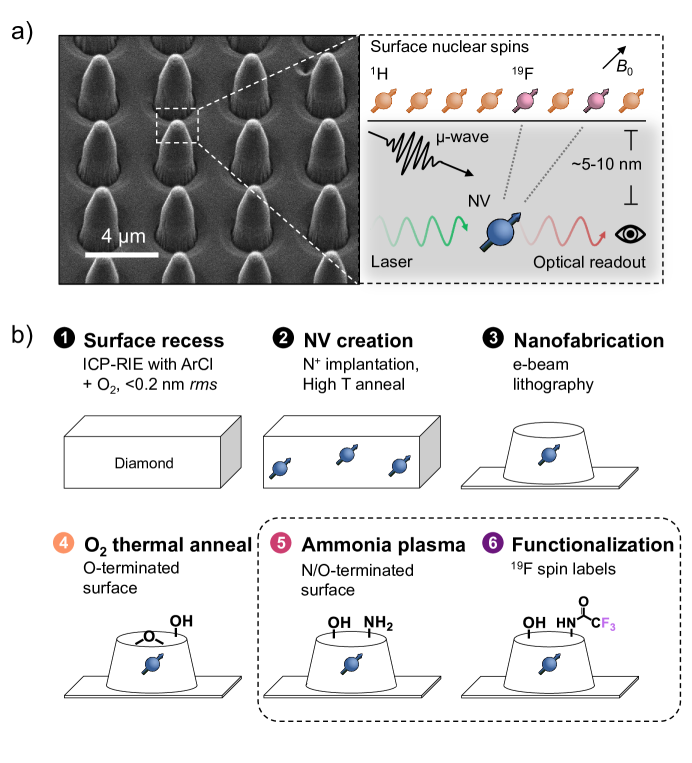
Here, we demonstrate direct chemical functionalization of mixed nitrogen- and oxygen- (N/O-) terminated diamond for surface NMR spectroscopy with single NV centers (Figure 1). Starting with O-terminated diamond obtained through thermal annealing, the composition of mixed N/O surface termination is controlled with exposure to ammonia (NH3) plasma. We show reversible surface functionalization of amine groups introduced by plasma exposure using derivatization; N-hydroxysuccinimide (NHS) ester crosslinking chemistry is used to attach fluorescent dye molecules by stable amide bond formation. We find at short plasma exposure times that the mixed N/O-terminated surfaces improve the coherence time of 10-nm-deep NV centers compared to only O-annealed surfaces. We finally functionalize diamond nanopillars hosting single NV centers with trifluoromethyl tags to detect the magnetic field variance arising from surface-bound 19F spins. Within the sensing volume of each NV, we estimate detection of ca. 50–100 surface-bound molecules. This surface functionalization demonstrates the generalizability of our immobilization protocol, as well as the persistent stability of shallow NV centers after chemical attachment of proximal molecules for nanoscale NMR.
Low diamond surface roughness is essential for surface sensing as it facilitates the formation of highly ordered chemical termination, which increases the coherence time of near-surface NV centers.23 Bulk and membrane samples (the latter for nanofabrication of pillar arrays) are recessed using Ar/Cl2 and and O2 inductively coupled plasma – reactive ion etching (ICP-RIE). This step is followed by cleaning in a tri-acid mixture of H2SO4:HClO4:HNO3 at 120 °C to remove graphitic carbon on the surface as a result of the O2 etching (Step 1, Figure 1b). Using this procedure we achieve surface roughness 0.2 nm-rms (Supplementary Information Figure S2).
To create shallow NV centers, recessed diamond samples are implanted with 15N+ ions with controllable density and depth (Step 2, Figure 1b). Implantation energies of 5 or 7 keV and fluences of are used, yielding an expected average implantation depth of 8.0(31) and 10.8(40) nm respectively (Supplementary Information Figure S3).42 Annealing at 800 °C for 2 h under high vacuum () converts implanted 15N+ ions into NV centers due to vacancy migration. Nanopillar arrays are then patterned on membrane samples via electron-beam lithography (Step 3, Figure 1b) for higher photoluminescence collection efficiency and to provide a map to selectively address and revisit the same NV centers for repeated characterization.43
Samples are subsequently thermally annealed under an oxygen atmosphere at 460 °C so as to remove further graphitic carbon and to terminate the diamond surface with oxgyen (Step 4, Figure 1b). Using an analogous oxygen annealing procedure, Sangtawesin et al. recently demonstrated improvements in coherence (T2) times of shallow NV centers of up to a factor of four compared to acid-cleaned diamond surfaces, which was attributed to a highly ordered, predominantly ether-terminated surface.23 Similarly, we see an improvement in NV optical contrast using both continuous wave (cw) and pulsed optically detected magnetic resonance (ODMR) measurements, and factor of two improvement in coherence and Rabi decay times of NV centers following oxygen annealing (Supplementary Information Figure S4).
Although suggested to be beneficial for near-surface NV stability, the resulting scarcity of hydroxyl or carboxylic acid functional groups on O-terminated surfaces by thermal annealing in oxygen precludes straightforward functionalization with molecules of interest. Here, we incorporate additional reactive amine terminal groups for subsequent molecular attachment by introducing a 50 W rf of 13.56 MHz NH3 plasma treatment to O-annealed surfaces for mixed N/O-termination (Step 5, Figure 1b). This approach mitigates full conversion to a surface that exhibits negative electron affinity (characteristic of fully amine-terminated diamond),44 which can be detrimental for NV stability. We note that alternative diamond amination procedures using NH3, N2, or mixed-source plasmas have been developed previously, but with hydrogen-terminated diamond as a starting surface or without characterizing their influence on the photophysical properties of near-surface NV centers.45, 46, 47, 48, 49
![[Uncaptioned image]](/html/2202.03969/assets/x2.png)
Changes to the chemical composition of the surfaces exposed to 0, 20, 40, and 60 s of NH3 plasma are characterized using X-ray photoelectron spectroscopy (XPS) on bulk oxygen-annealed diamond samples. A decrease in elemental percentage of oxygen in the sampling depth (ca. 6 nm) is accompanied by an increase in nitrogen to 3% after 60 s (Figure 2a,b). These percentages are obtained by integration of O 1s, N 1s, and C 1s signals after adjusting for photoionization cross sections of the core electron species.50 The emergent N 1s peak position at 398.4 eV suggests predominately single-bonded C–N and/or double-bonded C=N, while nitrile groups may be excluded.51, 19 Analysis of high-resolution C 1s spectra shows a dominant peak at 284.8 assigned to sp3 carbon, a shoulder at ca. 1 eV lower binding energy attributed to sp2 carbon, and satellite peaks at higher binding energy due to carbon–oxygen binding (Figure 2c).23, 52 The NH3 plasma treatment reduces the already low amount of sp2 carbon on the diamond surfaces with increasing exposure time (Figure 2d). The high-resolution O 1s signals can be fit with a C–O–C (ether) assignment at 531.9 eV and C–O–H (hydroxyl) contribution at ca. 1 eV lower binding energy (Figure 2e).23 The relative ratio of these two peaks for oxygen-annealed diamond is ca. 5:1, and decreases to ca. 2:1 after 60 s plasma exposure (Figure 2f).
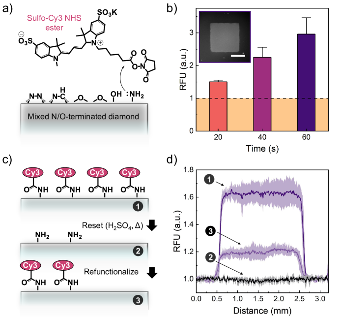
The presence of reactive amines on the mixed N/O-terminated diamond surface is determined using complementary fluorescence microscopy characterization (Figure 3a,b). Amine groups are derivitized by incubating surfaces with a sulfo-Cy3 NHS ester dye designed to bind selectively via amine-reactive crosslinking. From 20 s to 60 s exposure time, increasing relative fluorescence intensity is observed above the O-terminated background regions. These results indicate clearly the tunability of available amine groups and surface density of adsorbed molecules. This functionalization is also reversible (Figure 3c,d). We show that surfaces can be reset by treating with 75% H2SO4 at 60 °C for 5 h in order to break the amide bonds formed between the adsorbed molecules and aminated surface, indicated by total loss of fluorescence relative to the background region. A repeated functionalization of the surface by incubating with sulfo-Cy3 NHS ester again shows fluorescence from the region originally exposed to NH3 plasma, albeit at lower intensity. The reduction in intensity is likely due to partial loss of surface-bound amine groups following the reset, resulting in a lower surface density of dye molecules. Still, this proof-of-principle recyclability may be optimized to preserve a higher percentage of reactive functional groups for repeated analysis.
We next test how the NH3 plasma treatment affects the sensing properties of shallow NV centers. Figure 4a shows representative confocal fluorescence images of bulk diamond surfaces containing individually resolvable NV centers before and after 20 s plasma exposure. The plasma treatment causes background fluorescence to decrease, making it easier to distinguish fluorescent NV centers from bright surface impurities. Next, individual NV centers are identified and characterized using OMDR, wherein the NV center is first illuminated with a green laser ( nm) to polarize the electron spin into the state. Applying a subsequent microwave field at the Larmor frequency of the electron spin drives transitions between the optically polarized (bright) state and the 1 (dark) states. Finally, readout is performed by integrating the emitted photons during a second green laser pulse. The measurement sensitivity scales linearly with ODMR contrast, which is defined as the ratio of integrated fluorescence of the bright vs dark states 6.
We find that increased plasma exposure time is accompanied by a decrease in the number of NV centers that display an ODMR signal (Figure 4b). The survival rate is calculated by determining the number of NV centers with an ODMR signal detected per scan area after plasma treatment, and normalized to the number of detected NV centers prior to exposure (Supporting Information Figure S5). Approximately two-thirds of NV centers remain following 20 s plasma exposure, which drops below 50% after 40 and 60 s. Consequently, we opted to use a nanostructured diamond membrane for further testing such that we could track the same NV centers before and after treatment. Figure 4c shows a representative fluorescence map of one nanopillar array after a 20 s plasma treatment. Analogous to the bulk samples, a decrease in fluorescence background is observed, in addition to a decrease in the number of pillars containing NV candidates with detectable ODMR signals. Encouragingly, no significant difference in the mean pulsed-ODMR contrast nor the mean photoluminescence of NV centers is determined before vs after 20 s plasma exposure (Figure 4d,e).
![[Uncaptioned image]](/html/2202.03969/assets/x4.png)
We compare the coherence properties of NV centers using a Carr-Purcell-Meiboom-Gill-type (CPMG) multi-pulse dynamical decoupling protocol,53, 54 which is illustrated in Figure 4f. After an initial pulse, a train of pulses follows which refocuses the spin. A final pulse maps the coherence back to the initialized bright state. When increasing the evolution time between the pulses, we observe a signal decay with which we associate the time. To compensate for brightness variations, we collect reference traces of an initialization of the and state in parallel. The sequence also allows us to perform NMR spectroscopy. By tuning the inter-pulse delay of the pulses a CPMG sequence, we match the nuclear Larmor frequency and detect the noise generated by external nuclear spins in a lock-in manner.55 By quantifying the signal, we estimate the root mean square value of the field fluctuations produced by external spins. This signal can then be used to infer the depth of the NV center. Fit analysis providing is performed by a Monte-Carlo model as well as by fitting an analytical model. 16, 56, 57 From the extracted we infer the depth by inverting16
| (1) |
where denotes the magnetic field constant, is the reduced Planck’s constant, is the gyromagnetic ratio of protons, is the density of the surface proton layer, is the thickness of this layer and is the NV center depth. We assume an adsorbed water layer of 1 nm thickness and proton density of .16, 58, 59, 60, 61
The T2,CPMG coherence times are shown as a function of NV center depth in Figure 4g. We observe a monotonic decrease in NV center coherence time with increasing proximity to the surface, consistent with previous reports.62, 63, 23, 64, 28 Promisingly, the T2,CPMG values are larger on average after 20 s plasma exposure. In analysis of five NV centers directly compared before and after plasma treatment, we observe increases in T2,CPMG by as much as 1.8(3) times. Depths and T2,CPMG values of these NV centers are summarized in Table 1. In addition, from the data set characterized prior to plasma exposure, we could not measure an ODMR signal from three of the NV centers following 20 s plasma, indicated by red crosses in Figure 4g. We note that these NV centers, with calculated depths of ca. 4.5(8), 5.4(8), and 9.6(28) nm, each exhibited below-average coherence times, suggesting that while the plasma reduces the number of observed NV centers (Figure 4b), it preserves those with the highest stability and sensitivity for performing surface NV-NMR measurements.
| Deptha (nm) | T2,CPMG () | ||
|---|---|---|---|
| O-annealed | NH3 plasma | ||
| NV 1 | 4.1(2) | 30.6(9) | 46.0(42) |
| NV 2 | 5.1(4) | 45.1(17) | 39.0(58) |
| NV 3 | 5.5(4) | 34.0(8) | 60.0(9) |
| NV 4 | 6.4(2) | 44.8(14) | 65.0(13) |
| NV 5 | 6.8(3) | 41.9(7) | 44.0(22) |
| a Depth values are calculated from the average of | |||
| measurements before and after plasma treatment. | |||
Finally, we test our N/O-termined diamond NV sensing platform for nanoscale-NMR of surface-bound molecules (Step 6, Figure 1b). Following plasma treatment, a diamond nanopillar sample is incubated with 2,5-dioxopyrrolidin-1-yl 2,2,2-trifluoroacetate to attach trifluoromethyl groups to the surface via amide bond formation (Figure 5a). Functionalization is confirmed by detection of fluorine on the surface via XPS.
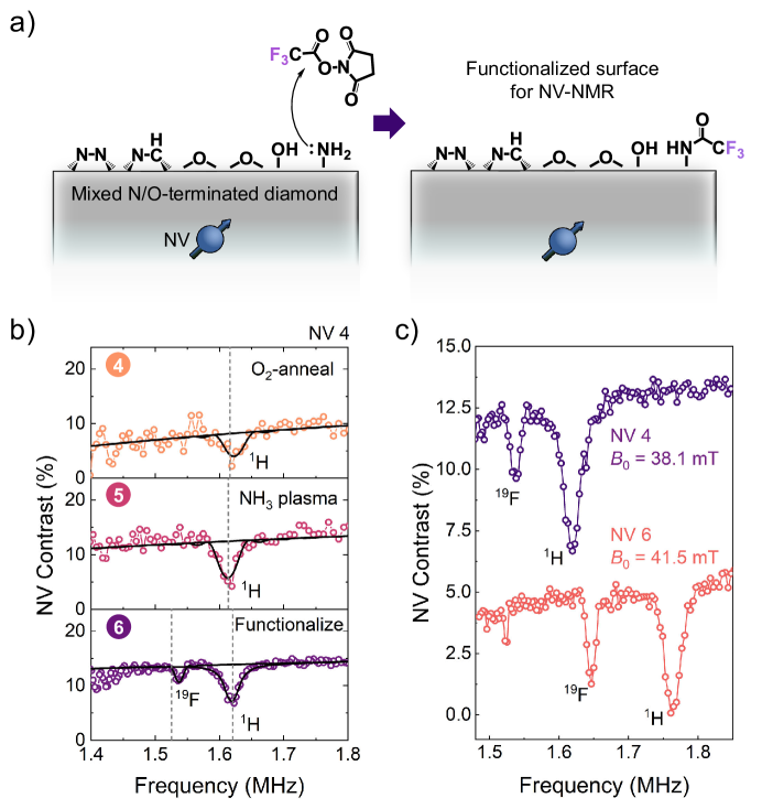
Surface NV-NMR spectra are shown in Figure 5b that correspond to the same NV center (NV 4) measured after oxygen-annealing, 20 s plasma treatment, and surface functionalization (Steps 4–6, Figure 1b). Prior to attachment of trifluoromethyl moieties to the surface, the dynamical decoupling spectrum shows only dips in contrast at the frequency corresponding to spins, attributed mainly to the presence of an adsorbed water layer. After exposure to 20 s plasma, an improvement in coherence time of the NV is evident by reduced signal decay with decreasing frequency and a further pronounced proton signal due to this slower decay. Following attachment of trifluoromethyl tags, a second dip appears in the vicinity of the expected resonance frequency. Assignment in the NV-NMR spectra is validated by measuring two separate NV centers (NV 4 and NV 6) independently and at distinct magnetic field biases, which shifts the Larmor frequencies of both and spins (Figure 5c). We find for NV 4 at the frequency a of 245(5) nT and 102(5) nT at the frequency. For NV 6, we find 331(12) nT and 163(7) nT for these two resonances, respectively. From the proton signals, we estimate the NV depths to be 6.4(2) nm and 6.3(2) nm (Equation (1)).16, 58, 59, 60, 61 Modeling the spin bath as a 2D layer,
| (2) |
where is the density of the 2D layer and is the gyromagnetic ratio of . By using the depths determined by NMR, we estimate the surface density of to be 5(1) and 12(2) for NV 4 and NV 6, respectively. Since each attached sub-unit contains three fluorine atoms per molecule, this corresponds to a molecular surface density of 1.7(5) and 4.0(1) molecules . By using the sensitive surface area for a NV center at depth which produces first 70% of the signal, (Supporting Information Figure S6), we conclude that 51(15) and 117(10) molecules contribute to the detected signal by each NV center, respectively.16
In summary, we demonstrate a diamond surface preparation strategy that enables direct chemical functionalization of amine functional groups while improving coherence times of near-surface NV centers for surface NMR spectroscopy. Mixed N/O-terminated surfaces are prepared by thermal annealing under an oxygen atmosphere at elevated temperature and subsequent exposure to NH3 plasma. This process yields robust and reproducible control over the elemental percentages of nitrogen and oxygen on diamond surfaces. We identify an experimental regime in which two-thirds of shallow NV centers are preserved following NH3 plasma treatment with improved sensing properties compared to oxygen-annealed surfaces. In particular, we find significant reduction in the background luminescence and up to 1.8(3) times improvement in the coherence times of near-surface NV centers. The use of nanostructured diamond allowed us to compare the same NV centers at different stages of processing, with the additional benefit of improved collection efficiency. Finally, we bind trifluoromethyl tags to surface-bound amine groups and perform NMR spectroscopy at the few-molecule (ca. 50–100 molecules) regime using dynamical-decoupling noise spectroscopy with single NV centers. The presented approach enables a precise preparation of molecular systems of interest at diamond surfaces that may be applied for emergent NV center-based quantum sensing of chemical functionality.65, 66, 67, 68
1 Supporting Information
The Supporting Information provides additional materials and experimental methods, as well as additional data used to evaluate the conclusions in the paper; Figures S1–S7 and Table S1 show data related to XPS characterization and NV-NMR with silanized surfaces, SRIM (The Stopping and Range of Ions in Matter) calculations, atomic force microscopy images, characterization of NV centers following thermal annealing under oxygen, additional characterization of NV centers following exposure to ammonia plasma, integration of the sensitive slice on the surface above each NV center for NMR measurements, and estimated errors from magnetic field measurements and NV center depths.
2 Author Contributions
J.M.A. and K.H. contributed equally to this work. J.M.A., K.H., and C.L.D. conceived and designed the experiments. T.Z. developed the nano-structure fabrication. Data were collected by J.M.A., K.H., and E.J. All authors discussed the results. The manuscript was co-written by J.M.A. and K.H. with assistance by E.J., T.Z., L.A.V., and C.L.D.
3 Notes
The authors declare no competing financial interest.
4 Acknowledgment
The authors thank Dr. Andrea Arcifa from EMPA, and Dr. Viraj Damle and Dr. Jan Rhensius from QZabre AG for insightful discussions and their help. This work has been supported by Swiss National Science Foundation (SNSF) Project Grant No. 200020 175600, the National Center of Competence in Research in Quantum Science and Technology (NCCR QSIT), and the Advancing Science and TEchnology thRough dIamond Quantum Sensing (ASTERIQS) program, Grant No. 820394, of the European Commission. J.M.A. acknowledges funding from an ETH Zurich Career Seed Grant and from a SNSF Ambizione Grant [PZ00P2_201590].
References
- Lee et al. 2014 Lee, J. H.; Okuno, Y.; Cavagnero, S. Sensitivity Enhancement in Solution NMR: Emerging Ideas and New Frontiers. J. Magn. Reson. 2014, 241, 18–31
- Mamin et al. 2013 Mamin, H. J.; Kim, M.; Sherwood, M. H.; Rettner, C. T.; Ohno, K.; Awschalom, D. D.; Rugar, D. Nanoscale Nuclear Magnetic Resonance with a Nitrogen-Vacancy Spin Sensor. Science 2013, 339, 557–560
- Staudacher et al. 2013 Staudacher, T.; Shi, F.; Pezzagna, S.; Meijer, J.; Du, J.; Meriles, C. A.; Reinhard, F.; Wrachtrup, J. Nuclear Magnetic Resonance Spectroscopy on a (5-Nanometer)3 Sample Volume. Science 2013, 339, 561–563
- Aslam et al. 2017 Aslam, N.; Pfender, M.; Neumann, P.; Reuter, R.; Zappe, A.; de Oliveira, F. F.; Denisenko, A.; Sumiya, H.; Onoda, S.; Isoya, J.; Wrachtrup, J. Nanoscale Nuclear Magnetic Resonance with Chemical Resolution. Science 2017, 357, 67–71
- Jelezko et al. 2004 Jelezko, F.; Gaebel, T.; Popa, I.; Gruber, A.; Wrachtrup, J. Observation of Coherent Oscillations in a Single Electron Spin. Phys. Rev. Lett. 2004, 92, 076401
- Schirhagl et al. 2014 Schirhagl, R.; Chang, K.; Loretz, M.; Degen, C. L. Nitrogen-Vacancy Centers in Diamond: Nanoscale Sensors for Physics and Biology. Annu. Rev. Phys. Chem. 2014, 65, 83–105
- Gali 2019 Gali, A. Ab initio Theory of the Nitrogen-Vacancy Center in Diamond. Nanophotonics 2019, 8, 1907–1943
- Härtl et al. 2004 Härtl, A.; Schmich, E.; Garrido, J. A.; Hernando, J.; Catharino, S. C. R.; Walter, S.; Feulner, P.; Kromka, A.; Steinmüller, D.; Stutzmann, M. Protein-Modified Nanocrystalline Diamond Thin Films for Biosensor Applications. Nat. Mater. 2004, 3, 736–742
- Stavis et al. 2011 Stavis, C.; Clare, T. L.; Butler, J. E.; Radadia, A. D.; Carr, R.; Zeng, H.; King, W. P.; Carlisle, J. A.; Aksimentiev, A.; Bashir, R.; Hamers, R. J. Surface Functionalization of Thin-Film Diamond for Highly Stable and Selective Biological Interfaces. Proc. Natl. Acad. Sci. U.S.A. 2011, 108, 983–988
- Nebel et al. 2007 Nebel, C. E.; Shin, D.; Rezek, B.; Tokuda, N.; Uetsuka, H.; Watanabe, H. Diamond and Biology. J. R. Soc. Interface 2007, 4, 439–461
- Raymakers et al. 2019 Raymakers, J.; Haenen, K.; Maes, W. Diamond Surface Dunctionalization: From Gemstone to Photoelectrochemical Applications. J. Mater. Chem. C 2019, 7, 10134–10165
- Hauf et al. 2011 Hauf, M. V.; Grotz, B.; Naydenov, B.; Dankerl, M.; Pezzagna, S.; Meijer, J.; Jelezko, F.; Wrachtrup, J.; Stutzmann, M.; Reinhard, F.; Garrido, J. A. Chemical Control of the Charge State of Nitrogen-Vacancy Centers in Diamond. Phys. Rev. B 2011, 83, 081304
- Bluvstein et al. 2019 Bluvstein, D.; Zhang, Z.; McLellan, C. A.; Williams, N. R.; Jayich, A. C. B. Extending the Quantum Coherence of a Near-Surface Qubit by Coherently Driving the Paramagnetic Surface Environment. Phys. Rev. Lett. 2019, 123, 146804
- Rondin et al. 2010 Rondin, L.; Dantelle, G.; Slablab, A.; Grosshans, F.; Treussart, F.; Bergonzo, P.; Perruchas, S.; Gacoin, T.; Chaigneau, M.; Chang, H.-C.; Jacques, V.; Roch, J.-F. Surface-Induced Charge State Conversion of Nitrogen-Vacancy Defects in Nanodiamonds. Phys. Rev. B 2010, 82, 115449
- Ofori-Okai et al. 2012 Ofori-Okai, B. K.; Pezzagna, S.; Chang, K.; Loretz, M.; Schirhagl, R.; Tao, Y.; Moores, B. A.; Groot-Berning, K.; Meijer, J.; Degen, C. L. Spin Properties of Very Shallow Nitrogen Vacancy Defects in Diamond. Phys. Rev. B 2012, 86, 081406
- Loretz et al. 2014 Loretz, M.; Pezzagna, S.; Meijer, J.; Degen, C. L. Nanoscale Nuclear Magnetic Resonance with a 1.9-nm-Deep Nitrogen-Vacancy Sensor. Appl. Phys. Lett. 2014, 104, 033102
- Rosskopf et al. 2014 Rosskopf, T.; Dussaux, A.; Ohashi, K.; Loretz, M.; Schirhagl, R.; Watanabe, H.; Shikata, S.; Itoh, K. M.; Degen, C. L. Investigation of Surface Magnetic Noise by Shallow Spins in Diamond. Phys. Rev. Lett. 2014, 112, 147602
- Cui and Hu 2013 Cui, S.; Hu, E. L. Increased Negatively Charged Nitrogen-Vacancy Centers in Fluorinated Diamond. Appl. Phys. Lett. 2013, 103, 051603
- Stacey et al. 2015 Stacey, A.; O’Donnell, K. M.; Chou, J.-P.; Schenk, A.; Tadich, A.; Dontschuk, N.; Cervenka, J.; Pakes, C.; Gali, A.; Hoffman, A.; Prawer, S. Nitrogen Terminated Diamond. Adv. Mater. Interfaces 2015, 2, 1500079
- Chou et al. 2017 Chou, J.-P.; Retzker, A.; Gali, A. Nitrogen-Terminated Diamond (111) Surface for Room-Temperature Quantum Sensing and Simulation. Nano Lett. 2017, 17, 2294–2298
- Kawai et al. 2019 Kawai, S.; Yamano, H.; Sonoda, T.; Kato, K.; Buendia, J. J.; Kageura, T.; Fukuda, R.; Okada, T.; Tanii, T.; Higuchi, T.; Haruyama, M.; Yamada, K.; Onoda, S.; Ohshima, T.; Kada, W.; Hanaizumi, O.; Stacey, A.; Teraji, T.; Kono, S.; Isoya, J.; Kawarada, H. Nitrogen-Terminated Diamond Surface for Nanoscale NMR by Shallow Nitrogen-Vacancy Centers. J. Phys. Chem. C 2019, 123, 3594–3604
- Kaviani et al. 2014 Kaviani, M.; Deák, P.; Aradi, B.; Frauenheim, T.; Chou, J.-P.; Gali, A. Proper Surface Termination for Luminescent Near-Surface NV Centers in Diamond. Nano Lett. 2014, 14, 4772–4777
- Sangtawesin et al. 2019 Sangtawesin, S.; Dwyer, B. L.; Srinivasan, S.; Allred, J. J.; Rodgers, L. V. H.; De Greve, K.; Stacey, A.; Dontschuk, N.; O’Donnell, K. M.; Hu, D.; Evans, D. A.; Jaye, C.; Fischer, D. A.; Markham, M. L.; Twitchen, D. J.; Park, H.; Lukin, M. D.; de Leon, N. P. Origins of Diamond Surface Noise Probed by Correlating Single-Spin Measurements with Surface Spectroscopy. Phys. Rev. X 2019, 9, 031052
- Ohashi et al. 2013 Ohashi, K.; Rosskopf, T.; Watanabe, H.; Loretz, M.; Tao, Y.; Hauert, R.; Tomizawa, S.; Ishikawa, T.; Ishi-Hayase, J.; Shikata, S.; Degen, C. L.; Itoh, K. M. Negatively Charged Nitrogen-Vacancy Centers in a 5 nm Thin 12C Diamond Film. Nano Letters 2013, 13, 4733–4738
- Schlipf et al. 2017 Schlipf, L.; Oeckinghaus, T.; Xu, K.; Dasari, D. B. R.; Zappe, A.; de Oliveira, F. F.; Kern, B.; Azarkh, M.; Drescher, M.; Ternes, M.; Kern, K.; Wrachtrup, J.; Finkler, A. A Molecular Quantum Spin Network Controlled by a Single Qubit. Sci. Adv. 2017, 3, e1701116
- Lovchinsky et al. 2016 Lovchinsky, I.; Sushkov, A. O.; Urbach, E.; de Leon, N. P.; Choi, S.; Greve, K. D.; Evans, R.; Gertner, R.; Bersin, E.; Müller, C.; McGuinness, L.; Jelezko, F.; Walsworth, R. L.; Park, H.; Lukin, M. D. Nuclear Magnetic Resonance Detection and Spectroscopy of Single Proteins using Quantum Logic. Science 2016, 351, 836–841
- Liu et al. 2022 Liu, K. S.; Henning, A.; Heindl, M. W.; Allert, R. D.; Bartl, J. D.; Sharp, I. D.; Rizzato, R.; Bucher, D. B. Surface NMR using Quantum Sensors in Diamond. Proc. Natl. Acad. Sci. U.S.A. 2022, 119
- Xie et al. 2021 Xie, M.; Yu, X.; Rodgers, L. V. H.; Xu, D.; Chi-Duran, I.; Toros, A.; Quack, N.; de Leon, N. P.; Maurer, P. C. Biocompatible Surface Functionalization Architecture for a Diamond Quantum Sensor. arXiv preprint arXiv:2108.04843 2021,
- Kolkowitz et al. 2012 Kolkowitz, S.; Unterreithmeier, Q. P.; Bennett, S. D.; Lukin, M. D. Sensing Distant Nuclear Spins with a Single Electron Spin. Phys. Rev. Lett. 2012, 109, 137601
- Müller et al. 2014 Müller, C.; Kong, X.; Cai, J.-M.; Melentijević, K.; Stacey, A.; Markham, M.; Twitchen, D.; Isoya, J.; Pezzagna, S.; Meijer, J.; Du, J. F.; Plenio, M. B.; Naydenov, B.; McGuinness, L. P.; Jelezko, F. Nuclear Magnetic Resonance Spectroscopy with Single Spin Sensitivity. Nat. Commun. 2014, 5, 4703
- Sushkov et al. 2014 Sushkov, A. O.; Lovchinsky, I.; Chisholm, N.; Walsworth, R. L.; Park, H.; Lukin, M. D. Magnetic Resonance Detection of Individual Proton Spins Using Quantum Reporters. Phys. Rev. Lett. 2014, 113, 197601
- Boss et al. 2016 Boss, J. M.; Chang, K.; Armijo, J.; Cujia, K.; Rosskopf, T.; Maze, J. R.; Degen, C. L. One- and Two-Dimensional Nuclear Magnetic Resonance Spectroscopy with a Diamond Quantum Sensor. Phys. Rev. Lett. 2016, 116, 197601
- Cujia et al. 2019 Cujia, K. S.; Boss, J. M.; Herb, K.; Zopes, J.; Degen, C. L. Tracking the Precession of Single Nuclear Spins by Weak Measurements. Nature 2019, 571, 230–233
- Notsu et al. 2001 Notsu, H.; Fukazawa, T.; Tatsuma, T.; Tryk, D. A.; Fujishima, A. Hydroxyl Groups on Boron-Doped Diamond Electrodes and Their Modification with a Silane Coupling Agent. Electrochem. Solid-State Lett. 2001, 4, H1
- Ohta et al. 2004 Ohta, R.; Saito, N.; Inoue, Y.; Sugimura, H.; Takai, O. Organosilane Self-Assembled Monolayers Directly Linked to the Diamond Surfaces. J. Vac. Sci. Technol. A 2004, 22, 2005–2009
- Hernando et al. 2007 Hernando, J.; Pourrostami, T.; Garrido, J. A.; Williams, O. A.; Gruen, D. M.; Kromka, A.; Steinmüller, D.; Stutzmann, M. Immobilization of Horseradish Peroxidase via an Amino Silane on Oxidized Ultrananocrystalline Diamond. Diam. Relat. Mater. 2007, 16, 138–143
- Acres et al. 2012 Acres, R. G.; Ellis, A. V.; Alvino, J.; Lenahan, C. E.; Khodakov, D. A.; Metha, G. F.; Andersson, G. G. Molecular Structure of 3-Aminopropyltriethoxysilane Layers Formed on Silanol-Terminated Silicon Surfaces. J. Phys. Chem. C 2012, 116, 6289–6297
- Naik et al. 2013 Naik, V. V.; Crobu, M.; Venkataraman, N. V.; Spencer, N. D. Multiple Transmission-Reflection IR Spectroscopy Shows that Surface Hydroxyls Play Only a Minor Role in Alkylsilane Monolayer Formation on Silica. J. Phys. Chem. Lett. 2013, 4, 2745–2751
- Stine et al. 2007 Stine, R.; Cole, C. L.; Ainslie, K. M.; Mulvaney, S. P.; Whitman, L. J. Formation of Primary Amines on Silicon Nitride Surfaces: A Direct, Plasma-Based Pathway to Functionalization. Langmuir 2007, 23, 4400–4404
- Asenath Smith and Chen 2008 Asenath Smith, E.; Chen, W. How To Prevent the Loss of Surface Functionality Derived from Aminosilanes. Langmuir 2008, 24, 12405–12409
- Vandenberg et al. 1991 Vandenberg, E. T.; Bertilsson, L.; Liedberg, B.; Uvdal, K.; Erlandsson, R.; Elwing, H.; Lundström, I. Structure of 3-Aminopropyl Triethoxy Silane on Silicon Oxide. J. Colloid Interface Sci. 1991, 147, 103–118
- Ziegler et al. 2010 Ziegler, J. F.; Ziegler, M.; Biersack, J. SRIM – The Stopping and Range of Ions in Matter (2010). Nucl. Instrum. Methods Phys. Res. B: Beam Interact. Mater. At. 2010, 268, 1818–1823
- Hausmann et al. 2010 Hausmann, B. J.; Khan, M.; Zhang, Y.; Babinec, T. M.; Martinick, K.; McCutcheon, M.; Hemmer, P. R.; Lončar, M. Fabrication of Diamond Nanowires for Quantum Information Processing Applications. Diam. Relat. Mater. 2010, 19, 621–629
- Zhu et al. 2016 Zhu, D.; Bandy, J. A.; Li, S.; Hamers, R. J. Amino-Terminated Diamond Surfaces: Photoelectron Emission and Photocatalytic Properties. Surf. Sci. 2016, 650, 295–301
- Coffinier et al. 2007 Coffinier, Y.; Szunerits, S.; Jama, C.; Desmet, R.; Melnyk, O.; Marcus, B.; Gengembre, L.; Payen, E.; Delabouglise, D.; Boukherroub, R. Peptide Immobilization on Amine-Terminated Boron-Doped Diamond Surfaces. Langmuir 2007, 23, 4494–4497
- Wei et al. 2015 Wei, J.; Liu, J.; Chen, L.; Hei, L.; Lv, F.; Li, C. Amination of Diamond Film by Ammonia Microwave Plasma Treatment. Diam. Relat. Mater. 2015, 54, 34–38
- Torrengo et al. 2011 Torrengo, S.; Miotello, A.; Minati, L.; Bernagozzi, I.; Ferrari, M.; Dipalo, M.; Kohn, E.; Speranza, G. The Role of Oxygen in the One Step Amination Process of Nanocrystalline Diamond Surface. Diam. Relat. Mater. 2011, 20, 990–994
- Wang et al. 2012 Wang, Q.; Kromka, A.; Houdkova, J.; Babchenko, O.; Rezek, B.; Li, M.; Boukherroub, R.; Szunerits, S. Nanomolar Hydrogen Peroxide Detection Using Horseradish Peroxidase Covalently Linked to Undoped Nanocrystalline Diamond Surfaces. Langmuir 2012, 28, 587–592
- Chandran et al. 2015 Chandran, M.; Shasha, M.; Michaelson, S.; Hoffman, A. Nitrogen Termination of Single Crystal (100) Diamond Surface by Radio Frequency N2 Plasma Process: An in-situ X-Ray Photoemission Spectroscopy and Secondary Electron Emission Studies. Appl. Phys. Lett. 2015, 107, 111602
- Yeh and Lindau 1985 Yeh, J.; Lindau, I. Atomic Subshell Photoionization Cross Sections and Asymmetry Parameters: 1 Z 103. At. Data Nucl. Data Tables 1985, 32, 1–155
- Graf et al. 2009 Graf, N.; Yegen, E.; Gross, T.; Lippitz, A.; Weigel, W.; Krakert, S.; Terfort, A.; Unger, W. E. XPS and NEXAFS Studies of Aliphatic and Aromatic Amine Species on Functionalized Surfaces. Surf. Sci. 2009, 603, 2849–2860
- Baldwin et al. 2014 Baldwin, C. G.; Downes, J. E.; McMahon, C. J.; Bradac, C.; Mildren, R. P. Nanostructuring and Oxidation of Diamond by Two-Photon Ultraviolet Surface Excitation: An XPS and NEXAFS Study. Phys. Rev. B 2014, 89, 195422
- Carr and Purcell 1954 Carr, H. Y.; Purcell, E. M. Effects of Diffusion on Free Precession in Nuclear Magnetic Resonance Experiments. Phys. Rev. 1954, 94, 630–638
- Meiboom and Gill 1958 Meiboom, S.; Gill, D. Modified Spin‐Echo Method for Measuring Nuclear Relaxation Times. Rev. Sci. Instrum. 1958, 29, 688–691
- Degen et al. 2017 Degen, C. L.; Reinhard, F.; Cappellaro, P. Quantum Sensing. Rev. Mod. Phys. 2017, 89, 035002
- Pham et al. 2016 Pham, L. M.; DeVience, S. J.; Casola, F.; Lovchinsky, I.; Sushkov, A. O.; Bersin, E.; Lee, J.; Urbach, E.; Cappellaro, P.; Park, H.; Yacoby, A.; Lukin, M.; Walsworth, R. L. NMR Technique for Determining the Depth of Shallow Nitrogen-Vacancy Centers in Diamond. Phys. Rev. B 2016, 93, 045425
- Herb and Welter 2022 Herb, K.; Welter, P. Parallel Time Integration using Batched BLAS (Basic Linear Algebra Subprograms) Routines. Comput. Phys. Commun. 2022, 270, 108181
- Degen et al. 2009 Degen, C. L.; Poggio, M.; Mamin, H. J.; Rettner, C. T.; Rugar, D. Nanoscale Magnetic Resonance Imaging. Proc. Natl. Acad. Sci. U.S.A. 2009, 106, 1313–1317
- Mamin et al. 2009 Mamin, H. J.; Oosterkamp, T. H.; Poggio, M.; Degen, C. L.; Rettner, C. T.; Rugar, D. Isotope-Selective Detection and Imaging of Organic Nanolayers. Nano Lett. 2009, 9, 3020–3024
- Xue et al. 2011 Xue, F.; Weber, D. P.; Peddibhotla, P.; Poggio, M. Measurement of Statistical Nuclear Spin Polarization in a Nanoscale GaAs Sample. Phys. Rev. B 2011, 84, 205328
- Grob et al. 2019 Grob, U.; Krass, M. D.; Héritier, M.; Pachlatko, R.; Rhensius, J.; Košata, J.; Moores, B. A.; Takahashi, H.; Eichler, A.; Degen, C. L. Magnetic Resonance Force Microscopy with a One-Dimensional Resolution of 0.9 Nanometers. Nano Lett. 2019, 19, 7935–7940
- Myers et al. 2014 Myers, B. A.; Das, A.; Dartiailh, M. C.; Ohno, K.; Awschalom, D. D.; Bleszynski Jayich, A. C. Probing Surface Noise with Depth-Calibrated Spins in Diamond. Phys. Rev. Lett. 2014, 113, 027602
- Fávaro de Oliveira et al. 2017 Fávaro de Oliveira, F.; Antonov, D.; Wang, Y.; Neumann, P.; Momenzadeh, S. A.; Häußermann, T.; Pasquarelli, A.; Denisenko, A.; Wrachtrup, J. Tailoring Spin Defects in Diamond by Lattice Charging. Nat. Commun. 2017, 8, 15409
- Findler et al. 2020 Findler, C.; Lang, J.; Osterkamp, C.; Nesládek, M.; Jelezko, F. Indirect Overgrowth as a Synthesis Route for Superior Diamond Nano Sensors. Sci. Rep. 2020, 10, 22404
- Krečmarová et al. 2021 Krečmarová, M.; Gulka, M.; Vandenryt, T.; Hrubý, J.; Fekete, L.; Hubík, P.; Taylor, A.; Mortet, V.; Thoelen, R.; Bourgeois, E.; Nesládek, M. A Label-Free Diamond Microfluidic DNA Sensor Based on Active Nitrogen-Vacancy Center Charge State Control. ACS Appl. Mater. Interfaces 2021, 13, 18500–18510
- Kayci et al. 2021 Kayci, M.; Fan, J.; Bakirman, O.; Herrmann, A. Multiplexed Sensing of Biomolecules with Optically Detected Magnetic Resonance of Nitrogen-Vacancy Centers in Diamond. Proc. Natl. Acad. Sci. U.S.A. 2021, 118
- Li et al. 2022 Li, C.; Soleyman, R.; Kohandel, M.; Cappellaro, P. SARS-CoV-2 Quantum Sensor Based on Nitrogen-Vacancy Centers in Diamond. Nano Lett. 2022, 22, 43–49
- Mzyk et al. 2022 Mzyk, A.; Ong, Y.; Ortiz Moreno, A. R.; Padamati, S. K.; Zhang, Y.; Reyes-San-Martin, C. A.; Schirhagl, R. Diamond Color Centers in Diamonds for Chemical and Biochemical Analysis and Visualization. Anal. Chem. 2022, 94, 225–249