Ultrafast Collective Excited State Dynamics of a Virus-supported Fluorophore Antenna
Abstract
Radiation brightening was recently observed in a multi-fluorophore-conjugated brome mosaic virus (BMV) particle, at room temperature under pulsed excitation. Based on its nonlinear dependence on the number of fluorophores, the origins of the phenomenon were attributed to a collective relaxation. However, the mechanism remains unknown. We present ultrafast transient absorption and fluorescence spectroscopic studies which shed new light on the collective nature of the relaxation dynamics in such radiation-brightened, multi-fluorophore particles. Our findings indicate that the emission dynamics is consistent with a superradiance mechanism. The ratio between the rates of competing radiative and non-radiative relaxation pathways depends on the number of fluorophores per virus. We also discuss the evidence of coherent oscillations in the transient absorption trace from multi-fluorophore conjugated which last for s of picoseconds, at room temperature. The findings suggest that small icosahedral virus shells provide a unique biological scaffold for developing non-classical, deep subwavelength light sources, and may open new realms for the development of photonic probes for medical imaging applications.
keywords:
biophotonics, nanolaser, sub-wavelength, superradiance, quantum coherenceDepartment of Chemistry] Department of Chemistry, Indiana University, Bloomington, IN 47405, U.S. \altaffiliationContributed equally to this work Department of Chemistry] Department of Chemistry, Indiana University, Bloomington, IN 47405, U.S. \altaffiliationContributed equally to this work Department of Chemistry] Department of Chemistry, Indiana University, Bloomington, IN 47405, U.S. Physics Department] Physics Department, Indiana University, Bloomington, IN 47405, U.S. CNM] The Center for Nanoscale Materials at Argonne National Laboratory, Lemont, IL 60439, U.S. Department of Chemistry] Department of Chemistry, Indiana University, Bloomington, IN 47405, U.S.
![[Uncaptioned image]](/html/2202.01733/assets/Figures/TOC_1.png)
High-contrast luminescent nanoprobes enable a myriad applications including biological detection1, therapeutics2, 3, sensing4, 5, optogenetics6, 7, and anti-counterfeiting8, 9. For the vast majority of current probes, radiation is the result of random, spontaneous relaxation. Consequently, emission dynamics obeys the classical exponential decay 10. Since background emission has similar dynamics, time-domain background removal to improve contrast is seldom a viable option. Increasing the number of emitters per nanoprobe to augment brightness generally results in self-quenching due to inter-emitter distances becoming short enough ( nm) for efficient resonant energy transfer to occur. Both challenges could be addressed by constructing a multi-emitter nanoprobe with correlated, non-classical emission. In this letter we present experimental evidence for this behavior, which occurs after pulsed excitation of a dense array of hundreds of fluorescent dyes, deterministically-arranged on a 28 nm diameter icosahedral virus template.
Instead of the several ns exponential decay expected from individual fluorophores in solution, emission from a multi-fluorophore virus particle, at saturation coverage, occurs as a short burst of ps. Peak intensity is attained at ps after the ultrafast excitation pulse. Instead of being nearly quenched like under cw excitation, the estimated quantum yield in burst-mode is comparable to that of free, individual fluorophores. These unusual characteristics occur at room temperature, in a biocompatible setting. Therefore, such viromimetic probes are promising to overcome some of the current limitations of classical biophotonic probes and open new venues in fluorescence imaging.
Radiation brightening from dye-conjugated fluorescent virus-like particles (fVLPs) was first reported by Tsvetkova et al 11. It was found that when the fluorophores are conjugated with reactive residues of the brome mosaic virus (BMV) capsid interfaces, emission by the complex is strongly accelerated with respect to that of the free dye 11. The brightening effect was found to be a nonlinear function of , the average number of fluorophores per particle. Thus, the origins of the phenomenon were attributed to a collective effect 11, 12. However, emission dynamics, which carries potential clues about the mechanism, remained unknown. To obtain additional information about the mechanism of radiation brightening in fVLPs, we performed measurements of the emission and the excited state relaxation dynamics.
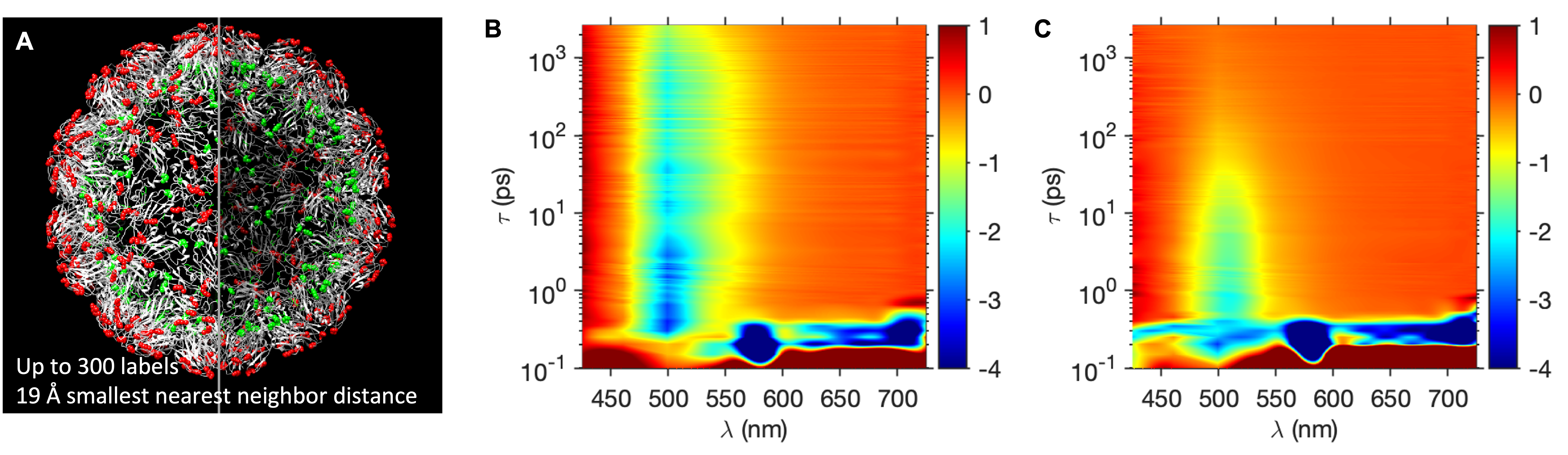
In this work, a fluorescein-derived dye, Oregon GreenTM 488, was covalently bound to the BMV capsid via NHS ester labelling of exposed lysines11. Maximum labeling density was dyes/virus11(see Figure SI1). Figure 1A shows a molecular model of BMV, with the dye-accessible external and internal lysines colored in red and green respectively12. Here ”internal” means located between the lumenal or outer capsid surfaces. The minimum nearest neighbour distance between lysines was estimated to be nm11.
To follow the excited state dynamics of coupled fluorophores in fVLPs, we performed pump-probe femtosecond, transient absorption (TA) spectroscopy. Differential absorption spectra of isolated fluorophores and fVLP samples with an average number of 278 dyes per particle (hereby called BMV-OG278) are presented as a function of the wavelength and time-delay in Figure 1B and Figure 1C, respectively. The ultrafast excitation pulse at 488 nm was provided by an optical parametric amplifier pumped by a regenerative amplifier ( fs pulse width).
In both the control and BMV-OG278 sample, two intense negative signals are evident at very early times in the transient spectra. One appears at nm and it can be attributed to Raman scattering of water13. Since this event is simultaneous with the excitation pulse, its appearance defines the zero delay between pump and probe pulses. The second, early negative signal is due to the coherent interaction between pump and probe pulses, which leads to stimulated Raman amplification appearing at 650-750 nm14. Both Raman signals vanish within ps from the pump pulse.
A spectral region of particular interest is nm, where ground state bleach (GSB) is expected. Indeed, both sample and control exhibit a prominent feature in this spectral region. However, there are also some stark differences between the two, Figure 1B and Figure 1C: In particular, the excited state decay is much faster for the BMV-OG278 sample than for the free dye solution.
Minima in spectra were at nm for free dye and nm for VLP-bound fluorophores, and are consistent with the peak wavelength in steady-state absorption spectra (Figure SI1). The TA spectral evolution of the free dye solution and BMV-OG278 samples depends on pump energy, Figures SI3-7. The nm shift between free and bound fluorophores is attributable to the difference in dielectric constant between protein and water.
For quantitative time-domain analysis and comparison, TA spectra were integrated from 520 to 530 nm and fitted with an exponential model from fs to ns (Figure SI8-12). For analysis, the wavelength and time-delay range were chosen past the ground state bleach minimum to avoid artifacts from pump laser scattering as well to remove coherent artifacts which arise around the zero delay time where the pump and probe beams are temporally overlapping15, 16. Exponential fit parameters (amplitude and decay time) were obtained by means of nonlinear least squares, and the estimates of the errors from the model were computed from the sample covariance matrix. Best fit kinetic models for the transients were found to be predominantly mono-exponential for the free fluorophore and bi-exponential for the virus-bound fluorophore. Normalized amplitudes () and decay times () obtained from the fit procedure are presented as a function of in the plots in Figure 2, with corresponding to free fluorophores in solution.
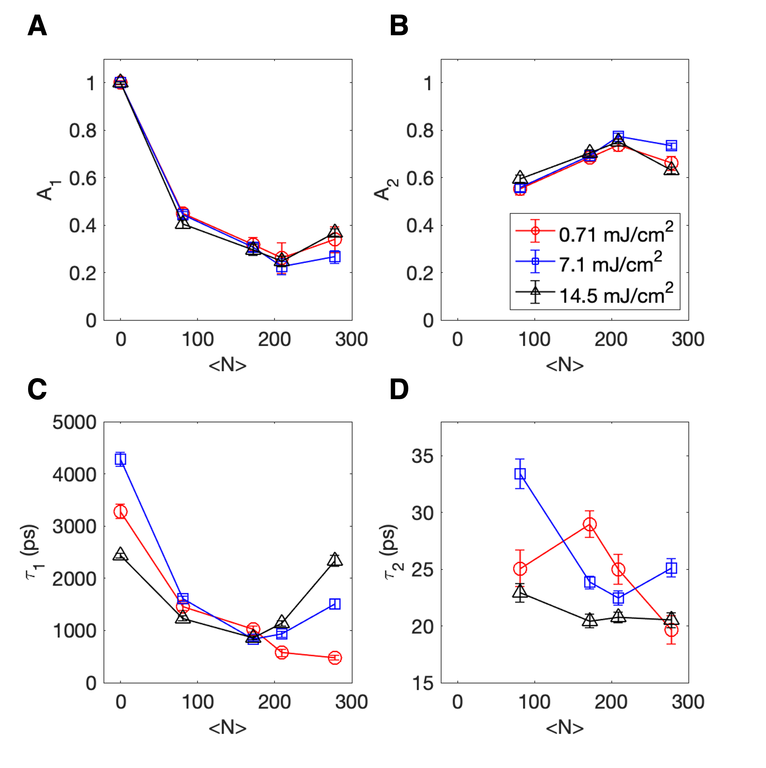
At low power, the excited state lifetime () of free dyes measured by TA was in average ns, which matches the spontaneous emission lifetime of OG in solution. Since the quantum yield is high for this dye, fluorescence emission is the dominant pathway in the relaxation dynamics of the free fluorophore excited at nm. The long lifetime component () exhibits a monotonic shortening as the number of bound dyes increases (Figure 2C, red curve). At high labeling density, the is reduced by a factor of when compared to the free fluorophore. In previous fluorescence lifetime experiments at similar excitation fluence and the fluorescence lifetime was short, and the photon counts were quenched 11. A possible explanation is that at low pump power, only a few fluorophores are excited while the majority are in the ground state and there is a high probability for homo- resonance energy transfer (RET) to occur, with the result of an increase in fluorescence quenching. In any event, we note a correlation between the evolution of the TA evolution and that of the fluorescence decay measured previously.
As the pump fluence increases, decreases for the free dye, possibly due to photobleaching or to an increase in inter-system crossing and triplet state formation 17. Indeed, some photobleaching was observed in amount of between the first and second run for each sample. However, at high labeling density () increases from ps at lowest pump fluence up to ps for highest pump fluence. Thus, it appears that the nonradiative rate, which was deemed responsible for the initial shortening of the lifetime at low fluences and high density 11, slows down to a value that is observed at lower labeling densities.
The second fit component (parameters, and ) dominates BMV-OG dynamics at short times suggesting a new relaxation channel that only operates in the multi-fluorophore VLP. The associated amplitude is significantly greater than , Figure 2B. Its time constant is ps for low and medium fluence and ps for the highest pump fluence, Figure 2D. This component appears to decrease with the number of dyes per virus although the trends are noisy (Figure 2D).
To summarize up to this point, two factors appear to lead to changes in the excited state dynamics: i) pump fluence at fixed , and ii) at all fluences. Specifically, relaxation dynamics is accelerated when increases.
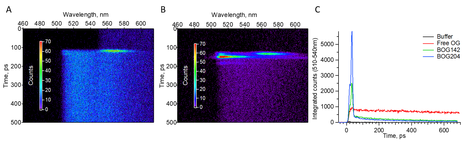
While fluorophore coupling clearly affects relaxation lifetimes, the result of the competition between nonradiative and radiative relaxation channels cannot be solely gauged based on TA. To obtain further spectral and temporal information on fluorescence emission, we have performed time-resolved fluorescence spectroscopy with a streak-camera detector. Figures 3A and B present spectrally and time-resolved fluorescence emission for free fluorophore and BMV-OG204, respectively. The pump pulse arrives at ps. Raman scattering from water, at nm, is simultaneous with the pump pulse. Also, a faint spot at can be observed at nm in free OG and BMV-OG samples. This is due to elastic scattering of the laser line being incompletely suppressed by laser rejection filters. The scattering peak is very faint in the case of free OG because this sample does not scatter as strongly as the virus particles. The inelastic and the elastic scattering peaks are convenient because they facilitate the timing of fluorescence emission with respect to the pump pulse.
For free dye samples emission is spread in time and decays uniformly along the entire measured time interval, Figure 3 A,C. In stark contrast, the fluorescence from the BMV-OG samples is emitted as a burst of ps duration. The burst is delayed with respect to the pump pulse, with a maximum occurring at ps after excitation, Figure 3B,C. At this point, we note that the value of from TA measurements is close to the characteristic time of the fluorescence burst observed in the streak camera experiment. This observation is consistent with the short relaxation pathway observed in TA corresponding to radiative relaxation. The wavelength-integrated peak amplitude of the burst shows a increase when is increased from to dyes per virus, Figure 3C.
What phenomena could potentially be responsible for delayed, non-exponential, accelerated radiation in VLPs? We consider superfluorescence (SF), superradiance (SR), and amplified spontaneous emission (ASE)18, 19. Although we are not completely excluding the possibility of lasing, i.e. light amplification by stimulated emission, we note that it is less likely, at least in the conventional sense, because of the absence of a resonator and the extremely small VLP volume and thus, small Purcell factor20.
There are qualitative differences between ASE and SF in both time and frequency domain. In the frequency domain, SF(SR) emission occurs in a broader window21 while in ASE, spectral narrowing is expected as pump intensity increases22. We have not observed spectral narrowing in our experiments11.
In the time domain, the SF pulse is sharp and develops after a time delay 23. In ASE, the time delay is vanishingly small, the output pulse is longer than in SF and noisy, and the pulse duration is insensitive to dephasing factors (e.g. local viscosity and temperature)24, 25, 26. From previous work 11, 12 we know that the radiation brightening from multi-fluorophore VLPs is very sensitive to the local environment, unlike ASE. Therefore, the significant shortening of excited state lifetime observed in TA and streak camera experiments, the delayed intense pulse in fluorescence emission, the broad fluorescence spectrum, the sensitivity to the fluorophore local environment, all point to a superradiance-like mechanism responsible for radiation brightening. We discuss a simple superradiance model in the SI, which illustrates the burst emission.
An intriguing feature here is the fact that the phenomenon occurs at room temperature. SR emission dynamics was experimentally studied first in atomic gases at very low temperatures 27. Later, it was detected in low-temperature solids28, 29, 30, 31, 32. Excitonic superradiance was studied extensively in molecular aggregates, at cryogenic temperatures as well. 33, 34.
Only recently SR was shown to occur at room temperature in photosynthetic complexes 35, 36. The structured environment within the photosynthetic complex is believed to promote collective fluorophore behavior since rigidity and proper orientation can alleviate thermal dephasing 37. In support of this idea, alterations in the relative position and orientation of fluorophores in the protein pockets of an artificial photosynthetic system were found to strongly affect the excited state dynamics of protein-bound fluorophores 38, 39. However, it is worth emphasizing that interfluorophore interactions in light harvesting complexes and in most molecular J-aggregates are characterized by strong coupling 40, 41, 42. By comparison, the nearest-neighbor fluorophore distances on BMV-OG are much longer than that of molecular aggregates, which precludes electron tunnelling.
In our case, collective emission of radiation arise from weak coupling at room temperature. Until now, this coupling range has not been given much attention. Dipole-dipole coupling is considered detrimental to the formation of coherent multi-fluorophore states 27. Could the virus template play a role in coupling? The original model of Dicke superradiance (see SI) does not take into account the spatial organization or orientation of the fluorophores. However, from previous work it was clear that the nature of the template is important for the radiation brightening effect 11, 12. Below we provide further evidence for it from a study of relaxation dynamics.
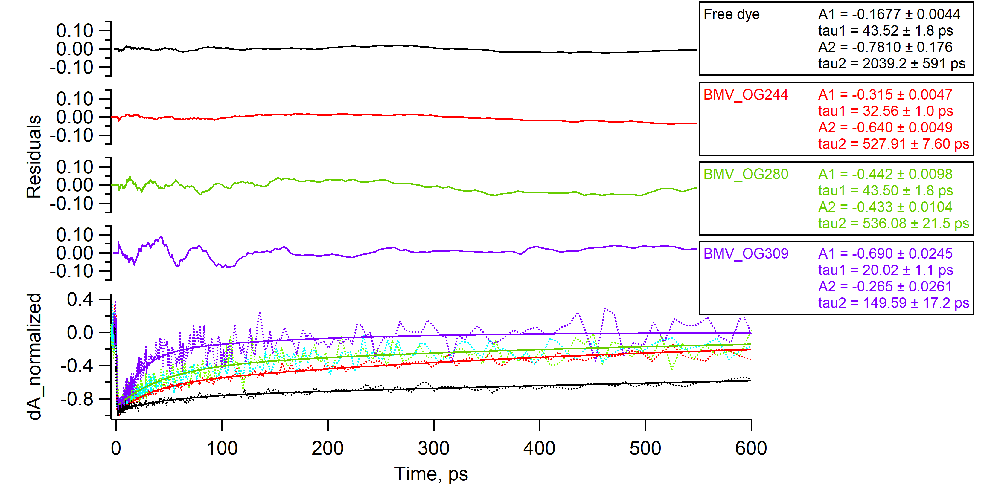
We performed additional TA experiments with a set-up in which pump and probe beams were counter-propagating, at a small angle. This set-up change allowed us to reduce laser scattering artifacts and measure the excited state dynamics at high laser powers with the probe wavelength closer to ground state bleach peak position nm.
The higher signal-to-noise ratio afforded by this setup change has allowed us to make an interesting observation. Figure 4 presents normalized absorbance difference curves obtained for free dye and BMV-OG with ranging from 200 to 300 dyes/virus. The data were tail-fitted with double exponential decay and the residuals from the fitting are presented on the top panels of the graph. At high dye loads, oscillations in TA can be observed, even in the raw data but most prominently in the residuals.
Oscillating TA signals previously observed in photosynthetic complexes at room temperature were attributed to vibrations of the protein matrix encapsulating the fluorophores43, 44. The oscillations observed on high-load VLPs in Figure 4 persist for a longer time, i.e. up to 100 ps, and have lower frequency. They cannot be assigned to quantum beats from closely spaced levels since those are not expected to be observed at room temperature. We hypothesise that the oscillations have to do with the template. Two mechanisms could be envisioned by which the virus template may affect the collective emission of the multi-fluorophore antenna. First, we note that the frequency of the observed oscillations falls within the range of computed low-frequency modes (vibrational/breathing) of the virus shells 45, 46, 47. Since coupling in near-field is very sensitive to inter-fluorophore distance and orientation, such low-frequency vibrations could modulate the TA trace. Second, the template may modulate the radiative rate of the antenna via oscillations in polarizability due to virus shell vibrations 48. Could such virus-shell modes be involved in maintaining fluorophore correlations? At this point such questions concerning the role of spatial symmetry and the nature of coupling remain open, and warrant future studies.
In conclusion, the symmetric BMV capsids provides a biological scaffold for a multi-fluorophore array that acts an antenna with accelerated emission rate at room temperature. We have employed ultrafast, time-resolved transient absorption and fluorescence spectroscopy to provide further evidence for the mechanism of radiation brightening. Taken together with results of previous work, the new data strongly suggest that the virus-supported multi-fluorophore array exhibits a form of superradiant emission. Moreover, coherent, low-frequency virus shell vibrations could be observed as they modulate the relaxation rate for nearly 100 ps at room temperature. This finding highlights the involvement of the virus scaffold in the excited state relaxation dynamics. Enhanced light intensity, non-classical emission dynamics, and biocompatibility make these viromimetic superfluorescent particles interesting for biological imaging at high background conditions, for instance as deep tissue optical probes.
1 Experimental
1.1 BMV-OG sample preparation
BMV-OG samples were prepared according to protocols previously described 11. Briefly, the lysines of wtBMV were modified by covalent conjugation with Oregon Green™ 488 carboxylic acid, succinimidyl ester49. wtBMV stock in SAMA buffer (50 mM NaOAc, 8 mM \ceMg(OAc)2(pH 4.6)) was mixed with sodium bicarbonate buffer (100 mM, pH 8.2) in 1:1 ratio to a final concentration of particles/ml. Dye stock was freshly prepared in DMSO and was added to virus solution at varied ratios ranging from 500 to 10000 dyes/capsid. The DMSO concentration in reaction mixture was kept constant at w/v%. The reaction was left for 1-2 hours at room temperature, followed by removal of free dye via filter wash or dialysis with SAMA buffer. UV-Vis spectroscopy was performed to estimate the number of dyes per virus, and fluorescence spectroscopy was used to confirm absence of free dyes in the purified samples.
1.2 Transmission Electron Microscopy
Electron-transparent samples were prepared by placing the dilute sample (10 L) onto a carbon-coated copper grid. After 10 mins, excess solution on the grid was removed with filter paper. The grid was stained with uranyl acetate (10 L of 2% solution) for 10 minutes and the excess solution was removed by blotting with filter paper. The sample was then left to dry for several minutes. Images were acquired at an accelerating voltage of 80 kV on a JEOL JEM 1010 Transmission electron microscope and analyzed with the ImageJ Processing Toolkit (see SI2).
1.3 Time-resolved Pulsed Excitation Fluorescence Spectroscopy with Streak camera
The sample was excited by a pulse generated through an optical parametric amplifier using a Ti:sapphire femtosecond laser (Spectra-Physics Spitfire Pro). The pulses have a wavelength of nm, a duration of 35 fs, and a repetition rate of 2000 Hz. The focused spot size was measured to be 160 m and the average laser power varied from 1 to 6.5 mW (or 0.5 to 3.25 J/pulse). The excitation beam was directed at the cuvette at a small angle to prevent cavity effect. The sample fluorescence was collected with a lens, directed through long pass filter (LP508) and a 150 mm spectrograph, then was detected as a function of wavelength and time after excitation using a photon counting streak camera (Hamamatsu C5680). Data was collected and pre-processed by HPDTA software and further analysis was done with Igor Pro software.
1.4 Transient absorption spectroscopy
Ultrafast transient absorption measurements were carried out using an output of regeneratively amplified Ti:sapphire laser (800 nm, 120 fs, 2 kHz repetition rate) which was split into two beams. The first beam, containing 10% power, was focused into a sapphire crystal to generate a white light continuum (440-750 nm), which serves as the probe laser. The other beam, containing 90% of the power, was sent into an optical parametric amplifier to generate the 480 nm pump beam. After passing through a depolarizer, the pump beam is focused and overlapped with the probe beam at the sample (focal diameter being 300 m). The pump pulse fluence was varied from 0.71 mJ/ to 14.5 mJ/, while the probe pulse fluence was significantly smaller and kept constant in all experiments. The upper and lower limits over the range of excitation energies correspond to photon densities that were close to but greater than the number of fluorophores attached in VLP antennas. Prior to any analysis, spectra were corrected for chirp and group dispersion velocity using Surface Explorer (Ultrafast Systems).
1.5 Transient absorption set-up
The tunable wavelength output of optical parametric amplifier pumped by a Ti:sapphire regenerative amplifier at 250 kHz repetition rate(180 fs pulsewidth), seeded by a mode-locked oscillator at 76 MHz (Coherent Inc.) was set at 480 nm and used as the pump pulse for transient absorption measurements. The part of white light output from OPA passed through a 490/20 optical filter and used as probe pulse. The fluence of the pump was varied from from 0.1 to 3.5 mJ/cm2 and the probe pulse was set at J/mm2. For measurements of relaxation decay, the probe pulse arrived at a delayed time after the pump that was continuously adjusted using a variable length delay line (600 ps max.). A mechanical chopper was used to modulate the pump beam at 1000 Hz. The lock-in amplifier was used to measure probe signal at the modulation frequency. The transient absorption data processing included a background subtraction step.
The work was supported by the Army Research Office, under awards W911NF2010072 and W911NF2010071, and by the National Science Foundation, under award CBET-1803440. This work was performed, in part, at the Center for Nanoscale Materials, a U.S. Department of Energy Office of Science User Facility, and supported by the U.S. Department of Energy, Office of Science, under Contract No. DE-AC02-06CH11357. We are grateful to the Center for Bioanalytical Metrology (CBM), an NSF Industry-University Cooperative Research Center, for providing funding for this project under grant NSF IIP 1916645, and to members of the industry advisory board of the CBM for valuable discussions and feedback.
The Supporting Information is available free of charge on the ACS Publications website at DOI: 10.1021/acs.jpclett.0000000. Experimental methods, sample characterization, TA fitting results and model of superradiant emission from N fluorophores are presented in the supporting information file.
References
- Wolfbeis 2015 Wolfbeis, O. S. An overview of nanoparticles commonly used in fluorescent bioimaging. Chem. Soc. Rev. 2015, 44, 4743–4768
- Yi et al. 2014 Yi, X.; Wang, F.; Qin, W.; Yang, X.; Yuan, J. Near-infrared fluorescent probes in cancer imaging and therapy: an emerging field. International Journal of nanomedicine 2014, 9, 1347
- Vats et al. 2017 Vats, M.; Mishra, S. K.; Baghini, M. S.; Chauhan, D. S.; Srivastava, R.; De, A. Near infrared fluorescence imaging in nano-therapeutics and photo-thermal evaluation. International journal of molecular sciences 2017, 18, 924
- Ueno and Nagano 2011 Ueno, T.; Nagano, T. Fluorescent probes for sensing and imaging. Nature methods 2011, 8, 642–645
- Gao et al. 2021 Gao, L.; Wang, W.; Wang, X.; Yang, F.; Xie, L.; Shen, J.; Brimble, M. A.; Xiao, Q.; Yao, S. Q. Fluorescent probes for bioimaging of potential biomarkers in Parkinson’s disease. Chemical Society Reviews 2021, 50, 1219–1250
- Leopold et al. 2019 Leopold, A. V.; Shcherbakova, D. M.; Verkhusha, V. V. Fluorescent biosensors for neurotransmission and neuromodulation: engineering and applications. Frontiers in cellular neuroscience 2019, 13, 474
- Oheim et al. 2014 Oheim, M.; van’t Hoff, M.; Feltz, A.; Zamaleeva, A.; Mallet, J.-M.; Collot, M. New red-fluorescent calcium indicators for optogenetics, photoactivation and multi-color imaging. Biochimica et Biophysica Acta (BBA)-Molecular Cell Research 2014, 1843, 2284–2306
- Abdollahi et al. 2020 Abdollahi, A.; Roghani-Mamaqani, H.; Razavi, B.; Salami-Kalajahi, M. Photoluminescent and chromic nanomaterials for anticounterfeiting technologies: recent advances and future challenges. ACS nano 2020, 14, 14417–14492
- Li et al. 2018 Li, M.; Feng, Y.; Tian, Q.; Yao, W.; Liu, L.; Li, X.; Wang, H.; Wu, W. Tunable and ultra-stable UV light-switchable fluorescent composites for information hiding and storage. Dalton Transactions 2018, 47, 11264–11271
- Valeur and Berberan-Santos 2013 Valeur, B.; Berberan-Santos, M. r. N. John Wiley Sons, 2nd ed.; Molecular Fluorescence: Principles and Applications; John Wiley & Sons: New York, 2013
- Tsvetkova et al. 2019 Tsvetkova, I. B.; Anil Sushma, A.; Wang, J. C.-Y.; Schaich, W. L.; Dragnea, B. Radiation Brightening from Virus-like Particles. ACS Nano 2019, 13, 11401–11408
- Anil Sushma et al. 2021 Anil Sushma, A.; Zhao, B.; Tsvetkova, I. B.; Pérez-Segura, C.; Hadden-Perilla, J. A.; Reilly, J. P.; Dragnea, B. Subset of Fluorophores Is Responsible for Radiation Brightening in Viromimetic Particles. J. Phys. Chem. B 2021, 125, 10494–10505
- Parker 1959 Parker, C. Raman spectra in spectrofluorimetry. Analyst 1959, 84, 446–453
- Lorenc et al. 2002 Lorenc, M.; Ziolek, M.; Naskrecki, R.; Karolczak, J.; Kubicki, J.; Maciejewski, A. Artifacts in femtosecond transient absorption spectroscopy. Applied Physics B 2002, 74, 19–27
- Vardeny and Tauc 1981 Vardeny, Z.; Tauc, J. Picosecond coherence coupling in the pump and probe technique. Optics Communications 1981, 39, 396–400
- Hui et al. 2011 Hui, L.; Hang, Z.; Jin-Hai, S.; Li-He, Y.; Feng, C.; Xun, H. Elimination of the coherent artifact in a pump-probe experiment by directly detecting the background-free diffraction signal. Chinese Physics Letters 2011, 28, 086602
- Demchenko 2020 Demchenko, A. P. Photobleaching of organic fluorophores: quantitative characterization, mechanisms, protection. Methods and Applications in Fluorescence 2020, 8, 022001
- Cong et al. 2016 Cong, K.; Zhang, Q.; Wang, Y.; Noe, G. T.; Belyanin, A.; Kono, J. Dicke Superradiance in Solids [Invited]. J. Opt. Soc. Am. B 2016, 33, C80
- Dai 2011 Dai, D. Brief comment: Dicke superradiance and superfluorescence find application for remote sensing in air. arXiv preprint arXiv:1108.5360 2011,
- Samuel et al. 2009 Samuel, I. D. W.; Namdas, E. B.; Turnbull, G. A. How to Recognize Lasing. Nat. Photonics 2009, 3, 546–549
- Dicke 1954 Dicke, R. H. Coherence in Spontaneous Radiation Processes. Phys. Rev. 1954, 93, 99–110
- Allen and Peters 1973 Allen, L.; Peters, G. Amplified spontaneous emission and external signal amplification in an inverted medium. Physical review A 1973, 8, 2031
- Malcuit et al. 1987 Malcuit, M. S.; Maki, J. J.; Simkin, D. J.; Boyd, R. W. Transition from Superfluorescence to Amplified Spontaneous Emission. Phys. Rev. Lett. 1987, 59, 1189–1192
- Wilk et al. 1983 Wilk, S. R.; Boyd, R. W.; Teegarden, K. J. Laser characteristics of KCL: O-2. Optics communications 1983, 47, 404–406
- Rai and Bowden 1992 Rai, J.; Bowden, C. M. Quantum-statistical analysis of superfluorescence and amplified spontaneous emission in dense media. Physical Review A 1992, 46, 1522
- Maki et al. 1989 Maki, J. J.; Malcuit, M. S.; Raymer, M. G.; Boyd, R. W.; Drummond, P. D. Influence of collisional dephasing processes on superfluorescence. Physical Review A 1989, 40, 5135
- Gross and Haroche 1982 Gross, M.; Haroche, S. Superradiance: An Essay on the Theory of Collective Spontaneous Emission. Phys. Rep. 1982, 93, 301–396
- Miyajima et al. 2017 Miyajima, K.; Kumagai, Y.; Ishikawa, A. Ultrashort radiation of biexcitonic superfluorescence from high-density assembly of semiconductor quantum dots. The Journal of Physical Chemistry C 2017, 121, 27751–27757
- Tighineanu et al. 2016 Tighineanu, P.; Daveau, R. S.; Lehmann, T. B.; Beere, H. E.; Ritchie, D. A.; Lodahl, P.; Stobbe, S. Single-photon superradiance from a quantum dot. Physical review letters 2016, 116, 163604
- Rainò et al. 2018 Rainò, G.; Becker, M. A.; Bodnarchuk, M. I.; Mahrt, R. F.; Kovalenko, M. V.; Stöferle, T. Superfluorescence from lead halide perovskite quantum dot superlattices. Nature 2018, 563, 671–675
- Krieg et al. 2020 Krieg, F.; Sercel, P. C.; Burian, M.; Andrusiv, H.; Bodnarchuk, M. I.; St0̈ferle, T.; Mahrt, R. F.; Naumenko, D.; Amenitsch, H.; Rainò, G., et al. Monodisperse long-chain sulfobetaine-capped CsPbBr3 nanocrystals and their superfluorescent assemblies. ACS central science 2020, 7, 135–144
- Haider et al. 2021 Haider, G.; Sampathkumar, K.; Verhagen, T.; Nádvorník, L.; Sonia, F. J.; Valeš, V.; Sỳkora, J.; Kapusta, P.; Němec, P.; Hof, M., et al. Superradiant Emission from Coherent Excitons in van Der Waals Heterostructures. Advanced Functional Materials 2021, 2102196
- Palacios et al. 2002 Palacios, M. A.; de Weerd, F. L.; Ihalainen, J. A.; van Grondelle, R.; van Amerongen, H. Superradiance and Exciton (De)localization in Light-Harvesting Complex II from Green Plants? The Journal of Physical Chemistry B 2002, 106, 5782–5787
- Engel et al. 2007 Engel, G. S.; Calhoun, T. R.; Read, E. L.; Ahn, T.-K.; Mančal, T.; Cheng, Y.-C.; Blankenship, R. E.; Fleming, G. R. Evidence for wavelike energy transfer through quantum coherence in photosynthetic systems. Nature 2007, 446, 782–786
- Monshouwer et al. 1997 Monshouwer, R.; Abrahamsson, M.; van Mourik, F.; van Grondelle, R. Superradiance and Exciton Delocalization in Bacterial Photosynthetic Light-Harvesting Systems. J. Phys. Chem. B 1997, 101, 7241–7248
- Malina et al. 2021 Malina, T.; Koehorst, R.; Bína, D.; Pšenčík, J.; van Amerongen, H. Superradiance of bacteriochlorophyll c aggregates in chlorosomes of green photosynthetic bacteria. Scientific Reports 2021, 11, 8354
- Rolczynski et al. 2018 Rolczynski, B. S.; Zheng, H.; Singh, V. P.; Navotnaya, P.; Ginzburg, A. R.; Caram, J. R.; Ashraf, K.; Gardiner, A. T.; Yeh, S.-H.; Kais, S., et al. Correlated protein environments drive quantum coherence lifetimes in photosynthetic pigment-protein complexes. Chem 2018, 4, 138–149
- Noriega et al. 2015 Noriega, R.; Finley, D. T.; Haberstroh, J.; Geissler, P. L.; Francis, M. B.; Ginsberg, N. S. Manipulating excited-state dynamics of individual light-harvesting chromophores through restricted motions in a hydrated nanoscale protein cavity. J. Phys. Chem. B 2015, 119, 6963–6973
- Delor et al. 2018 Delor, M.; Dai, J.; Roberts, T. D.; Rogers, J. R.; Hamed, S. M.; Neaton, J. B.; Geissler, P. L.; Francis, M. B.; Ginsberg, N. S. Exploiting chromophore–protein interactions through linker engineering to tune photoinduced dynamics in a biomimetic light-harvesting platform. Journal of the American Chemical Society 2018, 140, 6278–6287
- Spano and Mukamel 1989 Spano, F. C.; Mukamel, S. Superradiance in Molecular Aggregates. J. Chem. Phys. 1989, 91, 683–700
- Meinardi et al. 2003 Meinardi, F.; Cerminara, M.; Sassella, A.; Bonifacio, R.; Tubino, R. Superradiance in molecular H aggregates. Physical review letters 2003, 91, 247401
- Ratner 2003 Ratner, M. Superradiance of J-aggregates: correspondence between an infinite disordered chain and a regular finite chain. Low Temperature Physics 2003, 29, 602–605
- Panitchayangkoon et al. 2010 Panitchayangkoon, G.; Hayes, D.; Fransted, K. A.; Caram, J. R.; Harel, E.; Wen, J.; Blankenship, R. E.; Engel, G. S. Long-lived quantum coherence in photosynthetic complexes at physiological temperature. Proceedings of the National Academy of Sciences 2010, 107, 12766–12770
- Engel 2011 Engel, G. S. Quantum coherence in photosynthesis. Procedia Chemistry 2011, 3, 222–231
- van Vlijmen and Karplus 2005 van Vlijmen, H. W.; Karplus, M. Normal mode calculations of icosahedral viruses with full dihedral flexibility by use of molecular symmetry. J Mol Biol 2005, 350, 528–42
- Dykeman and Sankey 2008 Dykeman, E. C.; Sankey, O. F. Low Frequency Mechanical Modes of Viral Capsids: An Atomistic Approach. Phys. Rev. Lett. 2008, 100, 028101
- Hadden et al. 2018 Hadden, J. A.; Perilla, J. R.; Schlicksup, C. J.; Venkatakrishnan, B.; Zlotnick, A.; Schulten, K. All-atom molecular dynamics of the HBV capsid reveals insights into biological function and cryo-EM resolution limits. Elife 2018, 7
- Young et al. 2018 Young, G. et al. Quantitative mass imaging of single biological macromolecules. Science 2018, 360, 423–427
- Johnson and Spence 2010 Johnson, I., Spence, M., Eds. The Molecular Probes Handbook : a guide to fluorescent probes and labeling technologies., 11th ed.; Life Technologies, 2010