Few-shot Unsupervised Domain Adaptation for Multi-modal Cardiac Image Segmentation
Abstract
Unsupervised domain adaptation (UDA) methods intend to reduce the gap between source and target domains by using unlabeled target domain and labeled source domain data, however, in the medical domain, target domain data may not always be easily available, and acquiring new samples is generally time-consuming. This restricts the development of UDA methods for new domains. In this paper, we explore the potential of UDA in a more challenging while realistic scenario where only one unlabeled target patient sample is available. We call it Few-shot Unsupervised Domain adaptation (FUDA). We first generate target-style images from source images and explore diverse target styles from a single target patient with Random Adaptive Instance Normalization (RAIN). Then, a segmentation network is trained in a supervised manner with the generated target images. Our experiments demonstrate that FUDA improves the segmentation performance by 0.33 of Dice score on the target domain compared with the baseline, and it also gives 0.28 of Dice score improvement in a more rigorous one-shot setting. Our code is available at https://github.com/MingxuanGu/Few-shot-UDA.
1 Introduction
As manual contouring of medical images is tedious and time-consuming, automatic medical image segmentation is more desirable [1]. While deep learning methods often suffer from performance degradation when a domain gap is observed between training (source) and testing (target) data. UDA methods tackle this problem by reducing the domain gap with a variety of techniques, for example, discrepancy reduction [2], adversarial learning [3], image translation [4], etc. These methods are conditioned on the availability of a large amount of target data which, however, is quite scarce.
In this work, we consider a more realistic and practical scenario where we still have sufficient labeled source data, while we only have one unlabeled target data for training. To this end, a style transfer method called Random Adaptive Instance Normalization (RAIN) [5] is used to generate diverse target-style images from a single target patient data. Then, a segmentation module can be trained in a supervised manner with generated images. Our contributions are: (1) we explore the potential of FUDA for multi-modal cardiac CMR segmentation and it shows better performance compared with its baseline model and other recent UDA methods, (2) we extend our method to one-shot learning and demonstrate the possibility of FUDA with only one slice of target data available.
2 Materials and methods
2.1 Dataset
We assess the proposed FUDA on MS-CMRSeg [6] 2019 challenge dataset, which consists of 45 short-axis bSSFP, T2-weighted and LGE scans from patients diagnosed with cardiomyopathy. The ground-truth contours are generated by two experts and include right ventricle (RV) cavity, left ventricle (LV) cavity, and myocardium (Myo) region. Only affine transformation (rotation, translation, shearing, etc.) is applied and the sequences are normalized using min-max normalization. Then, the sequences are center-cropped to pixels to have only region-of-interest (ROIs) areas.
2.2 Problem statement
In UDA for semantic segmentation, a set of labeled data in source domain is given, where represents one sample, and the corresponding label in . Whereas for target domain () only unlabeled target images () are given. The goal is to improve the performance of the segmentation by reducing the distribution gap between the source and target domain. In our case, we consider only one unlabeled target patient data () being available.
2.3 RAIN Module
RAIN is developed on the basis of Adaptive Instance Normalization (AdaIN) [7]. AdaIN has an encoder-decoder architecture. It generates stylized images which have the appearance of the style image while preserving the structure of the content images by re-normalizing the features of content images with Eq. 1:
| (1) |
where , are the latent features of the content and style image, and denote channel-wise mean and standard deviation. To achieve realistic image transfer, a content loss and a style loss [7] is employed.
RAIN takes advantage of the style transfer in AdaIN and involves a style variational auto-encoder (VAE) in between the encoder and the decoder. The style-VAE is composed of an encoder and a decoder . encodes to , where denotes concatenation, and aims to reconstruct the original style with , where . Kullback-Leibler (KL) divergence loss is applied to enforce to be normal distributed. Furthermore, L2 loss is applied between and to force identity reconstruction .
Thus, the overall loss function to train RAIN can be formulated as where , and denote the weights for the losses.
2.4 Few-shot UDA for cardiac MRI segmentation
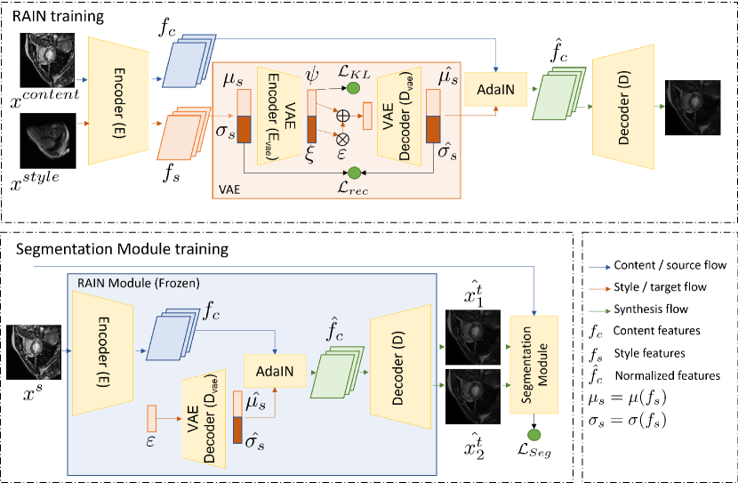
The proposed FUDA framework is constructed based on ASM [5] implementation and is shown in Fig. 1. To train the segmentation module and explore styles from a single target image, we first generate the latent distribution from the input image . Then we sample an from . After that, we can generate stylized images from any source image . Then the style transfer can be formulated as:
| (2) |
Furthermore, the segmentation module can be trained in a supervised manner with and the corresponding .
We train the segmentation module with a combination of cross-entropy loss (CE) and jaccard distance loss (JD) as . To enforce the segmentation module to produce domain invariant features between and the corresponding , is applied as a consistency loss, where are the latent features of the source and the corresponding generated target image from the bottleneck layer of the segmentation module. Then the overall loss function for the segmentation module is:
| (3) |
where is the weight of . Finally, to generate more diverse and increasingly difficult target images , is updated in the direction that makes the segmentation module perform worse on segmentation with:
| (4) |
where is the iteration number, represents the corresponding source label for and denotes the learning rate.
| DC () | HD\ts[mm] () | ||||||||
| Method | Data | Myo | LV | RV | AVG | Myo | LV | RV | AVG |
| W/o UDA | N/A | 0.24 | 0.40 | 0.27 | 0.30 | 31.7 | 31.0 | 45.0 | 35.9 |
| AdaptSeg | Few | 0.39 | 0.63 | 0.58 | 0.53 | 39.1 | 28.7 | 25.6 | 31.1 |
| ADVENT | Few | 0.39 | 0.59 | 0.52 | 0.50 | 38.4 | 35.3 | 37.9 | 37.2 |
| Proposd | Few | 0.46 | 0.77 | 0.65 | 0.63 | 24.5 | 13.7 | 22.4 | 20.2 |
| AdaptSeg | One | 0.45 | 0.65 | 0.52 | 0.54 | 32.3 | 34.3 | 36.4 | 34.3 |
| ADVENT | One | 0.37 | 0.61 | 0.51 | 0.50 | 42.7 | 26.9 | 35.0 | 34.9 |
| Proposd | One | 0.39 | 0.73 | 0.63 | 0.58 | 36.2 | 19.0 | 25.6 | 26.9 |
| Observer | N/A | 0.76 | 0.88 | 0.81 | 0.82 | 12.0 | 14.3 | 21.5 | 15.9 |
2.5 Training
The training process has two stages. First, we use bSSFP-MRI as content images and T2-weighted-MRI as style images to pretrain the RAIN module. Since it does not involve any target images (LGE), this process makes it convenient to train RAIN anytime before training the segmentation module. In the second stage, the RAIN model is frozen, and we employ Dilated-Residual UNet (DR-UNet) [9] as the segmentation module. We first pretrain the DR-UNet with bSSFP images and the generated LGE-style images in a supervised manner for 40-50 iterations. Then the model is trained together with being iteratively updated for another 40-50 iterations. We used stochastic gradient descent (SGD) with a momentum of and a weight decay of as the optimizer. We empirically set the hyper-parameters of , and . The proposed method is trained and tested on one Geforce 1080Ti GPU. The training of RAIN and segmentation module takes 2 hours and 21 hours respectively. Overall inference takes 23 seconds in average for each patient.
3 Results
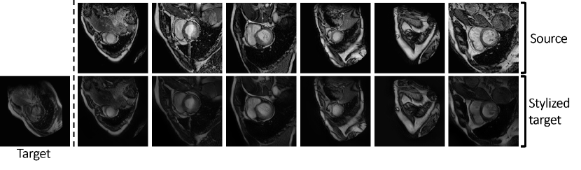
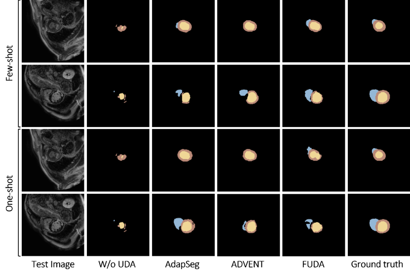
Fig. 2 shows the qualitative results of the pretrained RAIN. We can observe that RAIN successfully captures the features of target images. Tab. 1 summarizes the quantitative results of different methods. Baseline method achieved the lowest average volumetric Dice (0.30) and an HD (35.9\tsmm). With a few-shot UDA setting, our proposed method achieved the best overall Dice (0.63) and the best lowest HD (20.2\tsmm). Subsequently, we demonstrate the results for one-shot UDA, and our proposed method achieved the highest Dice (0.58) and the lowest HD (26.9\tsmm). Fig. 3 illustrates qualitative examples of different segmentation approaches. Compared with other methods, the proposed method is able to produce more complete and precise segmentation maps.
4 Discussion
In this work, we presented a few-shot UDA (FUDA) for multi-modal CMR image segmentation while restricting the experiments in a more challenging yet realistic scenario where only one target sample is available. By comparing the proposed method with other approaches under the same settings, we show that FUDA highly reduced the domain gap with only a few target slices. We also demonstrated that the proposed few-shot method produces promising results with the more rigorous one-shot setting. We find for conventional UDA methods like AdaptSeg and ADVENT, performance on one-shot and few-shot settings only has a slight difference. This can be attributed to the fact that the slices of one patient only have a small distribution shift, hence the knowledge learned by the model from target slices of one patient is limited. While for FUDA, the model has the ability to explore unseen styles of the target images, hence more data provided results in more diverse target styles. Consequently, better segmentation performance could be achieved. Furthermore, we believe there is still room to improve the quality of the segmentation prediction. As a result, in the future, we will explore feasible techniques like contrastive learning and attention to improve the performance of the proposed FUDA.
References
- [1] Tanja Kurzendorfer et al. “Fully automatic segmentation of left ventricular anatomy in 3-D LGE-MRI” In Computerized Medical Imaging and Graphics 59 Elsevier, 2017, pp. 13–27
- [2] Arthur Gretton et al. “A kernel method for the two-sample-problem” In Advances in neural information processing systems, 2007, pp. 513–520
- [3] T. Vu et al. “ADVENT: Adversarial Entropy Minimization for Domain Adaptation in Semantic Segmentation” In CVPR, 2019, pp. 2512–2521 DOI: 10.1109/CVPR.2019.00262
- [4] J. Zhu, T. Park, P. Isola and A.. Efros “Unpaired Image-to-Image Translation Using Cycle-Consistent Adversarial Networks” In ICCV, 2017, pp. 2242–2251
- [5] Yawei Luo et al. “Adversarial Style Mining for One-Shot Unsupervised Domain Adaptation” In Advances in Neural Information Processing Systems 33 Curran Associates, Inc., 2020, pp. 20612–20623
- [6] X. Zhuang “Multivariate mixture model for myocardial segmentation combining multi-source images” In IEEE Trans Pattern Anal Mach Intell, 2019, pp. 1–1
- [7] X. Huang and S. Belongie “Arbitrary Style Transfer in Real-Time with Adaptive Instance Normalization” In ICCV, 2017, pp. 1510–1519
- [8] Y. Tsai et al. “Learning to Adapt Structured Output Space for Semantic Segmentation” In CVPR, 2018, pp. 7472–7481
- [9] Sulaiman Vesal, Nishant Ravikumar and Andreas Maier “Automated Multi-sequence Cardiac MRI Segmentation Using Supervised Domain Adaptation” In STACOM, 2020, pp. 300–308