In-plane magnetic structure and exchange interactions in the high-temperature antiferromagnet Cr2Al
Abstract
The ordered tetragonal intermetallic Cr2Al forms the same structure type as Mn2Au, and the latter has been heavily investigated for its potential in antiferromagnetic spintronics due to its degenerate in-plane Néel vector. We present the single crystal flux growth of Cr2Al and orientation-dependent magnetic properties. Powder neutron diffraction of Cr2Al and first-principles simulations reveal that the magnetic ordering is likely in-plane and therefore identical to Mn2Au, providing a second material candidate in the MoSi2 structure type to evaluate the fundamental interactions that govern spintronic effects. The single ordering transition seen in thermal analysis and resistivity indicates that no canting of the moments along the axis is likely. Magnetometry, resistivity, and differential scanning calorimetry measurements confirm the Néel temperature to be K. First-principles simulations indicate that the system has a small density of states at the Fermi energy and confirm the lowest-energy magnetic ground state ordering, while Monte Carlo simulations match the experimental Néel temperature.
I Introduction
Antiferromagnets have attracted considerable attention due to fundamentally different proposed magnetization switching mechanisms and optically-probed spin relaxation dynamics in the THz range,Kimel et al. (2005); Little et al. (2017); Kampfrath et al. (2011) compared to GHz for ferromagnets and ferrimagnets.Viala et al. (2004); Lee et al. (2016); Chai et al. (2013); Sharma et al. (2016) Recent studies have demonstrated partial switching of the Néel vector of antiferromagnetic CuMnAs and Mn2AuBhattacharjee et al. (2018); Barthem et al. (2013); Jourdan et al. (2015), perhaps due to current-induced spin-orbit torques.Olejník et al. (2018); Saidl et al. (2017); Wadley et al. (2015, 2016, 2018) The magnetic-field-induced spin rotation and spin-flop behavior of epitaxial CuMnAs thin films has been probed using X-ray magnetic linear dichroism and signatures of the Néel vector reorientation have been seen in the anisotropic magnetoresistance with multiterminal devices.Wang et al. (2020); Wadley et al. (2016); Olejník et al. (2018) Néel vector reorientations have also been observed in femtosecond pump-probe magneto-optical Kerr effect experiments.Saidl et al. (2017)
Intriguingly, Chien et al. have shown that the purported spin-orbit torque magnetoresistance effects may be artifacts of excessive current-induced heating through multiterminal devices, thus unequivocal detection of the Néel vector before and after SOT switching at low currents is required to effectively prove magnetic switching.Chiang et al. (2019) The in-plane current-pules switching experiments on Mn2Au quantified the anisotropic magnetoresistance and planar Hall effect of thin films.Bodnar et al. (2018) A spin-orbit-torque-driven antiferromagnetic resonance of Mn2Au has not been observed to date by time-domain THz spectroscopy, despite early reports.Bhattacharjee et al. (2020)
Mn2Au remains antiferromagnetic until it forms a disordered (Mn,Au) solid solution at 953 K,Cahn (1991) with a Néel temperature predicted to be above 1600 K, due to its large anisotropy energy, calculated based on first-principles calculations.Khmelevskyi and Mohn (2008) Such large anisotropy may not be desired in all spintronic applications. In particular, the ability to observe a Néel-order-driven effect vanish at , without excessive current flow and Joule heating, would be a strong confirmation of true spintronic effects. To understand the underlying physics and capabilities, there is a pressing need to expand the library of intermetallic antiferromagnets that share features with CuMnAs (which itself has not been grown as bulk single crystals, likely due to a competing orthorhombic phase)Uhlířová et al. (2019) and Mn2Au. Single crystals of such materials are needed to study the orientation dependence of their magnetic dynamics.
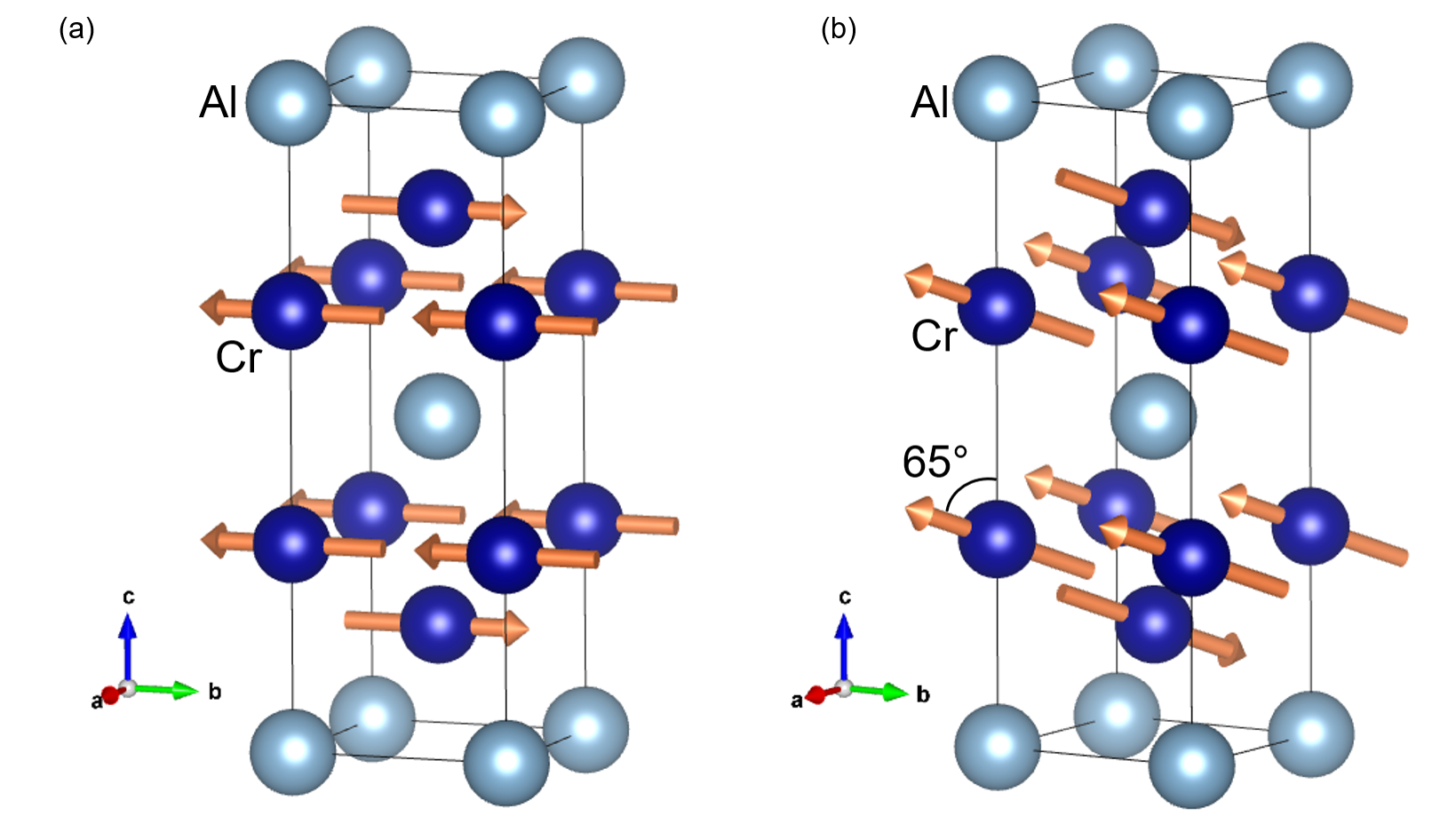
Cr2Al is an incongruently-melting intermetallic antiferromagnet with the MoSi2 structure type, tetragonal space group , isostructural to Mn2Au and Au2Mn. There is no reported single crystal growth or measurements of its anisotropic properties. Atoji presented variable-temperature powder neutron diffraction of Cr2Al in 1965. Atoji (1965) In that study, the (001) neutron diffraction peak intensity was used to determine the K. The magnetic structure was claimed to be inclined by an angle toward the axis, though no explanation of that determination was given. We show here that the in-plane (zero canting) spin configuration is far more plausible.
In 1988, Semukhin et al. investigated the magnetic phase transition of Cr2Al by high-temperature X-ray diffraction and calorimetry.Semukhin et al. (1988) Sharp changes in the and axis lattice parameters and a heat capacity anomaly occurred K. Variation in the reported may be due to a compositional width of the Cr2Al phase, which has been reported to be about 10 %.Mahdouk and Gachon (2000); Rank et al. (2019) Variable can also arise from further alloying, as when Susner et al. showed that the resistivity anomaly at shifts to lower temperatures and is more pronounced with increased Fe substitution for Cr.Susner et al. (2015)
Here we show the first steps of understanding the potential of Cr2Al as a spintronic material, with single crystal growth via tin flux and melt decanting. We consider the allowed magnetic orderings and examine the sensitivity to a -axis component of the local moments. Magnetometry measurements with varying temperature and external field show the predicted magnetic susceptibility and anisotropy, while resistivity and calorimetry confirm the presence of a single , without a second metamagnetic transition that would be expected in the case of spin canting as proposed by Atoji.Atoji (1965) First-principles simulations test multiple magnetic configurations, with the experimental ordering found to be the predicted ground state. Exchange coefficients and band structures show how Cr2Al has a small density of states near the Fermi energy, and confirm the experimental susceptibility and .
II Methods
Tetragonal single crystals with edge lengths around 0.5 mm and mass 1-2 mg were grown from Sn flux. Cr (99.99% metals basis) and Al (99.9999% metals basis) powders were mixed in 2:1.5 molar ratio along with 95 at% Sn powder (99.98% metals basis) inside an Ar filled glove box and vacuum sealed inside a 13 mm inner diameter quartz tube. A stainless-steel mesh was inserted inside the tube just above the powder mixture. The tube was heated to 1173 K at 10 K/min and held for 12 hours, slowly cooled to 933 K at 2 K/hr, and then the tube was taken out, flipped, and centrifuged to filter the grown crystals with the inserted mesh, followed by an air quench to room temperature. The collected crystals were sonicated in 1 M nitric acid for 2 hours to remove residual Sn. Separately, Cr2Al powder was prepared by annealing a mixture of Cr and Al powders with molar ratio of 2:1 that was pressed into a pellet and heated at 1073 K for 72 hours.
Scanning electron microscopy was performed in a JEOL 6060 LV SEM, and X-ray diffraction was performed in a Bruker D8 ADVANCE diffractometer with Mo K radiation. Powder neutron diffraction was performed on the NOMAD instrument at the Spallation Neutron Source at Oak Ridge National Laboratory.Calder et al. (2018) Nuclear and magnetic structures were refined using GSAS-IIToby and Von Dreele (2013) and structures were visualized using VESTA.Momma and Izumi (2008) After alignment by XRD, single crystal magnetometry was conducted using a standard quartz rod (2 to 400 K) and oven attachment (up to 750 K) on a Quantum Design MPMS3 vibrating sample magnetometer. High-temperature resistivity up to 773 K was measuring using a two-point pressed contact configuration shown in Figure S1.sup A separate thermocouple was placed on the sample stage to measure the temperature and the measurement was performed under flowing nitrogen atmosphere. Differential scanning calorimetry was performed with 2.5 mg of crystals on a TA DSC 2500.
First-principles density functional theory (DFT) simulations were performed using the Vienna Ab-Initio Simulation Package Kresse and Furthmüller (1996); Kresse and Joubert (1999) (VASP). In solving the Kohn-Sham equation, we used the generalized-gradient approximation (GGA) formulated by Perdew, Burke, and Ernzerhof Perdew et al. (1996), to describe exchange and correlation. The electron-ion interaction was described by the projector-augmented wave Blöchl (1994) (PAW) method. For the Brillouin zone sampling, Monkhorst-Pack (MP) Monkhorst and Pack (1976) -points were used. The kinetic energy cutoff of the plane-wave basis was chosen as 600 eV by convergence testing. Phonon dispersion calculations within the finite displacement method were performed using the phonopy package Togo and Tanaka (2015), a supercell of , and a MP -point grid. We performed all energy dispersion calculations accounting for noncollinear magnetism and spin-orbit coupling Steiner et al. (2016).
Exchange coefficients are calculated using the spin polarized relativistic Korringa-Kohn-Rostoker (SPR-KKR) code Ebert et al. (2011) and the relaxed atomic structure from the DFT calculations described above. To integrate over the Brillouin zone, 1000 randomly chosen -points are used in our energy convergence test with a criterion of meV/atom. Next, the exchange coefficients are extracted by Lichtenstein’s approach, as implemented in the SPR-KKR code Liechtenstein et al. (1984). The exchange coefficients follow from the isotropic exchange term in a Heisenberg model,
| (1) |
The exchange Hamiltonian () consists of isotropic exchange coefficients () and the unit vector of all magnetic moments at site and ( and ). is calculated up to an relative interaction distance , where is the axis lattice parameter of Cr2Al.
We calculate atomistic spin dynamics using a Monte Carlo approach implemented in UppASDEriksson et al. (2017) to estimate the Néel temperature. To simulate microscopic magnetism at finite temperature, the stochastic Landau-Lifshitz-Gilbert (LLG) equationEriksson et al. (2017) is solved,
| (2) |
where is an isotropic Gilbert damping constant and is the renormalized gyromagnetic ratio. is the magnetic moment at site and here we use the value from our DFT ground state simulations. is the effective magnetic field containing exchange, anisotropy, and magnetic dipolar interactions at magnetic site . The stochastic term is introduced by the thermally fluctuating magnetic field term . This term is shaped based on a central limit theorem as a form of Gaussian distribution with zero mean and a variance that depends on temperature. All atomistic spin dynamics calculations use a supercell.
III Results and Discussion
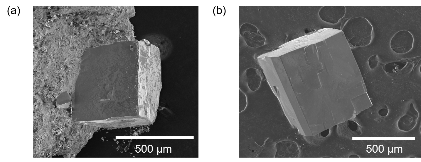
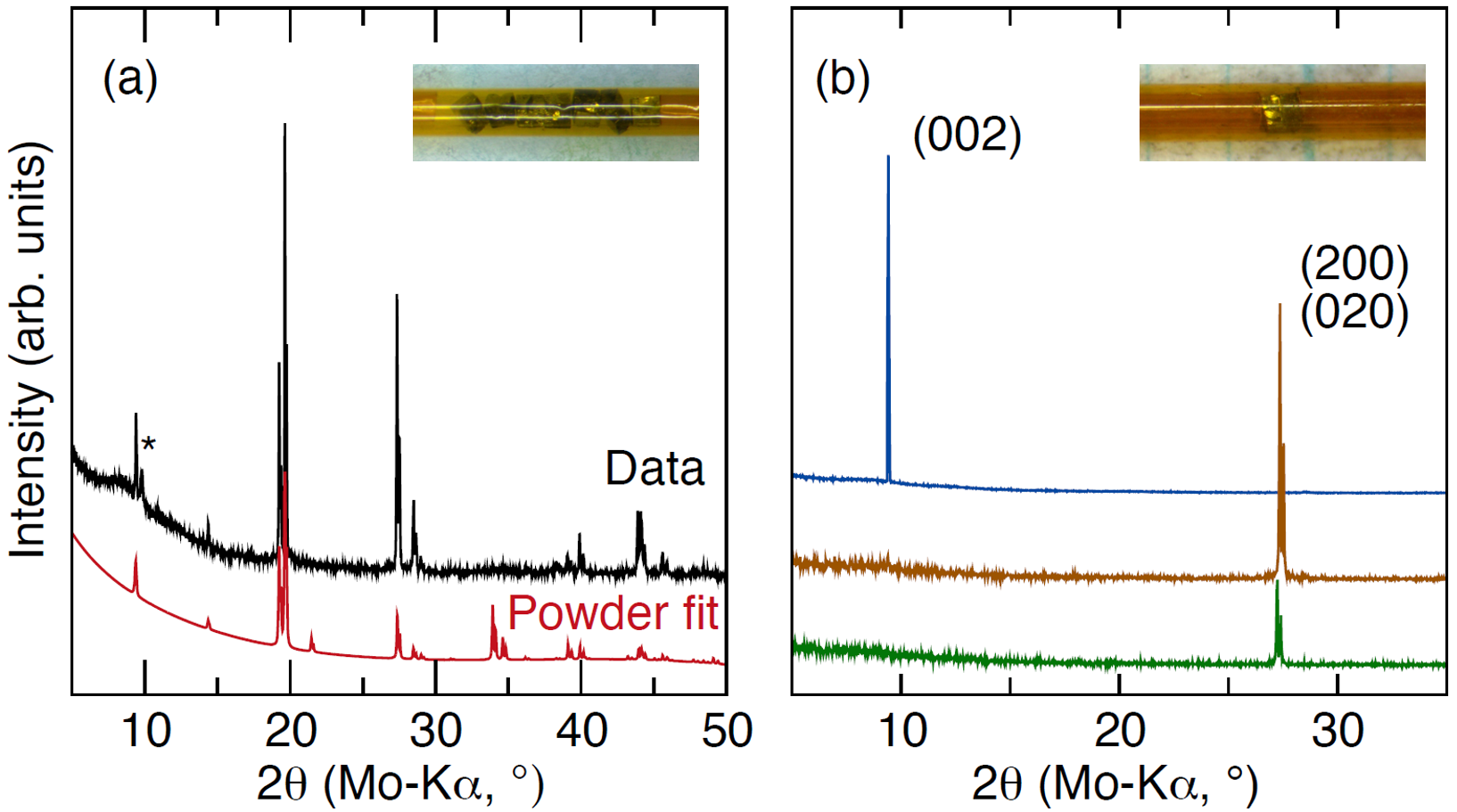
The crystals obtained from melt centrifuging were metallic silver and had rectangular prismatic shapes, with a typical appearance shown in Figure 2, before and after etching excess Sn in nitric acid. The phase purity was confirmed by powder XRD of several aligned crystals in a capillary and measured while rotating, which gives peaks that more fully reproduce the powder pattern of Cr2Al in Figure 3(a), with some expected difference in peak intensity due to preferred orientation. Collection of XRD data on stationary single crystals was used to determine the direction of the axis, with XRD shown for a typical crystal measured on three orientations shown in Figure 3(b). Energy-dispersive X-ray spectroscopy gave an average Cr:Al ratio of 2.0(2), shown in Figure S2.sup
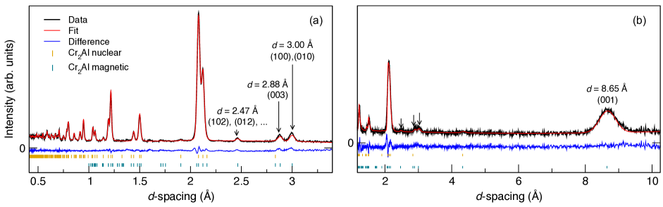
Figures 4(a) and 4(d) show the neutron diffraction pattern of a Cr2Al powder at 298 K. The nuclear peaks give a clear fit to the ordered structure, with the magnetic peak (100) clearly visible at Å. The magnetic propagation vector reproduces all magnetic peaks, which confirms the magnetic unit cell exhibits the same dimension as the crystallographic unit cell. The magnetic space group of Cr2Al with the Néel vector along [100] is in Belov-Neronova-Smirnova (BNS) notation and in Opechowski-Guccione (OG) notation, and the refinement in Figure 4 is thus constrained with Cr1 moments on the body center sites opposite to the Cr2 moments on the edges of the cell.
The refined magnetic moments on Cr positions are 1.06(19). The obtained magnetic structure resolved is visualized in Figure 1. Powder diffraction is not sensitive to the orientation of the Cr moments within the plane. The refined structure, with the nearest-neighbor Cr spins antiferromagnetic, which are in turn ferromagnetic across the Al plane, gives a significantly improved fit over a model where the Cr bilayers have ferromagnetic alignment within the layers, or antiferromagnetic alignment from layer to layer. These fits to the neutron diffraction data are shown Figures S4-5.sup The latter configurations relax to a nonmagnetic state in first-principles simulations. Likewise, a uniaxial configuration with spins along does not reproduce the neutron data (Figure S6-8).sup A small canting of the moments toward , as suggested by Atoji,Atoji (1965) does not significantly improve the fit, even with the addition of an extra free parameter, shown in Figure S9.sup Small canting is, thus, unlikely due to the additional irreducible representation that would be required to observe it, along with the presence of a single Néel transition, which we will discuss subsequently. Canting the moment along also leads to a calculated increase in the total energy, as shown in Figure S10.
First-principles density functional theory simulations for antiferromagnetic Cr2Al are implemented for ground state calculations, electronic band structure, phonon dispersion, magnetic susceptibility, and exchange coefficients. The calculated ground state confirms the structure with Å and Å, in good agreement with the room temperature lattice parameters Å and Å from neutron diffraction in Figure 4. Magnetic moments on Cr sites shown in Figure 1 converged to 1.311 . The discrepancy of 23 % likely arises from decrease of the low-temperature moment upon heating to 300 K. Ground state DFT simulations for other possible magnetic orderings (see Figure S4-5)sup conclude that there is no stable state for those.
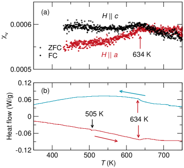
The temperature dependence of the magnetic susceptibility of an aligned single crystal from 430 to 750 K with T is shown in Figure 5(a). A small hump is observed at K for measurements with and . The splitting of the two orientations and the lower susceptibility with field along indicates that the magnetic ordering is in the plane. Further confirmation of is seen in calorimetry, shown in Figure 5(b). The small event at K is the melting of a small amount of residual Sn. The fraction of residual Sn is estimated to be 2 wt% based on peak integration of the DSC measurement (see Figure S11).sup ; Grønvold (1993)
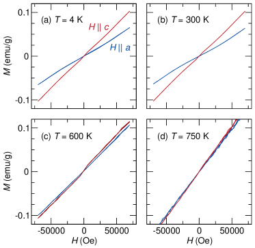
The magnetization as a function of applied field and orientation is shown in Figure 6. The anisotropy consistently shows that the susceptibility along is greater than along , which is to be expected for moments lying in the plane. No evidence of a spin flop transition is observed up to T. When the temperature of the experiment is raised to 750 K, the anisotropy between and directions vanishes, as expected above . This is the first measurement of the magnetic anisotropy of a bulk MoSi2-type antiferromagnet in the vicinity of its , and understanding the temperature dependence will be a crucial topic of future work.
The phenomenon that the measured moment of Cr2Al is greater with over the entire field and temperature range can be compared to similar measurements on another in-plane degenerate antiferromagnet Fe2As.Yang et al. (2020a) In Fe2As, the low-field susceptibility mirrors that of Cr2Al. Above approximately 0.7 T, the susceptibility of Fe2As with becomes greater than with , indicating rotation of the antiferromagnetic domains into a single-domain state with all moments likely along . This domain rotation is absent in Cr2Al for the fields we are able to achieve here. This lack of domain rotation is most likely due to the small intrinsic susceptibility of Cr2Al versus Fe2As.
The magnetic susceptibility of Cr2Al can be extracted from total energy calculations within DFT for magnetic configurations with tilted magnetic moments. Since an explicit magnetic field is not implemented in our DFT simulations, we use the tilting of magnetic moments to mimic the magnetic structure under an applied external field Kang et al. (2020). Within this approach, we obtain the lowest energy for a given tilting angle. Constraining the tilting angle of magnetic moments changes the total energy changes due to exchange interactions in the tilted state (see Figure S10 sup ). We compute the magnetic susceptibility from a quadratic fit to the resulting total-energy curve and the equation
| (3) |
where is the vacuum permeability and the quadratic fit coefficient Kang et al. (2020). Since DFT implements the calculation at K, should be zero and is non-zero for a single crystal with a single domain. Thus, we assume an experiment in which the field is oriented along the axis and moments are tilted within the -plane. The calculated magnetic susceptibility is 6.88, which shows good agreement with the scale of measured value of 1.07 at K, two orders of magnitude smaller than 0.015 for Fe2As.Yang et al. (2020b)
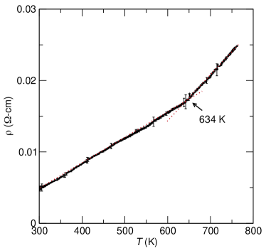
The two-point resistivity of Cr2Al from 298 to 733 K is shown in Figure 7. Since contact resistance between the Al pads and the Cr2Al sample is present, this data serves as an upper bound for the intrinsic resistivity of Cr2Al. A clear second-order transition can be observed at 634 K where the slope increases, indicating an increase in scattering due to the disappearance of AF domains.
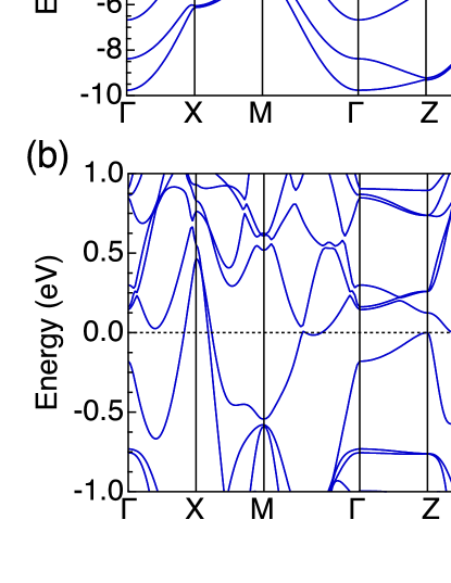
The calculated electronic band structure shown in Figure 8 demonstrates the metallicity of Cr2Al, with a low electronic density of states near the Fermi level, which leads to high resistivity, especially when some antisite defects may be present due to the compositional phase width of Cr2Al. The number of states at the Fermi level of Cr2Al is 0.0155 states/(eVÅ), smaller than that of Fe2As (0.0770 states/(eVÅ))Yang et al. (2019). This explains why Cr2Al shows higher resistivity than Fe2As. The projected density of states shows that a wide energy range of states originates from Cr orbitals, while or orbital contributions from Cr and Al are small.
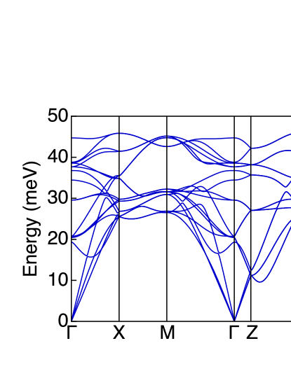
The structural stability of Cr2Al is confirmed by the phonon dispersion, computed using the finite difference method including the antiferromagnetic configuration and the effect of spin-orbit coupling, shown in Figure 9. There are no states with imaginary energy/frequency, which confirms the dynamic stability. There are a total of 18 phonon bands, spanning the energy up to 50 meV. The phonon density of states shows two peaks around 30 and 45 meV.
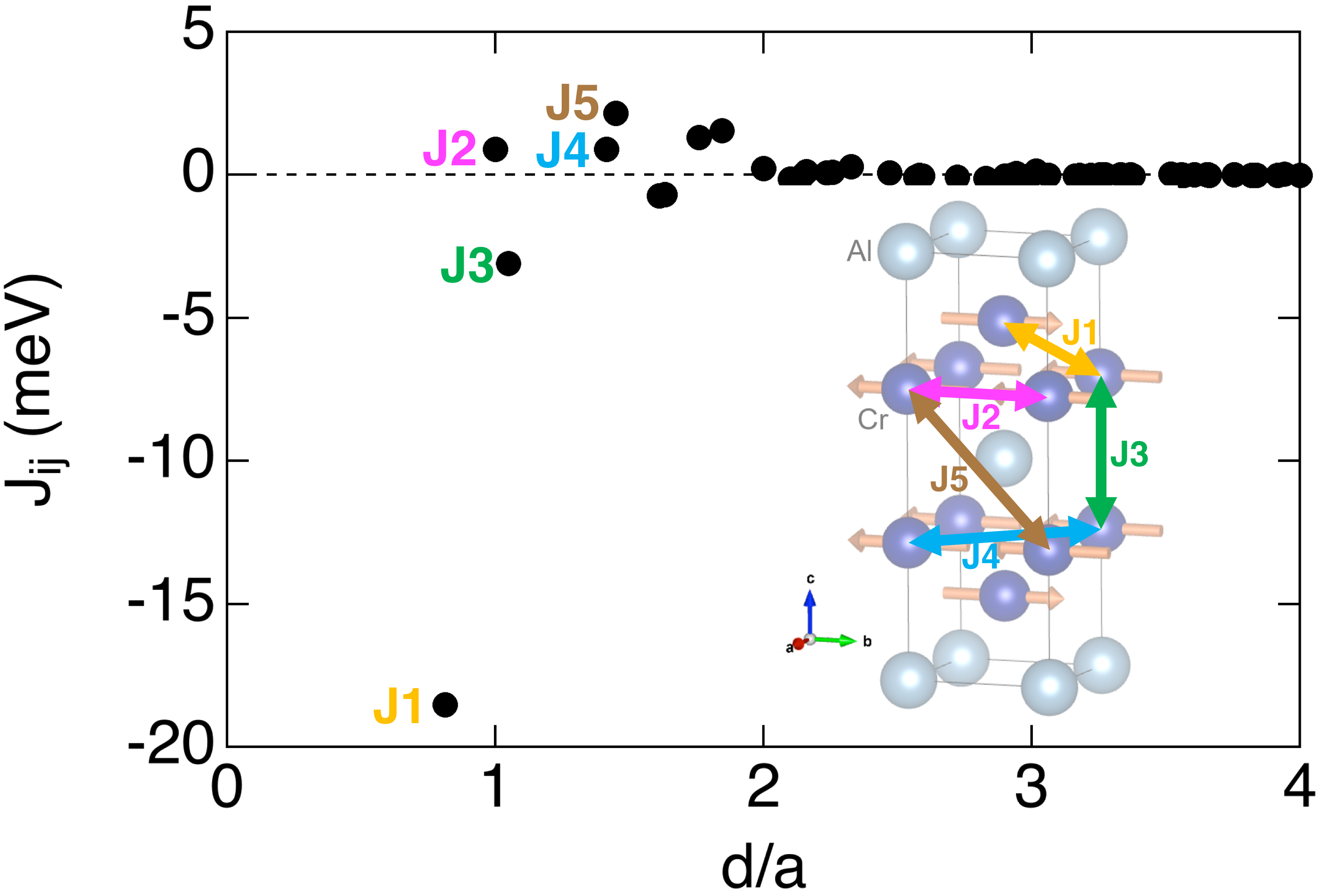
Exchange coefficients describe the interaction between magnetic moments and can further explain the magnetic structure of Cr2Al. These exchange coefficients can be utilized for atomistic spin dynamics simulations, as has been done for Fe2As.Karigerasi et al. (2020) The exchange coefficients extracted in this work are shown as a function of relative distance () in Figure 10. Positive and negative exchange parameters indicate ferromagnetic and antiferromagnetic couplings, respectively. The nearest-neighbor exchange interaction ( meV, shown as a yellow arrow in Figure 10) is the primary driving force for the AF ordering in Cr2Al. Even though the third-nearest neighbor exchange interaction also shows a negative value ( meV), the corresponding moments show parallel alignment. This is because the interaction is overcompensated by meV, i.e., ferromagnetic coupling, which has a multiplicity four, while has only one interaction. Interactions along and directions are all ferromagnetic ( meV and meV). Numerical uncertainties of the exchange integrals mainly originate from k-point sampling and we determined these to be lower than 2.7%, except for J4, where due to the small absolute value the relative error is larger (14.2%).
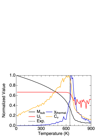
Based on the calculated exchange coefficients, was estimated using three thermodynamic observables that were computed from Monte Carlo (MC) simulations, see details in Ref. Eriksson et al., 2017. The observables, computed using the stochastic LLG equation, are shown as a function of in Figure 11. Sub-lattice magnetization (, black solid line) is not sharp enough to determine the critical temperature because of finite size effects, but it does reproduce the 23% difference between the calculated K and the experimental at 300 K from neutron scattering. Instead, we use the Binder cumulant , which indicates as a transition from the normalized value of 0.667 to 0.444. The isothermal susceptibility and the heat capacity show a peak at the critical temperature. This leads to predictions for the transition temperature of 660, 650, and 630 K, respectively, which are all in good agreement with the experimental value of K.
IV Conclusions
Neutron diffraction and magnetometry are consistent with an in-plane magnetic structure of Cr2Al, identical to that of Mn2Au, but with light elements that minimize spin-orbit coupling. Magnetometry, calorimetry, and transport all confirm to be K, in agreement with previous studies. The ability to access this without decomposing the compound enables a more stringent test for evaluating spintronic phenomena that should vanish in the paramagnetic regime. Aligned measurements provide some early evidence for anisotropy in Cr2Al, which will be the focus of further studies. The ability to engineer the transport and morphology, for example by thin film deposition, should provide a promising platform for spintronic investigation.
V Acknowledgments
This work was undertaken as part of the Illinois Materials Research Science and Engineering Center, supported by the National Science Foundation MRSEC program under NSF Award No. DMR-1720633. The characterization was carried out in part in the Materials Research Laboratory Central Research Facilities, University of Illinois. This work made use of the Illinois Campus Cluster, a computing resource that is operated by the Illinois Campus Cluster Program (ICCP) in conjunction with the National Center for Supercomputing Applications (NCSA) and which is supported by funds from the University of Illinois at Urbana-Champaign. This research is part of the Blue Waters sustained-petascale computing project, which is supported by the National Science Foundation (Awards No. OCI-0725070 and No. ACI-1238993) and the state of Illinois. Blue Waters is a joint effort of the University of Illinois at Urbana-Champaign and its National Center for Supercomputing Applications. This research used resources of the Spallation Neutron Source, a DOE Office of Science User Facility operated by Oak Ridge National Laboratory. The authors thank Jue Liu for additional assistance with the neutron scattering experiment.
References
- Kimel et al. (2005) A. V. Kimel, A. Kirilyuk, P. A. Usachev, R. V. Pisarev, A. M. Balbashov, and T. Rasing, Nature 435, 655 (2005).
- Little et al. (2017) A. Little, L. Wu, P. Lampen-Kelley, A. Banerjee, S. Patankar, D. Rees, C. A. Bridges, J. Q. Yan, D. Mandrus, S. E. Nagler, and J. Orenstein, Phys. Rev. Lett. 119, 227201 (2017).
- Kampfrath et al. (2011) T. Kampfrath, A. Sell, G. Klatt, A. Pashkin, S. Mährlein, T. Dekorsy, M. Wolf, M. Fiebig, A. Leitenstorfer, and R. Huber, Nature Photonics 5, 31 (2011).
- Viala et al. (2004) B. Viala, G. Visentin, and P. Gaud, IEEE Trans. Magn. 40, 1996 (2004).
- Lee et al. (2016) S. Lee, S. Grudichak, J. Sklenar, C. C. Tsai, M. Jang, Q. Yang, H. Zhang, and J. B. Ketterson, J. Appl. Phys. 120, 033905 (2016).
- Chai et al. (2013) G. Chai, N. N. Phuoc, and C. K. Ong, Appl. Phys. Lett. 103, 042412 (2013).
- Sharma et al. (2016) V. Sharma, J. Saha, S. Patnaik, and B. K. Kuanr, AIP Advances 7, 056405 (2016).
- Bhattacharjee et al. (2018) N. Bhattacharjee, A. A. Sapozhnik, S. Y. Bodnar, V. Y. Grigorev, S. Y. Agustsson, J. Cao, D. Dominko, M. Obergfell, O. Gomonay, J. Sinova, M. Kläui, H. J. Elmers, M. Jourdan, and J. Demsar, Phys. Rev. Lett. 120, 237201 (2018).
- Barthem et al. (2013) V. M. T. S. Barthem, C. V. Colin, H. Mayaffre, M. H. Julien, and D. Givord, Nature Commun. 4, 2892 (2013).
- Jourdan et al. (2015) M. Jourdan, H. Bräuning, A. Sapozhnik, H. J. Elmers, H. Zabel, and M. Kläui, J. Phys. D: Appl. Phys. 48, 385001 (2015).
- Olejník et al. (2018) K. Olejník, T. Seifert, Z. Kašpar, V. Novák, P. Wadley, R. P. Campion, M. Baumgartner, P. Gambardella, P. Němec, J. Wunderlich, J. Sinova, P. Kužel, M. Müller, T. Kampfrath, and T. Jungwirth, Science Advances 4, eaar3566 (2018).
- Saidl et al. (2017) V. Saidl, P. Němec, P. Wadley, V. Hills, R. P. Campion, V. Novák, K. W. Edmonds, F. Maccherozzi, S. S. Dhesi, B. L. Gallagher, F. Trojánek, J. Kuneš, J. Železný, P. Malý, and T. Jungwirth, Nature Photonics 11, 91 (2017).
- Wadley et al. (2015) P. Wadley, V. Hills, M. R. Shahedkhah, K. W. Edmonds, R. P. Campion, V. Novák, B. Ouladdiaf, D. Khalyavin, S. Langridge, V. Saidl, P. Nemec, A. W. Rushforth, B. L. Gallagher, S. S. Dhesi, F. Maccherozzi, J. Železný, and T. Jungwirth, Scientific Reports 5, 17079 (2015).
- Wadley et al. (2016) P. Wadley, B. Howells, J. Zelezny, C. Andrews, V. Hills, R. P. Campion, V. Novak, K. Olejnik, F. Maccherozzi, S. S. Dhesi, S. Y. Martin, T. Wagner, J. Wunderlich, F. Freimuth, Y. Mokrousov, J. Kunes, J. S. Chauhan, M. J. Grzybowski, A. W. Rushforth, K. W. Edmonds, B. L. Gallagher, and T. Jungwirth, Science 351, 587 (2016).
- Wadley et al. (2018) P. Wadley, S. Reimers, M. J. Grzybowski, C. Andrews, M. Wang, J. S. Chauhan, B. L. Gallagher, R. P. Campion, K. W. Edmonds, S. S. Dhesi, F. Maccherozzi, V. Novak, J. Wunderlich, and T. Jungwirth, Nat. Nanotechnol. 13, 362 (2018).
- Wang et al. (2020) M. Wang, C. Andrews, S. Reimers, O. J. Amin, P. Wadley, R. P. Campion, S. F. Poole, J. Felton, K. W. Edmonds, B. L. Gallagher, A. W. Rushforth, O. Makarovsky, K. Gas, M. Sawicki, D. Kriegner, J. Zubáč, K. Olejník, V. Novák, T. Jungwirth, M. Shahrokhvand, U. Zeitler, S. S. Dhesi, and F. Maccherozzi, Phys. Rev. B 101, 094429 (2020).
- Chiang et al. (2019) C. C. Chiang, S. Y. Huang, D. Qu, P. H. Wu, and C. L. Chien, Phys. Rev. Lett. 123, 227203 (2019).
- Bodnar et al. (2018) S. Y. Bodnar, L. Šmejkal, I. Turek, T. Jungwirth, O. Gomonay, J. Sinova, A. A. Sapozhnik, H. J. Elmers, M. Kläui, and M. Jourdan, Nature Commun. 9, 348 (2018).
- Bhattacharjee et al. (2020) N. Bhattacharjee, A. A. Sapozhnik, S. Y. Bodnar, V. Y. Grigorev, S. Y. Agustsson, J. Cao, D. Dominko, M. Obergfell, O. Gomonay, J. Sinova, M. Kläui, H.-J. Elmers, M. Jourdan, and J. Demsar, Phys. Rev. Lett. 124, 039901 (2020).
- Cahn (1991) R. W. Cahn, Adv. Mater. 3, 628 (1991).
- Khmelevskyi and Mohn (2008) S. Khmelevskyi and P. Mohn, Appl. Phys. Lett. 93, 162503 (2008).
- Uhlířová et al. (2019) K. Uhlířová, E. Duverger-Nédellec, R. H. Colman, J. Volný, B. Vondráčková, and K. Carva, J. Alloys Compd. 771, 680 (2019).
- Atoji (1965) M. Atoji, J. Chem. Phys. 43, 222 (1965).
- Semukhin et al. (1988) B. S. Semukhin, V. M. Kushnarenko, and E. V. Kozlov, Soviet Phys. J. 31, 267 (1988).
- Mahdouk and Gachon (2000) K. Mahdouk and J.-C. Gachon, J. Phase Equilibria 21, 157 (2000).
- Rank et al. (2019) M. Rank, P. Franke, J. Hoffmann, and H. J. Seifert, Calphad 66, 101638 (2019).
- Susner et al. (2015) M. A. Susner, D. S. Parker, and A. S. Sefat, J. Magn. Magn. Mater. 392, 68 (2015).
- Calder et al. (2018) S. Calder, K. An, R. Boehler, C. R. Dela Cruz, M. D. Frontzek, M. Guthrie, B. Haberl, A. Huq, S. A. J. Kimber, J. Liu, J. J. Molaison, J. Neuefeind, K. Page, A. M. dos Santos, K. M. Taddei, C. Tulk, and M. G. Tucker, Rev. Sci. Instrum. 89, 092701 (2018).
- Toby and Von Dreele (2013) B. H. Toby and R. B. Von Dreele, J. Appl. Cryst. 46, 544 (2013).
- Momma and Izumi (2008) K. Momma and F. Izumi, J. Appl. Cryst. 41, 653 (2008).
- (31) Supplementary Material available online.
- Kresse and Furthmüller (1996) G. Kresse and J. Furthmüller, Phys. Rev. B 54, 11169 (1996).
- Kresse and Joubert (1999) G. Kresse and D. Joubert, Phys. Rev. B 59, 1758 (1999).
- Perdew et al. (1996) J. P. Perdew, K. Burke, and M. Ernzerhof, Phys. Rev. Lett. 77, 3865 (1996).
- Blöchl (1994) P. E. Blöchl, Phys. Rev. B 50, 17953 (1994).
- Monkhorst and Pack (1976) H. J. Monkhorst and J. D. Pack, Phys. Rev. B 13, 5188 (1976).
- Togo and Tanaka (2015) A. Togo and I. Tanaka, Scr. Mater. 108, 1 (2015).
- Steiner et al. (2016) S. Steiner, S. Khmelevskyi, M. Marsmann, and G. Kresse, Phys. Rev. B 93, 224425 (2016).
- Ebert et al. (2011) H. Ebert, D. Kodderitzsch, and J. Minar, Rep. Prog. Phys. 74, 096501 (2011).
- Liechtenstein et al. (1984) A. I. Liechtenstein, M. I. Katsnelson, and V. A. Gubanov, J. Phys. F: Metals Phys. 14, L125 (1984).
- Eriksson et al. (2017) O. Eriksson, A. Bergman, L. Bergqvist, and J. Hellsvik, Atomistic Spin Dynamics: Foundations and Applications (Oxford University Press, 2017).
- Grønvold (1993) F. Grønvold, The Journal of Chemical Thermodynamics 25, 1133 (1993).
- Yang et al. (2020a) K. Yang, K. Kang, Z. Diao, M. H. Karigerasi, D. P. Shoemaker, A. Schleife, and D. G. Cahill, Phys. Rev. B 102, 064415 (2020a).
- Kang et al. (2020) K. Kang, K. Yang, K. Puthalath, D. G. Cahill, and A. Schleife, arXiv:2012.02090 (2020).
- Yang et al. (2020b) K. Yang, K. Kang, Z. Diao, M. H. Karigerasi, D. P. Shoemaker, A. Schleife, and D. G. Cahill, Phys. Rev. B 102, 064415 (2020b).
- Yang et al. (2019) K. Yang, K. Kang, Z. Diao, A. Ramanathan, M. H. Karigerasi, D. P. Shoemaker, A. Schleife, and D. G. Cahill, Phys. Rev. Mater. 3, 124408 (2019).
- Karigerasi et al. (2020) M. H. Karigerasi, K. Kang, G. E. Granroth, A. Banerjee, A. Schleife, and D. P. Shoemaker, Phys. Rev. Mater. 4, 114416 (2020).