Can biophysical models give insight into the synaptic changes associated with addiction?
Mayte Bonilla-Quintana and Padmini Rangamani*
Department of Mechanical and Aerospace Engineering,
University of California San Diego, La Jolla CA 92093.
∗To whom correspondence must be addressed: prangamani@ucsd.edu
Abstract
Effective treatments that prevent or reduce drug relapse vulnerability should be developed to relieve the high burden of drug addiction to society. This will only be possible by enhancing the understanding of the molecular mechanisms underlying the neurobiology of addiction. Recent experimental data have shown that dendritic spines, small protrusions from the dendrites that receive input from excitatory neurons, from spiny neurons in the nucleus accumbens exhibit morphological changes during drug exposure and withdrawal. Moreover, these changes relate to the characteristic drug-seeking behavior of addiction. However, due to the complexity of the dendritic spines, we do not yet fully understand the processes underlying their structural changes in response to different inputs. We propose that biophysical models can enhance the current understanding of these processes by incorporating different, and sometimes, discrepant experimental data to identify the shared underlying mechanisms and generate experimentally testable hypotheses. This review aims to give an up-to-date report on biophysical models of dendritic spines, focusing on those models that describe their shape changes, which are well-known to relate to learning and memory. Moreover, it examines how these models can enhance our understanding of the effect of the drugs and the synaptic changes during disease progression.
1 Introduction
The highest burden of drug addiction to all societies is the immense loss of human capital – from unfulfilled personal ambitions to loss of family structure. According to the National Institute on Drug Abuse [1], in 2019 more than 70,000 Americas died from a drug-involved overdose, including illicit drugs and prescription opioids. However, this number only represents a small percentage of drug-related deaths. According to a statistical study [2], in 2016 the drug-associated deaths was 2.2 times the number of drug-coded deaths. Besides these losses, the estimated medical cost in hospitals of substance use disorder in 2017 was $13.7 billion [3].
A continued medical aspiration for relieving the high burden of drug addiction in society is to develop and tailor effective treatment to prevent relapse. Assuming that drug addiction is a neuropsychiatric disease whose behavioral pathology consists in the propensity to relapse, even after periods of abstinence, then an effective treatment should prevent or reduce relapse vulnerability [4]. Because current behavioral and pharmacological therapy only helps a small percentage of patients [4], we need to understand the molecular mechanisms underlying the neurobiology of addiction. In the past decade, studies have focused on the neurons in the nucleus accumbens (NAc), a brain region that mediates goal-directed behavior, and their structural changes after drug exposure [4, 5, 6, 7]. These studies have noted that structural changes in neurons could be an important readout of addictive behavior. For example, when animals are exposed to an environment associated with previous cocaine consumption, it triggers cocaine-seeking behavior that is induced by structural changes in NAc neurons [7]. Another study showed that structural alterations to these neurons are only present in animals that show a persistent increase in the psychomotor activating effects of cocaine after repeated exposure to it, i.e., psychomotor sensitization [8]. However, there is no consensus on whether and how the structural changes in neurons derived from drug exposure drive addictive behavior [5, 4] due to opposing experimental data which show that the structural changes in NAc neurons after cocaine exposure are not required for locomotor sensitization and suggest that, instead, these changes may represent a compensatory process [9]. Complicating matters, studies have also shown that different drugs have different effects on these neurons [4, 5]. For example, stimulants, such as cocaine, amphetamine, and nicotine, enhance the structural complexity of the neurons, while narcotics, like morphine, decrease it [10].
In this review, we explore the idea that experimentally-constrained biophysical computational models that relate structural changes of neurons to different drug conditions may be able to narrow down the possible mechanisms that may underlie the neurobiology of drug addiction. Although there are a repertoire of theoretical models of drug use and addiction, they rely on theories of how the brain solves computational, information-processing, and control problems [11, 12]. Some of these models link these computations with specific brain areas [12]. However, to our knowledge, they do not consider the structural and molecular changes of the neurons and, instead, focus on drug use behavior. Recently, biophysical models have informed the structural and molecular mechanisms related to learning and memory [13, 14, 15, 16, 17, 18, 19]. Here, we review such models and describe how they can broaden our understanding of the mechanisms behind addiction and relapse to aid the treatments for drug addicts. We focus on dendritic spines, small protrusions from the dendrites that receive excitatory input, and hence, form the postsynaptic part of the synapse. Dendritic spines are thought to be standalone biochemical computational units [20, 21, 22] and it is well-documented that they undergo structural plasticity during learning and memory formation [23] and during drug consumption and withdrawal [24, 7, 5]. Therefore, biophysical analyses of spines may provide us with the key links between these fundamental neurological processes.
This work begins by giving an overview of the structure and dynamics of dendritic spines. Then, we introduce the most studied forms of synaptic changes, namely, long-term potentiation (LTP) and long-term depression (LTD), and relate them with the modifications associated with drug addiction. After this biological background, we review different types of biophysical models and discuss how they can enhance our understanding of drug addiction and other neurological diseases. We finish by describing how these biophysical models can be extended to study memory formation and storage, and identifying the opportunities for predictive model development.
2 Dendritic Spines
2.1 Morphology
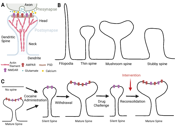
Although dendritic spines were first described by Santiago Ramón y Cajal over a century ago, it has only recently become possible, through the development of novel imaging techniques, to investigate their structure-function relationships. The dendritic spine is a bulbous protrusion from the dendritic shaft often connected by a thin neck, creating a spine head. The postsynaptic density (PSD) is located at the tip of the spine head, close to the presynaptic terminal, and it is enriched with receptors, proteins, and signaling molecules (Fig. 1A). The quantitative characteristics of the spine morphology are broadly described by: the length and width of its neck; the volume of its head; and the surface area of the PSD. These geometric features alone have been studied in terms of correlations between the morphology and function of the spine [25, 26, 27]. For example, the spine head volume correlates with the area of the PSD, the number of postsynaptic receptors, the readily-releasable pool of transmitters, and the neck width [25, 26, 27]. Moreover, dendritic spines that connect to the same axon have similar size and head volume demonstrating that they share the same activation history [28].
Dendritic spines also have diverse shapes. Thus, early studies classified them into three categories: 1) spines with long, thin necks and small heads (thin spines), 2) spines with thicker necks and large heads (mushroom spines), and 3) short spines without well-defined necks (stubby spines) [29] (Fig.1B). Experiments found that spine shapes were more distinct in adults than in young animals, which suggested that stubby spines form from the dendritic shaft when the axon contacts the membrane (shaft synapse) and then turn into thin or mushroom spines [30]. However, the current view is that a filopodia-like structure that emerges from the dendritic shaft may connect to the axon to form a synapse [31, 32] evolving to a thin spine which later matures to a mushroom spine [33]. Moreover, thin spines are more prone to respond to synaptic activity and change their morphology accordingly, suggesting that they are learning spines that can mature to mushroom-like spines, which are more stable and thus, thought to be memory spines [33]. While mushroom spines can persist for months, thin spines only last a few days [33]. Dendritic spines have also been divided depending on the size of their heads [34]. On one hand, spines with small heads (filopodia and thin spines) are motile and unstable. On the other hand, spines with large heads (mushroom and stubby spines) are stable and have stronger connections. Thus, if we see spines as memory devices, large-headed spines are “write protected”: they maintain preexisting connections and prevent new memory formation, while small-headed spines are “write-enabled” because they allow the acquisition of new memories [34].
In addition to their unique morphological features, spines have attracted considerable attention of the signaling community because it has been demonstrated they compartmentalize calcium and that spine necks can filter membrane potentials, thereby isolating biochemical and electrical synaptic input from each other [35]. These findings have led to the hypothesis that spines implement input-specific learning rules [25]. The shape changes in spines, in response to synaptic input, are possible by a reconfiguration of the spine cytoskeleton, mainly composed of actin filaments; actin remodeling is triggered by a cascade of chemical reactions due to calcium influx into the spine [36, 37, 38, 39, 40, 41]. Changes in spine shape and size not only occur in development but also in adulthood [42], for example, during motor skill learning [43] and fear learning and extinction [44]. This dynamic change to spine morphology is known as structural plasticity [45]. Note that due to this plasticity, the shapes of spines form a continuum instead of distinct categories [26].
2.2 Structural Plasticity
The connection between neurons can be strengthened or weakened in a process called synaptic plasticity, and this change in the connections has a direct impact on learning and memory formation [46, 42, 47, 48]. The most studied forms of these synaptic changes are long-term potentiation (LTP), which strengthens the synapse, and long-term depression (LTD), which weakens it. In LTP, glutamate released due to the high-frequency stimulation of the presynaptic neuron is captured by the -amino-3-hydroxy-5-methyl-4-isoxazolepropionic acid receptors (AMPARs) located at the PSD of the dendritic spine (Fig. 1A). Due to this glutamate influx, the spine depolarizes and releases the Magnesium (Mg2+) ion of the N-methyl-D-aspartate receptors (NMDARs), which allows the calcium (Ca2+) influx into the spine and triggers a cascade of reactions that increase the number of AMPARs at the PSD and AMPARs mediated current. Such increase facilitates the subsequent uptake of glutamate, and hence, the synapse strengthens. Moreover, dendritic spines show an associated increase in size, thus, linking their function and morphology [47, 22, 22, 45, 46]. LTD also depends on the activation of NMDARs, but it is induced by low-frequency stimulation of the presynaptic cell, causing glutamate release. However, the amount of glutamate is not enough to depolarize the postsynaptic cell and remove all the Mg2+ ions blocking the NMDARs, but only a few. Hence, the Ca2+ influx is lower than in LTP and triggers a different set of chemical reactions resulting in a decrease in the number of AMPARs at the PSD, and spine shrinkage [48].
How is plasticity impacted by drugs? It has been shown that cocaine use impairs the induction of LTP in spines of medium spiny neurons (MSNs) in the NAc [7]. These spines are depressed after exposure to cocaine, which generates silent synapses that lack AMPARs but have NMDARs [4, 7, 24, 5]. The lack of AMPARs impairs LTP because these spines cannot respond to glutamate release. Because silent synapses are highly abundant during development, it has been hypothesized that the brain returns to a more juvenile state after drug exposure, which may underlie pathological drug-seeking behaviors [49]. Interestingly, AMPARs are reinserted into these silent synapses only when a stimulus related to cocaine consumption is present [7, 24]. This synaptic potentiation induces drug craving, and thus, may explain the high rates of relapse [7]. Moreover, re-silencing these synapses via optogenetic removal of AMPARs inhibits relapse-like behaviors [49]. A recent study shows that there is a time window in which previously silent synapses that are potentiated due to exposure to cues related to drug consumption can be temporally destabilized and vulnerable to disruptions before being consolidated (becoming mature) [24]. Moreover, this window relates to the dynamics of signaling molecules that alter the spine cytoskeleton. Preventing spine maturation during this window decreases cue-induced cocaine seeking, which can be used for anti-relapse treatments [24] (Fig. 1C).
However, the precise neuronal and molecular substrates encoding the dynamics of drug memories have not been fully identified [24, 4]. Moreover, until recently, new imaging techniques allowed for the study of the mechanisms underlying the structural and molecular changes of the synapses [42], but they remain incompletely understood due to the complexity of the synapse and the diverse temporal and spatial scales of the experiments and underlying events.
2.3 Signaling Pathways associated with structural plasticity
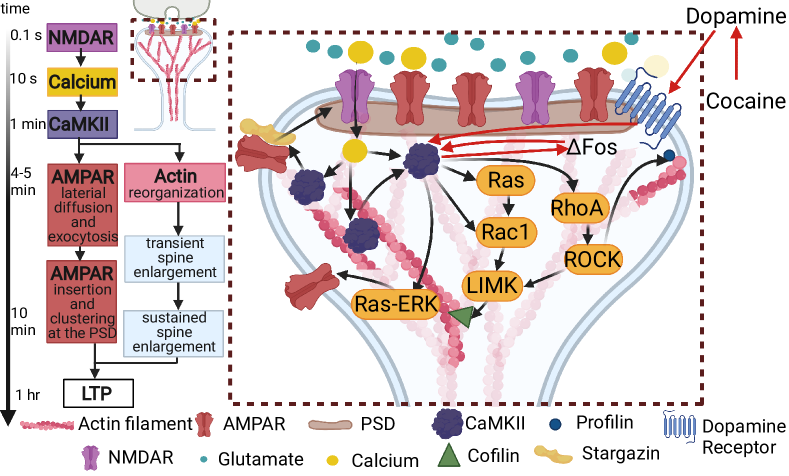
Upon LTP induction, there is an increase of the number of AMPARs in the PSD due to the Ca2+/ calmodulin-dependent protein kinase II (CaMKII) mediated phosphorylation of AMPAR auxiliary protein stargazin, which allows extrasynaptic diffusive receptors to bind to PSD95, a PSD molecule [50, 51, 52]. The exocytosis of extrasynaptic AMPARs into the plasma membrane depends on the rat sarcoma virus-extracellular signal-regulated kinase (RAS–ERK) pathway, RAB-GTPase proteins, soluble N-ethylmaleimide-sensitive-factor attachment protein receptors (SNARE proteins), syntaxin 4 and 13, and the motor protein myosin V [50, 53, 54, 51]. The RAS-ERK pathway is stimulated by CaMKII upon LTP induction [55, 54]. Thus, CaMKII has a dual function upon LTP: it immobilizes extracellular AMPARs in the PSD and replenishes the pool of extrasynaptic receptors via exocytosis. Moreover, CaMKII is one of the most important molecules for LTP. It is activated by calcium-calmodulin, which in turn is activated by Ca2+ influx into the spine through NMDARs and it can act as a protein switch due to its autophosphorylation property. Such a state lasts longer than calcium elevation, thus CAMKII acts as a biochemical integrator of the multiple calcium pulses during LTP induction [50].
In addition to its involvement with AMPARs, CaMKII also affects the dendritic spine cytoskeleton by stabilizing actin filaments [56]. Actin is a globular protein (G-actin) that forms filaments (F-actin) and it is the main component of the spine cytoskeleton. F-actin is a polar structure that continuously polymerizes G-actin at its plus end and it is depolymerized at its minus end. In the spine, there is a dynamic equilibrium between G-actin and F-actin that is modulated by actin-binding proteins (ABPs) which promote F-actin depolymerization and G-actin polymerization [57]. Moreover, the actin cytoskeleton is restructured upon LTP by an orchestrated interplay between actin and ABPs [58, 59], which allows for spine enlargement. In the basal state, F-actin is bundled by CaMKII, which stabilizes the bundle. Upon NMDARs activation, CaMKII autophosphorylates and detaches from actin filaments, which allows CaMKII to interact with other molecules and remodel the actin cytoskeleton [56, 60]. Furthermore, CaMKII regulates actin dynamics by signaling pathways involving the Rho family of small GTPases, such as RhoA, Rac1, or Cdc42 [56]. The activity of these GTPases is triggered by guanine-nucleotide-exchange factors (GEFs) and suppressed by GTPase activating proteins (GAPs), and they coordinately regulate ABPs activity. For example, RhoA controls profilin II and cofilin [61, 62, 56]. The former binds to G-actin and facilitates the polymerization of F-actin at its plus end. Cofilin, which is also controlled by Rac1, has a dual function depending on its concentration. At low concentrations, cofilin severes F-actin and at high concentrations, it promotes F-actin nucleation [63]. Note that the nucleation and severing events produce more actin filaments, and the combination of both events is likely to be responsible for keeping a steady distribution of the F-actin length. Thus, cofilin regulation is critical for actin dynamics. These pathways are depicted in Figure 2.
It has been shown that cocaine decreases active RhoA in the NAc MSNs, which could lead to a decrease in cofilin [64], affecting the spine cytoskeleton. Moreover, activation of CaMKII by D1-like dopamine receptors in the NAc reinstates cocaine-seeking behavior; this is associated with an increase of AMPARs in the membrane [65]. CaMKII also phosphorylates Fos and it is required for the cocaine-mediated accumulation of Fos in NAc [66] (red lines in Fig. 2). Fos is a Fos family transcription factor that shows long-lasting accumulation in the NAc after chronic administration of any drug of abuse, thereby supporting the view that changes in gene expression contribute to drug addiction. Interestingly, it has been shown that Fos upregulates the transcription of the CaMKII gene selectively in the NAc shell and that CaMKII stabilizes Fos, allowing for a greater accumulation of Fos that induces more CaMKII [66]. However, how this positive feedback alters the spines is not fully understood, because of the complexity of the signaling pathways. Moreover, the signaling pathways activated by LTP induction have diverse temporal and spatial extents [46], and the coupling between the signaling pathways and spine structure is also complex (Fig.2). In order to disentangle the multiple spatial and temporal complexities involved in LTP induction and effects of drugs, we propose the theoretical models that incorporate biophysical details at various levels can shed light into the emergent behaviors of spines. These modeling approaches can elucidate the mechanisms underlying adaptation to addiction by allowing the incorporation of different experimental observations, eliminate possibilities that are not physicochemically feasible, and generate experimentally testable hypotheses.
3 Biophysical Models of Dendritic Spines
Addictive behaviors share common features with learning models [67], and hence, learning theories [68] can be adapted to investigate these behaviors. However, in this work, we focus on the structural and functional changes in dendritic spines that can inform relapse propensity. We first present a review of the models that investigate signaling pathways in the dendritic spine relevant to the mechanisms involved in drug addiction, particularly those pathways that result in structural LTP or LTD due to an increase or decrease of AMPARs and CaMKII dynamics. Then, we revise models that have incorporated idealized and realistic spine shapes linking the structural and functional characteristics of dendritic spines.
3.1 Signaling Models
Early models of synaptic plasticity, based on Ca+2 influx through the NMDARs, describe how different levels of Ca+2 [69, 70] or induction protocols [71] lead to an increase or decrease in the synaptic weight, which describes the strength of the synaptic connection. Later models provide a more detailed description of the biochemical reactions leading to either LTP or LTD [72], the volume regulation of dentric spines upon LTP [73], or the change in AMPARs mediated current promoting synaptic modifications [74]. These signaling models use ordinary differential equations (ODEs) to describe the biochemical reactions. They span from simple descriptions based on key assumptions [71] to detailed descriptions of the different chemical pathways [74]. As technological advances allow for the study of specific signaling pathways, models have incorporated these findings to test synaptic plasticity hypotheses [75]. The increase in computational power has also allowed the use of agent-based models [76], in which the molecules are agents that follow a set of rules, mimicking chemical reactions leading to LTP. Some of these models include the CaMKII activity [72, 70, 76, 77] and GTPases [73, 74].
These models test different hypotheses based on the available experimental findings and create theories about how long-term information can be stored in the brain. For example, early models test the idea that CaMKII provides a bistable switch [69] where “off” CaMKII is turned “on” when there is a synaptic event and stays on for long periods due to the autophosphorylation property of the kinase. Furthermore, this hypothesized switch can stay on for longer times than protein turnover because the newly synthesized unphosphorylated CaMKII subunit could become phosphorylated [78]. The mathematical models show the feasibility of this hypothesis [69], and expand it to add a phosphatase switch involved in LTD, providing a tristable system to examine how a synapse can be bidirectionally modified [72]. Models have also been used to systematically investigate how the interaction between CaMKII and other molecules can control the sensitivity of synapses to calcium signals [76, 73]. Moreover, the incorporation of actin remodeling revealed that it added robustness to the dendritic spine response upon LTP [73]. One common difficulty for these models is parameter fitting since not all the reaction rates or protein concentrations are known in the spines, due to experimental difficulties. Thus, informed assumptions and decisions are made. One common assumption is that reaction rates are the same across cell types, and protein concentrations are cell-type dependent; and hence, protein concentrations are fitted in the models [74]. Although parameters are fitted when unknown and varied to see their effects in the model dynamics, they have to be within physiological regimes [74, 73].
As experiments began to investigate the source of the AMPARs that incorporate into the PSD upon LTP, i.e., whether they are exocytosed or laterally diffused into the PSD [79, 80, 81, 51], theoretical studies examined the implications of the different sources to plasticity [15, 82, 83, 84, 85]. Moreover, models started to explore how synapses are maintained over time periods that are longer than the average lifetime of these receptors and other molecules [86, 87, 88], and aided the study of AMPARs clusters formation [89, 88]. These models propose different hypotheses for memory maintenance. Instead of relying on a bistable molecular switch, like that of CaMKII, a model of clusters of AMPARs show that they are metastable with longer lifetimes than that of a single receptor, and that synaptic weights, which depend on the number of AMPARs, can be bidirectionally changed [88]. An alternative hypothesis is that AMPARs exhibit a self-sustained switch related to their movement to and from the membrane [86]. In this model, the presence of receptors at the synapse enhances the recruitment of more AMPARs, resembling the conversion from silent to active synapses [86]. Moreover, a model based on simplified biophysical assumptions shows cooperative binding and unbinding of proteins at the PSD, replicating experimental observations [87]. By performing model simulations, the interplay between AMPARs and CaMKII dynamics has been examined [86, 85]. Another important player of structural plasticity, the actin filaments dynamics [36, 38] and its dynamic equilibrium [57], has also been investigated providing a quantitative description of the change of the filamentous to globular actin ratio upon LTP observed experimentally [90]. Moreover, computer simulations have aid the understanding of the F-actin binding and unbinding to CaMKII [60].
3.2 Models Incorporating Spine Shape
Given the structural and morphological complexity of spine shapes, the incorporation of geometrical detail is a critical aspect of computational modeling for LTP. Models started examining the role of the spatial localization of signaling molecules in synaptic plasticity [91, 92, 19], the effect of their diffusion on nearby spines [92, 93, 18] or the localization of other Ca2+ sources, for example the endoplasmatic reticulum or the mitochondria [18, 17, 94, 28], or astrocytes, a glia type cell of the central nervous system that contacts the synapse [95]. Many of these models use idealized, simplified geometries where the spine head is represented by an ellipsoid or a sphere and the spine neck by a cylinder connecting the spine head with the dendrite [17, 18, 75, 91, 92]. The dynamics of the signaling molecules are described by reaction-diffusion models that use partial differential equations (PDEs). Because some molecules are scarce in the dendritic spines, they cannot be described by their concentration amount, as is done for deterministic reaction-diffusion models, and thus, stochastic simulations were implemented to better represent their dynamics [91, 92]. Also, the localization and diffusion of AMPARs were examined in idealized geometries [96, 83]. Recent models have used reconstructed dendritic segments with spines from electron micrographs to give an insight into how the shape of dendritic spines influences calcium dynamics [28, 97, 98].
Overall, the incorporation of spatial detail in the models allowed examination of how molecular organization in the dendritic spines affect the response of signaling molecules to calcium [19], and the function of proteins [91]. Moreover, different hypotheses, that are not possible to experimentally test yet, about how organelle size and contact between organelles affect Ca2+ dynamics have been tested [18, 17]. Although including the spatial component in the models increases their complexity and the computational burden of the simulations, it is necessary to shed light on mechanisms that rely on the protein distribution and translocation, as structural plasticity.
Models started to explore the role of actin cytoskeleton remodeling into spine maturation, how different configurations of actin filaments derive in the different types of spines due to a balance between a force generated by actin polymerization and the resistance offered by the spine membrane [16, 99], and show the importance of the spine-neck constriction by structures like actin rings [100] for spine stability. These models assumed isotropic forces generated by actin pushing the lipid membrane forward, which is represented as a thin elastic shell that minimizes its bending energy [101], to investigate the equilibrium configurations of symmetric spine shapes.
However, models that incorporate spatio-temporal dynamics of signaling often neglect the dynamics of actin remodeling because of the inherently different types of governing equations. Signaling models usually focus on reaction and diffusion in a spatio-temporal manner [17, 19, 18, 91, 92]. Models incorporating actin remodeling usually involve force balances and movement of the boundary (spine membrane in this case) in response to those forces [99, 16]. A model coupling the signaling spatio-temporal dynamics and the membrane dynamics resulting from a force imbalance is hard to analyze because it is not always clear how its steady state is defined: the protein location affects the spine shape that, in turn, affects the protein dynamics. Moreover, in basal conditions, proteins are not steady, they diffuse and react with other molecules, and the spine shape fluctuates [102, 103]. This increases the complexity of computational simulations because, at each time step, the chemical reactions and spine shape have to be updated. Nonetheless, some efforts have been made to push the boundaries on these biophysical models forward. New models were proposed to investigate these spontaneous spine shape fluctuations [104, 105] and their implications in LTP [14, 105] by continuously changing the membrane according to the actin dynamics. These models couple the biochemical and mechanical properties of dendritic spines, and thus, their function and structure. This allows the comparison with time-lapse microscopy data and shed light into the underlying mechanisms for spine maintenance and changes upon LTP. Moreover, it promotes testing different hypothesis regarding to the most efficient mechanisms behind synaptic function.
3.3 Design considerations for model building
Although we focus on models of dendritic spines, they span across different temporal scales. For example, Ca+2 dynamics are fast (tens to hundreds of milliseconds) [28, 98, 94, 17, 71] while kinases like CaMKII or more complex signaling dynamics are slower (seconds to minutes) [72, 74, 75, 19, 76, 86, 91, 92, 18]. AMPARs dynamics also span from seconds, in studies of AMPARs lateral diffusion [83], to minutes, when investigating different sources of AMPARs [15, 96]. Moreover, models investigating spontaneous fluctuations of spine shape [104, 105] or AMPARs [87] can span from seconds to minutes, respectively (Fig. 2).
The level of detail also varies across studies, from simple models based on a few assumptions [71] to detailed models accounting for all the signaling pathways [74]. Most of the models look at the reaction induced by Ca2+ influx through NMDARs, but also other sources of calcium such as organelles [17, 18]. Moreover, the spatial representation of the dendritic spine also expands from a volume [73] or number of AMPARs readout [74] to the embodiment in static idealized [17, 28, 18, 83, 16] and real geometries [97] or motile shapes [14, 104, 105].
The dynamics are described in different ways depending on the studied phenomena. To select which type of model is the most suited to investigate a biological phenomenon, it is critical to know the readout of the model and whether it is comparable with experiments. If there are no available experiments, at least there has to be evidence that such experiments can be performed with the current technology. This way, the model predictions can be tested, and the model can be used to motivate more experiments. Most signaling pathways use ODEs to describe chemical reactions [75] or PDEs when accounting for molecular diffusion [75]. When the number of the studied molecules inside the spine is low, stochastic ODEs or PDEs are used instead [91, 92]. Other models take molecules [76] or filaments [90] as agents that follow experimentally verified rules that mimic their reactions.
Overall, there is always a trade-off between the level of detail, the spatial and temporal scale, and computational burden when proposing theoretical models. For example, for modeling the relapse propensity that lasts from hours to days [24], most of the fast molecular dynamics can be assumed to reach a semi-steady state. Hence, the model would only account for deviations of this state due to drug exposure. In this top-down approach, only chemical pathways affected by drug consumption are considered, and the rest are assumed to be unaffected. The affected pathways involve cytoskeleton proteins which induce remodeling. Thus, not only the variation of the AMPARs should be included, but the spine size and shape changes. Moreover, mechanical properties, such as surface area tension that regulates cell functions and triggers signals [106], should be considered in the models.
4 Perspective: Can we use models to enhance our understanding of synaptic changes during disease progression and to study the effect of drugs?
We consider that biophysical models can act as a computational microscope that incorporates diverse experimental findings to perform in silico experiments and test hypotheses that are not yet accessible experimentally. Therefore, they provide a platform to study the molecular effects of drug consumption in dendritic spines and simulate the impact of relapse treatments that inhibits or enhance certain signaling pathways. Although currently there is vast experimental measurements that can be use to restrict these models, these are sensitive to the the different experimental setups of drug administration: whether the drug is self-administrated or administrated by an experimenter [4, 5]. Thus, the models should also consider these discrepancies. Models should be suited to incorporate different spatial and temporal extents of the signal transduction underlying LTP [46] that is affected by cocaine. As a modeling community, this can be achieved by building modular, shareable models for different time- and spatial scales that can be coupled. By developing biophysical models to understand the changes in the synapse during drug abuse, we can gain insight into mechanisms that may be involved in other neurological diseases. For example, dendritic spine alterations have been found in autism spectrum disorders, schizophrenia, and Alzheimer’s disease [107, 49]. Such alterations correlate with the expression of proteins associated with the cytoskeleton [108], or the number of AMPARs [109].
The models presented here can be extended to study the complex interplay between membrane mechanics, cytoskeleton dynamics, and protein interaction during endo- and exocytosis of extracellular vesicles (EVs) containing AMPARs that are inserted and removed from the spine membrane, respectively. Although such mechanisms have been modeled for other types of cells [110, 111, 112], they have not been incorporated into dendritic spine studies. Furthermore, EVs containing proteins and RNAs are also endo- and exocytosed. Recent evidence shows EVs are involved in pathologies of neurological diseases [113, 114], and that they can potentially serve as diagnostic tools [115, 113, 114] or as vehicles for medicine delivery [116]. Therefore, biophysical models can be used to complement the efforts in understanding the underlying mechanisms to propose new avenues to diagnose and treat neurological diseases.
5 Outlook
We have assumed that dendritic spines are capable of storing memories: they protrude from dendrites and then connect to presynaptic sites. After LTP, the number of AMPARs at the PSD increase, enhancing the AMPAR-mediated conductivity and enlarging the spine. These mature spines can last over days or disappear to promote the creation of new memories, allowing for dynamic memory [117]. But the idea that memories reside on synapses has not been conclusively proven yet [118]. Moreover, synaptic proteins have half-lives of a few days and there is a continuous turnover of spines [118, 119], but memories can last years. This leads to an alternative hypothesis stating that memories are rather stored in engrams, that are populations of neurons activated by an experience, across different brain regions [120]. There have been modeling efforts to understand these different mechanisms, from the study of different learning rules that dictate the strength changes of the connection between neurons [121, 68] to investigate how memories can be stored and recalled in different cell assemblies [122] or how these networks can learn without disrupting memories [123]. Besides spanning across the spatial scale of the synapse and cell assemblies, the mechanisms underlying learning and memory storage span across different time scales [124]. Although models are addressing how memories can be stored despite high spine turnover [13] or how multiple spines in the dendrite contribute to single neuron computations and how it is affected by dendritic morphology [125, 126], more studies are needed to address the pressing problems facing society.
Acknowledgements
We thank members of the Rangamani Lab for discussion about dendritic spines. This work was supported by an Air Force Office of Scientific Research Grant FA9550-18-1-0051 to P.R.
References
- [1] National Institute on Drug Abuse “Overdose Death Rates”, 2021 URL: https://www.drugabuse.gov/drug-topics/trends-statistics/overdose-death-rates
- [2] Dana A. Glei and Samuel H. Preston “Estimating the impact of drug use on US mortality, 1999-2016” In PLOS ONE 15.1, 2020, pp. e0226732 DOI: 10.1371/journal.pone.0226732
- [3] Cora Peterson et al. “Assessment of Annual Cost of Substance Use Disorder in US Hospitals” In JAMA Network Open 4.3, 2021, pp. e210242 DOI: 10.1001/jamanetworkopen.2021.0242
- [4] M.. Scofield et al. “The Nucleus Accumbens: Mechanisms of Addiction across Drug Classes Reflect the Importance of Glutamate Homeostasis” In Pharmacological Reviews 68.3, 2016, pp. 816–871 DOI: 10.1124/pr.116.012484
- [5] Scott J. Russo et al. “The addicted synapse: mechanisms of synaptic and structural plasticity in nucleus accumbens” In Trends in Neurosciences 33.6, 2010, pp. 267–276 DOI: 10.1016/j.tins.2010.02.002
- [6] Saturnino Spiga, Giovanna Mulas, Francesca Piras and Marco Diana “The “addicted” spine” In Frontiers in Neuroanatomy 8, 2014, pp. 110 DOI: 10.3389/fnana.2014.00110
- [7] Cassandra D. Gipson et al. “Relapse Induced by Cues Predicting Cocaine Depends on Rapid, Transient Synaptic Potentiation” In Neuron 77.5, 2013, pp. 867–872 DOI: 10.1016/j.neuron.2013.01.005
- [8] Yilin Li, Martin J. Acerbo and Terry E. Robinson “The induction of behavioural sensitization is associated with cocaine-induced structural plasticity in the core (but not shell) of the nucleus accumbens” In European Journal of Neuroscience 20.6, 2004, pp. 1647–1654 DOI: 10.1111/j.1460-9568.2004.03612.x
- [9] Suprabha Pulipparacharuvil et al. “Cocaine regulates MEF2 to control synaptic and behavioral plasticity” In Neuron 59.4, 2008, pp. 621–633 DOI: 10.1016/j.neuron.2008.06.020
- [10] Terry E. Robinson and Bryan Kolb “Structural plasticity associated with exposure to drugs of abuse” In Neuropharmacology 47, Frontiers in Addiction Research: Celebrating the 30th Anniversary of the National Institute on Drug Abuse., 2004, pp. 33–46 DOI: 10.1016/j.neuropharm.2004.06.025
- [11] Boris Gutkin and Serge H. Ahmed “Computational Neuroscience of Drug Addiction” Springer Science & Business Media, 2011
- [12] Jessica A. Mollick and Hedy Kober “Computational models of drug use and addiction: A review.” In Journal of Abnormal Psychology 129.6, pp. 544 DOI: 10.1037/abn0000503
- [13] Michael Fauth, Florentin Wörgötter and Christian Tetzlaff “Formation and Maintenance of Robust Long-Term Information Storage in the Presence of Synaptic Turnover” In PLOS Computational Biology 11.12, 2015, pp. e1004684 DOI: 10.1371/journal.pcbi.1004684
- [14] Mayte Bonilla-Quintana and Florentin Wörgötter “Exploring new roles for actin upon LTP induction in dendritic spines” In Scientific Reports 11.1, 2021, pp. 7072 DOI: 10.1038/s41598-021-86367-z
- [15] Moritz F.. Becker and Christian Tetzlaff “The biophysical basis underlying the maintenance of early phase long-term potentiation” In PLOS Computational Biology 17.3, 2021, pp. e1008813 DOI: 10.1371/journal.pcbi.1008813
- [16] C.. Miermans, R… Kusters, C.. Hoogenraad and C. Storm “Biophysical model of the role of actin remodeling on dendritic spine morphology” In PLOS ONE 12.2, 2017, pp. e0170113 DOI: 10.1371/journal.pone.0170113
- [17] Miriam Bell, Tom Bartol, Terrence Sejnowski and Padmini Rangamani “Dendritic spine geometry and spine apparatus organization govern the spatiotemporal dynamics of calcium” In The Journal of General Physiology 151.8, 2019, pp. 1017–1034 DOI: 10.1085/jgp.201812261
- [18] A. Leung, D. Ohadi, G. Pekkurnaz and P. Rangamani “Systems modeling predicts that mitochondria ER contact sites regulate the postsynaptic energy landscape” In npj Systems Biology and Applications 7.1, 2021, pp. 1–14 DOI: 10.1038/s41540-021-00185-7
- [19] Donya Ohadi and Padmini Rangamani “Geometric Control of Frequency Modulation of cAMP Oscillations due to Calcium in Dendritic Spines” In Biophysical Journal 117.10, 2019, pp. 1981–1994 DOI: 10.1016/j.bpj.2019.10.004
- [20] K Svoboda, D W Tank and W Denk “Direct measurement of coupling between dendritic spines and shafts” In Science (New York, N.Y.) 272.5262, 1996, pp. 716–719 DOI: 10.1126/science.272.5262.716
- [21] Rafael Yuste and Winfried Denk “Dendritic spines as basic functional units of neuronal integration” In Nature 375.6533, 1995, pp. 682–684 DOI: 10.1038/375682a0
- [22] Rafael Yuste and Tobias Bonhoeffer “Morphological Changes in Dendritic Spines Associated with Long-Term Synaptic Plasticity” In Annual Review of Neuroscience 24.1, 2001, pp. 1071–1089 DOI: 10.1146/annurev.neuro.24.1.1071
- [23] Raphael Lamprecht and Joseph LeDoux “Structural plasticity and memory” In Nature Reviews Neuroscience 5.1, 2004, pp. 45–54 DOI: 10.1038/nrn1301
- [24] William J. Wright et al. “Silent synapses dictate cocaine memory destabilization and reconsolidation” In Nature Neuroscience 23.1, 2020, pp. 32–46 DOI: 10.1038/s41593-019-0537-6
- [25] Jon Arellano, Ruth Benavides-Piccione, Javier DeFelipe and Rafael Yuste “Ultrastructure of dendritic spines: correlation between synaptic and spine morphologies” In Frontiers in Neuroscience 1, 2007, pp. 10 DOI: 10.3389/neuro.01.1.1.010.2007
- [26] Netanel Ofer et al. “Ultrastructural analysis of dendritic spine necks reveals a continuum of spine morphologies” In Developmental Neurobiology 81.5, 2021, pp. 746–757 DOI: 10.1002/dneu.22829
- [27] Jan Tønnesen, Gergely Katona, Balázs Rózsa and U. Nägerl “Spine neck plasticity regulates compartmentalization of synapses” In Nature Neuroscience 17.5, 2014, pp. 678–685 DOI: 10.1038/nn.3682
- [28] Thomas M. Bartol et al. “Computational reconstitution of spine calcium transients from individual proteins” In Frontiers in Synaptic Neuroscience 7, 2015, pp. 17 DOI: 10.3389/fnsyn.2015.00017
- [29] Alan Peters and Ita R. Kaiserman-Abramof “The small pyramidal neuron of the rat cerebral cortex. The perikaryon, dendrites and spines” In American Journal of Anatomy 127.4, 1970, pp. 321–355 DOI: 10.1002/aja.1001270402
- [30] K.. Harris, F.. Jensen and B. Tsao “Three-dimensional structure of dendritic spines and synapses in rat hippocampus (CA1) at postnatal day 15 and adult ages: implications for the maturation of synaptic physiology and long-term potentiation [published erratum appears in J Neurosci 1992 Aug;12(8):following table of contents]” In Journal of Neuroscience 12.7, 1992, pp. 2685–2705 DOI: 10.1523/JNEUROSCI.12-07-02685.1992
- [31] Yi Zuo, Aerie Lin, Paul Chang and Wen-Biao Gan “Development of Long-Term Dendritic Spine Stability in Diverse Regions of Cerebral Cortex” In Neuron 46.2, 2005, pp. 181–189 DOI: 10.1016/j.neuron.2005.04.001
- [32] Noam E. Ziv and Stephen J Smith “Evidence for a Role of Dendritic Filopodia in Synaptogenesis and Spine Formation” In Neuron 17.1, 1996, pp. 91–102 DOI: 10.1016/S0896-6273(00)80283-4
- [33] Jennifer Bourne and Kristen M Harris “Do thin spines learn to be mushroom spines that remember?” In Current Opinion in Neurobiology 17.3, Signalling mechanisms, 2007, pp. 381–386 DOI: 10.1016/j.conb.2007.04.009
- [34] Haruo Kasai et al. “Structure–stability–function relationships of dendritic spines” In Trends in Neurosciences 26.7, 2003, pp. 360–368 DOI: 10.1016/S0166-2236(03)00162-0
- [35] Brenda L. Bloodgood and Bernardo L. Sabatini “Neuronal activity regulates diffusion across the neck of dendritic spines” In Science (New York, N.Y.) 310.5749, 2005, pp. 866–869 DOI: 10.1126/science.1114816
- [36] Lorenzo A. Cingolani and Yukiko Goda “Actin in action: the interplay between the actin cytoskeleton and synaptic efficacy” In Nature Reviews Neuroscience 9.5, 2008, pp. 344–356 DOI: 10.1038/nrn2373
- [37] Naoki Honkura et al. “The Subspine Organization of Actin Fibers Regulates the Structure and Plasticity of Dendritic Spines” In Neuron 57.5, 2008, pp. 719–729 DOI: 10.1016/j.neuron.2008.01.013
- [38] Jelena Borovac, Miquel Bosch and Kenichi Okamoto “Regulation of actin dynamics during structural plasticity of dendritic spines: Signaling messengers and actin-binding proteins” In Molecular and Cellular Neuroscience 91, Membrane Trafficking and Cytoskeletal Dynamics in Neuronal Function, 2018, pp. 122–130 DOI: 10.1016/j.mcn.2018.07.001
- [39] Anja Konietzny, Julia Bär and Marina Mikhaylova “Dendritic Actin Cytoskeleton: Structure, Functions, and Regulations” In Frontiers in Cellular Neuroscience 11, 2017, pp. 147 DOI: 10.3389/fncel.2017.00147
- [40] Farida Korobova and Tatyana Svitkina “Molecular Architecture of Synaptic Actin Cytoskeleton in Hippocampal Neurons Reveals a Mechanism of Dendritic Spine Morphogenesis” In Molecular Biology of the Cell 21.1, 2010, pp. 165–176 DOI: 10.1091/mbc.e09-07-0596
- [41] Pirta Hotulainen and Casper C. Hoogenraad “Actin in dendritic spines: connecting dynamics to function” In Journal of Cell Biology 189.4, 2010, pp. 619–629 DOI: 10.1083/jcb.201003008
- [42] Nathalie L Rochefort and Arthur Konnerth “Dendritic spines: from structure to in vivo function” In EMBO Reports 13.8, 2012, pp. 699–708 DOI: 10.1038/embor.2012.102
- [43] Xinzhu Yu and Yi Zuo “Spine plasticity in the motor cortex” In Current Opinion in Neurobiology 21.1, Developmental neuroscience, 2011, pp. 169–174 DOI: 10.1016/j.conb.2010.07.010
- [44] Cora Sau Wan Lai, Thomas F. Franke and Wen-Biao Gan “Opposite effects of fear conditioning and extinction on dendritic spine remodelling” In Nature 483.7387, 2012, pp. 87–91 DOI: 10.1038/nature10792
- [45] Yoshihisa Nakahata and Ryohei Yasuda “Plasticity of Spine Structure: Local Signaling, Translation and Cytoskeletal Reorganization” In Frontiers in Synaptic Neuroscience 10, 2018, pp. 29 DOI: 10.3389/fnsyn.2018.00029
- [46] Hideji Murakoshi and Ryohei Yasuda “Postsynaptic signaling during plasticity of dendritic spines” In Trends in Neurosciences 35.2, 2012, pp. 135–143 DOI: 10.1016/j.tins.2011.12.002
- [47] Masanori Matsuzaki, Naoki Honkura, Graham C.. Ellis-Davies and Haruo Kasai “Structural basis of long-term potentiation in single dendritic spines” In Nature 429.6993, 2004, pp. 761–766 DOI: 10.1038/nature02617
- [48] Qiang Zhou, Koichi J. Homma and Mu-ming Poo “Shrinkage of Dendritic Spines Associated with Long-Term Depression of Hippocampal Synapses” In Neuron 44.5, 2004, pp. 749–757 DOI: 10.1016/j.neuron.2004.11.011
- [49] C.. Gipson and M.. Olive “Structural and functional plasticity of dendritic spines – root or result of behavior?” In Genes, Brain and Behavior 16.1, 2017, pp. 101–117 DOI: 10.1111/gbb.12324
- [50] John Lisman, Ryohei Yasuda and Sridhar Raghavachari “Mechanisms of CaMKII action in long-term potentiation” In Nature reviews. Neuroscience 13.3, 2012, pp. 169–182 DOI: 10.1038/nrn3192
- [51] Daniel Choquet “Linking Nanoscale Dynamics of AMPA Receptor Organization to Plasticity of Excitatory Synapses and Learning” In Journal of Neuroscience 38.44, 2018, pp. 9318–9329 DOI: 10.1523/JNEUROSCI.2119-18.2018
- [52] Cecile Bats, Laurent Groc and Daniel Choquet “The Interaction between Stargazin and PSD-95 Regulates AMPA Receptor Surface Trafficking” In Neuron 53.5, 2007, pp. 719–734 DOI: 10.1016/j.neuron.2007.01.030
- [53] Michael A. Patterson, Erzsebet M. Szatmari and Ryohei Yasuda “AMPA receptors are exocytosed in stimulated spines and adjacent dendrites in a Ras-ERK–dependent manner during long-term potentiation” In Proceedings of the National Academy of Sciences 107.36, 2010, pp. 15951–15956 DOI: 10.1073/pnas.0913875107
- [54] Seok-Jin R. Lee, Yasmin Escobedo-Lozoya, Erzsebet M. Szatmari and Ryohei Yasuda “Activation of CaMKII in single dendritic spines during long-term potentiation” In Nature 458.7236, 2009, pp. 299–304 DOI: 10.1038/nature07842
- [55] J. Zhu et al. “Ras and Rap Control AMPA Receptor Trafficking during Synaptic Plasticity” In Cell 110.4, 2002, pp. 443–455 DOI: 10.1016/S0092-8674(02)00897-8
- [56] Kenichi Okamoto, Miquel Bosch and Yasunori Hayashi “The Roles of CaMKII and F-Actin in the Structural Plasticity of Dendritic Spines: A Potential Molecular Identity of a Synaptic Tag?” In Physiology 24.6, 2009, pp. 357–366 DOI: 10.1152/physiol.00029.2009
- [57] Ken-Ichi Okamoto, Takeharu Nagai, Atsushi Miyawaki and Yasunori Hayashi “Rapid and persistent modulation of actin dynamics regulates postsynaptic reorganization underlying bidirectional plasticity” In Nature Neuroscience 7.10, 2004, pp. 1104–1112 DOI: 10.1038/nn1311
- [58] Miquel Bosch et al. “Structural and Molecular Remodeling of Dendritic Spine Substructures during Long-Term Potentiation” In Neuron 82.2, 2014, pp. 444–459 DOI: 10.1016/j.neuron.2014.03.021
- [59] Shigeo Okabe “Regulation of actin dynamics in dendritic spines: Nanostructure, molecular mobility, and signaling mechanisms” In Molecular and Cellular Neurosciences 109, 2020, pp. 103564 DOI: 10.1016/j.mcn.2020.103564
- [60] Qian Wang et al. “Assemblies of calcium/calmodulin-dependent kinase II with actin and their dynamic regulation by calmodulin in dendritic spines” In Proceedings of the National Academy of Sciences 116.38, 2019, pp. 18937–18942 DOI: 10.1073/pnas.1911452116
- [61] Midori Maekawa et al. “Signaling from Rho to the Actin Cytoskeleton Through Protein Kinases ROCK and LIM-kinase” In Science, 1999 DOI: 10.1126/science.285.5429.895
- [62] Walter Witke “The role of profilin complexes in cell motility and other cellular processes” In Trends in Cell Biology 14.8, 2004, pp. 461–469 DOI: 10.1016/j.tcb.2004.07.003
- [63] Ernesto Andrianantoandro and Thomas D. Pollard “Mechanism of Actin Filament Turnover by Severing and Nucleation at Different Concentrations of ADF/Cofilin” In Molecular Cell 24.1, 2006, pp. 13–23 DOI: 10.1016/j.molcel.2006.08.006
- [64] W.. Kim et al. “Cocaine regulates ezrin–radixin–moesin proteins and RhoA signaling in the nucleus accumbens” In Neuroscience 163.2, 2009, pp. 501–505 DOI: 10.1016/j.neuroscience.2009.06.067
- [65] Sharon M. Anderson et al. “CaMKII: a biochemical bridge linking accumbens dopamine and glutamate systems in cocaine seeking” In Nature Neuroscience 11.3, 2008, pp. 344–353 DOI: 10.1038/nn2054
- [66] Alfred J. Robison et al. “Behavioral and Structural Responses to Chronic Cocaine Require a Feedforward Loop Involving ΔFosB and Calcium/Calmodulin-Dependent Protein Kinase II in the Nucleus Accumbens Shell” In The Journal of Neuroscience 33.10, 2013, pp. 4295–4307 DOI: 10.1523/JNEUROSCI.5192-12.2013
- [67] Susan Jones and Antonello Bonci “Synaptic plasticity and drug addiction” In Current Opinion in Pharmacology 5.1, 2005, pp. 20–25 DOI: 10.1016/j.coph.2004.08.011
- [68] Jeffrey C. Magee and Christine Grienberger “Synaptic Plasticity Forms and Functions” In Annual Review of Neuroscience 43.1, 2020, pp. 95–117 DOI: 10.1146/annurev-neuro-090919-022842
- [69] J.. Lisman and M.. Goldring “Feasibility of long-term storage of graded information by the Ca2+/calmodulin-dependent protein kinase molecules of the postsynaptic density” In Proceedings of the National Academy of Sciences 85.14, 1988, pp. 5320–5324 DOI: 10.1073/pnas.85.14.5320
- [70] J. Lisman “A mechanism for the Hebb and the anti-Hebb processes underlying learning and memory” In Proceedings of the National Academy of Sciences 86.23, 1989, pp. 9574–9578 DOI: 10.1073/pnas.86.23.9574
- [71] Harel Z. Shouval, Mark F. Bear and Leon N. Cooper “A unified model of NMDA receptor-dependent bidirectional synaptic plasticity” In Proceedings of the National Academy of Sciences 99.16, 2002, pp. 10831–10836 DOI: 10.1073/pnas.152343099
- [72] Hyun Jae Pi and John E. Lisman “Coupled Phosphatase and Kinase Switches Produce the Tristability Required for Long-Term Potentiation and Long-Term Depression” In Journal of Neuroscience 28.49, 2008, pp. 13132–13138 DOI: 10.1523/JNEUROSCI.2348-08.2008
- [73] Padmini Rangamani, Michael G. Levy, Shahid Khan and George Oster “Paradoxical signaling regulates structural plasticity in dendritic spines” In Proceedings of the National Academy of Sciences 113.36, 2016, pp. E5298–E5307 DOI: 10.1073/pnas.1610391113
- [74] Tuomo Mäki-Marttunen et al. “A unified computational model for cortical post-synaptic plasticity” In eLife 9, 2020, pp. e55714 DOI: 10.7554/eLife.55714
- [75] Donya Ohadi et al. “Computational Modeling Reveals Frequency Modulation of Calcium-cAMP/PKA Pathway in Dendritic Spines” In Biophysical Journal 117.10, 2019, pp. 1963–1980 DOI: 10.1016/j.bpj.2019.10.003
- [76] Mariam Ordyan et al. “Interactions between calmodulin and neurogranin govern the dynamics of CaMKII as a leaky integrator” In PLOS Computational Biology 16.7, 2020, pp. e1008015 DOI: 10.1371/journal.pcbi.1008015
- [77] Yuri E. Rodrigues et al. “A stochastic model of hippocampal synaptic plasticity with geometrical readout of enzyme dynamics”, 2021, pp. 2021.03.30.437703 DOI: 10.1101/2021.03.30.437703
- [78] Stephen G. Miller and Mary B. Kennedy “Regulation of brain Type II Ca2+calmodulin-dependent protein kinase by autophosphorylation: A Ca2+-triggered molecular switch” In Cell 44.6, 1986, pp. 861–870 DOI: 10.1016/0092-8674(86)90008-5
- [79] Mikyoung Park “AMPA Receptor Trafficking for Postsynaptic Potentiation” In Frontiers in Cellular Neuroscience 12, 2018, pp. 361 DOI: 10.3389/fncel.2018.00361
- [80] Hiroshi Makino and Roberto Malinow “AMPA Receptor Incorporation into Synapses during LTP: The Role of Lateral Movement and Exocytosis” In Neuron 64.3, 2009, pp. 381–390 DOI: 10.1016/j.neuron.2009.08.035
- [81] Mikyoung Park et al. “Recycling Endosomes Supply AMPA Receptors for LTP” In Science 305.5692, 2004, pp. 1972–1975 DOI: 10.1126/science.1102026
- [82] V.. Burlakov, N. Emptage, A. Goriely and P.. Bressloff “Synaptic Bistability Due to Nucleation and Evaporation of Receptor Clusters” In Physical Review Letters 108.2, 2012, pp. 028101 DOI: 10.1103/PhysRevLett.108.028101
- [83] Max Adrian et al. “Probing the Interplay between Dendritic Spine Morphology and Membrane-Bound Diffusion” In Biophysical Journal 113.10, 2017, pp. 2261–2270 DOI: 10.1016/j.bpj.2017.06.048
- [84] Daniel Choquet and Antoine Triller “The role of receptor diffusion in the organization of the postsynaptic membrane” In Nature Reviews Neuroscience 4.4, 2003, pp. 251–265 DOI: 10.1038/nrn1077
- [85] M.. Bell and P. Rangamani “Crosstalk between biochemical signaling and trafficking determines AMPAR dynamics in synaptic plasticity”, 2021, pp. 2021.12.23.473965 DOI: 10.1101/2021.12.23.473965
- [86] Arnold Hayer and Upinder S. Bhalla “Molecular Switches at the Synapse Emerge from Receptor and Kinase Traffic” In PLOS Computational Biology 1.2, 2005, pp. e20 DOI: 10.1371/journal.pcbi.0010020
- [87] Aseel Shomar, Lukas Geyrhofer, Noam E. Ziv and Naama Brenner “Cooperative stochastic binding and unbinding explain synaptic size dynamics and statistics” In PLOS Computational Biology 13.7, 2017, pp. e1005668 DOI: 10.1371/journal.pcbi.1005668
- [88] Harel Z. Shouval “Clusters of interacting receptors can stabilize synaptic efficacies” In Proceedings of the National Academy of Sciences 102.40, 2005, pp. 14440–14445 DOI: 10.1073/pnas.0506934102
- [89] Deepak Nair et al. “Super-Resolution Imaging Reveals That AMPA Receptors Inside Synapses Are Dynamically Organized in Nanodomains Regulated by PSD95” In Journal of Neuroscience 33.32, 2013, pp. 13204–13224 DOI: 10.1523/JNEUROSCI.2381-12.2013
- [90] Max R. Bennett, Les Farnell and William G. Gibson “A Model of NMDA Receptor Control of F-actin Treadmilling in Synaptic Spines and Their Growth” In Bulletin of Mathematical Biology 73.9, 2011, pp. 2109–2131 DOI: 10.1007/s11538-010-9614-4
- [91] Myungsook Kim et al. “Colocalization of Protein Kinase A with Adenylyl Cyclase Enhances Protein Kinase A Activity during Induction of Long-Lasting Long-Term-Potentiation” In PLOS Computational Biology 7.6, 2011, pp. e1002084 DOI: 10.1371/journal.pcbi.1002084
- [92] Rodrigo F. Oliveira, MyungSook Kim and Kim T. Blackwell “Subcellular Location of PKA Controls Striatal Plasticity: Stochastic Simulations in Spiny Dendrites” In PLOS Computational Biology 8.2, 2012, pp. e1002383 DOI: 10.1371/journal.pcbi.1002383
- [93] Yinyun Li, Tomas Kulvicius and Christian Tetzlaff “Induction and Consolidation of Calcium-Based Homo- and Heterosynaptic Potentiation and Depression” In PLOS ONE 11.8, 2016, pp. e0161679 DOI: 10.1371/journal.pone.0161679
- [94] Andrea Cugno et al. “Geometric principles of second messenger dynamics in dendritic spines” In Scientific Reports 9.1, 2019, pp. 11676 DOI: 10.1038/s41598-019-48028-0
- [95] Audrey Denizot, Corrado Calì, Hugues Berry and Erik De Schutter “Stochastic Spatially-Extended Simulations Predict the Effect of ER Distribution on Astrocytic Microdomain Ca2+ Activity” In Proceedings of the Eight Annual ACM International Conference on Nanoscale Computing and Communication, NANOCOM ’21 New York, NY, USA: Association for Computing Machinery, 2021, pp. 1–5 DOI: 10.1145/3477206.3477456
- [96] Remy Kusters, Lukas C. Kapitein, Casper C. Hoogenraad and Cornelis Storm “Shape-Induced Asymmetric Diffusion in Dendritic Spines Allows Efficient Synaptic AMPA Receptor Trapping” In Biophysical Journal 105.12, 2013, pp. 2743–2750 DOI: 10.1016/j.bpj.2013.11.016
- [97] Christopher T. Lee et al. “3D mesh processing using GAMer 2 to enable reaction-diffusion simulations in realistic cellular geometries” In PLOS Computational Biology 16.4, 2020, pp. e1007756 DOI: 10.1371/journal.pcbi.1007756
- [98] M.. Holst, M.. Bell, C.. Lee and P. Rangamani “Stochastic simulations reveal that dendritic spine morphology regulates synaptic plasticity in a deterministic manner”, 2021, pp. 2021.05.06.442994 DOI: 10.1101/2021.05.06.442994
- [99] Haleh Alimohamadi, Miriam K. Bell, Shelley Halpain and Padmini Rangamani “Mechanical Principles Governing the Shapes of Dendritic Spines” In Frontiers in Physiology 12, 2021, pp. 836 DOI: 10.3389/fphys.2021.657074
- [100] Julia Bär, Oliver Kobler, Bas Bommel and Marina Mikhaylova “Periodic F-actin structures shape the neck of dendritic spines” In Scientific Reports 6.1, 2016, pp. 37136 DOI: 10.1038/srep37136
- [101] W. Helfrich “Elastic Properties of Lipid Bilayers: Theory and Possible Experiments” In Zeitschrift für Naturforschung C 28.11, 1973, pp. 693–703 DOI: 10.1515/znc-1973-11-1209
- [102] Maria Fischer, Stefanie Kaech, Darko Knutti and Andrew Matus “Rapid Actin-Based Plasticity in Dendritic Spines” In Neuron 20.5, 1998, pp. 847–854 DOI: 10.1016/S0896-6273(00)80467-5
- [103] Nicholas A. Frost et al. “Single-Molecule Discrimination of Discrete Perisynaptic and Distributed Sites of Actin Filament Assembly within Dendritic Spines” In Neuron 67.1, 2010, pp. 86–99 DOI: 10.1016/j.neuron.2010.05.026
- [104] Mayte Bonilla-Quintana, Florentin Wörgötter, Christian Tetzlaff and Michael Fauth “Modeling the Shape of Synaptic Spines by Their Actin Dynamics” In Frontiers in Synaptic Neuroscience 12, 2020, pp. 9 DOI: 10.3389/fnsyn.2020.00009
- [105] Mayte Bonilla-Quintana et al. “Reproducing asymmetrical spine shape fluctuations in a model of actin dynamics predicts self-organized criticality” In Scientific Reports 11.1, 2021, pp. 4012 DOI: 10.1038/s41598-021-83331-9
- [106] Nils C. Gauthier, Thomas A. Masters and Michael P. Sheetz “Mechanical feedback between membrane tension and dynamics” In Trends in Cell Biology 22.10, 2012, pp. 527–535 DOI: 10.1016/j.tcb.2012.07.005
- [107] Peter Penzes et al. “Dendritic spine pathology in neuropsychiatric disorders” In Nature Neuroscience 14.3, 2011, pp. 285–293 DOI: 10.1038/nn.2741
- [108] Alba Bellot et al. “The structure and function of actin cytoskeleton in mature glutamatergic dendritic spines” In Brain Research 1573, 2014, pp. 1–16 DOI: 10.1016/j.brainres.2014.05.024
- [109] Hongyu Zhang and Clive R. Bramham “Bidirectional Dysregulation of AMPA Receptor-Mediated Synaptic Transmission and Plasticity in Brain Disorders” In Frontiers in Synaptic Neuroscience 12, 2020, pp. 26 DOI: 10.3389/fnsyn.2020.00026
- [110] Matthew Akamatsu et al. “Principles of self-organization and load adaptation by the actin cytoskeleton during clathrin-mediated endocytosis” In eLife 9, 2020, pp. e49840 DOI: 10.7554/eLife.49840
- [111] Lionel Foret and Pierre Sens “Kinetic regulation of coated vesicle secretion” In Proceedings of the National Academy of Sciences 105.39, 2008, pp. 14763–14768 DOI: 10.1073/pnas.0801173105
- [112] Hiroshi Noguchi “Vesicle budding induced by binding of curvature-inducing proteins” In Physical Review E 104.1, 2021, pp. 014410 DOI: 10.1103/PhysRevE.104.014410
- [113] Yang You and Tsuneya Ikezu “Emerging roles of extracellular vesicles in neurodegenerative disorders” In Neurobiology of Disease 130, 2019, pp. 104512 DOI: 10.1016/j.nbd.2019.104512
- [114] Mea M. Holm, Julia Kaiser and Martin E. Schwab “Extracellular Vesicles: Multimodal Envoys in Neural Maintenance and Repair” In Trends in Neurosciences 41.6, 2018, pp. 360–372 DOI: 10.1016/j.tins.2018.03.006
- [115] Shin-ichi Kano, Eisuke Dohi and Indigo V L Rose “Extracellular Vesicles for Research on Psychiatric Disorders” In Schizophrenia Bulletin 45.1, 2019, pp. 7–16 DOI: 10.1093/schbul/sby127
- [116] Omnia M. Elsharkasy et al. “Extracellular vesicles as drug delivery systems: Why and how?” In Advanced Drug Delivery Reviews 159, Lipids in disease pathology, diagnosis & therapy, 2020, pp. 332–343 DOI: 10.1016/j.addr.2020.04.004
- [117] Haruo Kasai et al. “Structural dynamics of dendritic spines in memory and cognition” In Trends in Neurosciences 33.3, 2010, pp. 121–129 DOI: 10.1016/j.tins.2010.01.001
- [118] Wickliffe C. Abraham, Owen D. Jones and David L. Glanzman “Is plasticity of synapses the mechanism of long-term memory storage?” In npj Science of Learning 4.1, 2019, pp. 1–10 DOI: 10.1038/s41539-019-0048-y
- [119] Amir Minerbi et al. “Long-Term Relationships between Synaptic Tenacity, Synaptic Remodeling, and Network Activity” In PLOS Biology 7.6, 2009, pp. e1000136 DOI: 10.1371/journal.pbio.1000136
- [120] Sheena A. Josselyn and Susumu Tonegawa “Memory engrams: Recalling the past and imagining the future” In Science 367.6473, 2020, pp. eaaw4325 DOI: 10.1126/science.aaw4325
- [121] Per Jesper Sjöström and Sacha B Nelson “Spike timing, calcium signals and synaptic plasticity” In Current Opinion in Neurobiology 12.3, 2002, pp. 305–314 DOI: 10.1016/S0959-4388(02)00325-2
- [122] Jannik Luboeinski and Christian Tetzlaff “Organization and priming of long-term memory representations with two-phase plasticity”, 2021, pp. 2021.04.15.439982 DOI: 10.1101/2021.04.15.439982
- [123] Ben Tsuda, Kay M. Tye, Hava T. Siegelmann and Terrence J. Sejnowski “A modeling framework for adaptive lifelong learning with transfer and savings through gating in the prefrontal cortex” In Proceedings of the National Academy of Sciences 117.47, 2020, pp. 29872–29882 DOI: 10.1073/pnas.2009591117
- [124] Christian Tetzlaff, Christoph Kolodziejski, Irene Markelic and Florentin Wörgötter “Time scales of memory, learning, and plasticity” In Biological Cybernetics 106.11, 2012, pp. 715–726 DOI: 10.1007/s00422-012-0529-z
- [125] Panayiota Poirazi and Athanasia Papoutsi “Illuminating dendritic function with computational models” In Nature Reviews Neuroscience 21.6, 2020, pp. 303–321 DOI: 10.1038/s41583-020-0301-7
- [126] Panayiota Poirazi, Terrence Brannon and Bartlett W. Mel “Pyramidal Neuron as Two-Layer Neural Network” In Neuron 37.6, 2003, pp. 989–999 DOI: 10.1016/S0896-6273(03)00149-1