Transfer Learning for Retinal Vascular Disease Detection: A Pilot Study with Diabetic Retinopathy and Retinopathy of Prematurity
Abstract
Retinal vascular diseases affect the well-being of human body and sometimes provide vital signs of otherwise undetected bodily damage. Recently, deep learning techniques have been successfully applied for detection of diabetic retinopathy (DR). The main obstacle of applying deep learning techniques to detect most other retinal vascular diseases is the limited amount of data available.
In this paper, we propose a transfer learning technique that aims to utilize the feature similarities for detecting retinal vascular diseases. We choose the well-studied DR detection as a source task and identify the early detection of retinopathy of prematurity (ROP) as the target task. Our experimental results demonstrate that our DR-pretrained approach dominates in all metrics the conventional ImageNet-pretrained transfer learning approach, currently adopted in medical image analysis. Moreover, our approach is more robust with respect to the stochasticity in the training process and with respect to reduced training samples.
This study suggests the potential of our proposed transfer learning approach for a broad range of retinal vascular diseases or pathologies, where data is limited.
Index Terms— Medical image analysis, Transfer learning, Retinal vascular disease, Deep learning, Computer vision
1 Introduction
1.1 Problem
The health of eyes is beyond being simply an integral part of the well-being of human body. In particular, retinal vascular disease, referring to a condition that affects the blood vessels of the retina, is well recognized to provide early signals of bodily damage and is sometimes the only symptom of a person with a serious cardiovascular condition [1]. This is because the arrangement of blood vessels at the back of the eye, known as the retinal vasculature, is closely connected to the health of heart [2]. Diabetic retinopathy (DR) is another example of retinal vascular disease, with the lesions of retinal blood vessels caused by diabetes complications [3, 4].
Recently, deep learning techniques have been applied to detecting retinal vascular diseases. The most notable success is detection of DR [5]. One of the key reasons behind this success is the vast amount of available data sets, often in the order of several hundred thousands, for both the negative and positive images of DR. Indeed, developing high-performance deep learning algorithms for medical image analysis generally requires collections of large data sets with tens of thousands of abnormal (positive) cases.
Unfortunately, data sets for most retinal vascular diseases are far more limited (often less than several thousands), and generally imbalanced between negative and positive images. This is because acquiring a large number of labeled images is costly and time-consuming. The unavailability of a reasonable amount of data is one of the main obstacles that prohibit the replication of similar advances for detecting other retinal vascular diseases.
The problem is whether it is possible, and if so, how to build upon the techniques and knowledge of DR detection for other retinal vascular diseases, given their limited amount of data?
1.2 Our work
In this paper, we propose and apply a transfer learning technique for retinal vascular disease detection. The basic idea of transfer learning is to identify a well-studied source task that shares some similar features with the target task for which there is limited data. Here we choose the well-studied DR detection as a source task, and transfer the learned knowledge to the early detection of retinopathy of prematurity (ROP), as the target task.
The transfer learning approach proposed here is different from the traditional transfer learning approach widely adopted for medical image analysis. The former focuses on feature similarities between the source task and the target task, while the latter uses a large natural and generic image data set such as ImageNet [6] for pretraining, with the belief that transfer learning from a large image data set helps improve the model performance. Clearly, due to the large difference in image features, effectiveness and robustness of this ImageNet-pretrained transfer learning vary and depend on the size of the pretraining data set and the size of the architecture [7, 8].
To validate our DR-pretraining transfer learning approach, we compare its performance with the ImageNet-pretrained transfer learning approach, against the baseline results from the direct training approach. To investigate the robustness of our approach, we conduct a series of experiments with reduced training samples in the target task.
Our experimental results show the superior performance of the DR-pretrained approach, not only in all metrics of AUROC, accuracy, precision, and sensitivity, but also in robustness. The robustness is with respect to both the stochasticity in the training process and reduction in training samples.
Our studies suggest the effectiveness of our proposed transfer learning approach and its potential for a broad range of retinal vascular diseases or pathologies, where data is limited.
1.3 Why Retinopathy of Prematurity?
There are several reasons why ROP is chosen as the target task.
Firstly, ROP is a common retinal vascular disease. It is an abnormal blood vessel development in the retina of prematurely-born infants or infants with low birth weight [9]. ROP can lead to permanent visual impairment and is one of the leading causes of infant blindness globally. It is estimated that nineteen million children are visually impaired worldwide [10], among which ROP accounts for six to eighteen percent of childhood blindness [11]. Early treatment has confirmed the efficacy of treatment for ROP [12]. Therefore, it is crucial that at-risk infants receive timely retinal examinations for early detection of potential ROP.
Secondly, early detection of ROP is particularly challenging, due to infants’ inability of active participation in medical diagnosis. To minimize the number of missed diagnoses for ROP in infants, clinical screening for ROP requires exceptionally high sensitivity scores.
In light of these, ROP presents itself as an ideal test bed for the feasibility of transfer learning technique utilizing feature similarities for detecting retinal vascular diseases. And the success of transfer learning from DR to ROP, especially in comparison with existing approaches, is a barometer for the potential of this transfer learning technique.
1.4 Related Work
As mentioned earlier, earlier works on medical image data analysis with transfer learning are mostly ImageNet pretrained, including [7] in ophthalmology and radiology. Their experiment shows that ImagaNet-trained transfer learning offers little benefit to the performance of state-of-art models such as ResNet50 [13] and Inception-v3 [14]. [8] conducted similar and extended experiments at a larger scale in terms of the pretraining data set size and the architecture size. The data set size ranges from 1.3 million (the standard ImageNet data set) to 300 million, and the number of parameters of the architecture ranges from 24 million (ResNet50) to 380 million. Their results show that using larger architecture and a larger pretraining data set are the keys to benefit by transferring from natural image data sets.
Very limited studies have been done beyond ImageNet-pretrained transfer learning, except for [15] which combines several data sets to develop a 3D lung cancer segmentation model, and [16] which studies the transfer learning approach using an unlabeled pretraining data set.
To the best of our knowledge, our work is the first that applies the supervised transfer learning method from one retinal vascular disease to another.
2 Methodologies
2.1 Transfer Learning
Transfer learning (TL) is a technique in machine learning which aims to transfer the learned knowledge from a domain to another [17]. In transfer learning,
-
•
a domain is a pair of a measurable space and a probability distribution on this space: , where is called a feature space and is the distribution of the feature;
-
•
a task in a domain is a pair of a measurable space and a function from to : , where is called the label space and is called a decision function.
The problem that machine learning tries to solve in a domain for a task is to learn from the samples drawn from . Transfer learning utilizes the knowledge of a machine learning problem in a source domain for a task to improve performance of the learned decision function in a target domain for a task .
Fine-tuning is the most popular approach when transfer learning is applied to deep learning. In this approach, the network is first trained for the source task and then the weight is transferred to the target task. Namely, given the source domain , source task , and the network with the network weight parameter , transfer learning training is a bi-level optimization problem. The first step is to solve the following optimization problem
Suppose that an optimal weight is obtained from solving the above optimization problem, then the second step is to solve the following optimization problem
In other words, the trained network weight in the source task is used as the initial point of the optimization. This approach is very popular in computer vision problems because the image features such as edges or corners are universal across the image domains, and the fine-tuning approach promotes the reuse of these learned features.
Target task.
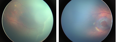
The target is to develop a deep neural network that correctly classifies input color fundus photograph (CFP) as ROP positive or ROP negative (Fig 1). In the transfer learning framework, the feature space is a space of 3D tensors, the feature distribution is the distribution of CFPs taken from infants, and the label space consists of ROP positive and ROP negative.
Source tasks.
There are two different source tasks: one for the conventional ImageNet-pretrained learning approach, and another for our proposed DR-pretrained transfer learning approach.
In the first one, the feature space is (again) a space of 3D tensors, the feature distribution is the color natural images, and the label space consists of 1000 classes.
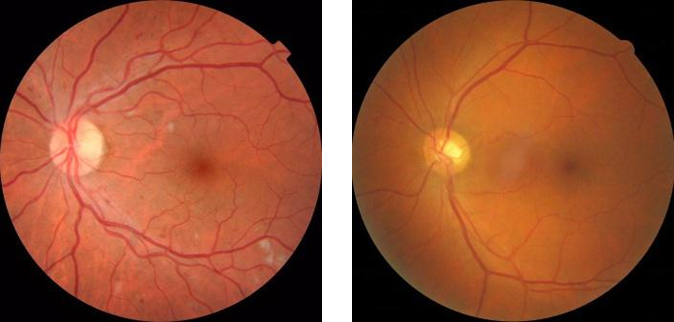
In the second one (Fig 2), the feature space is (again) a space of 3D tensors, the feature distribution is the distribution of CFPs taken from diabetes patients, and the label space consists of DR positive or DR negative.
3 Experiment
We investigate the effect of transfer learning by comparing our DR-pretrained approach with the ImageNet-pretrained transfer learning, against the baseline results from the direct training with random initialization. Throughout our experiment, ResNet50 architecture is used.
Data collection.
The CFPs taken from infants were collected from the Affiliated Eye Hospital of Nanchang University, which is an AAA (i.e., the highest ranked) hospital in China. All images were de-identified according to patient privacy protection policy, and the ethics review was approved by the ethical committee of Affiliated Eye Hospital of Nanchang University (ID: YLP202103012). The images were graded by 4 experienced ophthalmologists. As a result, the data set consists of 9,727 images (2,310 positive samples, 7,417 negative samples). The data set is randomly split into a training set and a test set by a ratio of 4:1.
The ImageNet data set consists of 1.3 million images collected online [6]. We used the public weight available in Keras [18].
The CFPs for DR were collected from the same hospital as the ROP data set, and the images were graded by experienced ophthalmologists whether DR positive or DR negative. The DR data set consists of 36,126 images with 26,548 positive samples and 9,578 negative samples.
Data augmentation.
Each image in ROP data set or DR data set is applied brightness adjustment and random flipping. Afterwards, each image is resized to 300300.
Class rebalance.
To mitigate the class imbalance issue in the DR and ROP data sets, we use a hybrid class balancing method: First, the class weight in the loss function is set to be for the negative class to the positive class; Second, when generating a minibatch, images from each class are sampled at the ratio of so that each class has an equal impact on the training process. The parameter is treated as one of the hyper parameters to be tuned.
Metrics for performance evaluation.
The trained models are evaluated by four metrics: the area under the receiver operator characteristic curve (AUROC), the accuracy, the precision, and the sensitivity. To account for the stochastic nature of the training, each experiment is iterated three times with different random seeds and the metrics are averaged.
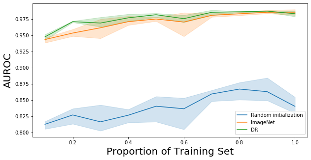
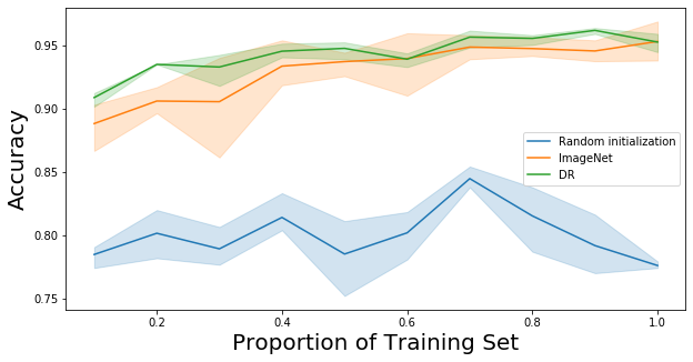
Experiment with reduced training samples.
To understand the effectiveness and robustness of transfer learning with limited data set, we further train the models with reduced training samples. In this series of experiments, the same pretrained weights are used for each training i.e., pretraining data set is fully utilized, but the training samples in the target task is reduced by factors ranging from 0% to 90% with 10% interval. The test set is kept the same to ensure consistency for comparison.
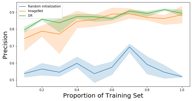
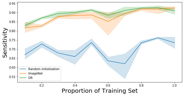
Results.
The results are shown in Figure 3 and tables 1, 2, and 3. We observe three critical advantages of our proposed approach via DR-pretraining over the traditional approach via ImageNet-pretraining. Firstly, DR-pretraining demonstrates superior performance compared with ImageNet-pretraining. Table 1 shows their mean percentage improvements from the direct training. DR-pretraining dominates ImageNet-pretraining by all four metrics. Secondly, the DR-pretraining is more robust with respect to the stochasticity in the training process. Table 2 shows the mean percentage reduction of standard deviation from direct training. DR-pretraining reduces the standard deviation by at least nearly 50% for all metrics. In contrast, ImageNet-pretraining adds more standard deviation (reduction of -46.6%) in precision and shows almost no improvement (reduction of 2.94%) in accuracy. Lastly, DR-pretraining is more robust with respect to the reduction of training sample size. The percentage changes of metrics from 100% training size to 10% training size are shown in Table 3.
These observations suggest 1) DR-pretraining dominates the traditional ImageNet-pretraining in all four metrics (AUROC, accuracy, precision, and sensitivity), 2) DR-pretraining is more robust with respect to both the stochasticity in the training process and reduced training samples.
| Pretraining | AUROC | Accuracy | Precision | Sensitivity |
|---|---|---|---|---|
| DR | 16.4% | 17.9% | 53.5% | 29.2% |
| ImageNet | 15.7% | 16.3% | 47.8% | 26.9% |
| Pretraining | AUROC | Accuracy | Precision | Sensitivity |
|---|---|---|---|---|
| DR | 75.4% | 64.3% | 47.5% | 53.7% |
| ImageNet | 57.0% | 2.94% | -46.6% | 25.1% |
| Pretraining | AUROC | Accuracy | Precision | Sensitivity |
|---|---|---|---|---|
| DR | 3.63% | 4.61% | 10.9% | 8.82% |
| ImageNet | 4.26% | 6.82% | 15.8% | 11.8% |
4 Conclusion
We propose a transfer learning approach that uses the detection of well-studied retinal vascular disease as a source task to transfer the learned knowledge to the detection of an under-studied retinal vascular disease as a target task. Experimental results demonstrate the superior performance of the DR-pretraining approach when compared with the traditional transfer learning and direct training approaches. Our study shows promises of transfer learning techniques utilizing feature similarities for general studies of retinal vascular diseases or other pathologies from different medical fields, where shortage of data is the main bottleneck for developing efficient deep-learning algorithms for medical image analysis.
References
- [1] Saud A. AlAnazi, Uchechukwu L. Osuagwu, Turki M. AlMubrad, Hany K. Ahmed, and Kelechi C. Ogbuehi, “Effectiveness of in-office blood pressure measurement by eye care practitioners in early detection and management of hypertension,” International Journal of Ophthalmology, vol. 8, no. 3, pp. 612–621, Jun 2015.
- [2] Carol Yim lui Cheung, Wan Ting Tay, M. Kamran Ikram, Yi Ting Ong, Deidre A. De Silva, Khuan Yew Chow, and Tien Yin Wong, “Retinal microvascular changes and risk of stroke,” Stroke, vol. 44, no. 9, pp. 2402–2408, Sept. 2013.
- [3] Diabetes Atlas et al., “International diabetes federation,” IDF Diabetes Atlas, 7th edn. Brussels, Belgium: International Diabetes Federation, 2015.
- [4] Justin Schaneman, Amy Kagey, Stephen Soltesz, and Julie Stone, “The role of comprehensive eye exams in the early detection of diabetes and other chronic diseases in an employed population,” Population Health Management, vol. 13, no. 4, pp. 195–199, Aug. 2010.
- [5] Varun Gulshan, Lily Peng, Marc Coram, Martin C. Stumpe, Derek Wu, Arunachalam Narayanaswamy, Subhashini Venugopalan, Kasumi Widner, Tom Madams, Jorge Cuadros, Ramasamy Kim, Rajiv Raman, Philip C. Nelson, Jessica L. Mega, and Dale R. Webster, “Development and Validation of a Deep Learning Algorithm for Detection of Diabetic Retinopathy in Retinal Fundus Photographs,” JAMA, vol. 316, no. 22, pp. 2402–2410, 12 2016.
- [6] Olga Russakovsky, Jia Deng, Hao Su, Jonathan Krause, Sanjeev Satheesh, Sean Ma, Zhiheng Huang, Andrej Karpathy, Aditya Khosla, Michael Bernstein, Alexander C. Berg, and Li Fei-Fei, “ImageNet Large Scale Visual Recognition Challenge,” International Journal of Computer Vision (IJCV), vol. 115, no. 3, pp. 211–252, 2015.
- [7] Maithra Raghu, Chiyuan Zhang, Jon Kleinberg, and Samy Bengio, “Transfusion: Understanding transfer learning for medical imaging,” in Advances in Neural Information Processing Systems, H. Wallach, H. Larochelle, A. Beygelzimer, F. d'Alché-Buc, E. Fox, and R. Garnett, Eds. 2019, vol. 32, Curran Associates, Inc.
- [8] Basil Mustafa, Aaron Loh, Jan Freyberg, Patricia MacWilliams, Megan Wilson, Scott Mayer McKinney, Marcin Sieniek, Jim Winkens, Yuan Liu, Peggy Bui, Shruthi Prabhakara, Umesh Telang, Alan Karthikesalingam, Neil Houlsby, and Vivek Natarajan, “Supervised transfer learning at scale for medical imaging,” 2021.
- [9] Walter M. Fierson, “Screening examination of premature infants for retinopathy of prematurity,” Pediatrics, vol. 142, no. 6, 2018.
- [10] Hannah Blencowe, Joy Lawn, Thomas Vazquez, Alistair Fielder, and Clare Gilbert, “Preterm-associated visual impairment and estimates of retinopathy of prematurity at regional and global levels for 2010,” Pediatric research, vol. 74 Suppl 1, pp. 35–49, 12 2013.
- [11] Clare Gilbert, Jugnoo Rahi, Michael Eckstein, Jane O’sullivan, and Allen Foster, “Retinopathy of prematurity in middle-income countries,” The Lancet, vol. 350, no. 9070, pp. 12–14, 1997.
- [12] Early Treatment For Retinopathy Of Prematurity Cooperative Group, “Revised Indications for the Treatment of Retinopathy of Prematurity: Results of the Early Treatment for Retinopathy of Prematurity Randomized Trial,” Archives of Ophthalmology, vol. 121, no. 12, pp. 1684–1694, 12 2003.
- [13] Kaiming He, Xiangyu Zhang, Shaoqing Ren, and Jian Sun, “Deep residual learning for image recognition,” in Proceedings of the IEEE conference on computer vision and pattern recognition, 2016, pp. 770–778.
- [14] C. Szegedy, V. Vanhoucke, S. Ioffe, J. Shlens, and Z. Wojna, “Rethinking the inception architecture for computer vision,” in 2016 IEEE Conference on Computer Vision and Pattern Recognition (CVPR), Los Alamitos, CA, USA, jun 2016, pp. 2818–2826, IEEE Computer Society.
- [15] Sihong Chen, Kai Ma, and Yefeng Zheng, “Med3d: Transfer learning for 3d medical image analysis,” 2019.
- [16] Laith Alzubaidi, Muthana Al-Amidie, Ahmed Al-Asadi, Amjad J. Humaidi, Omran Al-Shamma, Mohammed A. Fadhel, Jinglan Zhang, J. Santamaría, and Ye Duan, “Novel transfer learning approach for medical imaging with limited labeled data,” Cancers, vol. 13, no. 7, pp. 1590, Mar. 2021.
- [17] Fuzhen Zhuang, Zhiyuan Qi, Keyu Duan, Dongbo Xi, Yongchun Zhu, Hengshu Zhu, Hui Xiong, and Qing He, “A comprehensive survey on transfer learning,” 2020.
- [18] François Chollet et al., “Keras,” https://keras.io, 2015.