44email: M.Yap@mmu.ac.uk
Development of Diabetic Foot Ulcer Datasets: An Overview
Abstract
This paper provides conceptual foundation and procedures used in the development of diabetic foot ulcer datasets over the past decade, with a timeline to demonstrate progress. We conduct a survey on data capturing methods for foot photographs, an overview of research in developing private and public datasets, the related computer vision tasks (detection, segmentation and classification), the diabetic foot ulcer challenges and the future direction of the development of the datasets. We report the distribution of dataset users by country and year. Our aim is to share the technical challenges that we encountered together with good practices in dataset development, and provide motivation for other researchers to participate in data sharing in this domain.
1 Introduction
The Diabetic Foot Ulcer (DFU) is one of the major complications resulting from diabetes, which can lead to lower limb amputation [1]. Regular foot check by clinical professionals is required for patients with DFU development, which is often costly and / or requires referral to specialist care [2]. Research shows that healthcare services that treat DFU are unable to handle the growing number of patients due to inadequately trained medical staff [3], which is especially prevalent in low-income countries and rural areas [4, 5].
Over the past decade, the development of digital and information technology has enabled the creation of new computer-based solutions for healthcare, including wound care [6]. Figure 1 illustrates the timeline of development of DFU datasets, including the first use of computer vision methods in DFU detection. The focus of the earlier DFU research is in the design of new capturing tools [7], initiated in 2015. At the same time, computer vision methods (based on image processing algorithms) and conventional machine learning methods were used to analyse those images [8]. With the advent of deep learning in computer vision tasks, researchers began investigating the use of deep learning for DFU segmentation in 2016, where the first fully automated segmentation paper was published in 2017 by Goyal et al. [9].
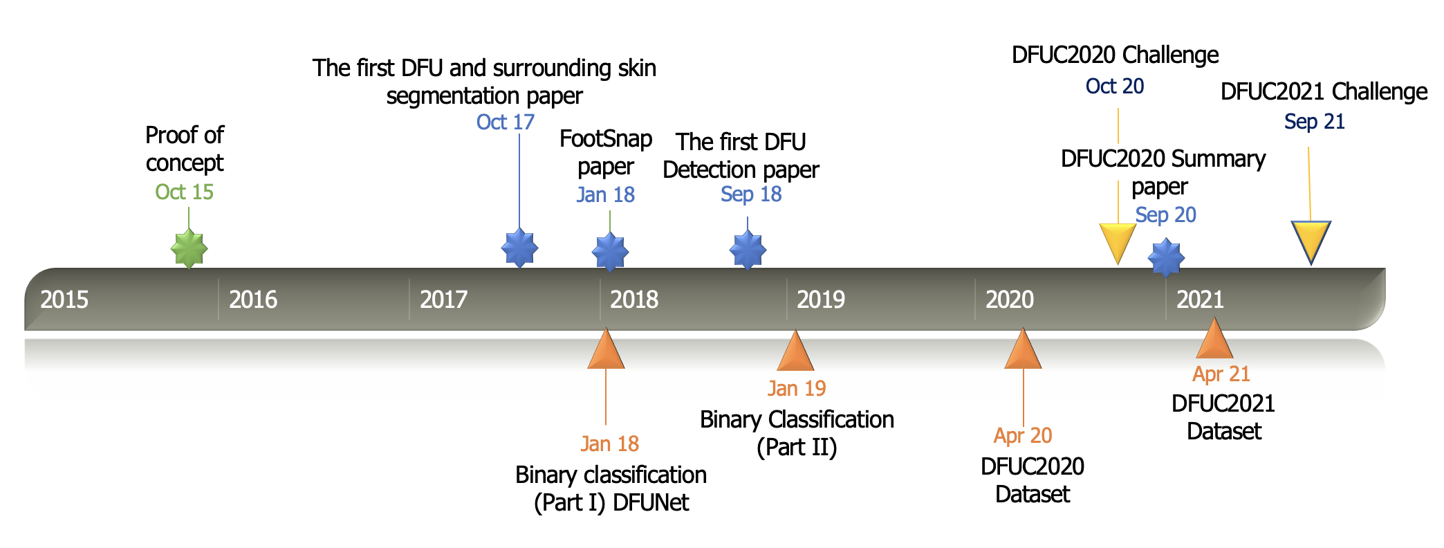
Motivated by the success of deep learning on DFU segmentation, the team at the Manchester Metropolitan University and Lancashire Teaching Hospitals NHS Foundation Trust had obtained ethical approval from the UK National Health Service Research Ethics Committee (reference number: 15/NW/0539) to create larger scale datasets of DFU images, with approval to share the datasets with the research community for research, provided that the users abide by the licence agreement. The first dataset (Part I) was on binary classification using normal and DFU patches, where the authors designed a new deep learning network (DFUNet) and benchmarked the datasets with popular networks at that time [10]. The second dataset (Part II) was also on binary classification, and focussed on ischaemic and infection skin patches. The binary classification of ischaemia-vs-all and infection-vs-all were benchmarked by Goyal et al. [11], and they proposed an ensemble method to increase the accuracy of infection and ischaemia recognition. In 2020, the team conducted the first inaugural research challenge in DFU detection, DFUC2020 [12], and the second challenge in DFU multi-class classification in 2021 [13].
The remaining sections of the paper are organised as follow: Section 2 provides a survey of DFU data capturing methods; Section 3 reviews the available DFU datasets; Section 4 describes the DFU research challenges conducted over the past years; Section 5 presents future work and research directions of DFU analysis; and Section 6 summarises the paper.
2 A Survey of Data Capturing Methods
In current clinical practices, podiatrists and consultants use a range of digital single-lens reflex (SLR) camera models to collect DFU photographs [10, 14, 15]. The photographs are transferred to a secured storage which is often isolated from the patients’ electronic health records. The process is operator dependent, with poor consistency across different clinic and care settings. Due to these inconsistencies and limitations of 2D images, it has not been possible to quantify the changes of the ulcers over time.
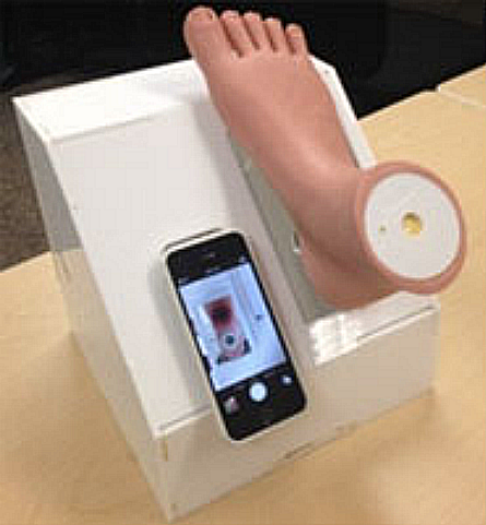
Over the past few years, several research teams have proposed new methods in standardising data capture of DFUs. The earliest attempts were conducted by Wang et al. [16] and Yap et al. [8]. Wang et al. [16] developed a smartphone app capable of image segmentation of DFU wounds using an accelerated mean-shift algorithm, which pre-dates current deep learning approaches. To improve and standardise the acquisition of DFU images, they created an image capture box, to be used in conjunction with the smartphone app. Their goal was to promote a more active role in patient self-monitoring. This approach used skin colour to determine foot boundaries, while wound area is determined by a simple connected region detection method. This system also assessed healing status using a red–yellow–black colour evaluation model and a quantitative trend analysis of time records for a given patient. The system was tested with 34 patients and 30 wound moulds. Figure 2 shows the design of the capture box111reproduced with permission from Peder C. Pedersen, Dept. of Electrical and Computer Engineering, Worcester Polytechnic Institute, Worcester, MA 01609, USA.
 |
 |
| (a) | (b) |
Wang et al. [17] later validated the capture box in a clinical study using 32 DFU photographs obtained from 12 diabetic patients. This system used a laptop to perform wound segmentation, calculation of the wound area and calculation of a healing score. They found a good correlation between human and automated wound area measurements, reporting a Matthews correlation coefficient value of 0.68 for the wound area determination algorithm, and a Krippendorff alpha coefficient within the range of 0.42 to 0.81.
Wang et al. [18] conducted a third study using their capture box, which collected 100 DFU photographs from 15 patients during a 2-year period. In this study, they utilised superpixel segmentation, using the Simple Linear Iterative Clustering (SLIC) algorithm, as inputs for a cascaded two-stage SVM-based (Support-Vector Machine) classifier to determine wound boundaries for DFU. Colour and bag-of-word representations of local dense scale invariant transformation features are used as descriptors for excluding non-wound regions. Wavelet-based features are used as descriptors to identify healthy tissue from wound tissue. Finally, wound boundaries are refined by applying a conditional random field method. This system ran on a Nexus 5 smartphone, and reported an average sensitivity of 73.3%, and an average specificity of 94.6% with a computation time of 15 to 20 seconds.
 |
 |
| (a) | (b) |
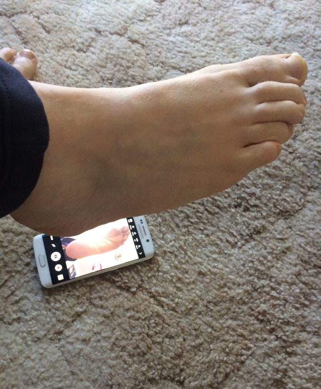
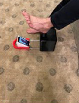
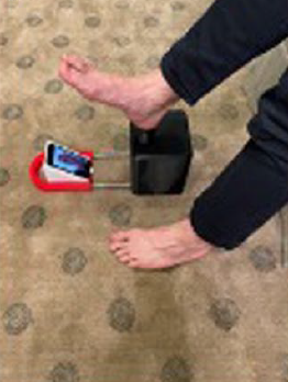
In 2015, Yap et al. [8] created a new mobile app called FootSnap which was used for standardising photographic capture of the plantar aspect of feet. This system generated an outline of a foot from an initial photograph of the patient’s foot. The outline could then be recalled on-screen to help align the foot in subsequent photographs. The app was initially tested on healthy feet, then later evaluated for standardisation on clinical data [7]. The app was then used as part of a clinical trial investigating DFU prevention using smart insole technology published in 2019 [20].
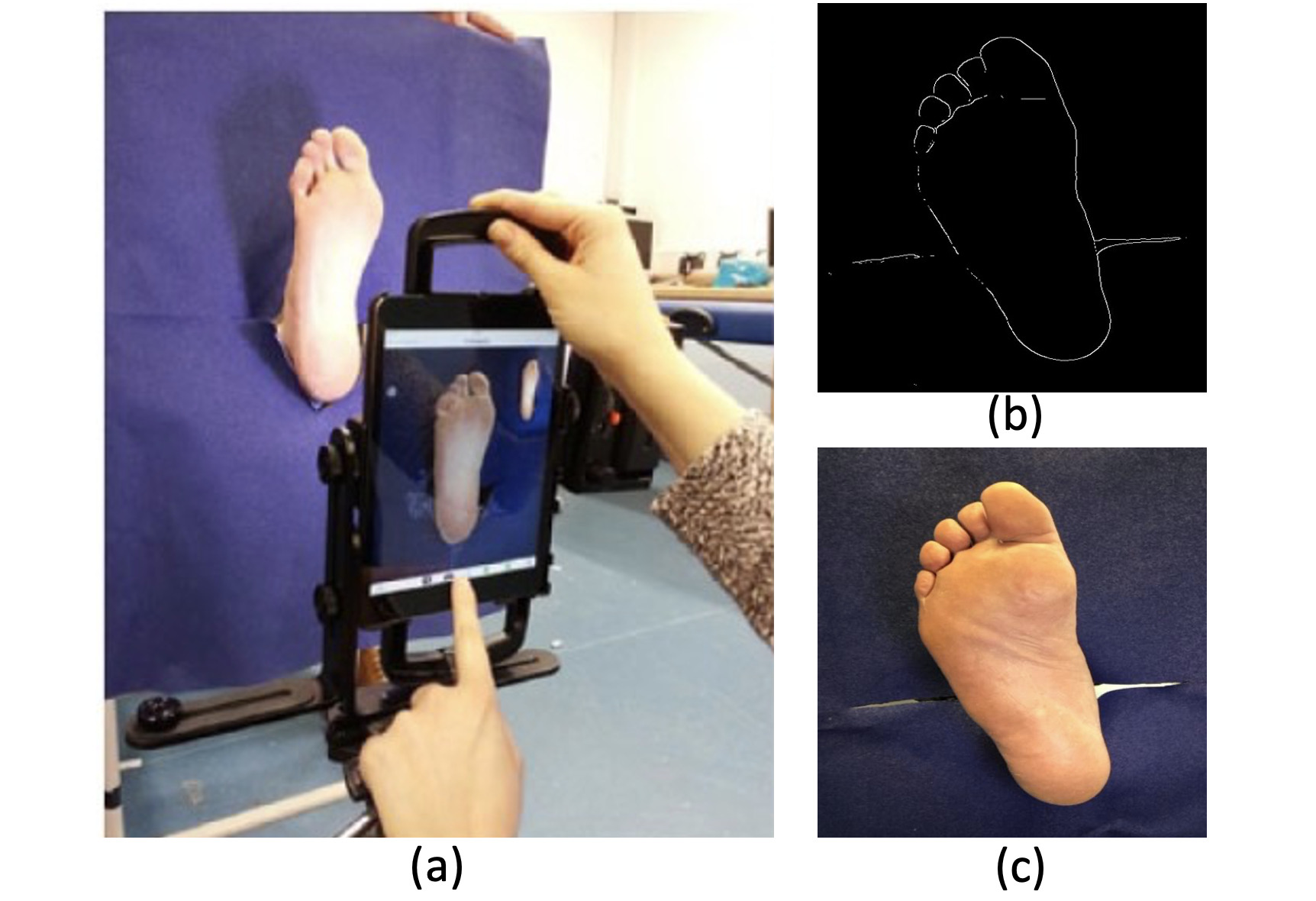
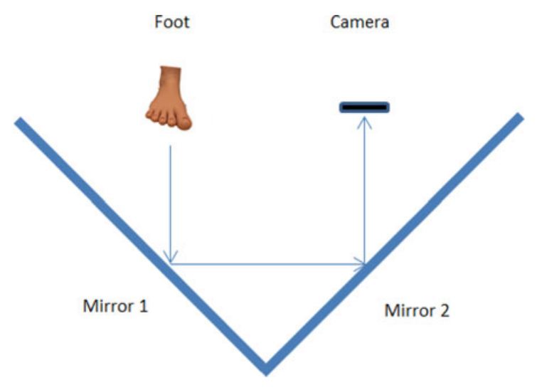
Researchers would continue to investigate the use of mobile technologies to address the growing problem of DFU. Brown et al. [19] developed the MyFootCare app - a self-monitoring tool that could be used by patients to promote self-care in home settings. The app was evaluated with 3 DFU patients, who reported it as useful for tracking wound progress and to assist in communications with clinicians. The app implements a number of features, including image capture, wound analysis (using OpenCV), diary reminders, and wound size tracking using a graph. The image capture allows the patient to place the phone onto the floor with the screen facing upwards. The patient can then guide their foot into the correct position, with voice feedback provided by the app. This allows the patient to remain seated while positioning their foot for image capture. However, this functionality was not completed in time for the evaluation with patients. Figure 3 shows the design of the app and its proposed use in DFU photograph acquisition222reproduced with permission from Ross Brown, School of Computer Science, Science and Engineering Faculty, Queensland University of Technology, Brisbane, Queensland, Australia.
More recent techniques for photographic acquisition of DFU have been proposed by Swerdlow et al. [21]. They devised a ”foot selfie” device comprising an elaborate assembly which helps to position the foot in front of a smartphone while minimising surface contact (see Figure 5)333reproduced with permission from David G. Armstrong, Southwestern Academic Limb Salvage Alliance (SALSA), Department of Surgery, Keck School of Medicine of University of Southern California, Los Angeles, California, USA. Compared to some of the earlier capture methods, this solution has the advantage of not requiring contact between wound and surface, and also allows for capture of more than just plantar DFU.
 |
 |
 |
| (a) | (b) | (c) |
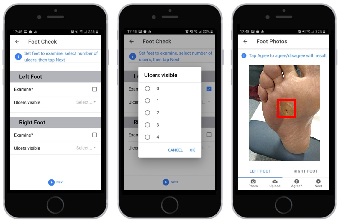
Cassidy et al. recently validated a fully automated DFU detection system which utilised mobile and cloud-based technologies [22, 23]. This system used a cross-platform mobile app to capture photographs of patient’s feet in clinical settings that could be uploaded to a cloud platform for inference to detect the presence of DFU. The system was validated in a proof-of-concept study at two UK hospitals over a six-month period. This technology is currently being adapted for use in additional clinical studies with the aim of replacing SLR cameras in the acquisition of DFU photographs. Figure 6 shows a selection of screens from the cross-platform mobile app.
3 A Review of DFU Image Datasets
This section reviews the available public DFU image datasets and the number of users (by countries, if known). Table 1 summarises and compares five publicly available datasets.
| Publication | Year | Dataset Name | Resolution | Task | Train | Test | Total |
| Goyal et al. [10] | 2018 |
Part A or
Part I |
varied | classification | NA | NA | 1,679 |
| Goyal et al. [11] | 2019 |
Part B or
Part II |
classification | NA | NA | 1,459 | |
| Cassidy et al. [15] | 2020 | DFUC2020 | detection | 2,000 | 2,000 | 4,000 | |
| Wang et al. [24] | 2020 | AZH wound care dataset | segmentation | 831 | 278 | 1,109 | |
| Thomas [25] | NA | Medetec |
|
segmentation | 152 | 8 | 160 |
| Wang et al. [26] | 2021 | FUSeg Challenge | segmentation | 1,010 | 200 | 1,210 | |
| Yap et al. [27] | 2021 | DFUC2021 | classification | 5,955 | 5,734 | 15,683* | |
| * 3.994 patches are unlabelled; NA indicates unknown | |||||||
3.1 Binary Classification
DFU patches and normal patches (Part A or Part I)
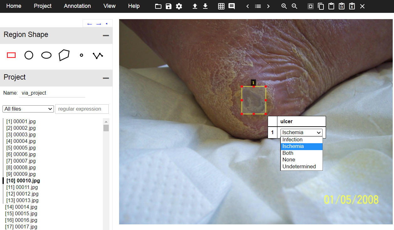
The Part A DFU Dataset [10] consists of 1038 DFU patches and 641 normal patches. This is the first binary classification dataset shared with the research community. The ground truth was produced by two healthcare professionals, specialising in DFU, using the annotation tool developed by Hewitt et al. [28]. The authors introduced this dataset and created a new deep learning model, DFUNet, to benchmark the performance on the dataset. DFUNet achieved an F1-score of 0.939. Since its initial release, the dataset has been used in later research, with the most recent publication achieving the best performance of 0.952 in F1-score [29].
Recognition of Infection and Ischaemia Datasets (Part B or Part II)
The first infection and ischaemia datasets were created by Goyal et al. [11], which consists of 1,459 DFUs: 645 with infection, 24 with ischaemia, 186 with infection and ischaemia, and 604 control DFU (presence of DFU, but without infection or ischaemia). This work focused on binary classification, i.e., infection-vs-all and ischaemia-vs-all. This dataset consists of:
-
•
4,935 patches (including augmented images) of ischaemia
-
•
4,935 patches (including augmented images) of non-ischaemia
-
•
2,946 patches (including augmented images) of infection
-
•
2,946 patches (including augmented images) of non-infection
Image labelling information:
-
•
00XXXX_1X.jpg - the original image patch
-
•
00XXXX_2X,jpg and 00XXX_3X.jpg - natural data augmentation, where M indicates mirroring; R1, R2 and R4 indicate rotations.
 |
 |
 |
 |
| (a) | (b) | (c) | (d) |
Goyal et al. [11] introduced an ensemble CNN method for binary classification and achieved an F1-score of 0.902 and 0.722 on ischaemia-vs-all and infection-vs-all respectively. Since the release of this dataset in early 2020, Al-Garaawi et al. [29] achieved the best F1-scores of 0.990 and 0.744 on ischaemia-vs-all and infection-vs-all, respectively. This demonstrates a significant challenge for machine learning algorithms in recognising infection from other classes.
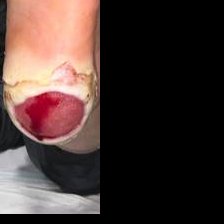
Figure 7 compares the Part A and Part B datasets. It is noted that the Part A dataset did not include the whole ulcer region, as it consists of image patches cropped from examples exhibiting ulcers and non-ulcers. In contrast, the Part B dataset consists of ulcers with different pathologies.
3.2 DFU Detection (DFUC2020)
In 2018, Goyal et al. [30] reported the performance of object detection algorithms on an in-house DFU dataset with 1,775 images. They proposed the use of a 2-tier transfer learning method using Faster R-CNN with InceptionV2 model. Overall, they achieved the best mean average precision (mAP) of 91.8% on 5-fold cross-validation.
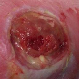
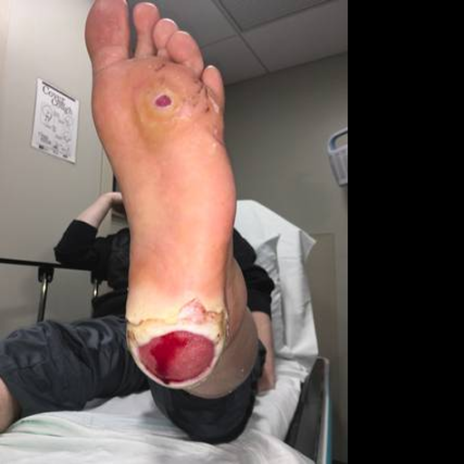
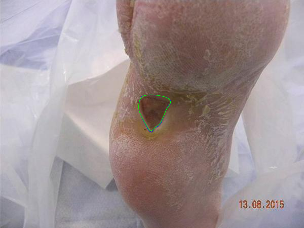
Cassidy et al. [15] introduced the largest DFU detection dataset to date, which was released on the 27th April 2020. This dataset contains 4,000 images (largely DFU images, with a small proportion of non-DFU images in the testing set), is highly heterogeneous and includes numerous challenging examples to help ensure robustness in algorithm development. Figure 8 shows an example of the expert labelling provided by podiatrists for this dataset.
3.3 Multi-class DFU Classification (DFUC2021)
The first multi-class DFU classification dataset was released on the 27th April 2021 by Yap et al. [27], with four types of DFU patches, i.e., control, ischaemia, infection and both (co-occurrence of ischaemia and infection). This dataset is comprised of cropped ulcer regions from the DFUC2020 dataset [15] and the Part B dataset [11].
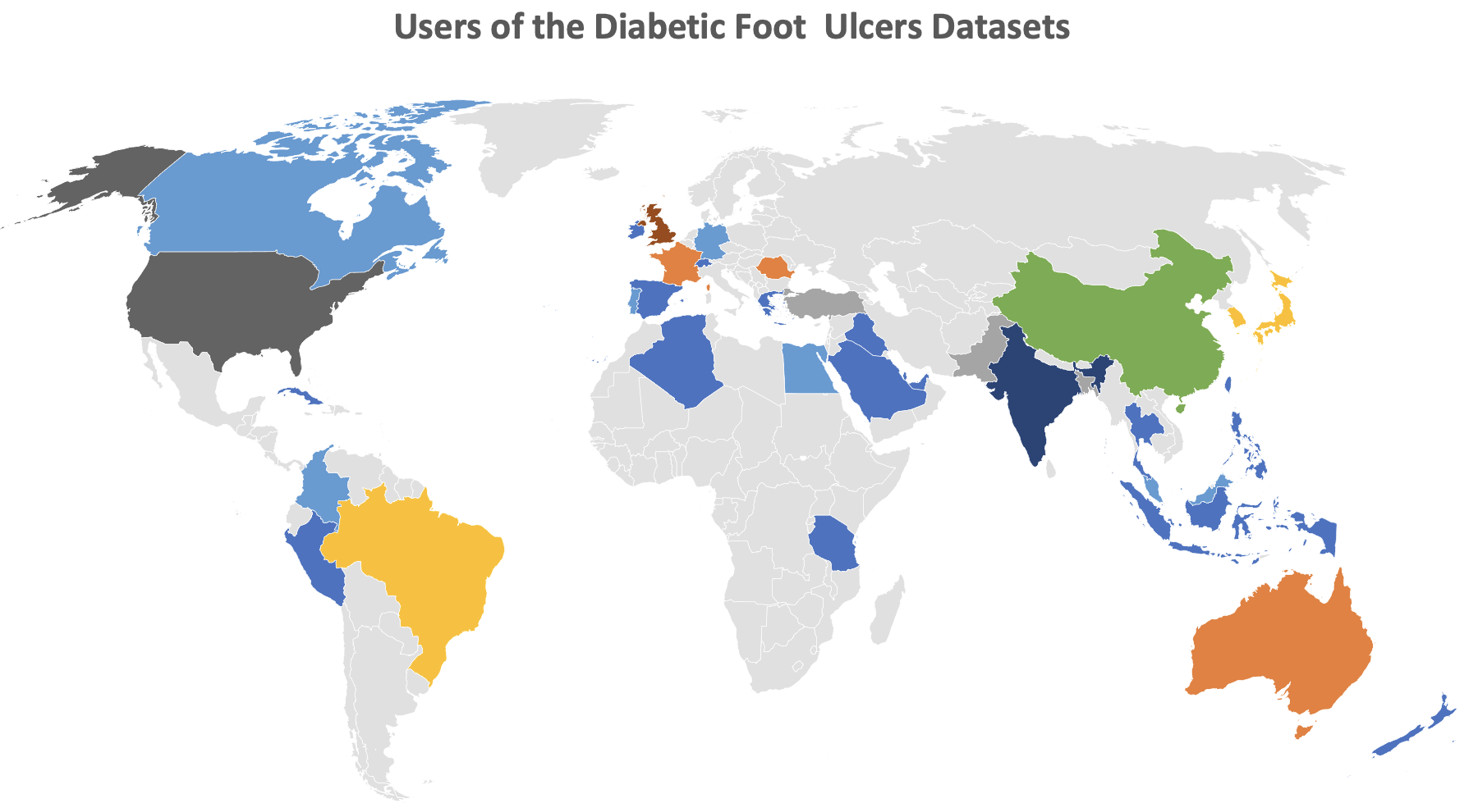
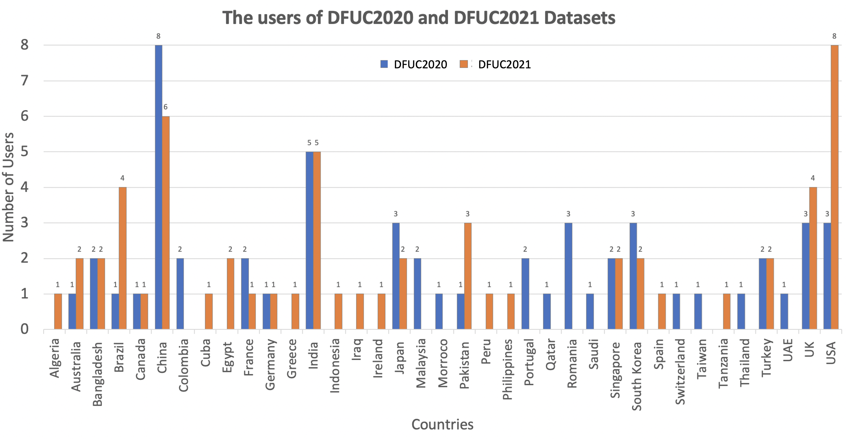
Since the release of the DFU Challenge datasets on the 27th April 2020, our datasets have been requested by users from 38 countries, as illustrated in Figure 9. To date, there are more requests from China, US, India and UK. When compared with the users of DFUC2020 and DFUC2021, we observe that DFUC2021 reached a wider research community as shown in Figure 10, including growing interest of researchers from Algeria, Cuba, Egypt, Greece, Indonesia, Iraq, Ireland, Peru, Philippines, Spain and Tanzania.
3.4 Other Datasets
Following the successful release of the Part A, Part B and DFUC2020 datasets by researchers at Manchester Metropolitan University and Lancashire Teaching Hospitals [15, 12, 32], other research groups have followed this path and released their own public DFU datasets. Wang et al. [24] released the Foot Ulcer Segmentation Challenge dataset which focuses on single class semantic segmentation of foot ulcer wounds and contains 1,210 images with 1,010 labels, of which 200 are used for testing. Images were captured in clinical settings at different angles, with many cases exhibiting background noise. The dataset contains images of the same ulcers at different angles at different time intervals. This dataset presents additional challenges for computer vision and deep learning algorithms as all images are padded with black pixels to maintain the aspect ratio and image size of pixels, as illustrated in Figure 11(a).
The AZH Wound Care Center Dataset was also introduced by Wang et al. in 2020 and contains 831 training images and 278 test images. All images in this dataset are pixels and contain only DFU patches. The majority of each image in this dataset contains padding using black pixels, as shown in Figure 11(b). A smaller dataset was also released (Medetec), as shown in Figure 11(c). The release date of this dataset is unknown. This is a collection of multiple wound types, and contains 46 DFU images with no labels. The image resolution of this dataset ranges from pixels to pixels.
 |
 |
 |
| (a) | (b) | (c) |
The segmentation dataset introduced by Wang et al. for the Foot UlcerSegmentation Challenge is bundled with 2 other datasets. First, is the Medetec wound dataset, resized to with black padding along the bottom. The Medetec dataset has 160 wound images, 152 labels and 46 DFU examples. These images focus on the wounds, but still exhibit some background details. The second dataset is the AZH wound care center dataset, which has 1,109 images and 831 labels. Similar to Medetec, these images are , but padded at the bottom and sides with black borders. However, these images where pre-cropped to focus on the lesion only so do not show background on the rest of the foot.
4 DFU Challenges
Once a year, The Medical Image Computing and Computer Assisted Intervention (MICCAI) Society conduct medical image challenges 444http://www.miccai.org/special-interest-groups/challenges/miccai-registered-challenges/ to support and lead to thoughtful research challenges. The registered MICCAI challenge is reviewed and evaluated by expert panels, criteria include the design, metrics and transparency toward higher quality challenges. Due to limited capacity and an increased number of proposals, some challenges were accepted as MICCAI endorsed events, which are online only challenges and are not associated with the conference.
The inaugural DFU challenge was initiated by Yap et al. [12] on the DFU detection task (DFUC2020). Lead by the Manchester Metropolitan University (UK) and Lancashire Teaching Hospitals (UK), together with other co-organisers including the University of Southern California (USA), University of Waikato (New Zealand), University of Manchester and Manchester Royal Infirmary (UK), Manipal College of Health Professions (India), Baylor College of Medicine (USA) and Waikato District Health Board (New Zealand). DFUC2020 was accepted as a MICCAI registered challenge, and was conducted in conjunction with MICCAI 2020. The DFUC2020 datasets [15] include 2,000 training images and 2,000 testing images. The summary of the challenge results were concluded by Yap et al. [32]. The organisers continue to support the research community with a live leaderboard on the Grand Challenge System 555https://dfu2020.grand-challenge.org. To date, the best result on the live leaderboard reports an mAP of 0.73.
DFUC2021 [13] was accepted as a MICCAI registered challenge, and was conducted in conjunction with MICCAI 2021. The focus on DFUC2021 was on classification, where the DFU patches were classified into control / none, infection, ischaemia and both conditions [27]. The organisers continue to support the research community with a live leaderboard on the Grand Challenge System 666https://dfu-2021.grand-challenge.org. At the time of writing this paper, the best macro F1-score on the live leaderboard is 0.6307 [33].
In the same period, another research group based in the US organised an online-only Foot Ulcer Segmentation Challenge (FUSeg) [26], which was conducted as a MICCAI endorsed event. The best performance of FUSeg is a Dice score of 0.8880. Since the evaluation is not on a live leaderboard and the participants were required to send their codes (docker/container) to the organiser. It is currently unclear if the organisers are still accepting submissions.
5 Future directions
In 2021, the DFUC2022 challenge proposal was accepted by MICCAI as a registered challenge [34]. The task concerns DFU segmentation 777https://dfu-challenge.github.io/. Compared to the online-only segmentation challenge conducted in 2021, DFUC2022 will comprise of large-scale higher resolution datasets, as illustrated in Figure 12. It will be conducted in conjunction with MICCAI 2022.
 |
 |
 |
| (a) | (b) | (c) |
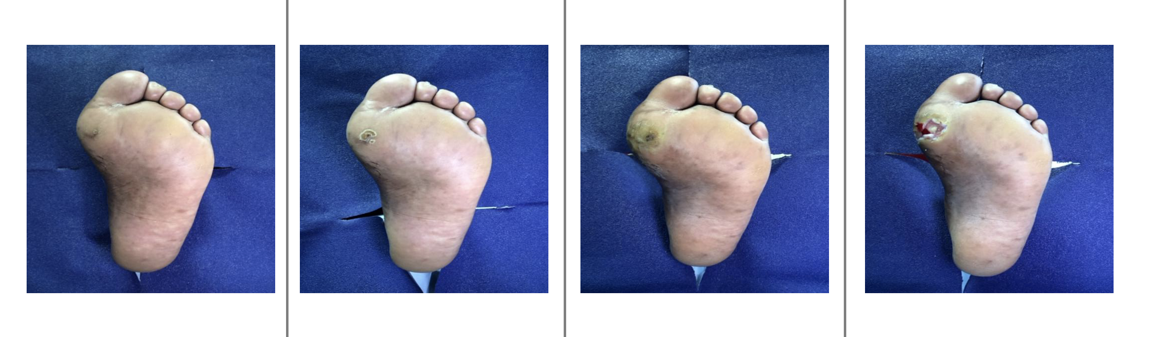
To monitor the healing progress of DFUs in clinical settings, podiatrists / consultants take photographs of the foot using standard SLR cameras. Due to a lack of standardisation, the photographs taken are operator dependent, with variations in angle, distance and illumination. Figure 13 shows photographs of the same wound taken at different time points.

The efforts shown in previous research [7] attempted to standardise the data capturing process, enabling improved observation on images captured at different timelines. Such longitudinal datasets, as illustrated in Figure 14, help computer vision techniques and human observers to better spot the subtle changes on feet.
In an effort to improve patient care and reduce the strain on healthcare systems, current research has been focused on the development of AI algorithms that can detect diabetic foot ulcers at different stages and grades on clinical wound / DFU images. These algorithms could potentially be used as part of a mobile application that patients could use themselves (or a carer / partner) to remotely monitor their foot condition and detect the appearance of DFU for timely clinical intervention. Also, clinicians and podiatrists can monitor the progress of DFU (detected by AI algorithms) through timeline images. This is an important step in deciding what therapeutic intervention is required depending upon the progression of DFU. Effective diagnosis of such wounds can lead to better treatments, which may lead to quicker healing, reduced amputation risk and a significant reduction in health care costs.
This paper discusses the challenges and opportunities in clinical DFU image datasets taken by different types of cameras and the role of AI algorithms in the detection and progress of DFU. Other than DFU images, researchers have used other imaging modalities such as infrared, Magnetic Resonance Imaging (MRI), and fluorescence imaging for the management of DFU. However, there are no such public datasets of other imaging modalities available for further research and development of AI solutions for multi-modality datasets.
In many research studies, thermal infrared imaging has been proven to be a useful technique in the clinical management of DFU. Several diabetic foot complications such as neuropathic ulcers, osteomyelitis and Charcot’s foot have been identified at locations with increased temperature [35, 36, 37, 38]. Increased plantar temperature is a strong indicator of pre-ulcer conditions and may present a week before an ulcer appears visually on the foot. Hence, regular monitoring of temperature (i.e. temperature difference (2.2 Celsius)) when comparing general foot temperature with suspected DFU sites can potentially help in early interventions.
Another imaging modality known as fluorescence imaging can detect the presence of clinically significant bacteria in diabetic foot ulcers by using a handheld device [39]. Potentially, fluorescence imaging can provide valuable information of DFU outcomes on whether a DFU is healing or not [40].
MRI is the modality of choice for imaging both Charcot foot and deep infection in the diabetic foot. In the early stages, MRI can demonstrate marrow oedema while plain films remain normal. the use of MRI is common with diabetic patients to rule out infection in the presence of an ulcer, to evaluate the severity of Charcot foot, or to distinguish between Charcot foot and infection [41, 42, 43].
In the current literature, there are no studies that combine AI interventions for the detection and management of DFUs in multi-modality imaging. Combining wound / ulcer images with different types of other imaging (such as MRI, Thermal Infrared, and fluorescence) can potentially help AI algorithms to provide a complete diagnosis and prognosis of DFUs and timely interventions in the treatment of diabetic foot ulcers to avoid amputations.
There are currently no publicly available datasets that combine the multi-modality imaging of diabetic foot patients. Great effort is needed for the collection of such datasets which combines different types of imaging such as thermal infrared (early detection of ulcers), clinical wound images (progression of ulcers), fluorescence (presence of clinically significant bacteria) and MRI (presence of infection and Charcot’s foot). Similarly, most of the current state-of-the-art AI algorithms rely on supervised learning, hence annotation of the dataset is another important step required for the development of AI algorithms for diagnosis and management of the diabetic foot. DFU datasets are prone to the same issues that have affected other medical imaging datasets, such as image duplication, feature over-representation and sourcing of large numbers of images from a relatively small pool of subjects [44, 45, 46].
6 Conclusion
This paper provides an overview of the development of DFU datasets and notable advances in the field that have led to the current use of deep learning techniques. The aim is to guide researchers in this domain to understand the breadth and depth of the processes involved in DFU classification, detection and segmentation, and to promote good practice in research and data sharing.
Collection, labelling and curation of DFU datasets is a challenging process requiring significant input from clinical experts at all stages of development. Comprehensive inter- and intra-rater analysis will prove to be key in refining the quality of datasets together with establishing new standards in this relatively new research domain. Researchers training deep learning models should pay particular attention to challenging examples within the datasets, as these will help to make networks more robust in real-world settings.
Acknowledgment
We gratefully acknowledge the support of NVIDIA Corporation who provided access to GPU resources for the DFUC2020 and DFUC2021 Challenges.
Author Index
Index
References
- [1] David G Armstrong, Lawrence A Lavery, and Lawrence B Harkless. Validation of a diabetic wound classification system: the contribution of depth, infection, and ischemia to risk of amputation. Diabetes care, 21(5):855–859, 1998.
- [2] Ll Prompers, M Huijberts, Jan Apelqvist, E Jude, A Piaggesi, K Bakker, M Edmonds, P Holstein, A Jirkovska, D Mauricio, et al. Delivery of care to diabetic patients with foot ulcers in daily practice: results of the eurodiale study, a prospective cohort study. Diabetic medicine, 25(6):700–707, 2008.
- [3] Peter Cavanagh, Christopher Attinger, Zulfiqarali Abbas, Arun Bal, Nina Rojas, and Zhang-Rong Xu. Cost of treating diabetic foot ulcers in five different countries. Diabetes/metabolism research and reviews, 28(S1):107–111, 2012.
- [4] Paul Z Zimmet, Dianna J Magliano, William H Herman, and Jonathan E Shaw. Diabetes: a 21st century challenge. The lancet Diabetes & endocrinology, 2(1):56–64, 2014.
- [5] Frank Vinicor. The public health burden of diabetes and the reality of limits. Diabetes Care, 21(Supplement 3):C15–C18, 1998.
- [6] Caroline Chanussot-Deprez and José Contreras-Ruiz. Telemedicine in wound care: a review. Advances in skin & wound care, 26(2):78–82, 2013.
- [7] Moi Hoon Yap, Katie E Chatwin, Choon-Ching Ng, Caroline A Abbott, Frank L Bowling, Satyan Rajbhandari, et al. A new mobile application for standardizing diabetic foot images. Journal of diabetes science and technology, 12(1):169–173, 2018.
- [8] Moi Hoon Yap, Choon-Ching Ng, Katie Chatwin, Caroline A Abbott, Frank L Bowling, Andrew JM Boulton, and Neil D Reeves. Computer vision algorithms in the detection of diabetic foot ulceration a new paradigm for diabetic foot care? Journal of diabetes science and technology, page 1932296815611425, 2015.
- [9] M. Goyal, M. H. Yap, N. D. Reeves, S. Rajbhandari, and J. Spragg. Fully convolutional networks for diabetic foot ulcer segmentation. In 2017 IEEE International Conference on Systems, Man, and Cybernetics (SMC), pages 618–623, Oct 2017.
- [10] Manu Goyal, Neil D Reeves, Adrian K Davison, Satyan Rajbhandari, Jennifer Spragg, and Moi Hoon Yap. Dfunet: Convolutional neural networks for diabetic foot ulcer classification. IEEE Transactions on Emerging Topics in Computational Intelligence, 4(5):728–739, 2018.
- [11] Manu Goyal, Neil D. Reeves, Satyan Rajbhandari, Naseer Ahmad, Chuan Wang, and Moi Hoon Yap. Recognition of ischaemia and infection in diabetic foot ulcers: Dataset and techniques. Computers in Biology and Medicine, 117:103616, 2020.
- [12] Moi Hoon Yap, Neil Reeves, Andrew Boulton, Satyan Rajbhandari, David Armstrong, Arun G. Maiya, Bijan Najafi, Eibe Frank, and Justina Wu. Diabetic foot ulcers grand challenge 2020, March 2020.
- [13] Moi Hoon Yap, Neil Reeves, Andrew Boulton, Satyan Rajbhandari, David Armstrong, Arun G. Maiya, Bijan Najafi, Eibe Frank, and Justina Wu. Diabetic foot ulcers grand challenge 2021, March 2020.
- [14] Manu Goyal and Moi Hoon Yap. Region of interest detection in dermoscopic images for natural data-augmentation. arXiv preprint arXiv:1807.10711, 2018.
- [15] Bill Cassidy, Neil D Reeves, Joseph M Pappachan, David Gillespie, Claire O’Shea, Satyan Rajbhandari, Arun G Maiya, Eibe Frank, Andrew J M Boulton, David G Armstrong, Bijan Najafi, Justina Wu, Rupinder Singh Kochhar, and Moi Hoon Yap. The dfuc 2020 dataset: Analysis towards diabetic foot ulcer detection. touchREVIEWS in Endocrinology, 17:5–11, 2021.
- [16] Lei Wang, Peder C Pedersen, Diane M Strong, Bengisu Tulu, Emmanuel Agu, and Ronald Ignotz. Smartphone-based wound assessment system for patients with diabetes. IEEE Transactions on Biomedical Engineering, 62(2):477–488, 2015.
- [17] Lei Wang, Peder Pedersen, Diane Strong, Bengisu Tulu, Emmanuel Agu, Ron Ignotz, and Qian He. An automatic assessment system of diabetic foot ulcers based on wound area determination, color segmentation, and healing score evaluation. Journal of diabetes science and technology, 10, 08 2015.
- [18] Lei Wang, Peder C. Pedersen, Emmanuel Agu, Diane M. Strong, and Bengisu Tulu. Area determination of diabetic foot ulcer images using a cascaded two-stage svm-based classification. IEEE Transactions on Biomedical Engineering, 64(9):2098–2109, 2017.
- [19] Ross Brown, Bernd Ploderer, Leonard Si Da Seng, Peter Lazzarini, and Jaap van Netten. Myfootcare: A mobile self-tracking tool to promote self-care amongst people with diabetic foot ulcers. In Proceedings of the 29th Australian Conference on Computer-Human Interaction, OZCHI ’17, page 462–466, New York, NY, USA, 2017. Association for Computing Machinery.
- [20] Caroline A Abbott, Katie E Chatwin, Philip Foden, Ahmad N Hasan, Chandbi Sange, Satyan M Rajbhandari, Prabhav N Reddy, Loretta Vileikyte, Frank L Bowling, Andrew JM Boulton, et al. Innovative intelligent insole system reduces diabetic foot ulcer recurrence at plantar sites: a prospective, randomised, proof-of-concept study. The Lancet Digital Health, 1(6):e308–e318, 2019.
- [21] Mark Swerdlow, Laura Shin, Karen D’Huyvetter, Wendy J. Mack, and David G. Armstrong. Initial clinical experience with a simple, home system for early detection and monitoring of diabetic foot ulcers: The foot selfie. Journal of Diabetes Science and Technology, 2021.
- [22] Bill Cassidy, Neil D. Reeves, Joseph M. Pappachan, Naseer Ahmad, Samantha Haycocks, David Gillespie, and Moi Hoon Yap. A cloud-based deep learning framework for remote detection of diabetic foot ulcers. arXiv preprint arXiv:2004.11853, 2021.
- [23] Neil D. Reeves, Bill Cassidy, Caroline A. Abbott, and Moi Hoon Yap. Chapter 7 - novel technologies for detection and prevention of diabetic foot ulcers. In Amit Gefen, editor, The Science, Etiology and Mechanobiology of Diabetes and its Complications, pages 107–122. Academic Press, 2021.
- [24] Chuanbo Wang, D. M. Anisuzzaman, Victor Williamson, Mrinal Kanti Dhar, Behrouz Rostami, Jeffrey Niezgoda, Sandeep Gopalakrishnan, and Zeyun Yu. Fully automatic wound segmentation with deep convolutional neural networks. Scientific Reports, 10(1):1–9, 2020.
- [25] Steve Thomas. Medetec, 2020. last access: 08/11/21.
- [26] Chuanbo Wang, Behrouz Rostami, Jeffrey Niezgoda, Sandeep Gopalakrishnan, and Zeyun Yu. Foot ulcer segmentation challenge 2021, March 2021.
- [27] Moi Hoon Yap, Bill Cassidy, Joseph M. Pappachan, Claire O’Shea, David Gillespie, and Neil D. Reeves. Analysis Towards Classification of Infection and Ischaemia of Diabetic Foot Ulcers. In Proceedings of the IEEE EMBS International Conference on Biomedical and Health Informatics (BHI 2021), pages 1–4, 2021.
- [28] Brett Hewitt, Moi Hoon Yap, and Robyn Grant. Manual whisker annotator (mwa): A modular open-source tool. Journal of Open Research Software, 4(1), 2016.
- [29] Nora Al-Garaawi, Raja Ebsim, Abbas F.H. Alharan, and Moi Hoon Yap. Diabetic foot ulcer classification using mapped binary patterns and convolutional neural networks. Computers in Biology and Medicine, 140:105055, 2022.
- [30] M. Goyal, N. D. Reeves, S. Rajbhandari, and M. H. Yap. Robust methods for real-time diabetic foot ulcer detection and localization on mobile devices. IEEE Journal of Biomedical and Health Informatics, 23(4):1730–1741, July 2019.
- [31] A. Dutta, A. Gupta, and A. Zissermann. VGG image annotator (VIA). https://github.com/ox-vgg/via, 2016. Version: 2.0.10., Accessed: July 2020.
- [32] Moi Hoon Yap, Ryo Hachiuma, Azadeh Alavi, Raphael Brüngel, Bill Cassidy, Manu Goyal, Hongtao Zhu, Johannes Rückert, Moshe Olshansky, Xiao Huang, Hideo Saito, Saeed Hassanpour, Christoph M. Friedrich, David B. Ascher, Anping Song, Hiroki Kajita, David Gillespie, Neil D. Reeves, Joseph M. Pappachan, Claire O’Shea, and Eibe Frank. Deep learning in diabetic foot ulcers detection: A comprehensive evaluation. Computers in Biology and Medicine, 135:104596, 2021.
- [33] Bill Cassidy, Connah Kendrick, Neil D. Reeves, Joseph M. Pappachan, Claire O’Shea, David G. Armstrong, and Moi Hoon Yap. Diabetic foot ulcer grand challenge 2021: Evaluation and summary. arXiv preprint arXiv:2111.10376, 2021.
- [34] Moi Hoon Yap, Neil Reeves, Andrew Boulton, Satyan Rajbhandari, David Armstrong, Arun G. Maiya, Bijan Najafi, Eibe Frank, and Justina Wu. Diabetic foot ulcers grand challenge 2022, March 2021.
- [35] JR Harding, DF Wertheim, RJ Williams, JM Melhuish, D Banerjee, and KG Harding. Infrared imaging in diabetic foot ulceration. In Proceedings of the 20th Annual International Conference of the IEEE Engineering in Medicine and Biology Society. Vol. 20 Biomedical Engineering Towards the Year 2000 and Beyond (Cat. No. 98CH36286), volume 2, pages 916–918. IEEE, 1998.
- [36] Jaap J van Netten, Jeff G van Baal, Chanjuan Liu, Ferdi van Der Heijden, and Sicco A Bus. Infrared thermal imaging for automated detection of diabetic foot complications, 2013.
- [37] JR Harding, D Banerjee, DF Wertheim, RJ Williams, JM Melhuish, and KG Harding. Infrared imaging in the long-term follow-up of osteomyelitis complicating diabetic foot ulceration. In Proceedings of the First Joint BMES/EMBS Conference. 1999 IEEE Engineering in Medicine and Biology 21st Annual Conference and the 1999 Annual Fall Meeting of the Biomedical Engineering Society (Cat. N, volume 2, pages 1104–vol. IEEE, 1999.
- [38] David G Armstrong, Andrew JM Boulton, and Sicco A Bus. Diabetic foot ulcers and their recurrence. New England Journal of Medicine, 376(24):2367–2375, 2017.
- [39] Yichao C Wu, Marlie Smith, Ashley Chu, Liis Lindvere-Teene, Danielle Starr, Kim Tapang, Rachel Shekhman, Olive Wong, Ron Linden, and Ralph S DaCosta. Handheld fluorescence imaging device detects subclinical wound infection in an asymptomatic patient with chronic diabetic foot ulcer: a case report. International wound journal, 13(4):449–453, 2016.
- [40] John W Lindberg. Predicting clinical outcomes in a diabetic foot ulcer population using fluorescence imaging. Advances in Skin & Wound Care, 34(11):596–601, 2021.
- [41] PL Tan and J Teh. Mri of the diabetic foot: differentiation of infection from neuropathic change. The British journal of radiology, 80(959):939–948, 2007.
- [42] B Schwegler, KDM Stumpe, D Weishaupt, K Strobel, GA Spinas, GK Von Schulthess, J Hodler, Thomas Böni, and MY Donath. Unsuspected osteomyelitis is frequent in persistent diabetic foot ulcer and better diagnosed by mri than by 18f-fdg pet or 99mtc-moab. Journal of internal medicine, 263(1):99–106, 2008.
- [43] Rachael O Forsythe and Robert J Hinchliffe. Assessment of foot perfusion in patients with a diabetic foot ulcer. Diabetes/metabolism research and reviews, 32:232–238, 2016.
- [44] David Wen, Saad M Khan, Antonio Ji Xu, Hussein Ibrahim, Luke Smith, Jose Caballero, Luis Zepeda, Carlos de Blas Perez, Alastair K Denniston, Xiaoxuan Liu, and Rubeta N Matin. Characteristics of publicly available skin cancer image datasets: a systematic review. The Lancet Digital Health, 2021.
- [45] Bill Cassidy, Connah Kendrick, Andrzej Brodzicki, Joanna Jaworek-Korjakowska, and Moi Hoon Yap. Analysis of the isic image datasets: Usage, benchmarks and recommendations. Medical Image Analysis, 2021.
- [46] Roxana Daneshjou, Catarina Barata, Brigid Betz-Stablein, M. Emre Celebi, Noel Codella, Marc Combalia, Pascale Guitera, David Gutman, Allan Halpern, Brian Helba, Harald Kittler, Kivanc Kose, Konstantinos Liopyris, Josep Malvehy, Han Seung Seog, H. Peter Soyer, Eric R. Tkaczyk, Philipp Tschandl, and Veronica Rotemberg. Checklist for Evaluation of Image-Based Artificial Intelligence Reports in Dermatology: CLEAR Derm Consensus Guidelines From the International Skin Imaging Collaboration Artificial Intelligence Working Group. JAMA Dermatology, 12 2021.