Absolute keV X-ray yield and conversion efficiency in over dense Si petawatt laser plasma
Abstract
Laser-produced plasmas are bright, short sources of X-rays often used for time-resolved imaging and spectroscopy. Absolute measurement requires accurate knowledge of laser-to-x-ray conversion efficiencies, spectrum, photon yield and angular distribution. Here we report on soft X-ray emission from a thin Si foil irradiated by a sub-PW picosecond laser pulse. These absolute measurements cover a continuous and broad spectral range that extends from 4.75 to 7.5 Å(1.7–2.6 keV). The X-ray spectrum consists of spectral line transitions from highly charged ions and broadband emission with contributions from recombination, and free-free processes that occur as electrons decelerate in plasma electromagnetic fields. These quantitative measurements are compared to particle-in-cell simulations allowing us to distinguish bremsstrahlung and synchrotron contributions to the free-free emission. We found that experiment and simulation estimations of laser-to-bremsstrahlung conversion efficiency are in a good agreement. This agreement illustrates the accuracy of experiment and physical interpretation of the measurements.
1 Introduction
Laser produced plasmas (LPP) are widely used as an X-rays source for both fundamental and applied research. This is due to its relatively short duration of the emission and possibility to precisely synchronise a measurement with phase of evolution of the probed object or experiment. It is possible to implement measurement techniques involving X-rays to investigate objects on temporal scales as short as femtoseconds. Broadband emission sources are often required, especially for bioimaging and absorption spectroscopy. LPP at the table-top scale are used as X-ray sources in commercial applications [1] and at the very large scale in inertial confinement fusion (ICF) research as very bright X-ray backlighters for capsule explosions. For this purposes petawatt-class (PW) lasers facilities (such as ARC [2] and PETAL [3] or any other mentioned in TABLE E.1 in [4]) able to generate picosecond duration pulses of about kJ are used. Even for ICF experiments, there is a need for bright short-duration sources of soft X-rays. For example, the backlighting of direct-drive cryogenic DT implosions [5] uses low energy photons of <2 keV due to low opacity of the plastic shell and deuterium-tritium fuel [6]. In addition, the low energy part of PW short-duration plasma sources spectrum is used to study warm-dense-matter via absorption spectroscopy [7].
These two applications, imaging of an ICF experiment and diagnosing warm-dense-matter, requires different spectral composition of the probing radiation. The source with narrow emission band, ideally monochromatic, is required for recording high-quality backlit images. In turn, absorption spectroscopy is most effective when using a source of radiation with continuous spectrum absent of spectral line features or sharp intensity drops [8]. Both types of the probe radiation can be obtained via LPP based due different processes illustrated in Fig. 1.
Plasma ions produce characteristic spectral lines during transitions of electrons between bound energy levels. For the case of highly charged ions of even relatively low-Z (around 10) the lines lie in keV range of photon energy. Typically, an element is chosen and stripped to the K-shell so that intense emission, usually transitions from the excited state with principal quantum number, n = 2, to the ground state in hydrogen-like (Lyα) and helium-like transition (Heα) is at the appropriate wavelength for high-contrast quasi-monochromatic backlighting imaging [6, 9]. The plasma spectrum also contains lower intensity continuous emission from free-bound (photorecombination) and free-free (bremsstrahlung and synchrotron emission). The contributions of each process depend on the element and irradiation conditions. As a result, a significant amount of experimental work is needed to characterize a of LPP source, including those produced by picosecond PW pulses. For example, Li et al., [10] estimated photon yield and conversion efficiency from laser- gas jet interaction up to 31019 W/cm2.
In this paper we investigate the soft X-ray emission properties of near solid density plasma created in a Si foil by a sub-PW laser pulse. The goal of the work is not only to present exact values for the X-ray source characteristics of photon yield in absolute units and angular distribution of the radiation, but also to describe the physical processes contributing to the emission.
2 Experimental setup
Experimental investigation of emissivity in soft X-ray range as a relativistic-intensity, picosecond duration laser pulse strikes a Si foil was carried out on Vulcan PW laser facility [11]. A schematic of the experimental setup is shown in Fig. 1. The plasma was created by irradiation of 2 m thick Si foil by laser pulses with intensity of 31020 W/cm2 ( = 1054 nm, 1 ps, on target energy 200 J, P = 200 TW) focused on foil surface. The laser beam was focused by an off-axis parabolic (OAP) mirror to a spot with diameter of 7 m with an angle of incidence of 45∘. The laser contrast was enhanced by using a plasma mirror.
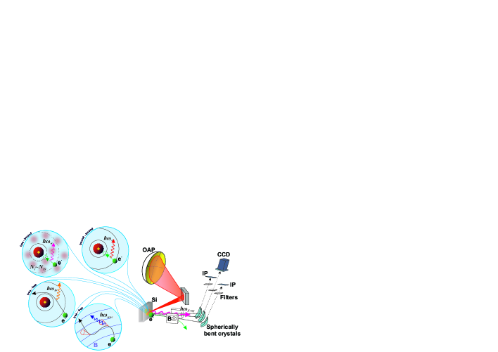
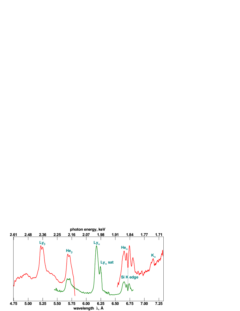
X-ray emission from a front side of the target was recorded using three focusing spectrometers with spatial resolution (FSSR) [12] at angles close to the target surface normal. A magnet was installed between the target and FSSR crystals to help prevent fast electrons produced during irradiation reaching the crystals and causing fluorescence. Fujifilm BAS-TR image plates and an Andor CCD DX-434 (for only one of the spectrometers) were used as X-ray detectors. Their sensitive layers were protected and shielded from visible light by filter foils made of Be, Al and polypropylene of different thicknesses. Observation ranges of the spectrometers overlap to provide cross-calibration between the separate FSSRs enabled measurement of continuous and high-resolution spectra across photon wavelengths from 4.7 to 7.3 Å(1.7 to 2.6 keV). Raw spectra registered by the individual FSSR spectrometers are shown in Fig. 2(a).
All the data gathered with the FSSRs were corrected for distance from the LPP, filtering, crystal reflectivity and detector response functions. Numerical modelling [13], via ray tracing through spectrometer, used the actual diffraction profiles or crystallographic rocking curves of the spherically bent crystals installed in the FSSRs. They were calculated with XOP [14] software. Rocking curves for the crystals used in experiments and image plates sensitivity functions are presented in the Supplemental Document uploaded with this Pre-print. Transmission functions calculated using the Henke tables [15] for filter foils was also taken into account. A summary of the response functions for each spectrometer is shown in 2(b). After convolution of the registered spectra with them an initial plasma radiation spectrum was restored (Fig.2(c)). The most significant corrections were needed around the Si K-edge at 6.7135 Å[16]. This causes a decrease in reflectivity of the dispersive -quartz (SiO2) crystals in the wavelength region close to the Heα line. The detailed Heα spectral line structure and the overlap between the two FSSRs in this region enables accurate calibration of the spectrometers.
(a)
(b)

(c)
3 Discussion
3.1 Registered spectrum description
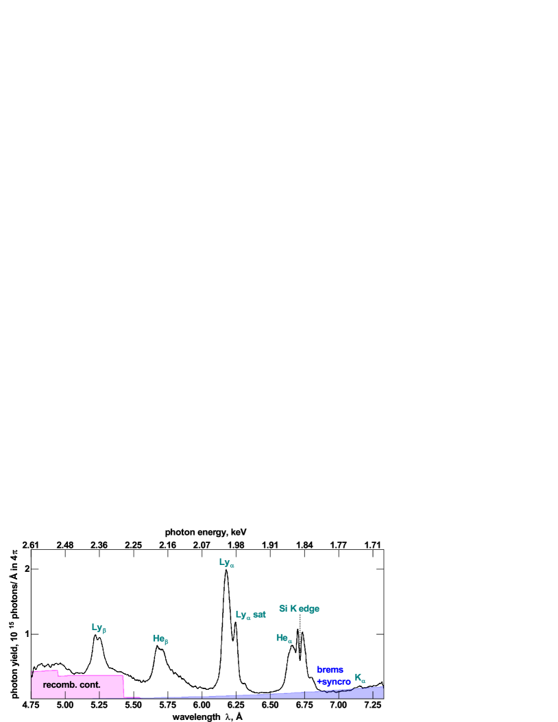
The spectrum shown in 2(c) contains both characteristic and continuous emission parts. Its shape is formed by all types of electron transitions: free-free, free-bound, bound-bound (1). Characteristic emission of the plasma ions is produced by transitions between energy states of H- and He-like Si ions. All of them are broadened due to strong Stark effect. The observed widths correspond to the electron density close to Si solid state value (N61023 cm-3). It was achieved both by extremely high value of the laser pulse temporal contrast of 10-10, this contrast was improved by a plasma mirror installed in a laser beam path. The most intense lines are the Lyα (2p1s transition in an H-like ion). About 21014 of photons (20% of the total number) were emitted in the narrow wavelength range from 6.05 to 6.35 Å (1.95–2.05 keV), which contains the line itself and its dielectronic satellites. This part of the plasma spectrum is the most suitable for implementation of quasi-monochromatic backlights schemes.
In the Heα region, from 6.53 to 6.89 Å (1.8–1.9 keV), the number of photons is smaller by approximately two: 1014. This spectral region contains a broad peak formed by overlapped Heα (1s2p 1P1s2 1S0 in He-like ion), intercombination line (1s2p 3P1s2 1S0) and dielectronic satellites produced by Li-like ions. In Lyβ (3p1s), and Heβ (1s3p 1P1s2 1S0) lines contain roughly 0.51014 and 0.71014 photons respectively. In comparison the Kα line at 7.2 Å is weak as most of the Si is highly ionised. The mentioned numbers of registered photons correspond to following values of conversion efficiency (CE) of laser energy to X-rays for Heα, Lyα, Lyβ, Heβ lines as 1.510-4, 3.210-4, 0.9410-4, 1.210-4 respectively.
In the short wavelength region of the spectrum there is a pedestal in intensity, this results from the recombination continuum produced by free-bound transitions of plasma electrons. This spectral region contains unresolved spectral lines corresponding to 4p1s transitions in H- and He-like ions. This is due to ionization potential depression [17] in the dense plasma. The shape of the recombination continuum calculated using the Hummer-Mihalas [18] model for He- and H-like ions in a solid-density Si plasma is shown in Fig. 2(c) by the magenta area. Approximately 31014 recombination continuum photons were registered between 4.75 and 5.4 Å. Simulations indicate that the recombination continuum extends to 2 Å. The conversion efficiency to this part of the spectrum is considerable and suggests that the recombination continuum produced by PW solid-density Si plasma is suitable for X-ray absorption spectroscopy in the range 5 Å, where the spectral lines are absent.
Other continuous emission results from free-free processes, this contributes to the spectrum through a monotonic growth in intensity in the long wavelength region of the spectrum. This is approximated by an exponential function shown by the blue line in Fig.2(c).
3.2 Modelling of the continuous emission.
There is no an obvious way to distinguish contributions of bremsstrahlung and synchrotron emission experimentally. It can be done analytically on the base of particle-in-cell (PIC) simulation results. There are several PIC codes that can self-consistently describe both synchrotron radiation and bremsstrahlung - EPOCH [19], OSIRIS [20], CALDER [21], PICLS [22] and some others [23, 24]. These codes were used in a variety of works [25, 26, 27, 28, 29] for theoretical investigation of high intensity (from 1019 to 1024 W/cm2) laser pulse energy conversion into continuous radiation. It was found that synchrotron emission dominates roughly at intensities exceeding 1022 W/cm2, but all these investigations are focused on a short laser pulse (30–120 fs).
We have performed two-dimensional (2D) PIC simulation via the EPOCH code for long 700 fs pulse. The code simulates bremsstrahlung radiation using a Monte Carlo method through the elastic and inelastic cross section for the electrons [28]. Synchrotron emission and radiation reaction are calculated in accordance with principles described in [30, 31]. All the modelling parameters were chosen close to experimental conditions: laser wavelength = 0.8 m, focal spot radius 4 mm, angle if incidence =45∘, intensity Ilas=31020 W/cm2. The laser pulse had Gaussian spatial and 3rd order super-Gauss temporal profile. A 2 m thick layer of fully ionized Si ions with solid-state density (51022 ions/cm-3) was used as a target. A long laser pulse requires a large simulation box to accurately describe the laser-target interaction and to accommodate the expanding plasma. In our simulations the box size was 120×120 m with 1010 nm grid. The simulation included a current smoothing algorithm and third order particle weighting to limit noise and numerical heating. All boundary conditions were absorbing for radiation and thermalizing for particles. The target was divided into 3 zones: Zone 1 with 50 ion macro-particles per cell (ppci) and 5014 electron-macro-particles per cell (ppcel), zone 2 with 25 ppci 2514 ppcel, and zone 3 with 10 ppci , 1014 ppcel. This allows efficient use of computational time without loss in accuracy. As a result, a temporally integrated angular distribution of bremsstrahlung and synchrotron X-rays in the wavelength range observed in the experiment was obtained, this is shown in Fig. 3.
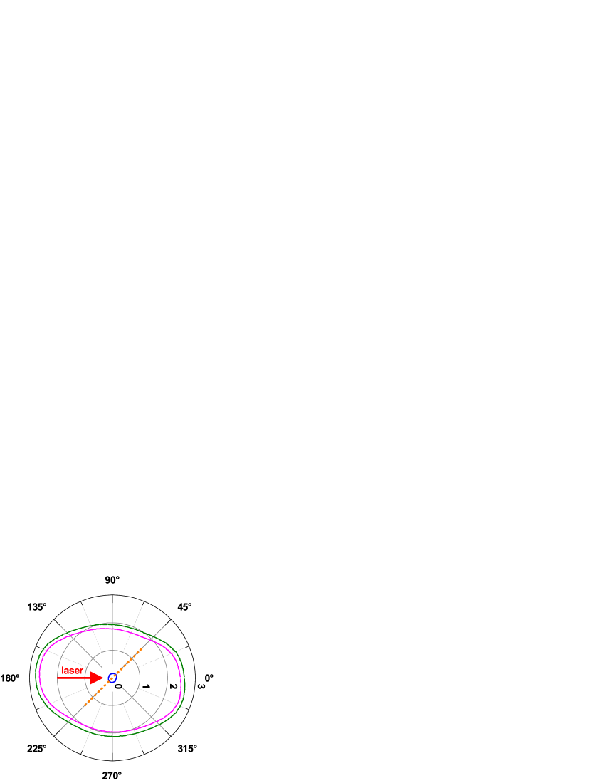
In summary the distribution is slightly elongated along the laser beam propagation axis. The ratio of maximum values for back (180∘) and transverse (90∘) directions is 0.8. For all directions the synchrotron radiation is significantly less intense than the bremsstrahlung. Thus 95% of 1.51014 photons associated with free-free emission corresponds to bremsstrahlung, i.e. integral over the blue curve in Fig.2(c). It contains about 63 mJ. Therefore, conversion efficiency of laser energy to the bremsstrahlung is 3.210-4, which is close to the value 510-4 predicted by the PIC simulation.
4 Conclusions
The laser plasma produced by sub-PW (1 ps, 200 J, 200 TW, with focal spot diameter 7 m, and on target intensity 31020 W/cm2) laser pulse in a 2 m thick Si foil is a very bright source of soft X-rays. We recorded about 1015 photons emitted by plasma in the wavelength range of 4.75–7.3 Å (1.7–2.6 keV) with total energy 0.33 J and a CE of about 1.510-3. About 40% of this energy were emitted in Si XIV (Si13+) Lyα, Lyβ and Si XIII (Si12+) Heα and Heβ resonance spectral lines and associated satellites. Lyα line is the most intense spectral line containing about a half of all emitted photons. This makes this line the best choice for quasi-monochromatic X-ray backlighter imaging and is sufficiently bright for use in appoint-project Bragg crystal imaging system.
About 170 mJ (CE8.410-4) of incident laser energy was re-emitted in continuous spectrum, of this approximately 64 mJ (CE 3.210-4) of it corresponds to bremsstrahlung radiation. The rest is associated with recombination continuum emission. The experimentally obtained laser-to-bremsstrahlung CE is close to 510-4 predicted by the EPOCH PIC code. The simulations are in qualitative and quantitative agreement with experimental results. The recombination continuum is sufficiently bright and featureless for use as an absorption spectroscopy source.
Acknowledgments The authors would like to thank the Central Laser Facility staff, whose dedication and expertise were essential to the success of their experiment. Calculations were carried out on the computational resources of the JSCC RAS. The reported study was funded by RFBR, project number 19-32-60050. This study was done in the frame of the State Assignment to JIHT RAS (topic #075-00892-20-00). The work of UK team received financial support from UK EPSRC grants EP/P026796/1, EP/L01663X/1 and EP/H012605/1.
Disclosures The authors declare no conflicts of interest.
References
- [1] C. Kleine, M. Ekimova, G. Goldsztejn, S. Raabe, C. Strüber, J. Ludwig, S. Yarlagadda, S. Eisebitt, M. J. J. Vrakking, T. Elsaesser, E. T. J. Nibbering, and A. Rouzée, “Soft X-ray Absorption Spectroscopy of Aqueous Solutions Using a Table-Top Femtosecond Soft X-ray Source,” \JournalTitleThe Journal of Physical Chemistry Letters 10, 52–58 (2019).
- [2] S. F. Khan, D. A. Martinez, D. H. Kalantar, R. K. Kirkwood, C. Santos, N. A. Ose, S. Johnson, D. A. Alessi, M. A. Prantil, D. T. Woods, S. G. Glendinning, R. Tommasini, A. J. Mackinnon, S. T. Prisbrey, T. R. Dittrich, M. W. Bowers, J. Cabral, J. Crane, J. M. Di Nicola, M. Hamamoto, S. Herriot, T. Lanier, R. Lowe-Webb, L. J. Pelz, C. C. Widmayer, W. Williams, and S. Yang, “A dual high-energy radiography platform with 15 m resolution at the National Ignition Facility,” \JournalTitleReview of Scientific Instruments 92, 043712 (2021).
- [3] A. Casner, T. Caillaud, S. Darbon, A. Duval, I. Thfouin, J. Jadaud, J. LeBreton, C. Reverdin, B. Rosse, R. Rosch, N. Blanchot, B. Villette, R. Wrobel, and J. Miquel, “LMJ/PETAL laser facility: Overview and opportunities for laboratory astrophysics,” \JournalTitleHigh Energy Density Physics 17, 2–11 (2015).
- [4] Opportunities in Intense Ultrafast Lasers (National Academies Press, Washington, D.C., 2018).
- [5] T. C. Sangster, R. Betti, R. S. Craxton, J. A. Delettrez, D. H. Edgell, L. M. Elasky, V. Y. Glebov, V. N. Goncharov, D. R. Harding, D. Jacobs-Perkins, R. Janezic, R. L. Keck, J. P. Knauer, S. J. Loucks, L. D. Lund, F. J. Marshall, R. L. McCrory, P. W. McKenty, D. D. Meyerhofer, P. B. Radha, S. P. Regan, W. Seka, W. T. Shmayda, S. Skupsky, V. A. Smalyuk, J. M. Soures, C. Stoeckl, B. Yaakobi, J. A. Frenje, C. K. Li, R. D. Petrasso, F. H. Séguin, J. D. Moody, J. A. Atherton, B. D. MacGowan, J. D. Kilkenny, T. P. Bernat, and D. S. Montgomery, “Cryogenic DT and D2 targets for inertial confinement fusion,” \JournalTitlePhysics of Plasmas 14, 058101 (2007).
- [6] C. Stoeckl, M. Bedzyk, G. Brent, R. Epstein, G. Fiksel, D. Guy, V. N. Goncharov, S. X. Hu, S. Ingraham, D. W. Jacobs-Perkins, R. K. Jungquist, F. J. Marshall, C. Mileham, P. M. Nilson, T. C. Sangster, M. J. Shoup, and W. Theobald, “Soft x-ray backlighting of cryogenic implosions using a narrowband crystal imaging system (invited),” \JournalTitleReview of Scientific Instruments 85, 11E501 (2014).
- [7] C. McGuffey, M. Dozières, J. Kim, A. Savin, J. Park, J. Emig, C. Brabetz, L. Carlson, R. F. Heeter, H. S. McLean, J. Moody, M. B. Schneider, M. S. Wei, and F. N. Beg, “Soft X-ray backlighter source driven by a short-pulse laser for pump-probe characterization of warm dense matter,” \JournalTitleReview of Scientific Instruments 89, 10F122 (2018).
- [8] A. S. Martynenko, S. A. Pikuz, I. Y. Skobelev, S. N. Ryazantsev, C. D. Baird, N. Booth, L. N. K. Döhl, P. Durey, A. Y. Faenov, D. Farley, R. Kodama, K. Lancaster, P. McKenna, C. D. Murphy, C. Spindloe, T. A. Pikuz, and N. Woolsey, “Optimization of a laser plasma-based x-ray source according to WDM absorption spectroscopy requirements,” \JournalTitleMatter and Radiation at Extremes 6, 014405 (2021).
- [9] B. Loupias, F. Perez, A. Benuzzi-Mounaix, N. Ozaki, M. Rabec, L. Gloahec, T. Pikuz, A. Faenov, Y. Aglitskiy, and M. Koenig, “Highly efficient, easily spectrally tunable X-ray backlighting for the study of extreme matter states,” \JournalTitleLaser and Particle Beams 27, 601–609 (2009).
- [10] Y. Li, J. Feng, J. Tan, J. Wang, D. Li, K. Dong, X. Zhang, B. Zhu, F. Tan, Y. Wu, Y. Gu, and L. Chen, “Electron beam and betatron x-ray generation in a hybrid electron accelerator driven by high intensity picosecond laser pulses,” \JournalTitleHigh Energy Density Physics 37, 100859 (2020).
- [11] C. Danson, P. Brummitt, R. Clarke, J. Collier, B. Fell, A. Frackiewicz, S. Hancock, S. Hawkes, C. Hernandez-Gomez, P. Holligan, M. Hutchinson, A. Kidd, W. Lester, I. Musgrave, D. Neely, D. Neville, P. Norreys, D. Pepler, C. Reason, W. Shaikh, T. Winstone, R. Wyatt, and B. Wyborn, “Vulcan Petawatt—an ultra-high-intensity interaction facility,” \JournalTitleNuclear Fusion 44, S239–S246 (2004).
- [12] A. Y. Faenov, S. A. Pikuz, A. I. Erko, B. A. Bryunetkin, V. M. Dyakin, G. V. Ivanenkov, A. R. Mingaleev, T. A. Pikuz, V. M. Romanova, and T. A. Shelkovenko, “High-Performance X-Ray Spectroscopic Devices for Plasma Microsources Investigations,” \JournalTitlePhysica Scripta 50, 333–338 (1994).
- [13] Y. S. Lavrinenko, I. V. Morozov, S. A. Pikuz, and I. Y. Skobelev, “Reflectivity and imaging capabilities of spherically bent crystals studied by ray-tracing simulations,” \JournalTitleJournal of Physics: Conference Series 653, 12027 (2015).
- [14] M. Sánchez del Río and R. J. Dejus, “XOP v2.4: recent developments of the x-ray optics software toolkit,” in Proc. SPIE, vol. 8141 M. Sanchez del Rio and O. Chubar, eds. (2011), p. 814115.
- [15] B. Henke, E. Gullikson, and J. Davis, “X-Ray Interactions: Photoabsorption, Scattering, Transmission, and Reflection at E = 50-30,000 eV, Z = 1-92,” \JournalTitleAtomic Data and Nuclear Data Tables 54, 181–342 (1993).
- [16] D. Li, G. Bancroft, M. Fleet, and X. Feng, “Silicon K-edge XANES spectra of silicate minerals,” \JournalTitlePhysics and Chemistry of Minerals 22, 115–122 (1995).
- [17] D. J. Hoarty, P. Allan, S. F. James, C. R. D. Brown, L. M. R. Hobbs, M. P. Hill, J. W. O. Harris, J. Morton, M. G. Brookes, R. Shepherd, J. Dunn, H. Chen, E. Von Marley, P. Beiersdorfer, H. K. Chung, R. W. Lee, G. Brown, and J. Emig, “Observations of the Effect of Ionization-Potential Depression in Hot Dense Plasma,” \JournalTitlePhysical Review Letters 110, 265003 (2013).
- [18] D. G. Hummer and D. Mihalas, “The equation of state for stellar envelopes. I - an occupation probability formalism for the truncation of internal partition functions,” \JournalTitleThe Astrophysical Journal 331, 794 (1988).
- [19] T. D. Arber, K. Bennett, C. S. Brady, A. Lawrence-Douglas, M. G. Ramsay, N. J. Sircombe, P. Gillies, R. G. Evans, H. Schmitz, A. R. Bell, and C. P. Ridgers, “Contemporary particle-in-cell approach to laser-plasma modelling,” \JournalTitlePlasma Physics and Controlled Fusion 57, 113001 (2015).
- [20] R. A. Fonseca, S. F. Martins, L. O. Silva, J. W. Tonge, F. S. Tsung, and W. B. Mori, “One-to-one direct modeling of experiments and astrophysical scenarios: pushing the envelope on kinetic plasma simulations,” \JournalTitlePlasma Physics and Controlled Fusion 50, 124034 (2008).
- [21] E. Lefebvre, N. Cochet, S. Fritzler, V. Malka, M.-M. Al onard, J.-F. Chemin, S. Darbon, L. Disdier, J. Faure, A. Fedotoff, O. Landoas, G. Malka, V. M ot, P. Morel, M. R. L. Gloahec, A. Rouyer, C. Rubbelynck, V. Tikhonchuk, R. Wrobel, P. Audebert, and C. Rousseaux, “Electron and photon production from relativistic laser–plasma interactions,” \JournalTitleNuclear Fusion 43, 629–633 (2003).
- [22] R. Mishra, P. Leblanc, Y. Sentoku, M. S. Wei, and F. N. Beg, “Collisional particle-in-cell modeling for energy transport accompanied by atomic processes in dense plasmas,” \JournalTitlePhysics of Plasmas 20, 072704 (2013).
- [23] F. Wan, C. Lv, M. Jia, H. Sang, and B. Xie, “Photon emission by bremsstrahlung and nonlinear Compton scattering in the interaction of ultraintense laser with plasmas,” \JournalTitleThe European Physical Journal D 71, 236 (2017).
- [24] D. Wu, X. T. He, W. Yu, and S. Fritzsche, “Particle-in-cell simulations of laser–plasma interactions at solid densities and relativistic intensities: the role of atomic processes,” \JournalTitleHigh Power Laser Science and Engineering 6, e50 (2018).
- [25] C. S. Brady, C. P. Ridgers, T. D. Arber, and A. R. Bell, “Synchrotron radiation, pair production, and longitudinal electron motion during 10-100 PW laser solid interactions,” \JournalTitlePhysics of Plasmas 21, 033108 (2014).
- [26] E. N. Nerush, I. Y. Kostyukov, L. Ji, and A. Pukhov, “Gamma-ray generation in ultrahigh-intensity laser-foil interactions,” \JournalTitlePhysics of Plasmas 21, 013109 (2014).
- [27] H. X. Chang, B. Qiao, Y. X. Zhang, Z. Xu, W. P. Yao, C. T. Zhou, and X. T. He, “Ultraintense laser absorption and -ray synchrotron radiation in near critical density plasmas,” \JournalTitlePhysics of Plasmas 24, 043111 (2017).
- [28] J. Vyskočil, O. Klimo, and S. Weber, “Simulations of bremsstrahlung emission in ultra-intense laser interactions with foil targets,” \JournalTitlePlasma Physics and Controlled Fusion 60, 054013 (2018).
- [29] J. Vyskočil, E. Gelfer, and O. Klimo, “Inverse Compton scattering from solid targets irradiated by ultra-short laser pulses in the 10 22 –10 23 W/cm 2 regime,” \JournalTitlePlasma Physics and Controlled Fusion 62, 064002 (2020).
- [30] R. Duclous, J. G. Kirk, and A. R. Bell, “Monte Carlo calculations of pair production in high-intensity laser–plasma interactions,” \JournalTitlePlasma Physics and Controlled Fusion 53, 015009 (2011).
- [31] N. V. Elkina, A. M. Fedotov, I. Y. Kostyukov, M. V. Legkov, N. B. Narozhny, E. N. Nerush, and H. Ruhl, “QED cascades induced by circularly polarized laser fields,” \JournalTitlePhysical Review Special Topics - Accelerators and Beams 14, 054401 (2011).