Characterization measurements of the TRISTAN multi-pixel silicon drift detector
Abstract
Sterile neutrinos are a minimal extension of the Standard Model of Particle Physics. A laboratory-based approach to search for this particle is via tritium \textbeta-decay, where a sterile neutrino would cause a kink-like spectral distortion. The Karlsruhe Tritium Neutrino (KATRIN) experiment extended by a multi-pixel Silicon Drift Detector system has the potential to reach an unprecedented sensitivity to the keV-scale sterile neutrino in a lab-based experiment. The new detector system combines good spectroscopic performance with a high rate capability. In this work, we report about the characterization of charge-sharing between pixels and the commissioning of a 47-pixel prototype detector in a MAC-E filter.
1 Introduction
Sterile neutrinos are a minimal extension of the Standard Model of particle physics, in which one or more additional neutrino mass eigenstates are introduced. The new mass eigenstate couples to the well-known active neutrino flavor eigenstates only via a small mixing. Depending on the mass of the new eigenstate , there are several motivations for the existence of sterile neutrinos. In case is in the range, sterile neutrinos are a viable dark matter candidate [1].
The kinematics of \textbeta-decay can be used to probe the neutrino mass eigenstates in a laboratory experiment. The Karlsruhe Tritium Neutrino (KATRIN) experiment makes use of this approach and determines the mass of the active neutrinos via a precision measurement of the endpoint region of the electron spectrum from tritium \textbeta-decay [2, 3]. A sterile mass eigenstate in the keV-regime would lead to a kink-like signature at the energy in the spectrum, where is the endpoint of the electron spectrum [4]. To detect such a signature, the spectral energy range measured by KATRIN must be extended from the endpoint region to the entire spectrum. This requires a new detector system for \textbeta-spectroscopy, which is currently being developed within the TRISTAN project. The new detector system needs to have an electron rate capability of cps. At the same time, excellent spectroscopic properties, like energy resolution and linearity, are required. To achieve these goals, we use the silicon drift detector (SDD) technology which allows an energy resolution of FWHM at at high rates of per pixel. The detector system will be implemented as a multi-pixel focal plane array covering an area of about with 3486 pixels of each. The development of the detector system follows a staged approach. Starting with 7-pixel detector prototypes with a simple mechanical design, the detector chip was scaled to a more complex module consisting of 47 pixels, which has already been tested successfully. The final focal plane array will consist of 9 (phase-1) and 21 (phase-2) detector modules of 166-pixels each.
2 Detector and readout scheme
The TRISTAN detector follows the general design idea of SDDs used for X-ray spectroscopy [5]. Each detector chip is made of up to 166 seamless hexagonal pixels with diameter each. The anode of every pixel is read out by a charge sensitive amplifier with a JFET transistor integrated into the anode structure of the chip, followed by a low-noise application-specific integrated circuit (ASIC) specifically developed for the TRISTAN project [6]. The integrated JFET allows one to place the ASIC chip at several distance to the detector chip, while keeping the total anode capacity at only . This provides an excellent signal to noise ratio and enables the operation at room temperature. Cooling the detector up to can further improve its performance. Fig. 1 shows two detector setups: A 7-pixel detector in a planar electronics board configuration was used for X-ray charge sharing measurements. For electron characterization measurements, a mechanically more complex 47-pixel detector, following the design of a final TRISTAN detector module, was used. In this design, all parts of the module are arranged behind the detector chip (area of ), so that 21 modules can be lined up with minimal distance to build a focal plane array. The 47-pixel detector module is an intermediate step toward the 166-pixel module.
3 Characterization of charge sharing
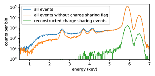
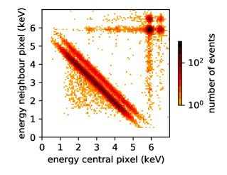
Due to the seamless and monolithic arrangement of the pixels, the charge cloud of an event at the pixel edge will be split between two adjacent pixels. If each pixel is read out independently, this adds a low-energy tail to the detector response, as part of the full charge cloud of an event is collected by the neighbouring pixel. To quantify this effect, events in a calibration measurement with an X-ray source were acquired with a 7-pixel TRISTAN detector at room temperature. As the central pixel is fully surrounded by six neighboring pixels, the time coincidence with the neighbours can be used to identify charge sharing events. A coincidence window with a duration of was chosen. Fig. 2 shows the recorded energy spectrum of the central pixel. By removing charge sharing-tagged events, the low-energy tail in the energy spectrum is reduced significantly. The energy distribution of both events causing the time coincidence is shown in Fig. 3. The energies detected with the central pixel and the neighboring pixels add up to the full X-ray energy. By adding both energies, the spectrum can be reconstructed. In this particular measurement, about of the events in the central pixel show charge sharing. Using the pixel geometry and the observed energy threshold of , this can be related to a Gaussian charge cloud with .
4 Module characterization with electrons
The 47-pixel TRISTAN detector module was tested at the monitor spectrometer beamline of the KATRIN experiment. Similar to the main beamline of KATRIN, an electron source is combined with a kinetic energy filter (MAC-E filter) followed by the detector. The electron source is a condensed, radioactive source providing several monoenergetic electron lines from internal conversion [7]. The source is set to a negative potential of , which adds to the kinetic energy of the electrons. In the measurement, the MAC-E filter was set to reject all electrons below the L3-32 line at , yielding a monoenergetic line from L3-32 conversion at and a superposition of L-32 and MN-32 conversion lines at around . For data acquisition we used the Kerberos system, a 48-channel analog pulse processing and data acquisition platform [8]. The acquired spectra are shown in Fig. 4. The detector was cooled to around . All pixels show a similar performance and detector response. The rate on the detector varied between due to the magnetic field configuration at the beamline. The L3-32 line is used to determine the energy resolution which is homogeneously distributed between FWHM over the entire detector chip.
5 Conclusion
Measurements of a 7-pixel TRISTAN detector prototype were analyzed regarding the charge sharing effect of neighboring pixels. Time coincidence could be used to obtain the energy distribution of charge sharing events. Furthermore, a mechanically more complex 47-pixel TRISTAN module showed the excellent performance of the SDD technology for high resolution \textbeta-spectroscopy in a realistic environment. The obtained energy resolution is better than FWHM for electrons on all read-out pixels. This new detector system, planned to be scaled to a pixel focal plane array, will enable the search for keV-scale sterile neutrinos at the KATRIN experiment.
References
- [1] A. Boyarsky, M. Drewes, T. Lasserre, S. Mertens and O. Ruchayskiy, Sterile neutrino dark matter, Progress in Particle and Nuclear Physics 104 (2019) 1.
- [2] M. Aker, K. Altenmüller, M. Arenz, M. Babutzka, J. Barrett, S. Bauer et al., Improved upper limit on the neutrino mass from a direct kinematic method by katrin, Physical Review Letters 123 (2019) 221802.
- [3] M. Aker, K. Altenmüller, J. F. Amsbaugh, M. Arenz, M. Babutzka, J. Bast et al., The design, construction, and commissioning of the katrin experiment, Journal of Instrumentation 16 (2021) T08015.
- [4] S. Mertens, A. Alborini, K. Altenmüller, T. Bode, L. Bombelli, T. Brunst et al., A novel detector system for katrin to search for kev-scale sterile neutrinos, Journal of Instrumentation 46 (2019) 065203.
- [5] P. Lechner, C. Fiorini, R. Hartmann, J. Kemmer, N. Krause, P. Leutenegger et al., Silicon drift detectors for high count rate x-ray spectroscopy at room temperature, Nuclear Instruments and Methods in Physics Research Section A: Accelerators, Spectrometers, Detectors and Associated Equipment 458 (2001) 281.
- [6] P. Trigilio, L. Bombelli, M. Carminati, R. Bisognin, A. Grande, M. Gugiatti et al., Ettore: a 12-channel front-end asic for sdds with integrated jfet, in 2018 IEEE Nuclear Science Symposium and Medical Imaging Conference Proceedings (NSS/MIC), pp. 1–4, IEEE, 10/11/2018 - 17/11/2018, DOI.
- [7] D. Vénos, J. Sentkerestiová, O. Dragoun, M. Slezák, M. Ryšavý and A. Špalek, Properties of 83 mkr conversion electrons and their use in the katrin experiment, Journal of Instrumentation 13 (2018) T02012.
- [8] P. King, M. Gugiatti, M. Carminati, L. Buonanno, G. Borghi, G. Pepponi et al., Design and characterization of kerberos: a 48-channel analog pulse processing and data acquisition platform, Journal of Instrumentation 16 (2021) T07007.