Radioactive nuclei for PET and theranostics:
selected candidates
Abstract
PET is an established medical diagnostic imaging method. Continuous improvements are aimed at refining image reconstruction, reducing the amount of radioactive tracer and combining with targeted therapy. TOF-PET provides the localization of the tracer through improved time resolution, nuclear physics may contribute to this goal via selection of radioactive nuclei emitting additional -rays. This additional radiation, when properly detected, localizes the decay of the tracer at the line of response determined by two detected 511 keV quanta. Selected candidates are presented. Some are particularly interesting, as they are strong candidates for theranostic applications.
Nowadays, it is obvious that society largely benefits from
the large investments done in basic Nuclear Physics research.
Recent achievements in particle- and radio-therapy within
the new paradigm of theranostic approach are some of
the most striking examples of the benefits
from Nuclear Physics. [1]
1 Introduction
Positron Emission Tomography (PET) is nowadays a standard medical diagnostic imaging technique. The average range of a positron from the decay of a radionuclide (tracer) is of the order of mm in the tissue. The spatial range of positrons following decay in water depends on the emission spectrum and is well described by the sum of two exponents [2]. The compounds with radioactive nuclei are predominantly used (above 90% of performed scans), but other tracers (containing or ) are found to have superior imaging properties [3] in certain cases. For the FWHM of the distribution is 0.102 mm [2]; the range is below 2.4 mm [4]. The annihilation of the positron with the electron creates usually two gamma quanta of 511 keV energy, emitted back-to-back, thus conserving null momentum of annihilating system. The mean free path of 511 keV photons in water is about 10.4 cm (computed from [5]). Detection of both 511 keV photons in coincidence in scintillation crystals enables the possibility to obtain the Line of Response (LoR) between the geometrical positions of fired detectors. The intersection of multiple LoRs provides, in the first approximation, the location of the tracer.
This standard procedure augmented by additional corrections due to attenuation and scattering of quanta) does not account for the position of the annihilation along LoR. This information is, in principle, stored in the time difference between 511 keV quanta interaction in detectors. Time-resolving power of detectors and associated electronic circuits are the key issue in this approach, as 3 cm difference along LoR corresponds to 200 ps. Important progress has been observed in this technology, as the time resolution of commercial scanners improved from 1000 ps two decades ago, to 550 ps a decade ago and 220 ps in currently available devices (four-fold increase of the axial dimension should also be noted). The Time-of-Flight (TOF) PET scanner, instead of full LoR, employs the concept of segment of response, determined by the time bin. The improvement of time-resolving power leads to the reduction of the background and allows the possibility to reduce the dose of tracer, important in several cases [6]. Coincidence resolving time in 10 ps range would lead to enormous improvement, allowing for on-line image reconstruction. The roadmap to achieve this goal has been recently published [7].
The decay vertex (lying close to the LoR) might be determined from the radioactive () decay of the daughter nucleus, provided the lifetime is short enough to stay within a coincidence window. The “third” photon should be detected via Compton scattering following the photoelectric absorption of the scattered quantum. This method provides localization of the decay vertex through the intersection of the reconstructed Compton cone with LoR on an event-by-event basis. The “third” photon should be around 1 MeV energy to increase the probability for Compton effect in detector material. Improved localisation of the vertex in this technique leads to the reduction of the global radioactive dose administered for the diagnosis, although some dose would come from this “third” (and additional) gamma.
2 Candidates for radionuclides in PET
The PET technique has been under development for more than two decades [8]. The approach seems to be more effective compared to the 3 decay of ortho-positronium, as this process is rare (typically below 1%) and requires more sophisticated treatment of the data. Suitable candidates for radionuclides to be used in PET should fulfil several conditions:
-
1.
Suitable lifetime (long enough for diagnostic procedure and short enough to reduce unwanted irradiation of the body after the medical procedure),
-
2.
High branching ratio (electron capture as the natural concurring process),
-
3.
Energy of the radiation favouring Compton scattering (i.e. MeV),
-
4.
Multiplicity of other -rays should in principle be low.
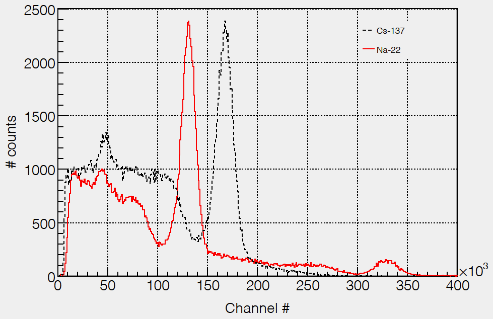
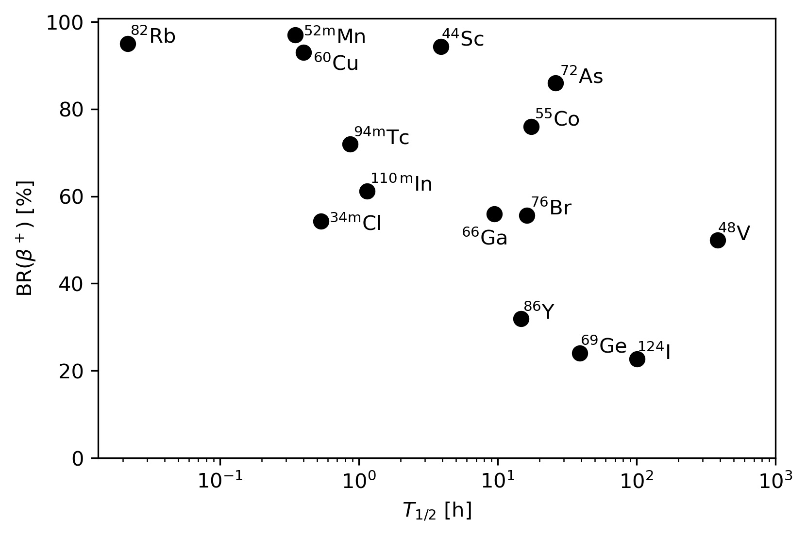
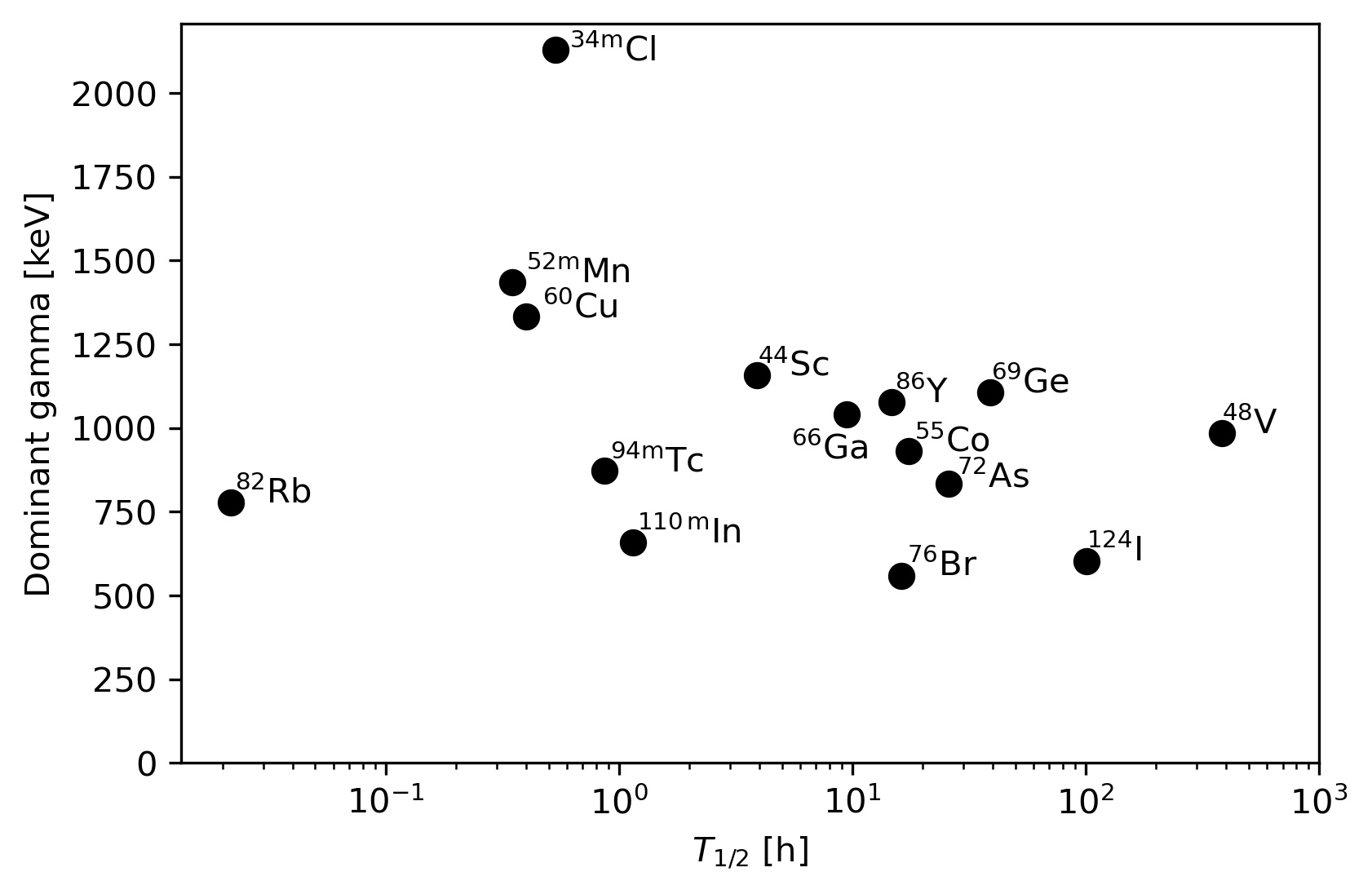
The candidates for those radionuclides have been recently reviewed by M. Sitarz et al. [8]. The basic properties of those nuclei are visualized in Figures 1 and 3. Their lifetime is in the order of hours. The branching ratio of considered nuclei (see Fig.1) makes them prospective candidates. Several of them have branching ratios well above 80%, so almost all radioactive decays could contribute to the emission, which is essential to the PET technique. The energy of the radiation emitted after the decay from the excited state of the daughter nuclei is an important factor for the envisaged technique. The lifetime of the excited state should allow for coincident measurement, so lifetimes in the range of ps are needed. In fact, this is the case of all considered nuclei. The -ray should be Compton-scattered, so MeV -rays are best suited. The energy spectra (Fig.2) for 22Na and 137Cs calibration sources registered by LYSO detector (typical for PET devices) shows how pronounced is the rise of Compton-scattering events when the -ray energy increases from 511 keV (annihilation of after the decay of 22Na) to 662 keV (after 137Cs decay). The energy of the most abundant -ray emitted after the decay of considered candidates is in the region of 1 MeV (see Fig.3).
The branching ratio of decay is the key factor for the standard PET operation method (almost 100% for18F). For the PET, the product of branching ratio and the fraction of so-called ”third” emission in the decay of the daughter nucleus, determines the efficiency of the process with respect to the dose of the tracer. Evidently (see Fig.4), 44Sc, 52mMn and 60Cu are three most suitable candidates.
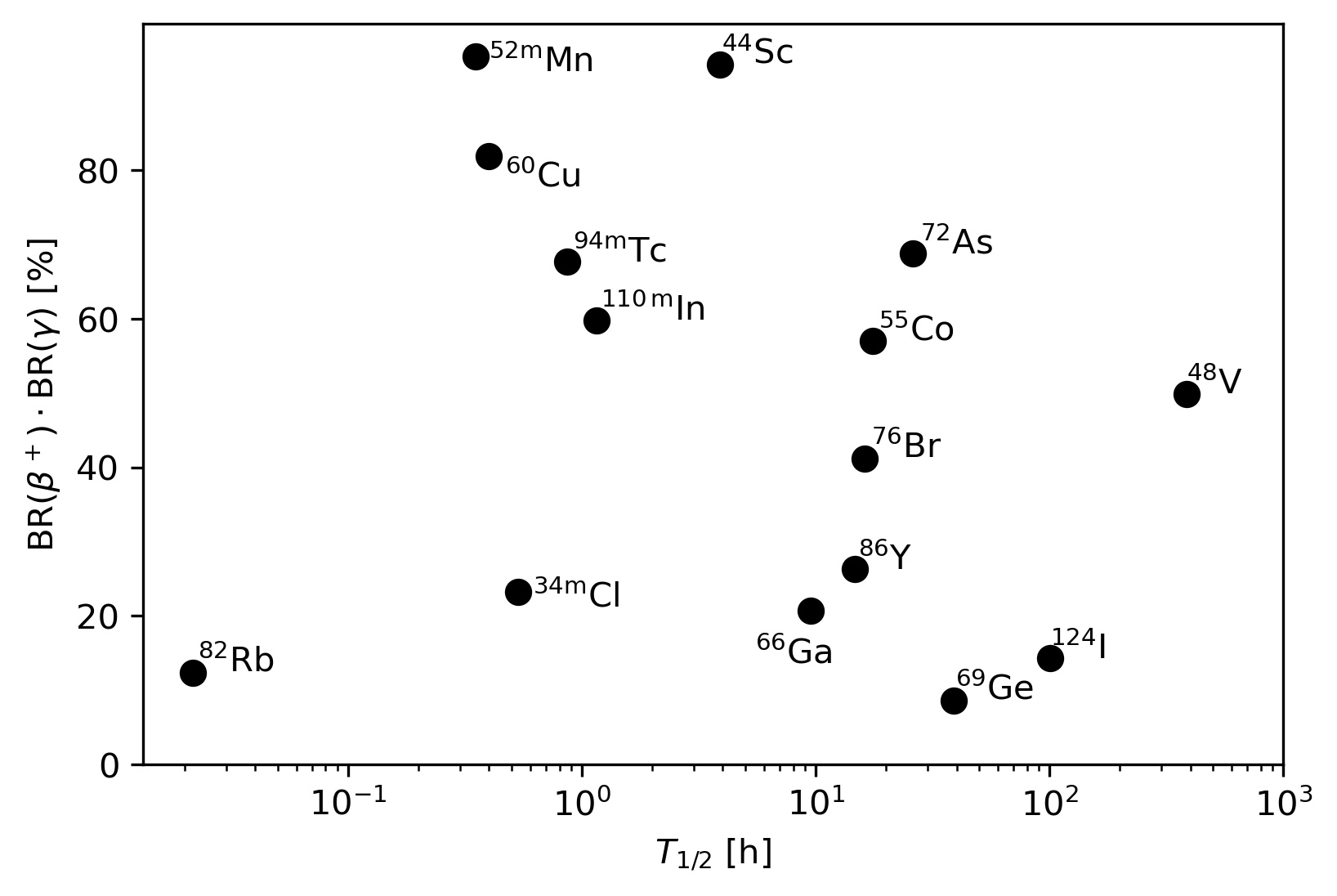
The three conditions mentioned above do not allow selection of the best nuclei for this technology. Apart from making suitable radio-pharmaceutic (chemistry), key issues are other properties of the decay and the method of producing the resulting sample. A very important aspect is the purity of the sample - the amount of co-produced (usually unwanted) radionuclides. For example, production of 34mCl requires beam of 65 MeV kinetic energy, beyond the range available in typical medical accelerators. Also, the decay of 34mCl populates many other excited states decaying via energetic -rays, which complicates the analysis and generates unwanted radiation effects. So, in spite of the high energy of emitted ”third” -ray and branching ratio above 50%, the 34mCl isotope is not one of the most appropriate candidates.
The production of medically interesting radioisotopes might provide unwanted activities of the same chemical element, especially in the case of non-monoisotopic targets, thus impossible to separate chemically. Short lifetime of unwanted activity would make the sample clear after a time, but this is rarely the case. Selective reactions, employing targets enriched with the appropriate isotope, are commonly used and efficient methods that have been developed for re-use of the target material. The production cross section for different reaction channels have been measured intensively in the past decades and numerical tools have been developed to evaluate the expected yields [10]. However, not all production routes have been fully experimentally verified or optimized and experimental activity is welcome. The numerous commercially available accelerators (now around 1500 worldwide [11]) provide the natural place to supply radioisotopes for this prospective PET technique. Their maximum energy is usually limited to 20 MeV protons, so those among the perspective nuclei for PET, which can be produced with this beam (or appropriately scaled in energy deuterons and -particles) are naturally preferred. Among them are 44Sc, 48V (for slow metabolic processes), 55Co, 60Cu, 66Ga, 94mTc and 124I (unfortunately low branching ratio). Some others may be obtained via nuclear generators, like 82Rb supplied from 82Sr (T1/2=25.4 d). Several of those radionuclides were already used in diagnosis of certain diseases.
3 Nuclei for theranostic applications
The theranostic approach aims to combine diagnosis and therapy to provide effective treatment at the very early stage of cancer [13]. The role of Nuclear Physics is to develop specific radioisotopes, providing diagnostics functionality together with therapeutic effect, so this activity has gone beyond standard PET tracers. These requests can be met with single radionuclide for imaging and therapy (like 117mSn) or by pairs of isotopes having the same chemical properties (like 44Sc/47Sc, 64Cu/67Cu and others) or similar (like 99mTc/188Re [14]). Evidently, 44Sc seems to be a natural link between PET and theranostic approach. The 44Sc/47Sc pair is the subject of wide-front research. The production of these radionuclides is studied intensively by very different methods (reports from only 5 last years):
- 1.
-
2.
Neutron irradiation of natural calcium in reactor [17],
- 3.
-
4.
3He reactions at low energies [21] and
-
5.
Spallation reaction induced by GeV protons [22].
There is also important progress in chemistry and medical applications of this pair of nuclei. The production, chemistry and in-vivo studies of Sc-isotopes were reviewed few years ago [23, 24]. Currently, around 20 papers are published yearly on the subject of production and medical applications of this theranostic pair of scandium nuclei, so any review soon might become outdated.
The progress in the development of other important theranostic pairs is recently significant, in particular concerning radioactive Cu isotopes. The photonuclear production of 67Cu was recently found to be an efficient route [25]. In this case the final product is free of co-produced isotopes, populated otherwise in proton, deuteron and alpha-induced reactions on zinc targets. The use of highly-enriched targets limits the production of long-lived 64Cu, what was recently demonstrated [26] in the measurement of deuteron-induced reactions on 70Zn. This progress is particularly important, as the therapeutic application of 67Cu has recently been demonstrated [27] and found efficacious in certain prostate cancers.
4 Conclusions
Not only nuclear aspects would select the best radioisotope for PET, as many other properties (e.g. chemistry) and technical restrictions would play a role. Special medical requests and imaging properties are also of primary concern. The pure nuclear aspects favour 44Sc and 60Cu, as their production is also possible in commercial PET-supplying cyclotrons. However, many others have already been used in practical diagnosis procedures and deserve further studies. The progress in theranostic application of 44Sc/47Sc pair is intensively studied and different production routes were recently evaluated. Other dedicated combinations are also of interest, particularly as their therapeutic efficiency was demonstrated. The demanding detection conditions of process can be effectively realized in a total-body PET [28], where the large geometrical acceptance of the device would be the key factor for making fast measurements with low-dose.
Acknowledgments
I am grateful to dr. Mateusz Sitarz and the late professor Jerzy Jastrzȩbski for pointing to me their vision of medical applications of nuclear physics. The help of Joanna Matulewicz in making graphics is acknowledged.
References
- [1] NuPECC Long Range Plans 2017, http://www.nupecc.org/lrp2016/Documents/lrp2017.pdf
- [2] Levin CS, Hoffman EJ. Calculation of positron range and its effect on the fundamental limit of positron emission tomography system spatial resolution. Phys. Med. Biol. 1999 44:781
- [3] Sharma A, McConathy J. Overview of PET Tracers for Brain Tumor Imaging. PET Clinics 2013 8:129-146 DOI:j.cpet.2013.02.001
- [4] Huang YY. An overview of PET radiopharmaceuticals in clinical use: Regulatory Regulatory, Quality and Pharmacopeia Monographs of the United States and Europe. in: Nuclear Medicine Physics, publisher: IntechOpen 2018 DOI:10.5772/intechopen.79227
- [5] NIST Standard Reference Database 8, DOI:10.18434/T48G6X
- [6] Lois C, Jakoby BW, Long MJ, Hubner KF, Barker DW, Casey ME, et al. An assessment of the impact of incorporating time-of-flight information into clinical PET/CT imaging. J Nucl Med 2010 51(2):237-45. DOI:10.2967/jnumed.109.068098.
- [7] Lecoq P, Morel C, O Prior J, Visvikis D, Gundacker S, Auffray E, et al. Roadmap toward the 10 ps time-of-flight PET challenge. Phys. Med. Biol. 2020 65:21RM01 DOI:10.1088/1361-6560/ab9500
- [8] Sitarz M, Cussonneau JP, Matulewicz T, Haddad F. Radionuclide candidates for coincidence PET: An overview. Applied Radiation and Isotopes 2020 155:108898 DOI:10.1016/j.apradiso.2019.108898, and references therein
- [9] Sæterstøl J. Characterization of Scintillation Crystals for Positron Emission Tomography. PhD Thesis U. Bergen 2010 https://wiki.uib.no/
- [10] Sitarz M. Radionuclide Yield Calculator https://www.arronax-nantes.fr/en/outil-telechargement/tool-radionuclide-yield-calculator/
- [11] Goethals PE, Zimmermann RG. Cyclotrons used in Nuclear Medicine 2020th ed; 2020 http://medraysintell.com
- [12] Synowiecki MA, Perk LR, Nijsen JFW. Production of novel diagnostic radionuclides in small medical cyclotrons. EJNMMI Radiopharmacy and Chemistry 2018 3:3 DOI: 10.1186/s41181-018-0038-z
- [13] L.Królicki, this issue
- [14] Alberto R, Schibli R, Waibel R, Abram U, Schubiger AP. Basic aqueous chemistry of directed towards radiopharmaceutical application. Coordination Chemistry Reviews 1999 190-192:901 DOI:/10.1016/S0010-8545(99)00128-9
- [15] Kazakov AG, Ekatova TY, Babenya JS. Photonuclear production of medical radiometals: a review of experimental studies. Journal of Radioanalytical and Nuclear Chemistry 2021 328:493-505 DOI:10.1007/s10967-021-07683-2
- [16] Loveless CS, Radford LL, Ferran SJ, Queern SL, Shepherd MR, Lapi SE. Photonuclear production, chemistry, and in vitro evaluation of the theranostic radionuclide Sc-47. EJNMMI Research 2019 9:42 DOI:10.1186/s13550-019-0515-8
- [17] Gizawy MA, Mohamed NMA, Aydia MI, Soliman MA, Shamsel-Din HA. Feasibility study on production of Sc-47 from neutron irradiated Ca target for cancer theranostics applications. Radiochimica Acta 2020 108:207-215 DOI:10.1515/ract-2018-3070
- [18] Carzaniga TS, Auger M, Braccini S, Bunka M, Ereditato A, Nesteruk KP, et al. Measurement of Sc-43 and Sc-44 production cross-section with an 18 MeV medical PET cyclotron. Applied Radiation and Isotopes 2017 129:96-102 DOI:10.1016/j.apradiso.2017.08.013
- [19] Sitarz M, Szkliniarz K, Jastrzebski J, Choinski J, Guertin A, Haddad F, et al. Production of Sc medical radioisotopes with proton and deuteron beams. Applied Radiation and Isotopes 2018 142:104-112 DOI:10.1016/j.apradiso.2018.09.025
- [20] Carzaniga TS, Braccini S. Cross-section measurement of Sc-44m,Sc-47, Sc-48 and Ca-47 for an optimized Sc-47 production with an 18 MeV medical PET cyclotron. Applied Radiation and Isotopes 2019 143:18-23 DOI:10.1016/j.apradiso.2018.10.015
- [21] Szelecsenyi F, Kovacs Z, Nagatsu K, Zhang MR, Suzuki K. Production cross sections of radioisotopes from He-3-particle induced nuclear reactions on natural titanium. Applied Radiation and Isotopes 2017 119:94-100 DOI:10.1016/j.apradiso.2016.10.016
- [22] Peplowski PN. Cross sections for the production of radionuclides via Cu-nat(p,X) spallation reactions for proton energies from 250 MeV to 2 GeV. Nucl. Phys. 2020 A1006:122067 DOI:10.1016/j.nuclphysa.2020.122067
- [23] Muller C, Domnanich KA, Umbricht CA, van der Meulen NP. Scandium and terbium radionuclides for radiotheranostics: current state of development towards clinical application. British Journal of Radiology 2018 91:20180074 DOI:10.1259/bjr.20180074
- [24] Huclier-Markai S, Alliot C, Kerdjoudj R, Mougin-Degraef M, Chouin N, Haddad F. Promising Scandium Radionuclides for Nuclear Medicine: A Review on the Production and Chemistry up to In Vivo Proofs of Concept. Cancer Biotherapy and Radiopharmaceuticals 2018 33:316-329 DOI:10.1089/cbr.2018.2485
- [25] Hovhannisyan GH, Bakhshiyan TM, Dallakyan RK. Photonuclear production of the medical isotope 67Cu. Nuclear Instruments and Methods in Physics Research 2011 B498:48-51 DOI:10.1016/j.nimb.2021.04.016
- [26] Nigron E, Guertin A, Haddad F, Sounalet T. Is 70Zn(d,x)67Cu the Best Way to Produce 67Cu for Medical Applications? Frontiers in Medicine 2021 8:1059 DOI:10.3389/fmed.2021.674617
- [27] McInnes LE, Cullinane C, Roselt PD, Jackson S, Blyth BJ, van Dam EM, et al. Therapeutic Efficacy of a Bivalent Inhibitor of Prostate-Specific Membrane Antigen Labeled with 67Cu. Journal of Nuclear Medicine 2021 62:829-832 DOI:10.2967/jnumed.120.251579
- [28] Vandenberghe S, Moskal P, Karp JS. State of the art in total body PET. EJNMMI Physics 2020 7:35 DOI:10.1186/s40658-020-00290-2