capbtabboxtable[][\FBwidth]
\jmlrvolume– Under Review
\jmlryear2021
\jmlrworkshopFull Paper – MIDL 2021 submission
\midlauthor\NameEkta Gavas\midljointauthortextEqual Contribution \Emailekta.gavas@research.iiit.ac.in
\addrIIIT Hyderabad, India
and \NameKaustubh Olpadkar\midlotherjointauthor \Emailkolpadkar@cs.stonybrook.edu
\addrStony Brook University, NY, USA
Deep CNNs for Peripheral Blood Cell Classification
Abstract
The application of machine learning techniques to the medical domain is especially challenging due to the required level of precision and the incurrence of huge risks of minute errors. Employing these techniques to a more complex subdomain of hematological diagnosis seems quite promising, with automatic identification of blood cell types, which can help in detection of hematologic disorders. In this paper, we benchmark 27 popular deep convolutional neural network architectures on the microscopic peripheral blood cell images dataset. The dataset is publicly available, with large number of normal peripheral blood cells acquired using the CellaVision DM96 analyzer and identified by expert pathologists into eight different cell types. We fine-tune the state-of-the-art image classification models pre-trained on the ImageNet dataset for blood cell classification. We exploit data augmentation techniques during training to avoid overfitting and achieve generalization. An ensemble of the top performing models obtains significant improvements over past published works, achieving the state-of-the-art results with a classification accuracy of 99.51%. Our work provides empirical baselines and benchmarks on standard deep-learning architectures for microscopic peripheral blood cell recognition task.
keywords:
Blood Cell Classification, Transfer Learning, Medical Imaging, Convolutional Neural Networks, Deep Learning.1 Introduction
Blood carries oxygen and nutrients to living cells in different organs and tissues. It carries away the waste for detoxification. It transports hormones to the desired site of action to fight infections and regulates body temperature. The ability to classify blood constituents can be critical in assessing the patient’s health. Plasma, which constitutes 55% of blood, is a colored liquid comprising mainly water (about 90%) and other essential substances such as proteins (albumin, clotting factors, antibodies, enzymes, and hormones), glucose, and fats. Rest 45% of blood is composed of white blood cells (WBCs/leukocytes), platelets (thrombocytes), and red blood cells (RBCs/erythrocytes), which float in the plasma[Fathima and Syeda(2017), L.(2005)]. All these cells are associated with different functionalities. RBCs are responsible for transporting gases () from lungs to tissues and maintaining systemic acid/base equilibria. Damage of red cell integrity, defined as hemolysis, has been shown to significantly contribute to severe pathologies, including endothelial dysfunction[Kuhn et al.(2017)Kuhn, Diederich, Keller, Kramer, Lückstädt, Panknin, Suvorava, Isakson, Kelm, and Cortese-Krott].
Based on the presence of visible granules in the microscopic view, WBCs can be classified into two broad categories: granulocytes and agranulocytes (nongranulocytes). Neutrophils, eosinophils, and basophils belong to the granulocytes category, while lymphocytes and monocytes belong to the agranulocytes category [Almezhghwi and Serte(2020), Acevedo et al.(2019)Acevedo, Alférez, Merino, Puigví, and Rodellar]. Various types of WBCs play a role in immune response [L.(2005)] and act as a defense mechanism in the body against illness-causing agents. Immature granulocytes (IG) are under-developed WBCs released from the bone marrow into the blood. Except for blood from newborn children or pregnant women, the appearance of IG (promyelocytes, myelocytes, and metamyelocytes) [Acevedo et al.(2019)Acevedo, Alférez, Merino, Puigví, and Rodellar] in the peripheral blood (PB) indicates an early-stage response to infection, inflammation, or other stimuli of the bone marrow. Similarly, erythroblasts are nucleated immature RBCs or erythroid precursors not seen after the neonatal period. Their appearance in PB of children and adults can signify bone marrow damage, stress, malignant neoplasms, or other potentially serious diseases [Constantino and Cogionis(2000)]. Platelets are anucleated cells in blood and get activated at the site of injury to form a blood clot. Besides, they play an important role in innate immunity and regulation of tumor growth and extravasations in the vessel[Holinstat(2017)]. They make up less than 1% of blood volume. Usually, the typical percentages of neutrophils in the blood are 0-6%. Eosinophils constitute 1–3%, basophils 0–1%, lymphocytes 25–33%, and monocytes 3–10% of the leukocytes circulating in the blood[Acevedo et al.(2019)Acevedo, Alférez, Merino, Puigví, and Rodellar].
Recognition of various blood cell types can reveal anomalous blood cell populations like immature cells (IG or erythroblasts). On accurate identification, differential blood cell count can suggest any possible abnormalities in the blood, or help diagnose an infection, inflammation, leukemia[Shafique and Tehsin(2018), Mathur et al.(2013)Mathur, Tripathi, and Kuse], or any immune system disorder. Analyzing the blood cell morphology is the outset for the diagnosis of 80% of hematological diseases[Acevedo et al.(2019)Acevedo, Alférez, Merino, Puigví, and Rodellar]. Quantitative morphological analysis can thus, help cytologists assess blood samples and conclude about the patient’s blood conditions. However, the above processes are complex and time-consuming, involving a specialist meticulously examining the blood smear under a microscope, subjecting it to human errors. For the past few years, several attempts have been made to automate these processes using image processing techniques and machine learning, making them time and cost-effective and substantially reducing the workload in laboratories.
Convolutional neural networks (CNNs) are known to show excellent results on image recognition tasks, and hence, there has been an extensive research for applying them in the medical domain. In this paper, we explore various deep CNNs for blood cell classification task with peripheral blood cell (PBC) images dataset containing samples of eight different cell types: neutrophils, eosinophils, basophils, lymphocytes, monocytes, IG, erythroblasts, and platelets [Acevedo et al.(2020)Acevedo, Merino, Alférez, Ángel Molina, Boldú, and Rodellar]. Our work provides state-of-the-art results on the PBC classification task without the need of manual feature extraction or designing complex and hybrid architectures. The main contributions of this paper are as follows:
-
1.
Train and evaluate an end-to-end deep learning-based classification system to recognize eight different blood cell types in peripheral blood smear.
-
2.
Explore and benchmark 27 standard deep CNN architectures for blood cell classification using transfer learning.
-
3.
Exploit data augmentation and ensembling techniques to further improve the model performance. Our model achieves state-of-the-art performance over previously published works.
2 Related Work
Computer-aided PBC classification is a challenging problem, and it has been studied extensively for the last few decades. Its solution can be used to develop tools and software that can assist doctors and radiologists in examining and diagnosing many blood-related diseases. Early attempts at this include designing the handcrafted features by experts, which can be further used to perform classification with the pattern recognition algorithms. The application of morphological analysis, including but not limited to morphological operators for PBC segmentation and classification, is the research area that has been explored in great detail in [Kim et al.(2001)Kim, Jeon, Choi, Kim, and Ho, Di Ruberto et al.(2002)Di Ruberto, Dempster, Khan, and Jarra, Piuri and Scotti(2004), Scotti(2005), Dorini et al.(2007)Dorini, Minetto, and Leite, Theera-Umpon and Dhompongsa(2007), Taherisadr et al.(2013)Taherisadr, Nasirzonouzi, Baradaran, and Mehdizade, Lee and Chen(2014), Rodellar et al.(2018)Rodellar, Alférez, Acevedo, Molina, and Merino] before the deep learning-based approach became popular. There has been a significant increase in the studies on the deep learning-based WBC classification approaches [Su et al.(2014)Su, Cheng, and Wang, Othman et al.(2017)Othman, Mohammed, and Baban, Jiang et al.(2018)Jiang, Cheng, Qin, Du, and Zhang, Macawile et al.(2018)Macawile, Quiñones, Ballado, Cruz, and Caya, Throngnumchai et al.(2019)Throngnumchai, Lomvisai, Tantasirin, and Phasukkit, Sharma et al.(2019)Sharma, Bhave, and Janghel, Banik et al.(2019)Banik, Saha, and Kim, Shahin et al.(2019)Shahin, Guo, Amin, and Sharawi, Almezhghwi and Serte(2020), Baydilli and Atila(2020), Sahlol et al.(2020)Sahlol, Kollmannsberger, and Ewees] in the last decade, especially after the successful application of CNNs on other computer vision problems. The works [Saraswat and Arya(2014), Rawat et al.(2015)Rawat, Bhadauria, Singh, and Virmani, Al-Dulaimi et al.(2018)Al-Dulaimi, Banks, Chandran, Tomeo-Reyes, and Nguyen Thanh, Patodia et al.(2020)Patodia, Nibhasya, and Chandraprabha], provide a review of the state-of-the-art methods of leukocyte segmentation, feature extraction, and classification published in the last two decades. The authors identified scope for improvement in several aspects compared to the classical approach involving image preprocessing and manual feature extraction, which achieved low performance and required expertise for labeling and handcrafting features. The usage of deep learning based techniques in the medical field has become popular due to the development of more efficient algorithms [Kumar et al.(2016)Kumar, Kim, Lyndon, Fulham, and Feng, Ravì et al.(2016)Ravì, Wong, Deligianni, Berthelot, Andreu-Perez, Lo, and Yang, Cabitza and Banfi(2018)]. There has also been a wide array of research in the area of RBC segmentation and classification [Tomari et al.(2014)Tomari, Zakaria, Jamil, Nor, and Fuad, Tyas et al.(2017)Tyas, Ratnaningsih, Harjoko, and Hartati, Xu et al.(2017)Xu, Papageorgiou, Abidi, Dao, Zhao, and Karniadakis, Alzubaidi et al.(2020)Alzubaidi, Fadhel, Al-Shamma, Zhang, and Duan] for diagnosis of many blood-related diseases. There are many different PBC sub-types; however, most studies in this direction focus only on the major types like leukocyte or RBC. Recently, there are attempts at blood cell analysis, encapsulating all different blood cells classes with the same model [Acevedo et al.(2019)Acevedo, Alférez, Merino, Puigví, and Rodellar, Ucar(2020)], and it can be more useful, having a large number of applications. Our models outperform the previously published works by successfully applying various standard deep CNN architectures on the large dataset [Acevedo et al.(2020)Acevedo, Merino, Alférez, Ángel Molina, Boldú, and Rodellar] of microscopic images of peripheral blood cells.
3 Methodology
3.1 Overview
In this work, our primary goal is to develop a deep learning-based end-to-end system to perform PBC recognition. We apply transfer learning to fine-tune standard state-of-the-art deep CNN architectures pre-trained on the ImageNet dataset for the PBC classification task. This approach leverages the already learned features and fine-tunes the models to learn specialized features of microscopic blood cell images. After fine-tuning these models, we apply ensemble techniques to improve the system’s overall performance utilizing the knowledge of all trained models, wherein our voting-based ensemble outperforms all other models and previously published works.
3.2 CNN Architectures
Deep CNNs have been demonstrated to outperform all other traditional machine learning algorithms for a variety of computer vision tasks in the last decade. Some of the exciting CNN application areas include Image Classification and Segmentation, Face Recognition, Object Detection, Video Processing, Natural Language Processing, and Speech Recognition. The powerful learning ability of deep CNNs is primarily due to multiple feature extraction stages that can automatically learn representations from the data. The CNN architecture generally consists of a feature extractor - containing several convolutional and pooling layers followed by a classifier - containing one or more fully connected layers where the last layer has softmax activation for classification. Depending upon the type of architectural modifications, CNNs can be broadly divided into seven different categories: spatial exploitation, depth, multi-path, width, feature-map exploitation, channel boosting, and attention-based CNNs [Khan et al.(2020b)Khan, Sohail, Zahoora, and Qureshi]. We utilize 27 standard CNN architectures and analyze their performance for the blood cell classification task. The details about these models can be found in Appendix B. For our experiments, we use the Python implementation of these architectures available in the PyTorch model zoo.
3.3 Transfer learning
In machine learning, transfer learning (TL) is a technique where the knowledge gained solving one problem is applied to a different but related problem [Ventura and Warnick(2007)]. In the last few years, several transfer learning techniques have been developed and successfully applied using deep neural networks in domains like computer vision, reinforcement learning and natural language processing [Shao et al.(2015)Shao, Zhu, and Li, Weiss et al.(2016)Weiss, Khoshgoftaar, and Wang, Csurka(2017), Tan et al.(2018)Tan, Sun, Kong, Zhang, Yang, and Liu, Zhuang et al.(2021)Zhuang, Qi, Duan, Xi, Zhu, Zhu, Xiong, and He, Taylor and Stone(2009)]. We employ transfer learning to solve the blood cell classification problem by fine-tuning 27 different standard deep CNN architectures pre-trained on the ImageNet dataset. As the size of our dataset is not as huge as ImageNet, transfer learning is a suitable choice for our use-case. We use the feature extractor from the pre-trained models and pass the extracted features through a single layer fully connected classifier with eight output units. The fine-tuning of the feature extractor allows us to take advantage of the learned knowledge to further learn new specialized features from blood cell images. This makes the end-to-end training process easier and faster compared to training the whole model from scratch.
4 Experiments
This section presents the details of the experiments performed along with the results for peripheral blood cell classification. All the experiments were performed using Google’s Colaboratory platform with GPU backend. The code is written in Python using Pytorch deep learning framework.
4.1 Dataset
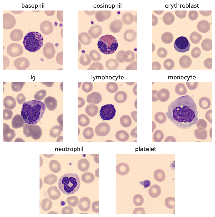
For the classification of blood cells, peripheral blood cells dataset [Acevedo et al.(2020)Acevedo, Merino, Alférez, Ángel Molina, Boldú, and Rodellar] was used which contains a total of 17,092 RGB images of normal blood cells, acquired using the analyser CellaVision DM96. The images are 360x363 dimensions each and were labelled by expert clinical pathologists at the Hospital Clinic of Barcelona. The traceability to the patient data was removed, resulting in an anonymized dataset. It is to be noted that the subjects were individuals without infection, hematologic or oncologic disease and free of any pharmacologic treatment at the moment of blood collection. No further filtering or image processing techniques were performed to the images in the dataset. Figure 2 shows a few sample images from the dataset. Number of instances in each class are presented in the Table 2. We split the dataset retaining 64% samples in each class as training samples, 24% as validation, and the rest 12% as testing samples while maintaining the sample per class ratio in all the sets as the original dataset.
| Cell Type | #Images | Percent(%) |
|---|---|---|
| Neutrophils | 3329 | 19.48 |
| Eosinophils | 3117 | 18.24 |
| Basophils | 1218 | 7.13 |
| Lymphocytes | 1214 | 7.1 |
| Monocytes | 1420 | 8.31 |
| Immature | 2895 | 16.94 |
| granulocytes(ig) | ||
| Erythroblasts | 1551 | 9.07 |
| Platelets | 2348 | 13.74 |
| Total | 17092 | 100 |
4.2 Training Configuration
We perform our experiments with 27 different deep CNN models pre-trained on the ImageNet dataset. We use the stochastic gradient descent optimizer with momentum of 0.9, and adopt the weight initialization for classifier as in [He et al.(2015)He, Zhang, Ren, and Sun]. These models are trained with a minibatch size of 32 on 1 GPU. We start with a learning rate of 0.001 and decay it by a factor of 0.1 every 7 epochs, and terminate training at 25 iterations as model converges. We use data augmentation techniques during training to deal with the limited amount of data which would help the model to generalize well on the unseen samples. The training set is augmented using common image transformations like random vertical and horizontal flips and rotating image by 60 degrees. At this stage, the images are also resized and centered-cropped to dimensions 299x299 for InceptionV3 model and 224x224 for rest of the architectures. For testing, we only evaluate the single view of the original 360×363 image. We use the softmax activation function at the output layer of the classifier and categorical cross-entropy as a loss function. We evaluate the performance of our models by calculating the confusion matrix and reporting evaluation metrics like accuracy, precision, sensitivity and specificity. The formulae of the parameters can be found in Appendix A.
4.3 Results
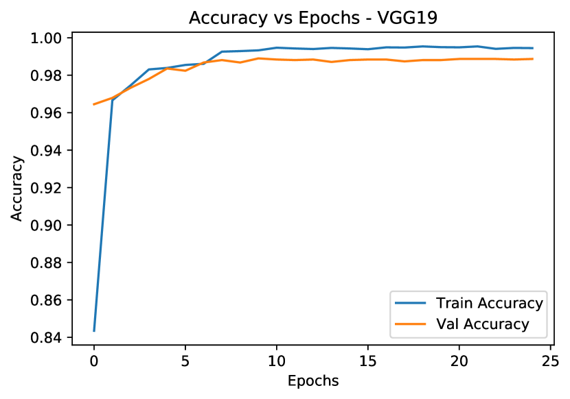
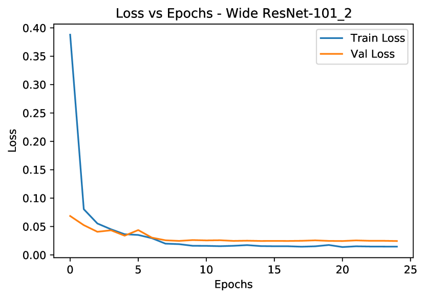
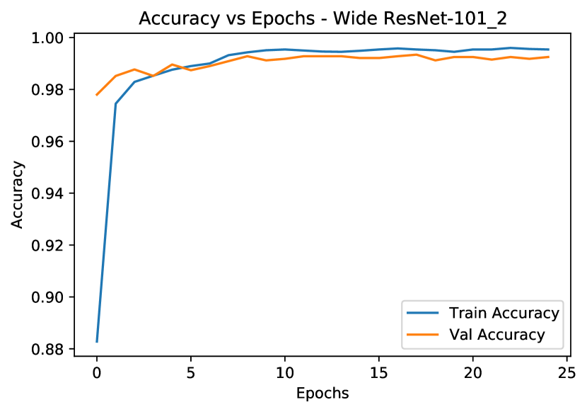
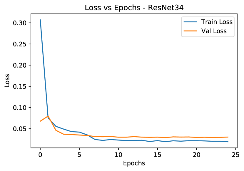
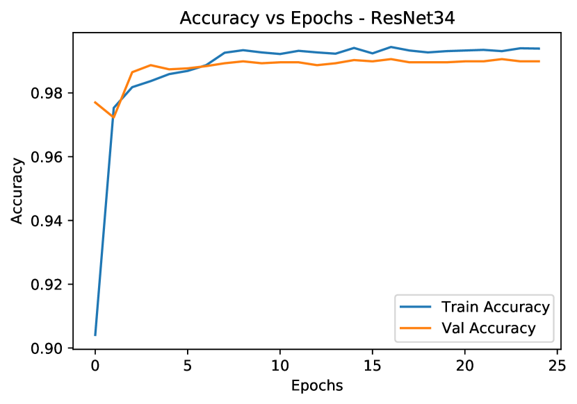
Table LABEL:tab:accuracy-table presents the class-wise and overall test accuracy metrics for all the models. The four top performing models are highlighted in bold which we use further for ensembling. We observe a minute difference (<1%) in overall accuracies of all the models. The lowest accuracy of 98.39% is reported on AlexNet and the highest accuracy is reported as 99.32% on Wide ResNet-50-2. The confusion matrices and training plots for the top models are presented in Appendix C.
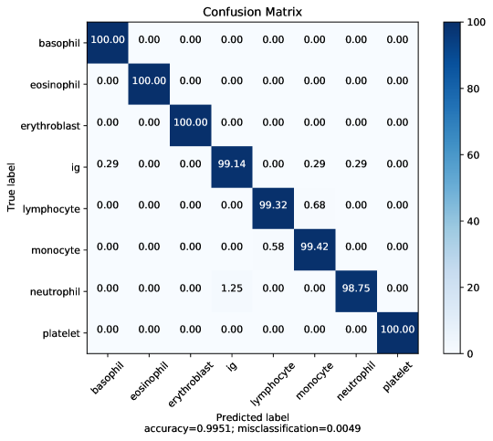
To further improve the performance, we employ voting-based ensemble by combining the top performing four models namely, Wide ResNet-50-2, VGG-19, Wide ResNet-101-2, and ResNet-34. For each instance in the test set, the class predicted by majority of the four classifiers is considered. This method improves the overall classification performance, achieving 99.51% overall accuracy, compared to the best model, Wide ResNet-50-2. It should be noted that there are five other models with overall accuracy equal to that of ResNet-34 that is, 99.17%, but we pick ResNet-34 over them because the validation performance of ensemble is highest with ResNet-34. Figure 3 shows the confusion matrix for the ensemble model. Table 2 reports the accuracy, precision, sensitivity and specificity metrics denoting that our ensemble outperforms the previously published works as well as the top performing models.
tab:accuracy-table Architecture Basophil Eosinophil Erythroblast IG Lymphocyte Monocyte Neutrophil Platelet Overall AlexNet 0.9864 0.9973 0.9893 0.9483 0.9932 0.9766 0.9875 1.0000 0.9839 Densenet-121 0.9932 0.9973 0.9893 0.9713 0.9932 0.9942 0.9850 1.0000 0.9893 Densenet-161 1.0000 0.9973 0.9947 0.9770 0.9863 0.9883 0.9775 1.0000 0.9888 Densenet-169 1.0000 0.9973 0.9893 0.9885 0.9863 0.9942 0.9775 1.0000 0.9908 Densenet-201 0.9864 0.9973 0.9947 0.9885 0.9863 0.9942 0.9775 1.0000 0.9903 VGG-11bn 0.9932 0.9973 0.9786 0.9799 0.9932 0.9825 0.9800 1.0000 0.9878 VGG-11 1.0000 0.9947 0.9947 0.9684 0.9932 0.9708 0.9875 1.0000 0.9878 VGG-13 1.0000 0.9973 0.9893 0.9770 0.9932 0.9708 0.9875 1.0000 0.9893 VGG-13bn 1.0000 0.9973 0.9893 0.9914 0.9932 0.9883 0.9800 1.0000 0.9917 VGG-16bn 1.0000 1.0000 0.9947 0.9856 0.9863 0.9825 0.9725 1.0000 0.9893 VGG-16 0.9932 0.9973 0.9786 0.9828 0.9932 0.9825 0.9775 0.9965 0.9874 VGG-19 1.0000 1.0000 1.0000 0.9799 0.9932 0.9883 0.9875 1.0000 0.9927 VGG-19bn 0.9864 0.9973 0.9893 0.9828 0.9795 0.9883 0.9775 1.0000 0.9878 ResNet-18 1.0000 1.0000 0.9947 0.9856 0.9932 0.9883 0.9800 1.0000 0.9917 ResNet-34 1.0000 0.9973 0.9947 0.9885 0.9932 0.9883 0.9800 1.0000 0.9917 ResNet-50 1.0000 0.9973 0.9947 0.9856 0.9932 0.9942 0.9775 1.0000 0.9912 ResNet-101 0.9932 1.0000 1.0000 0.9828 0.9863 0.9825 0.9875 1.0000 0.9917 ResNet-152 1.0000 0.9973 0.9893 0.9856 0.9863 0.9942 0.9750 1.0000 0.9898 ResNeXt-50-32x4d 0.9932 1.0000 0.9947 0.9770 0.9863 0.9942 0.9825 1.0000 0.9903 ResNeXt-101-32x8d 0.9864 0.9973 1.0000 0.9914 0.9863 0.9942 0.9800 1.0000 0.9917 SqueezeNet 1.0 0.9932 0.9973 0.9893 0.9799 0.9932 0.9708 0.9825 1.0000 0.9883 SqueezeNet 1.1 0.9932 0.9973 0.9786 0.9540 0.9932 0.9591 0.9925 1.0000 0.9839 Wide ResNet-50-2 0.9932 1.0000 0.9947 0.9856 0.9932 0.9942 0.9875 1.0000 0.9932 Wide ResNet-101-2 1.0000 1.0000 1.0000 0.9799 0.9932 0.9942 0.9825 1.0000 0.9922 GoogLeNet 1.0000 0.9973 0.9947 0.9856 0.9932 0.9825 0.9775 0.9965 0.9898 Inception-v3 1.0000 0.9973 1.0000 0.9828 0.9932 0.9825 0.9850 1.0000 0.9917 MobileNet-v2 0.9932 0.9973 1.0000 0.9856 0.9932 0.9883 0.9800 1.0000 0.9912
| Method | Accuracy | Precision | Sensitivity | Specificity |
|---|---|---|---|---|
| [Acevedo et al.(2019)Acevedo, Alférez, Merino, Puigví, and Rodellar] | 96.20 | 97.00 | 96.00 | 97.00 |
| [Ucar(2020)] | 97.94 | 97.94 | 97.94 | 99.71 |
| [Long et al.(2021)Long, Peng, Song, Xia, and Sang] | 99.30 | 99.17 | 99.16 | 99.88 |
| ResNet-34 (Ours) | 99.17 | 99.20 | 99.27 | 99.88 |
| Wide ResNet-101-2 (Ours) | 99.22 | 99.24 | 99.37 | 99.89 |
| VGG-19 (Ours) | 99.27 | 99.17 | 99.36 | 99.89 |
| Wide ResNet-50-2 (Ours) | 99.32 | 99.29 | 99.35 | 99.90 |
| Ensemble model (Ours) | 99.51 | 99.47 | 99.58 | 99.93 |
5 Discussion
The automatic peripheral blood cell classification is an interesting problem studied widely in recent decades, which can help in the diagnosis of several blood-related diseases. Early attempts to solve this problem involved applying traditional machine learning algorithms with manual feature extraction and morphological analysis. In recent years, deep learning techniques, especially CNNs are employed extensively in this direction. In this work, we solve this problem using deep CNNs, wherein we finetune 27 standard CNN architectures pre-trained on the ImageNet dataset. We further improve the overall performance using the voting-based ensemble of the top four models. Table 2 shows the comparison of our work with the previously published works where our ensemble model outperforms all of them, achieving a classification accuracy of 99.51%. It is worth noting that our model achieves a true positive rate (TPR) of 100% on basophils, eosinophil, erythroblasts, and platelets, 99.42% on monocytes, 99.32% on lymphocytes, 99.14% on IG, and 98.75% on neutrophil. From the confusion matrix in Figure 3, we can see that 1.25% of neutrophils are classified as IG. This may be due to the morphological similarities between the two classes, mainly in the nucleus shape. Moreover, single instances, each from classes lymphocyte and monocyte, are misclassified, which may be the anomalies in dataset.
In addition to our ensemble model, our finetuned CNN models AlexNet (98.39%), ResNet-18 (99.17%), VGG-16 (98.74%), InceptionV3 (99.17%) show considerable improvement in performance over the corresponding models in the past related works. [Acevedo et al.(2019)Acevedo, Alférez, Merino, Puigví, and Rodellar] achieve overall accuracy 96.2% on VGG-16 and 95% on InceptionV3 and [Long et al.(2021)Long, Peng, Song, Xia, and Sang] achieve overall accuracies of 81.5%, 95.9%, 97.8% and 98.4% on AlexNet, ResNet-18, VGG-16 and InceptionV3 respectively, which is considerably lower than our reported values. Such a significant difference in performance may be due to our careful selection of hyperparameters and weight initialization. Moreover, we notice that even with different architectures with varying configurations of depth, number of parameters, layers, or branches, models don’t show a vast difference in the overall performance. The reason behind it may be the simplicity and the smaller size of the PBC dataset compared to the ImageNet which has 1000 classes with over 14 million images making it a much harder dataset wherein the changes in architecture produce notable performance differences. It is noteworthy that the standard CNN architectures we fine-tuned, outperform the specialized architectures and techniques designed [Almezhghwi and Serte(2020), Long et al.(2021)Long, Peng, Song, Xia, and Sang] for the peripheral blood cell classification.
6 Conclusion
In this work, we solve peripheral blood cell classification problem by fine-tuning standard deep CNN architectures pre-trained on ImageNet dataset. We employ transfer learning to utilize features already learned by state-of-the-art deep learning models and fine-tune them to learn new specialized features of blood cells. In our experiments, all 27 models achieve 98% accuracy and 14 of them achieve 99% accuracy, showing significant improvements over past works. The Wide ResNet-50-2 achieves the highest accuracy of 99.32% and the VGG-19 achieves the second highest accuracy of 99.27%. An ensemble of the top performing models outperforms all other models, achieving state-of-the-art results with a classification accuracy of 99.51%. Our work provides an empirical baselines for deep learning-based blood cell classification against which the future works with specialized architectures and techniques can be compared. We think that our findings will help further advances in research in deep learning-based automatic blood cell classification.
References
- [Acevedo et al.(2019)Acevedo, Alférez, Merino, Puigví, and Rodellar] Andrea Acevedo, Santiago Alférez, Anna Merino, Laura Puigví, and José Rodellar. Recognition of peripheral blood cell images using convolutional neural networks. Computer Methods and Programs in Biomedicine, 180:105020, 2019. ISSN 0169-2607. https://doi.org/10.1016/j.cmpb.2019.105020. URL https://www.sciencedirect.com/science/article/pii/S0169260719303578.
- [Acevedo et al.(2020)Acevedo, Merino, Alférez, Ángel Molina, Boldú, and Rodellar] Andrea Acevedo, Anna Merino, Santiago Alférez, Ángel Molina, Laura Boldú, and José Rodellar. A dataset of microscopic peripheral blood cell images for development of automatic recognition systems. Data in Brief, 30:105474, 2020. ISSN 2352-3409. https://doi.org/10.1016/j.dib.2020.105474. URL https://www.sciencedirect.com/science/article/pii/S2352340920303681.
- [Al-Dulaimi et al.(2018)Al-Dulaimi, Banks, Chandran, Tomeo-Reyes, and Nguyen Thanh] Khamael Abbas Khudhair Al-Dulaimi, Jasmine Banks, Vinod Chandran, Inmaculada Tomeo-Reyes, and Kien Nguyen Thanh. Classification of white blood cell types from microscope images: Techniques and challenges. Microscopy science: Last approaches on educational programs and applied research (Microscopy Book Series, 8), pages 17–25, 2018.
- [Almezhghwi and Serte(2020)] Khaled Almezhghwi and S. Serte. Improved classification of white blood cells with the generative adversarial network and deep convolutional neural network. Computational Intelligence and Neuroscience, 2020, 2020.
- [Alzubaidi et al.(2020)Alzubaidi, Fadhel, Al-Shamma, Zhang, and Duan] Laith Alzubaidi, Mohammed A. Fadhel, Omran Al-Shamma, Jinglan Zhang, and Ye Duan. Deep learning models for classification of red blood cells in microscopy images to aid in sickle cell anemia diagnosis. Electronics, 9(3), 2020. ISSN 2079-9292. 10.3390/electronics9030427. URL https://www.mdpi.com/2079-9292/9/3/427.
- [Banik et al.(2019)Banik, Saha, and Kim] P. P. Banik, R. Saha, and K. Kim. Fused convolutional neural network for white blood cell image classification. In 2019 International Conference on Artificial Intelligence in Information and Communication (ICAIIC), pages 238–240, 2019. 10.1109/ICAIIC.2019.8669049.
- [Baydilli and Atila(2020)] Y.Y. Baydilli and Ü. Atila. Classification of white blood cells using capsule networks. Computerized Medical Imaging and Graphics, 80, 2020. 10.1016/j.compmedimag.2020.101699.
- [Cabitza and Banfi(2018)] Federico Cabitza and Giuseppe Banfi. Machine learning in laboratory medicine: waiting for the flood? Clinical Chemistry and Laboratory Medicine (CCLM), 56(4):516–524, 2018.
- [Chen et al.(2015)Chen, Wilson, Tyree, Weinberger, and Chen] Wenlin Chen, James T. Wilson, Stephen Tyree, Kilian Q. Weinberger, and Yixin Chen. Compressing neural networks with the hashing trick, 2015.
- [Constantino and Cogionis(2000)] Benie T Constantino and Bessie Cogionis. Nucleated rbcs—significance in the peripheral blood film. Laboratory Medicine, 31(4):223–229, 2000.
- [Csurka(2017)] Gabriela Csurka. A comprehensive survey on domain adaptation for visual applications. Domain adaptation in computer vision applications, pages 1–35, 2017.
- [Di Ruberto et al.(2002)Di Ruberto, Dempster, Khan, and Jarra] Cecilia Di Ruberto, Andrew Dempster, Shahid Khan, and Bill Jarra. Analysis of infected blood cell images using morphological operators. Image and Vision Computing, 20(2):133 – 146, 2002. ISSN 0262-8856. https://doi.org/10.1016/S0262-8856(01)00092-0. URL http://www.sciencedirect.com/science/article/pii/S0262885601000920.
- [Dorini et al.(2007)Dorini, Minetto, and Leite] Leyza Baldo Dorini, Rodrigo Minetto, and Neucimar Jerônimo Leite. White blood cell segmentation using morphological operators and scale-space analysis. In XX Brazilian Symposium on Computer Graphics and Image Processing (SIBGRAPI 2007), pages 294–304. IEEE, 2007.
- [Fathima and Syeda(2017)] Fathima and Syeda. Blood cells and leukocyte culture - a short review. Open Access Blood Research and Transfusion Journal, 1, 05 2017. 10.19080/OABTJ.2017.01.555559.
- [Frosst and Hinton(2017)] Nicholas Frosst and Geoffrey Hinton. Distilling a neural network into a soft decision tree. arXiv preprint arXiv:1711.09784, 2017.
- [Han et al.(2015)Han, Mao, and Dally] Song Han, Huizi Mao, and William J Dally. Deep compression: Compressing deep neural networks with pruning, trained quantization and huffman coding. arXiv preprint arXiv:1510.00149, 2015.
- [He et al.(2015)He, Zhang, Ren, and Sun] K. He, X. Zhang, S. Ren, and J. Sun. Delving deep into rectifiers: Surpassing human-level performance on imagenet classification. In 2015 IEEE International Conference on Computer Vision (ICCV), pages 1026–1034, 2015. 10.1109/ICCV.2015.123.
- [He et al.(2016)He, Zhang, Ren, and Sun] Kaiming He, Xiangyu Zhang, Shaoqing Ren, and Jian Sun. Deep residual learning for image recognition. In Proceedings of the IEEE conference on computer vision and pattern recognition, pages 770–778, 2016.
- [Holinstat(2017)] Michael Holinstat. Normal platelet function. Cancer and Metastasis Reviews, 36(2):195–198, 2017.
- [Huang et al.(2017)Huang, Liu, Van Der Maaten, and Weinberger] Gao Huang, Zhuang Liu, Laurens Van Der Maaten, and Kilian Q Weinberger. Densely connected convolutional networks. In Proceedings of the IEEE conference on computer vision and pattern recognition, pages 4700–4708, 2017.
- [Iandola et al.(2016)Iandola, Han, Moskewicz, Ashraf, Dally, and Keutzer] Forrest N Iandola, Song Han, Matthew W Moskewicz, Khalid Ashraf, William J Dally, and Kurt Keutzer. Squeezenet: Alexnet-level accuracy with 50x fewer parameters and¡ 0.5 mb model size. arXiv preprint arXiv:1602.07360, 2016.
- [Jiang et al.(2018)Jiang, Cheng, Qin, Du, and Zhang] Ming Jiang, Liu Cheng, Feiwei Qin, Lian Du, and Min Zhang. White blood cells classification with deep convolutional neural networks. International Journal of Pattern Recognition and Artificial Intelligence, 32, 02 2018. 10.1142/S0218001418570069.
- [Khan et al.(2020a)Khan, Sohail, and Ali] Asifullah Khan, Anabia Sohail, and Amna Ali. A new channel boosted convolutional neural network using transfer learning, 2020a.
- [Khan et al.(2020b)Khan, Sohail, Zahoora, and Qureshi] Asifullah Khan, Anabia Sohail, Umme Zahoora, and Aqsa Saeed Qureshi. A survey of the recent architectures of deep convolutional neural networks. Artificial Intelligence Review, 53(8):5455–5516, Apr 2020b. ISSN 1573-7462. 10.1007/s10462-020-09825-6. URL http://dx.doi.org/10.1007/s10462-020-09825-6.
- [Kim et al.(2001)Kim, Jeon, Choi, Kim, and Ho] Kyungsu Kim, Jeonghee Jeon, WanKyoo Choi, Pankoo Kim, and Yo-Sung Ho. Automatic cell classification in human’s peripheral blood images based on morphological image processing. In Australian Joint Conference on Artificial Intelligence, pages 225–236. Springer, 2001.
- [Krizhevsky(2014)] Alex Krizhevsky. One weird trick for parallelizing convolutional neural networks. arXiv preprint arXiv:1404.5997, 2014.
- [Kuhn et al.(2017)Kuhn, Diederich, Keller, Kramer, Lückstädt, Panknin, Suvorava, Isakson, Kelm, and Cortese-Krott] Viktoria Kuhn, Lukas Diederich, T.C. Stevenson Keller, Christian M. Kramer, Wiebke Lückstädt, Christina Panknin, Tatsiana Suvorava, Brant E. Isakson, Malte Kelm, and Miriam M. Cortese-Krott. Red blood cell function and dysfunction: Redox regulation, nitric oxide metabolism, anemia. Antioxidants & Redox Signaling, 26(13):718–742, 2017. 10.1089/ars.2016.6954. URL https://doi.org/10.1089/ars.2016.6954. PMID: 27889956.
- [Kumar et al.(2016)Kumar, Kim, Lyndon, Fulham, and Feng] Ashnil Kumar, Jinman Kim, David Lyndon, Michael Fulham, and Dagan Feng. An ensemble of fine-tuned convolutional neural networks for medical image classification. IEEE journal of biomedical and health informatics, 21(1):31–40, 2016.
- [L.(2005)] Dean L. Blood groups and red cell antigens, chapter 1, blood and the cells it contains., 2005. URL https://www.ncbi.nlm.nih.gov/books/NBK2263/.
- [Lee and Chen(2014)] Howard Lee and Yi-Ping Phoebe Chen. Cell morphology based classification for red cells in blood smear images. Pattern Recognition Letters, 49:155–161, 2014.
- [Long et al.(2021)Long, Peng, Song, Xia, and Sang] Fei Long, Jing-Jie Peng, Weitao Song, Xiaobo Xia, and Jun Sang. Bloodcaps: A capsule network based model for the multiclassification of human peripheral blood cells. Computer Methods and Programs in Biomedicine, page 105972, 2021.
- [Macawile et al.(2018)Macawile, Quiñones, Ballado, Cruz, and Caya] M. J. Macawile, V. V. Quiñones, A. Ballado, J. D. Cruz, and M. V. Caya. White blood cell classification and counting using convolutional neural network. In 2018 3rd International Conference on Control and Robotics Engineering (ICCRE), pages 259–263, 2018. 10.1109/ICCRE.2018.8376476.
- [Mathur et al.(2013)Mathur, Tripathi, and Kuse] Atin Mathur, Ardhendu S Tripathi, and Manohar Kuse. Scalable system for classification of white blood cells from leishman stained blood stain images. Journal of pathology informatics, 4(Suppl), 2013.
- [Othman et al.(2017)Othman, Mohammed, and Baban] Mazin Zeki Othman, Thabit Mohammed, and Alaa Baban. Neural network classification of white blood cell using microscopic images. (IJACSA) International Journal of Advanced Computer Science and Applications, 8:99–104, 06 2017. 10.14569/IJACSA.2017.080513.
- [Patodia et al.(2020)Patodia, Nibhasya, and Chandraprabha] Chandni Patodia, G. Nibhasya, and R. Chandraprabha. A survey on automated methods used for wbc classification. In International Journal of Engineering Technology and Management Sciences (IJETMS), 2020.
- [Peng et al.(2015)Peng, Huang, Qi, Zhao, Zhang, Zhao, Yuan, He, and Zhang] Yuxin Peng, Xin Huang, Jinwei Qi, Junjie Zhao, Junchao Zhang, Yunzhen Zhao, Yuxin Yuan, Xiangteng He, and Jian Zhang. Pku-icst at trecvid 2015: Instance search task. In TRECVID, 2015.
- [Piuri and Scotti(2004)] V. Piuri and F. Scotti. Morphological classification of blood leucocytes by microscope images. In 2004 IEEE International Conference onComputational Intelligence for Measurement Systems and Applications, 2004. CIMSA., pages 103–108, 2004. 10.1109/CIMSA.2004.1397242.
- [Ravì et al.(2016)Ravì, Wong, Deligianni, Berthelot, Andreu-Perez, Lo, and Yang] Daniele Ravì, Charence Wong, Fani Deligianni, Melissa Berthelot, Javier Andreu-Perez, Benny Lo, and Guang-Zhong Yang. Deep learning for health informatics. IEEE journal of biomedical and health informatics, 21(1):4–21, 2016.
- [Rawat et al.(2015)Rawat, Bhadauria, Singh, and Virmani] Jyoti Rawat, HS Bhadauria, Annapurna Singh, and Jitendra Virmani. Review of leukocyte classification techniques for microscopic blood images. In 2015 2nd International Conference on Computing for Sustainable Global Development (INDIACom), pages 1948–1954. IEEE, 2015.
- [Rodellar et al.(2018)Rodellar, Alférez, Acevedo, Molina, and Merino] J. Rodellar, S. Alférez, A. Acevedo, A. Molina, and A. Merino. Image processing and machine learning in the morphological analysis of blood cells. International Journal of Laboratory Hematology, 40(S1):46–53, 2018. https://doi.org/10.1111/ijlh.12818. URL https://onlinelibrary.wiley.com/doi/abs/10.1111/ijlh.12818.
- [Roy et al.(2017)Roy, Conjeti, Sheet, Katouzian, Navab, and Wachinger] Abhijit Guha Roy, Sailesh Conjeti, Debdoot Sheet, Amin Katouzian, Nassir Navab, and Christian Wachinger. Error corrective boosting for learning fully convolutional networks with limited data. In International Conference on Medical Image Computing and Computer-Assisted Intervention, pages 231–239. Springer, 2017.
- [Sahlol et al.(2020)Sahlol, Kollmannsberger, and Ewees] Ahmed T. Sahlol, P. Kollmannsberger, and A. A. Ewees. Efficient classification of white blood cell leukemia with improved swarm optimization of deep features. Scientific Reports, 10, 2020.
- [Sandler et al.(2018)Sandler, Howard, Zhu, Zhmoginov, and Chen] Mark Sandler, Andrew Howard, Menglong Zhu, Andrey Zhmoginov, and Liang-Chieh Chen. Mobilenetv2: Inverted residuals and linear bottlenecks. In Proceedings of the IEEE conference on computer vision and pattern recognition, pages 4510–4520, 2018.
- [Saraswat and Arya(2014)] Mukesh Saraswat and K.V. Arya. Automated microscopic image analysis for leukocytes identification: A survey. Micron, 65:20 – 33, 2014. ISSN 0968-4328. https://doi.org/10.1016/j.micron.2014.04.001. URL http://www.sciencedirect.com/science/article/pii/S0968432814000663.
- [Scotti(2005)] Fabio Scotti. Automatic morphological analysis for acute leukemia identification in peripheral blood microscope images. In CIMSA. 2005 IEEE International Conference on Computational Intelligence for Measurement Systems and Applications, 2005., pages 96–101. IEEE, 2005.
- [Shafique and Tehsin(2018)] Sarmad Shafique and Samabia Tehsin. Computer-aided diagnosis of acute lymphoblastic leukaemia. Computational and mathematical methods in medicine, 2018, 2018.
- [Shahin et al.(2019)Shahin, Guo, Amin, and Sharawi] Ahmed Ismail Shahin, Yanhui Guo, Khalid M Amin, and Amr A Sharawi. White blood cells identification system based on convolutional deep neural learning networks. Computer methods and programs in biomedicine, 168:69–80, 2019.
- [Shao et al.(2015)Shao, Zhu, and Li] L. Shao, F. Zhu, and X. Li. Transfer learning for visual categorization: A survey. IEEE Transactions on Neural Networks and Learning Systems, 26(5):1019–1034, 2015. 10.1109/TNNLS.2014.2330900.
- [Sharma et al.(2019)Sharma, Bhave, and Janghel] Mayank Sharma, Aishwarya Bhave, and Rekh Ram Janghel. White blood cell classification using convolutional neural network. In Jiacun Wang, G. Ram Mohana Reddy, V. Kamakshi Prasad, and V. Sivakumar Reddy, editors, Soft Computing and Signal Processing, pages 135–143, Singapore, 2019. Springer Singapore. ISBN 978-981-13-3600-3.
- [Simonyan and Zisserman(2014)] Karen Simonyan and Andrew Zisserman. Very deep convolutional networks for large-scale image recognition. arXiv preprint arXiv:1409.1556, 2014.
- [Srivastava et al.(2015)Srivastava, Greff, and Schmidhuber] Rupesh Kumar Srivastava, Klaus Greff, and Jürgen Schmidhuber. Highway networks. arXiv preprint arXiv:1505.00387, 2015.
- [Su et al.(2014)Su, Cheng, and Wang] M. Su, Chun-Yen Cheng, and P. Wang. A neural-network-based approach to white blood cell classification. The Scientific World Journal, 2014, 2014.
- [Szegedy et al.(2015)Szegedy, Liu, Jia, Sermanet, Reed, Anguelov, Erhan, Vanhoucke, and Rabinovich] Christian Szegedy, Wei Liu, Yangqing Jia, Pierre Sermanet, Scott Reed, Dragomir Anguelov, Dumitru Erhan, Vincent Vanhoucke, and Andrew Rabinovich. Going deeper with convolutions. In Proceedings of the IEEE conference on computer vision and pattern recognition, pages 1–9, 2015.
- [Szegedy et al.(2016)Szegedy, Vanhoucke, Ioffe, Shlens, and Wojna] Christian Szegedy, Vincent Vanhoucke, Sergey Ioffe, Jon Shlens, and Zbigniew Wojna. Rethinking the inception architecture for computer vision. In Proceedings of the IEEE conference on computer vision and pattern recognition, pages 2818–2826, 2016.
- [Taherisadr et al.(2013)Taherisadr, Nasirzonouzi, Baradaran, and Mehdizade] Mojtaba Taherisadr, Mona Nasirzonouzi, Behzad Baradaran, and Alireza Mehdizade. New approch to red blood cell classification using morphological image processing. Shiraz E-Medical Journal, 14(1):44–53, 2013.
- [Tan et al.(2018)Tan, Sun, Kong, Zhang, Yang, and Liu] Chuanqi Tan, Fuchun Sun, Tao Kong, Wenchang Zhang, Chao Yang, and Chunfang Liu. A survey on deep transfer learning. In International conference on artificial neural networks, pages 270–279. Springer, 2018.
- [Taylor and Stone(2009)] Matthew E Taylor and Peter Stone. Transfer learning for reinforcement learning domains: A survey. Journal of Machine Learning Research, 10(7), 2009.
- [Theera-Umpon and Dhompongsa(2007)] N. Theera-Umpon and S. Dhompongsa. Morphological granulometric features of nucleus in automatic bone marrow white blood cell classification. IEEE Transactions on Information Technology in Biomedicine, 11(3):353–359, 2007. 10.1109/TITB.2007.892694.
- [Throngnumchai et al.(2019)Throngnumchai, Lomvisai, Tantasirin, and Phasukkit] K. Throngnumchai, P. Lomvisai, C. Tantasirin, and P. Phasukkit. Classification of white blood cell using deep convolutional neural network. In 2019 12th Biomedical Engineering International Conference (BMEiCON), pages 1–4, 2019. 10.1109/BMEiCON47515.2019.8990301.
- [Tomari et al.(2014)Tomari, Zakaria, Jamil, Nor, and Fuad] Razali Tomari, Wan Nurshazwani Wan Zakaria, Muhammad Mahadi Abdul Jamil, Faridah Mohd Nor, and Nik Farhan Nik Fuad. Computer aided system for red blood cell classification in blood smear image. Procedia Computer Science, 42:206 – 213, 2014. ISSN 1877-0509. https://doi.org/10.1016/j.procs.2014.11.053. URL http://www.sciencedirect.com/science/article/pii/S1877050914014914. Medical and Rehabilitation Robotics and Instrumentation (MRRI2013).
- [Tyas et al.(2017)Tyas, Ratnaningsih, Harjoko, and Hartati] Dyah Aruming Tyas, Tri Ratnaningsih, Agus Harjoko, and Sri Hartati. The classification of abnormal red blood cell on the minor thalassemia case using artificial neural network and convolutional neural network. In Proceedings of the International Conference on Video and Image Processing, ICVIP 2017, page 228–233, New York, NY, USA, 2017. Association for Computing Machinery. ISBN 9781450353830. 10.1145/3177404.3177438. URL https://doi.org/10.1145/3177404.3177438.
- [Ucar(2020)] F. Ucar. Deep learning approach to cell classificatio in human peripheral blood. In 2020 5th International Conference on Computer Science and Engineering (UBMK), pages 383–387, 2020. 10.1109/UBMK50275.2020.9219480.
- [Ventura and Warnick(2007)] Dan Ventura and Sean Warnick. A theoretical foundation for inductive transfer. Brigham Young University, College of Physical and Mathematical Sciences, 19, 2007.
- [Wang et al.(2017)Wang, Jiang, Qian, Yang, Li, Zhang, Wang, and Tang] Fei Wang, Mengqing Jiang, Chen Qian, Shuo Yang, Cheng Li, Honggang Zhang, Xiaogang Wang, and Xiaoou Tang. Residual attention network for image classification. In Proceedings of the IEEE conference on computer vision and pattern recognition, pages 3156–3164, 2017.
- [Weiss et al.(2016)Weiss, Khoshgoftaar, and Wang] Karl Weiss, Taghi M Khoshgoftaar, and DingDing Wang. A survey of transfer learning. Journal of Big data, 3(1):1–40, 2016.
- [Woo et al.(2018)Woo, Park, Lee, and Kweon] Sanghyun Woo, Jongchan Park, Joon-Young Lee, and In So Kweon. Cbam: Convolutional block attention module. In Proceedings of the European conference on computer vision (ECCV), pages 3–19, 2018.
- [Wu et al.(2016)Wu, Leng, Wang, Hu, and Cheng] Jiaxiang Wu, Cong Leng, Yuhang Wang, Qinghao Hu, and Jian Cheng. Quantized convolutional neural networks for mobile devices. In Proceedings of the IEEE Conference on Computer Vision and Pattern Recognition, pages 4820–4828, 2016.
- [Xie et al.(2017)Xie, Girshick, Dollár, Tu, and He] Saining Xie, Ross Girshick, Piotr Dollár, Zhuowen Tu, and Kaiming He. Aggregated residual transformations for deep neural networks. In Proceedings of the IEEE conference on computer vision and pattern recognition, pages 1492–1500, 2017.
- [Xu et al.(2017)Xu, Papageorgiou, Abidi, Dao, Zhao, and Karniadakis] Mengjia Xu, Dimitrios P. Papageorgiou, Sabia Z. Abidi, Ming Dao, Hong Zhao, and George Em Karniadakis. A deep convolutional neural network for classification of red blood cells in sickle cell anemia. PLOS Computational Biology, 13(10):1–27, 10 2017. 10.1371/journal.pcbi.1005746. URL https://doi.org/10.1371/journal.pcbi.1005746.
- [Zagoruyko and Komodakis(2016)] Sergey Zagoruyko and Nikos Komodakis. Wide residual networks. arXiv preprint arXiv:1605.07146, 2016.
- [Zhang et al.(2018)Zhang, Zhou, Lin, and Sun] Xiangyu Zhang, Xinyu Zhou, Mengxiao Lin, and Jian Sun. Shufflenet: An extremely efficient convolutional neural network for mobile devices. In Proceedings of the IEEE conference on computer vision and pattern recognition, pages 6848–6856, 2018.
- [Zhuang et al.(2021)Zhuang, Qi, Duan, Xi, Zhu, Zhu, Xiong, and He] F. Zhuang, Z. Qi, K. Duan, D. Xi, Y. Zhu, H. Zhu, H. Xiong, and Q. He. A comprehensive survey on transfer learning. Proceedings of the IEEE, 109(1):43–76, 2021. 10.1109/JPROC.2020.3004555.
Appendix A Evaluation Metrics Formulae
Below are the performance metrics used to evaluate the classification model on PCB dataset. {align*} Overall Accuracy = Total Number of Correct PredictionsTotal Number of Samples {align*} Precision = TPTP+FP, Sensitivity = TPTP+FN, Specificity = TNTN+FP where TP (True Positive) is the value of principal diagonal of target class in confusion matrix, TN (True Negative) is the sum of all the elements except the row and column of target class, FP (False Positive) is the sum of all column elements of target class excluding its TP , and FN (False Negative) is the sum of all row elements of target class excluding its TP. For this multi-class classification problem, we considered one-vs-all approach for calculating specificity, sensitivity and precision from the confusion matrix.
Appendix B More details on CNN Architectures
The evolution of the CNN architectures in the last decade has been remarkable. Initially, different ideas such as information gating mechanism across multiple layers, skip connections, and cross-layer channel connectivity were introduced [Srivastava et al.(2015)Srivastava, Greff, and Schmidhuber, He et al.(2016)He, Zhang, Ren, and Sun, Huang et al.(2017)Huang, Liu, Van Der Maaten, and Weinberger]. Then, the focus of research shifted mainly on designing of generic blocks that can be inserted at any learning stage in CNN architecture to improve the network representation [Peng et al.(2015)Peng, Huang, Qi, Zhao, Zhang, Zhao, Yuan, He, and Zhang] and can be used to assign attention to spatial and feature map information [Wang et al.(2017)Wang, Jiang, Qian, Yang, Li, Zhang, Wang, and Tang, Roy et al.(2017)Roy, Conjeti, Sheet, Katouzian, Navab, and Wachinger, Woo et al.(2018)Woo, Park, Lee, and Kweon]. In 2018, an idea of channel boosting was introduced by Khan et al. [Khan et al.(2020a)Khan, Sohail, and Ali] to boost the performance of a CNN by learning distinct features and utilizing the already learned features using the TL. Some of the salient improvements involve knowledge distillation and compression or squeezing of pre-trained networks [Chen et al.(2015)Chen, Wilson, Tyree, Weinberger, and Chen, Han et al.(2015)Han, Mao, and Dally, Wu et al.(2016)Wu, Leng, Wang, Hu, and Cheng, Frosst and Hinton(2017)]. GoogleNet replaced the conventional convolution operation with point-wise group convolution operation to make it small and computationally efficient [Szegedy et al.(2015)Szegedy, Liu, Jia, Sermanet, Reed, Anguelov, Erhan, Vanhoucke, and Rabinovich]. ShuffleNet also used the point-wise group convolution but with a new idea of channel shuffle that reduced the number of operations significantly without impacting the performance [Zhang et al.(2018)Zhang, Zhou, Lin, and Sun]. For our experiments we used 27 standard CNN architectures, details of which are presented in Table 3.
| Architecture | Top-1 | Top-5 | Depth | #Params | Main Contribution |
| Error | Error | (millions) | |||
| AlexNet | 43.45 | 20.91 | 5 | 61 | - Deeper and wider than the LeNet |
| [Krizhevsky(2014)] | - Uses Relu, dropout and overlap Pooling | ||||
| - GPUs NVIDIA GTX 580 | |||||
| VGG-11 | 30.98 | 11.37 | 11 | 133 | |
| VGG-13 | 30.07 | 10.75 | 13 | 133 | |
| VGG-16 | 28.41 | 9.62 | 16 | 138 | |
| VGG-19 | 27.62 | 9.12 | 19 | 144 | - Analysis with different depths |
| VGG-11 bn | 29.62 | 10.19 | 11 | 133 | - Homogenous topology |
| VGG-13 bn | 28.45 | 9.63 | 13 | 133 | - Uses small size kernels |
| VGG-16 bn | 26.63 | 8.5 | 16 | 138 | |
| VGG-19 bn | 25.76 | 8.15 | 19 | 144 | |
| [Simonyan and Zisserman(2014)] | |||||
| ResNet-18 | 30.24 | 10.92 | 18 | 11 | |
| ResNet-34 | 26.7 | 8.58 | 34 | 21 | |
| ResNet-50 | 23.85 | 7.13 | 50 | 25 | - Residual learning framework |
| ResNet-101 | 22.63 | 6.44 | 101 | 44 | - Identity mapping based skip connections |
| ResNet-152 | 21.69 | 5.94 | 152 | 60 | - Ease the training of deeper networks |
| [He et al.(2016)He, Zhang, Ren, and Sun] | |||||
| SqueezeNet 1.0 | 41.9 | 19.58 | 18 | 1.24 | - 50x reduction in model size of AlexNet |
| SqueezeNet 1.1 | 41.81 | 19.38 | 18 | 1.23 | - Deep compression with 8-bit quantization |
| [Iandola et al.(2016)Iandola, Han, Moskewicz, Ashraf, Dally, and Keutzer] | |||||
| Densenet-121 | 25.35 | 7.83 | 121 | 8 | - Cross-layer information flow |
| Densenet-169 | 24 | 7 | 169 | 14 | - Alleviate the vanishing-gradient problem |
| Densenet-201 | 22.8 | 6.43 | 201 | 20 | - Reduced number of parameters |
| Densenet-161 | 22.35 | 6.2 | 161 | 28 | - Encourage Feature Reuse |
| [Huang et al.(2017)Huang, Liu, Van Der Maaten, and Weinberger] | |||||
| Inception v3 | 22.55 | 6.44 | 48 | 24 | - Handles representational bottleneck |
| [Szegedy et al.(2016)Szegedy, Vanhoucke, Ioffe, Shlens, and Wojna] | - Replace large size filters with small filters | ||||
| GoogleNet | 30.22 | 10.47 | 22 | 6 | - Introduced block concept |
| [Szegedy et al.(2015)Szegedy, Liu, Jia, Sermanet, Reed, Anguelov, Erhan, Vanhoucke, and Rabinovich] | - Split transform and merge idea | ||||
| MobileNet V2 | 28.12 | 9.71 | 53 | 3 | - Inverted residual structure |
| [Sandler et al.(2018)Sandler, Howard, Zhu, Zhmoginov, and Chen] | - Non-linearities in narrow layers removed | ||||
| ResNeXt-50-32x4d | 22.38 | 6.3 | 50 | 25 | - Cardinality |
| ResNeXt-101-32x8d | 20.69 | 5.47 | 101 | 88 | - Homogeneous topology |
| [Xie et al.(2017)Xie, Girshick, Dollár, Tu, and He] | - Grouped convolution | ||||
| Wide ResNet-50-2 | 21.49 | 5.91 | 50 | 68 | - Feature Reuse and Fast training |
| Wide ResNet-101-2 | 21.16 | 5.72 | 101 | 126 | - Width is increased and depth is decreased |
| [Zagoruyko and Komodakis(2016)] |
Appendix C Additional Evaluation Results
In this section, the training and validation plots for accuracy (Figures 9, 11, 13, 15), and loss (Figures 9, 11, 13, 15) are presented along with the confusion matrices (Figures 5, 5, 7, 7) of our four top performing models are presented.