Topological Defects and Unique Stacking Disorders in Honeycomb Layered Oxide Nanomaterials: Implications for Rechargeable Batteries
Abstract
Endowed with a multitude of exquisite properties such as rich electrochemistry, superb topology and eccentric electromagnetic phenomena, honeycomb layered oxides have risen to the top echelons of science with applications in diverse fields ranging from condensed matter physics, solid-state chemistry, materials science, solid-state ionics to electrochemistry. However, these oxides are vastly underutilised as their underlying atomistic mechanisms remain unexplored. Therefore, in this study, atomic-resolution imaging on pristine along multiple zone axes was conducted using spherical aberration-corrected scanning transmission electron microscopy (Cs-corrected STEM) to reveal hitherto unreported nanoscale topological defects and curvature which can be associated with various phase transitions. Furthermore, we discover the coexistence of a stacking variant with P3-type sequence alongside the well-reported P2-type stacking sequence in such honeycomb layered oxides. Our findings have the potential to inspire further experimental and theoretical studies into the role of stacking and topology in the functionality of honeycomb layered oxides , for instance, as high-performance electrode materials for rechargeable batteries.
Keywords: Honeycomb layered oxides, transmission electron microscopy (TEM), aberration-corrected STEM, topological defects, stacking disorders, dislocations, atomic-resolution imaging, curvature
AIST-Kyoto University Chemical Energy Materials Open Innovation Laboratory (ChEM-OIL), Sakyo-ku, Kyoto 606-8501, JAPAN
![[Uncaptioned image]](/html/2110.09233/assets/Rendition.png)
1 Introduction
In the quest for energy independence and ecological sustainability, honeycomb layered oxides1 have drawn enormous interest for their potential as rechargeable battery components owing to their fascinating two-dimensional (2D) ionic diffusion governed by phase transitions.1, 2 These compounds generally adopt the chemical composition , or (where can be divalent or trivalent transition metal atoms such as or some combination thereof; represents pentavalent or hexavalent pnictogen or chalcogen metal atoms such as ; and can be alkali atoms such as etc or coinage-metal atoms such as etc.)3, 4, 5, 6, 7, 8, 9, 10, 11, 12, 13, 14, 15, 16, 1, 17, 18, 19, 20, 21, 22, 23, 24, 25, 26, 27, 28 Such structures comprise an array of transition metal sheets consisting of octahedra surrounded by multiple octahedra in a distinct hexagonal (honeycomb) alignment. Oxygen atoms from the octahedra in turn coordinate with cations interposed between the layers to form heterostructures whose interlayer bonds are significantly weaker than the covalent in-plane bonds.
Progress towards their battery application has been hindered by the scarcity of evidence for their unique topologies, nanoscale defects and curvature effects that ought to accompany their otherwise well-reported stacking sequences. Given that the interlayer distance is inversely proportional to the strength of the interlayer bonds, the size of the resident atoms (Shannon-Prewitt radii)29 typically determines the structural topology and the resulting physicochemical properties of the honeycomb layered oxides. For instance, smaller atomic radii cations such as atoms in tend to form stronger tetrahedral coordinations between atoms and oxygen atoms, with 2 repetitive honeycomb layers in each unit cell. 4 This type of structure is typically referred to as T2-type in the Hagenmuller notation30 (where ‘T’ is the tetrahedral coordination and ‘2’ is the number of honeycomb slabs per unit cell). The research findings for other honeycomb layered oxide materials such as , , , amongst others31, 32, 33, 34, 35 are consistent with a dumbbell coordination of alkali atoms with oxygen and are classified as D-type structures.1 On the other hand, larger ionic radii atoms such as in adopt an O3-type layered framework (where ‘O’ is an octahedral coordination with a periodicity of three honeycomb slabs per unit cell) whilst and in and , respectively, adopt a P2-type framework (where ‘P’ is a prismatic coordination with a periodicity of two). 6, 7, 13, 16, 1, 36 Amongst the assorted honeycomb layered oxides reported so far,1 P-type honeycomb layered oxides have been noted to encompass the widest interlayer distances. 37 Thus, alkali atoms with larger ionic radii such as or typically result in P-type configurations.
Amongst honeycomb layered oxides entailing chalcogen or pnictogen atoms, P3-type structures have been found to have manifold electrochemical capabilities associated with multiple topological defects resulting from their weak interlayer bonds. 38, 1, 15, 13 However, these structures are difficult to synthesise and analyse in as-prepared honeycomb layered oxide materials due to their inferior stability. Thus, theoretical and experimental explorations into the relationship between topological defects innate to these structures and emergent nanoscale features remain underdeveloped. Although X-ray diffraction (XRD) has widely been employed to provide the average structural information of crystalline materials, it cannot be used on pristine materials to account for short-range structural evolutions occurring immediately after synthesis. Consequently, transmission electron microscopy (TEM) can be employed alongside XRD analyses to provide local atomistic information of the cathode, for instance the intermittent stacking evolutions before and after electrochemical operations. The high precision of TEM complements XRD by
Scheme 1: The solid state synthesis protocol of honeycomb layered oxide () showing the various stages (See experimental details furnished as Supporting Information)
![[Uncaptioned image]](/html/2110.09233/assets/Scheme_1.png)
identifying defects (disorders) beyond the detectable limits of the latter.
In order to ascertain the structural nature and topological behaviour of the enigmatic P-type honeycomb structures, we employ atomic-resolution scanning transmission electron microscopy (STEM) on pristine cathode material for rechargeable potassium-ion battery (prepared using the protocol details as displayed in Scheme 1) to reveal emergent structural disorders along -axis. Our analyses reveal partially missing honeycomb slabs of and octahedra as well as absence of some constituent atoms. Furthermore, we identify regions comprising both P2- and P3-type stacking sequences, which correlate with unique topological defects and nanoscale curvatures triggering phase transitions during battery operations.
2 Results and Discussion
Polycrystalline samples of were synthesised through the conventional high-temperature solid-state ceramics route (details provided in the EXPERIMENTAL section). The composition of the as-prepared material was ascertained using inductively coupled plasma-atomic emission spectroscopy ( Table S1), whereas the uniformity of the constituent elements and morphology were validated using scanning electron microscopy (shown in Figure S1). The obtained powder X-ray diffraction (XRD) pattern of as-prepared (Fig ure S2 and Table S2) could be indexed to the hexagonal lattice adopting the P2-type framework. 7 displays a layered crystal structure comprising atoms coordinated with oxygen atoms interposed between slabs of and as illustrated by Fig ure 1a. The slab is composed of divalent nickel atoms (as affirmed by X-ray absorption spectroscopy (Figure S3)) coordinated to six oxygen atoms, whereas the contains atoms also coordinated to six oxygen atoms. As shown in Fig ure 1b, a honeycomb configuration of and octahedra is formed with each surrounded by six octahedra. In each unit cell, there are two repetitive layers of and octahedra that are separated by atoms in a prismatic coordination with adjacent oxygen atoms. Even though humps indicating the presence of defects were detected (Fig ure S2), they could not be indexed using XRD, necessitating further characterisation using TEM.
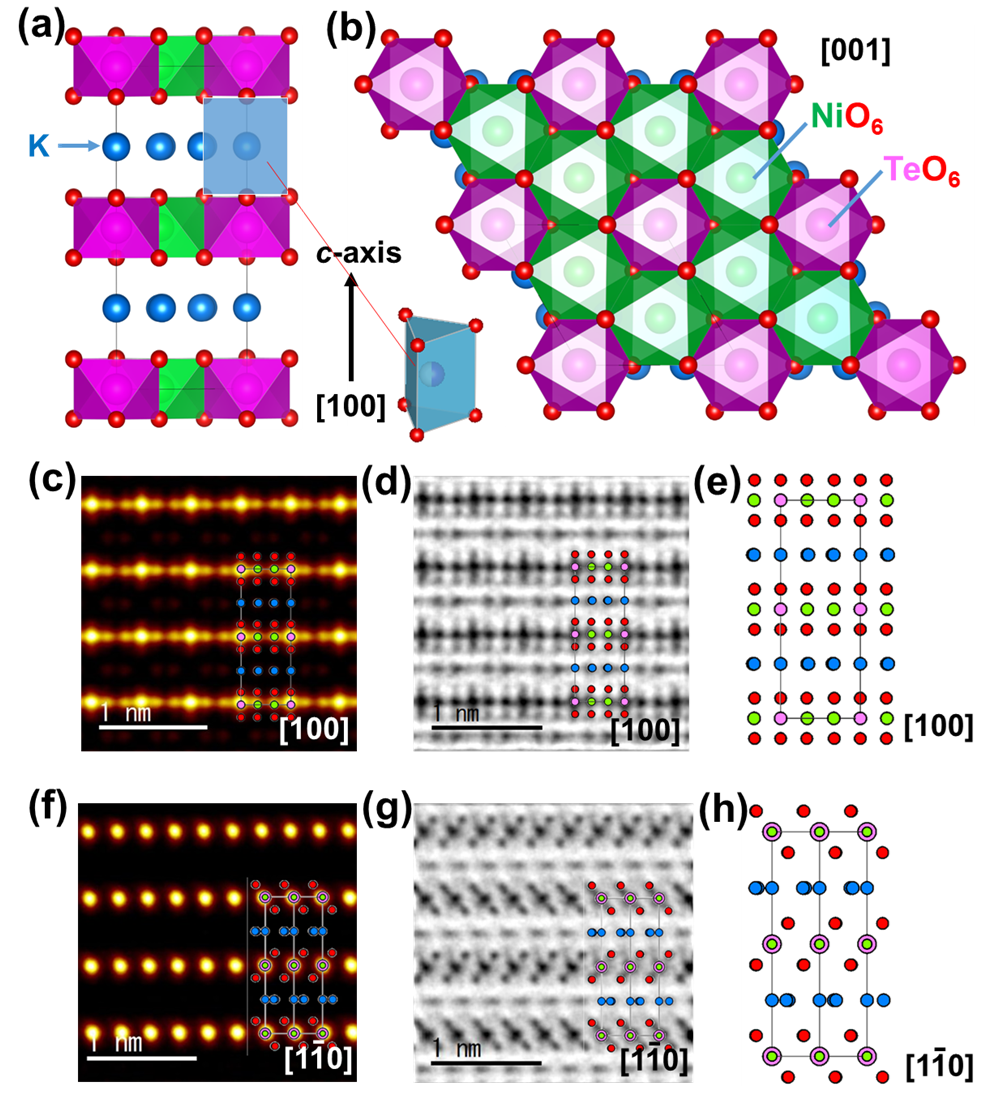
Therefore, to attain more nanostructural information on the honeycomb ordering and slab stacking sequences, aberration-corrected scanning transmission electron microscopy (STEM) was employed on . Considering that prolonged electron beam irradiation gradually leads to sample degradation (Fig ure S4), the sample was exposed to electron beams for short periods, sufficient enough to obtain reliable high-resolution TEM (HRTEM) imaging without compromising the stability of the crystal structure (details in the EXPERIMENTAL section). For clarity, the contrast () of the STEM images are proportional to the atomic number () of elements along the atomic arrangement (where ). 39, 40 The P2-type stacking of is explicitly verified by the atomic-resolution high-angle annular dark-field scanning TEM (HAADF-STEM) images viewed along the [100] zone axis as illustrated by Fig ure 1c. The brighter and bigger yellow spots correspond to Te atoms (), whilst the smaller yellow spots represent atoms (). As expected for a perfectly ordered P2-type honeycomb stacking sequence, the ––– sequence along the -axis is apparent, as illustrated by the crystal model (Fig ure 1e). The arrangement of the constituent atoms of is further validated by atomic-resolution elemental mapping, taken along the [100] zone axis (Fig ure S5). Close inspection using annular bright-field (ABF)-STEM, as shown in Fig ure 1d, shows atoms () positioned between the and slabs in a sandwich-like arrangement, as evinced by the atomistic model in Fig ure 1e. The atoms are also noted to appear in varying contrasts (Fig ure 1d), indicating their occupation of crystallographically distinct sites with varying occupancies ( Table S2), as can be further affirmed by HAADF- and ABF-STEM images taken along the [1-10] zone axis (Fig ures 1f and 1g). A crystal model of the P2-type viewed along the [1-10] zone axis is provided in Fig ure 1h.
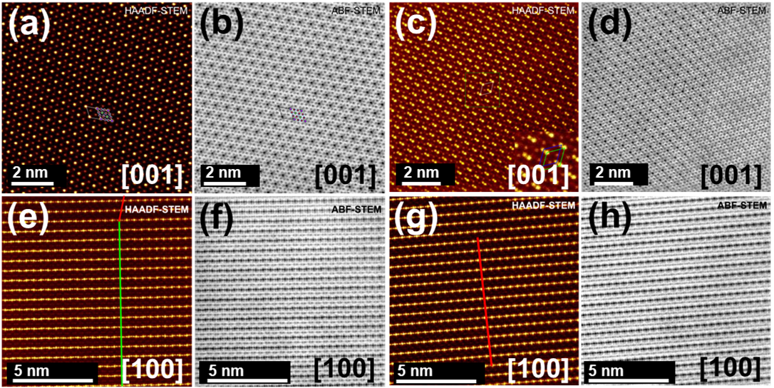
A defect chemistry involving the cationic mixing of transition metal has been reported in honeycomb layered oxides such as , where Ni and Sb have been observed to swap their crystallographic site positions.41 To investigate this behaviour in , HAADF-STEM images of the cathode material viewed along the [001] zone axis were obtained as shown in Fig ure 2a. The corresponding ABF-STEM images (Fig ure 2b) explicitly confirm the honeycomb configuration of atoms (depicted by the dark red spots) around atoms (brighter yellow spots) alongside atoms overlapping with oxygen atoms (). In this study, cationic mixing of and was not detected in any of the crystallites investigated. The honeycomb arrangement of the and atoms is further affirmed by atomic-resolution elemental mapping, taken along the [001] zone axis (Fig ure S6). Crystallite domains with doublets of and spots appear to form a peculiar diagonal-like pattern amongst some particles of as shown in Fig ures 2c and 2d. The enlarged STEM image (Fig ure 2c) clearly shows that, in these domains, adjacent honeycomb slabs deviate from the perfect vertical alignment of and atoms (Fig ures 1e and 1h) along the [100] and [1-10] zone axes. Based on the slab stacking sequence along the -axis ([001] direction), the adjacent slab shifts (by a translation vector of [1/3 1/3 0]) suggest that the new stacking variant in pristine material may best be observed along the -axis ([100]) or -axis ([010]).
Therefore, to discern the new stacking variants, HAADF-STEM and ABF-STEM images of were taken along the [100] zone axis, as seen in Fig ures 2e and 2f. The initial P2-type slab stacking (shown in green line) can be observed as well as a new slab stacking domain that appears to shift along the -axis. The new slab stackings are further elaborated by HAADF-STEM images shown in Fig ure 2g which show the -atom arrangement to be repeatable after three layers along the -axis (inset). The arrangement of the new stacking domain vis-à-vis the occupancy of the potassium atoms coordinated with oxygen atoms is also clearly seen in the corresponding ABF-STEM images (Fig ure 2h).
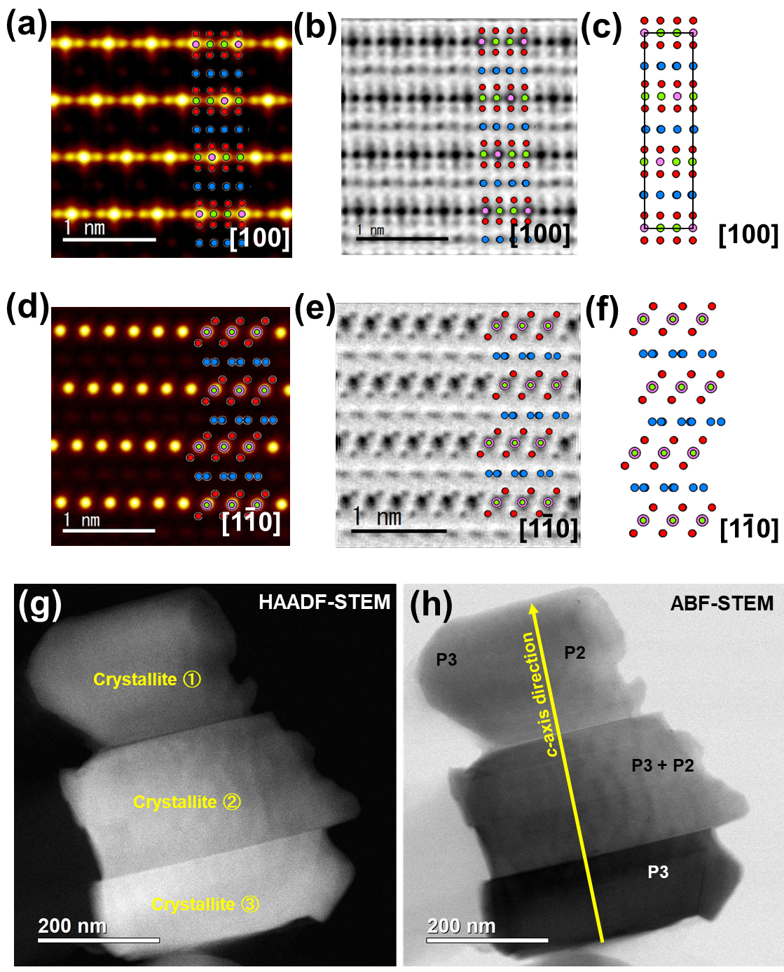
To further assess the nature of the new stacking, atomic-resolution HAADF-STEM and ABF-STEM images were taken as shown in Fig ures 3a and 3b. A new sequence with ––– alternating along the -axis is formed, indicating the formation of a new P3-type stacking by the slab-to-slab transition vector [1/3 1/3 0] relative to the main crystallographic axes, as displayed in the crystal model shown in Fig ure 3c. The new P3-stacking sequence was further verified by STEM images taken along the [1-10] zone axis (Fig ures 3d and 3e) and a projected crystal model (Fig ure 3f). The occupancy of K atoms in the various crystallographic sites of the new P3 stacking is also noted to be different, as indicated by the varying contrasts of the atoms coordinated with oxygen atoms along wavy-like columns in the plane as illustrated by the crystal model in Fig ure 3c. This is further ascertained by the ABF-STEM images. By locating the position of oxygen atoms along [1-10] zone axis, the salient atomic structural differences between P2- and P3-type frameworks can be distinguished. The oxygen atoms are arranged diagonally in a zig-zag orientation along the -axis in the P2-type framework (Fig ure 1g), whilst for the P3-type variant framework the oxygen atoms are aligned in the same direction on the plane (Fig ure 3e). This observation was further corroborated by the low-magnification STEM images (Fig ures 3g and 3h), where P3-type crystallites were located next to previously reported P2-type stackings. These observations not only demonstrate the emergence of a new type of stacking (P3-type) in pristine , but also explicitly show the existence of stacking variants (faults or disorders). Even so, an extensive examination into their stacking sequences is still necessary for a deeper insight into their crystallographic information.
As such, selected area electron diffraction (SAED) measurements were duly performed, exhibiting patterns of a crystallite with P2-stacking sequence along the [1-10] and [100] zone axes, as shown in Fig ures. S7a and S7c. The corresponding simulations are shown in Fig ures S7b and S7d, revealing a good match with the experimental diffractograms indexable to the hexagonal lattice of the P2-type phase. For comparison, the SAED patterns of a crystallite with P3-type stacking sequence are provided in Fig ures S7e and S7g along with their corresponding simulations (Fig ures S7f and S7h). The patterns of the domain with the P3-type stacking along [1-10] and [010] zone axes (Fig ures S7e and S7g), reveal arrays of pseudo-hexagonal symmetry dots that could be indexed into a slightly-distorted hexagonal cell with the approximate lattice parameters: Å, Å (interslab distance of Å). The P3-type lattice parameters are very close to the lattice parameters obtained from the P2-type stacking: Å, Å (interslab distance of Å). Indeed, this explains why this new stacking (P3-type) variant was undetected by the bulk XRD analyses owing to its isostructurality and exacerbated further by its crystallite concentration which is presumably rather subtle to be sufficiently distinguished. In the crystallites analysed, P2-type domains were predominantly seen with P3-type domains seldom being detected as shown in Fig ures 3g and 3h. A further inspection of the electron diffractograms of P3-type stacking (shown in Fig ures S7e and S7g) indicate angle deviations and from the adjacent main diffraction spots along the [1-10] and [010] zone axes, respectively. Typically, angle deviations are expected to fall around 90∘ with an accuracy range . 1% as such errors arising from experimental flaws can be ruled out from the inclinations observed in the electron diffractograms. This indicates that the P3-type stacking lattice is slightly distorted.
Further investigations into the nature of the pristine revealed for the first time, nanostructural defects entailing the disappearance or bending of the honeycomb slabs and potassium layers. In the low-magnification STEM images of the new P3-type stacking domains (Figures 4a and 4b), various crystallites oriented in different zone axes seem to be separated by grain boundary lines. However, close scrutiny of the HAADF-STEM images (Fig ure 4c) reveal the warping or bending of the honeycomb slabs. Corresponding ABF-STEM images (Fig ure 4d) further show similar warping, that could be mistaken for twin or tilt boundaries within the potassium layers. Moreover, high-magnification STEM images (Fig ures 4e and 4f) show an undulating topology of both the honeycomb slabs and potassium layers along the -axis, indicating defects that relate to curvature of the honeycomb slab surface. Such defects are exceedingly rare in layered oxides, prompting deeper scrutiny into the crystal structure.
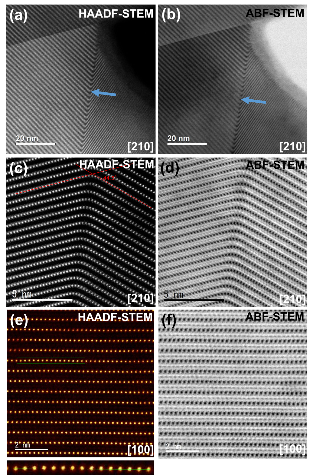
Topological defects such as (Taylor’s) dislocations and disclinations, where translational and rotational symmetry of the atoms in the crystal are destroyed, have often been associated with beneficial physicochemical properties amongst oxides. 42 As crystalline symmetries are intricately linked with momentum and angular momentum conservation law, such violations of can profoundly affect the dynamics of alkali cations during (de)intercalation processes.2 Generally, the origin (technically, dislocation core) of dislocations in the crystal lattice, is denoted by the symbol ‘T’. Figure 5a shows a HAADF-STEM image of a P2-type stacking domain along the [100] zone axis, where a curvature appears as a result of part of the honeycomb slab laying out of position (highlighted as ‘T’). The corresponding ABF-STEM images reveal considerable distortions on the lattice as shown in Fig ure 5b. Edge dislocations on the lattice are clearly discerned as highlighted in Fig ures 5c and 5d respectively. The images indicate that these dislocations are created by a shift along the -plane with a translation vector [1/3 1/3 0]. In principle, the magnitude and direction of the lattice distortion arising from such edge dislocation can be represented using a Burgers vector (b). The Burgers vector of the edge dislocation imaged in Fig ures 5a and 5b was determined to be [0 1 0], spanning along the -axis (slip plane). As for the edge dislocations appearing in Fig ures 5c and 5d, the Burgers vector was determined to be [1/3 1/3 1/2] spanning along the -axis. Other unique edge dislocations detected during the analysis were determined to shift with a Burgers vector of [–1/3 –1/3 1/2] as indicated in Fig ure S8. It is noteworthy that, despite proving elusive, other shifts occurring along the -axis cannot be ruled out. It must also be noted that the diversity and unpredictability of the edge dislocations observed in our investigation are unique to P3-type structures and are extremely rare amongst oxides.

The edge dislocations (Fig ure 5) and curvature (Fig ure 4) identified, may provide insight into the diffusion mechanisms of particularly during (de-)intercalation processes. Generally, the mechanism generating correlations between stacking sequences and topological defects such as ion vacancies and atomic rearrangements is often analysed within the context of slab shearing/gliding/sliding, which need not include curvature deformations.38, 43, 44, 13 However, our work explicitly demonstrates that curvature is an integral part of such topological defects. In fact, in a previous work, it has been theorised that there is a direct relation between such curvatures and the vacancy of cations such as .2, 1 Particularly, the integral of the 2D charge density of ionic vacancies () over a given patch of previously occupied by ions () is equal to the number of cationic vacancies () in the given patch. This is analogous to the well-known Gauss-Bonnet theorem () in 2D geometry, that relates curvature to topological ‘holes’ in the layer, where the (Gaussian) curvature () is proportional to the charge density of the ionic vacancies (). 45 For clarity to readers, a rendition representing such a mechanism is provided in Fig ure S9. The ‘holes’ act as cationic vacancies forming whenever sufficient activation energy is supplied in the material to offset the binding energy of cations that form the stable P2-type lattice, leading to stacking sequence phase transformations as has been noted in during (de)insertion. 38, 13 Similar to the aforementioned shearing/gliding/sliding mechanisms, sufficient activation energies can be provided by raising temperatures and/or by supplying other forms of energy-momentum via electrochemical processes within the material. In the case of the reported in this work, the temperatures (above C) applied during synthesis of these materials, was sufficient to offset the binding energies within the stable P2-type lattice. 7 Therefore, the phase transition to another stable P-type sequence (P3) in pristine (as further shown schematically in Fig ure S10) can possibly be traced to thermally-induced geometric shifts of adjacent honeycomb slabs (shear transformations) and the formation of cationic vacancies during heating, which are accompanied by the topological defects and weak curvatures given in Fig ures 4 and 5. However, the presence or absence of potassium vacancies could not be ascertained owing to the dislocations being accompanied by inter-layer displacement over a wide area (see Figure S11). The lattice strain hampered the clear visualisation of potassium atoms around the dislocation core. The large ionic radius of that gives rise to weaker interlayer bonds, also seemingly favours the retention of such topological defects and weak curvature even after adiabatic cool-down, as summarised in Fig ures 4 and 5. More information relating to the weak curvature identified in this study is given in Figure S12. HAADF-STEM images along the [210] zone axis in Fig ure 5c shows that is extremely malleable even under large stresses (for instance, elastic deformation) which result in even larger curvatures defects. We expect future work to encompass the influence of the synthesis conditions (thermal treatment temperature, annealing duration, precursor types, etc.) on the density (or volume fractions) of the nanoscale defects in such honeycomb layered oxides, as has been shown in layered oxides such as .46
3 Conclusion
In summary, we utilise the higher-order atomic-resolution in aberration-corrected scanning transmission electron microscopy (STEM), to explicitly reveal nanostructural disorders (variants) of honeycomb layers along -axis in pristine . We identify unique topological defects and curvatures due to partially missing honeycomb slabs of , octahedra as well as atoms. Moreover, a new stacking variant with P3-type stacking sequence is for the first time discovered to coexist with the predominant P2-type stacking of . In this study, it is postulated that the occurrence of the nanoscale topological defects and curvature is correlated with missing atoms (dislocations) leading to the emergence of the P3-type stacking. We expect this work will prompt rekindled interest in the study of the defects, curvature and their role in the functionality of such honeycomb layered oxides in applications related, for instance, to rechargeable batteries.
Author contributions
T. M. and H. S. contributed to the syntheses of the materials. Y.M., T.S., M.I. and T.T. performed the morphological characterisation of the materials using TEM. Y.M., G.K., T.M, H.S. and T.S. wrote the manuscript. G.K. and T.M. designed the graphic illustrations outlined in the work. G.K., H.S., T.M. and Y.M. provided input with the data analyses, helped with the discussion and assisted with the manuscript correction. T.T., M. I. and T.S. made the initiative to undertake this work. All authors have given approval to the final version of the manuscript.
Data availability
The data that support the findings of this study are available on request from the corresponding authors [T.M.], [Y. M.], [G. K.], [H. S.] and [T. S.].
Corresponding authors
Correspondence to Titus Masese or Yoshinobu Miyazaki or Godwill Kanyolo or Hiroshi Senoh or Tomohiro Saito.
4 Competing interests
The authors declare no competing interests.
T.M. thanks Ms. Shinobu Wada and Mr. Hiroshi Kimura for the unrelenting support in undertaking the entire study. T. M. also gratefully acknowledges Ms. Kumi Shiokawa, Mr. Masahiro Hirata and Ms. Machiko Kakiuchi for their advice and technical help as we conducted the syntheses, electrochemical and XRD measurements. Dr. Minami Kato is thanked for the assistance in designing the graphical images. This work was conducted in part under the auspices of the Japan Society for the Promotion of Science (JSPS KAKENHI Grant Number 19K15685), Sumika Chemical Analyses Services (SCAS) Co. Ltd., National Institute of Advanced Industrial Science and Technology (AIST) and Japan Prize Foundation.
The following files are available free of charge at https://pubs.acs.org/doi/10.1021/acsanm.0c02601.
-
•
Supporting Information: Experimental details and figures showing the material characterisation (XRD, SEM, TEM, ICP-AES and X-ray absorption spectroscopy (XAS) data) of pristine .
References
- Kanyolo et al. 2020 Kanyolo, G. M.; Masese, T.; Matsubara, N.; Chen, C.-Y.; Rizell, J.; Forslund, O. K.; Nocerino, E.; Papadopoulos, K.; Zubayer, A.; Kato, M.; Tada, K.; Kubota, K.; Senoh, H.; Huang, Z.-D.; Matsumoto, H. Honeycomb Layered Oxides: Structure, Energy Storage, Transport, Topology and Relevant Insights. arXiv 2020, arXiv:2003.03555
- Kanyolo and Masese 2020 Kanyolo, G. M.; Masese, T. An idealised approach of geometry and topology to the diffusion of cations in honeycomb layered oxide frameworks. Sci. Rep. 2020, 10, 13284
- Sathiya et al. 2013 Sathiya, M.; Ramesha, K.; Rousse, G.; Foix, D.; Gonbeau, D.; Guruprakash, K.; Prakash, A.; Doublet, M.; Tarascon, J.-M. \chLi4NiTeO6 as a positive electrode for Li-ion batteries. Chem. Commun. 2013, 49, 11376–11378
- Grundish et al. 2019 Grundish, N. S.; Seymour, L. D.; Henkelman, G.; Goodenough, J. B. Electrochemical Properties of Three \chLi2Ni2TeO6 Structural Polymorphs. Chem. Mater. 2019, 31, 9379–9388
- Yang et al. 2017 Yang, Z.; Jiang, Y.; Deng, L.; Wang, T.; Chen, S.; Huang, Y. A high-voltage honeycomb-layered \chNa4NiTeO6 as cathode material for Na-ion batteries. J. Power Sources 2017, 360, 319–323
- Yuan et al. 2014 Yuan, D.; Liang, X.; Wu, L.; Cao, Y.; Ai, X.; Feng, J.; Yang, H. A Honeycomb-Layered \chNa3Ni2SbO6: A high-rate and cycle-stable cathode for sodium-ion batteries. Adv. Mater. 2014, 26, 6301–6306
- Masese et al. 2018 Masese, T.; Yoshii, K.; Yamaguchi, Y.; Okumura, T.; Huang, Z.-D.; Kato, M.; Kubota, K.; Furutani, J.; Orikasa, Y.; Senoh, H.; Sakaebe, H.; Shikano, M. Rechargeable potassium-ion batteries with honeycomb-layered tellurates as high voltage cathodes and fast potassium-ion conductors. Nat. Commun. 2018, 9, 1–12
- Bhange et al. 2017 Bhange, D. S.; Ali, G.; Kim, D.-H.; Anang, D. A.; Shin, T. J.; Kim, M.-G.; Kang, Y.-M.; Chung, K. Y.; Nam, K.-W. Honeycomb-layer structured \chNa3Ni2BiO6 as a high voltage and long life cathode material for sodium-ion batteries. J. Mater. Chem. A 2017, 5, 1300–1310
- Gyabeng et al. 2017 Gyabeng, D.; Anang, D. A.; Han, J. I. Honeycomb layered oxide \chNa3Ni2SbO6 for high performance pseudocapacitor. J. Alloys Compd. 2017, 704, 734–741
- Yadav et al. 2019 Yadav, D. K.; Sethi, A.; Shalu,; Uma, S. New series of honeycomb ordered oxides, \chNa3M2SbO6 (\chM(ii) Mn, Fe, (Mn, Fe), (Mn, Co)): synthesis, structure and magnetic properties. Dalton Trans. 2019, 48, 8955–8965
- Yoshii et al. 2019 Yoshii, K.; Masese, T.; Kato, M.; Kubota, K.; Senoh, H.; Shikano, M. Sulfonylamide-Based Ionic Liquids for High-Voltage Potassium-Ion Batteries with Honeycomb Layered Cathode Oxides. ChemElectroChem 2019, 6, 3901–3910
- Zheng and Obrovac 2016 Zheng, L.; Obrovac, M. N. Honeycomb Compound \chNa3Ni2BiO6 as Positive Electrode Material in Na Cells. J. Electrochem. Soc. 2016, 163, A2362–A2367
- Ma et al. 2015 Ma, J.; Bo, S.-H.; Wu, L.; Zhu, Y.; Grey, C. P.; Khalifah, P. G. Ordered and Disordered Polymorphs of \chNa(Ni_2/3Sb_1/3)O2: Honeycomb-Ordered Cathodes for Na-Ion Batteries. Chem. Mater. 2015, 27, 2387–2399
- Wang et al. 2019 Wang, C.; Zhang, G.; Qu, W.; Wang, H.; Zhang, S.; Deng, C. The top-down synthesis of sequentially controlled architectures for honeycomb-layered \chNa3Ni2BiO6 towards high-voltage and superior performance cathodes for sodium-ion batteries. J. Mater. Chem. A 2019, 7, 1797–1809
- Dai et al. 2017 Dai, H.; Yang, C.; Ou, X.; Liang, X.; Xue, H.; Wang, W.; Xu, G. Unravelling the electrochemical properties and thermal behavior of \chNaNi_2/3Sb_1/3O2 cathode for sodium-ion batteries by in situ X-ray diffraction investigation. Electrochim. Acta 2017, 257, 146–154
- Evstigneeva et al. 2011 Evstigneeva, M. A.; Nalbandyan, V. B.; Petrenko, A. A.; Medvedev, B. S.; Kataev, A. A. A New Family of Fast Sodium Ion Conductors: \chNa2M2TeO6 (\chM Ni, Co, Zn, Mg). Chem. Mater. 2011, 23, 1174–1181
- Kumar et al. 2012 Kumar, V.; Bhardwaj, N.; Tomar, N.; Thakral, V.; Uma, S. Novel Lithium-Containing Honeycomb Structures. Inorg. Chem. 2012, 51, 10471–10473
- Bhardwaj et al. 2014 Bhardwaj, N.; Gupta, A.; Uma, S. Evidence of cationic mixing and ordering in the honeycomb layer of \chLi4MSbO6 (\chM(iii) Cr, Mn, Al, Ga) (S.G. C2/c) oxides. Dalton Trans. 2014, 43, 12050–12057
- Kumar et al. 2013 Kumar, V.; Gupta, A.; Uma, S. Formation of honeycomb ordered monoclinic \chLi2M2TeO6 (\chM Cu, Ni) and disordered orthorhombic \chLi2Ni2TeO6 oxides. Dalton Trans. 2013, 42, 14992–14998
- Berthelot et al. 2012 Berthelot, R.; Schmidt, W.; Sleight, A. W.; Subramanian, M. A. Studies on solid solutions based on layered honeycomb-ordered phases P2-Na2M2TeO6 (M=Co, Ni, Zn). J. Solid State Chem. 2012, 196, 225–231
- Berthelot et al. 2012 Berthelot, R.; Schmidt, W.; Muir, S.; Eilertsen, J.; Etienne, L.; Sleight, A. W.; Subramanian, M. A. New Layered Compounds with Honeycomb Ordering: \chLi3Ni2BiO6, \chLi3NiM’BiO6 (\chM’ Mg, Cu, Zn), and the Delafossite \chAg3Ni2BiO6. Inorg. Chem. 2012, 51, 5377–5385
- Berthelot et al. 2012 Berthelot, R.; Schmidt, W.; Sleight, A. W.; Subramanian, M. A. Studies on solid solutions based on layered honeycomb-ordered phases P2-\chNa2M2TeO6 (\chM Co, Ni, Zn). J. Solid State Chem. 2012, 196, 225–231
- Berthelot et al. 2012 Berthelot, R.; Schmidt, W.; Muir, S.; Eilertsen, J.; Etienne, L.; Sleight, A. W.; Subramanian, M. A. New Layered Compounds with Honeycomb Ordering: Li3Ni2BiO6, Li3NiM’BiO6 (M’ = Mg, Cu, Zn), and the Delafossite Ag3Ni2BiO6. Inorg. Chem. 2012, 51, 5377–5385
- Viciu et al. 2007 Viciu, L.; Huang, Q.; Morosan, E.; Zandbergen, H. W.; Greenbaum, N. I.; McQueen, T.; Cava, R. J. Structure and basic magnetic properties of the honeycomb lattice compounds \chNa2Co2TeO6 and \chNa3Co2SbO6. J. Solid State Chem. 2007, 180, 1060–1067
- He et al. 2017 He, Z.; Guo, W.; Cui, M.; Tang, Y. Synthesis and magnetic properties of new tellurate compounds \chNa4MTeO6 (\chM Co and Ni) with a ferromagnetic spin-chain structure. Dalton Trans. 2017, 46, 5076–5081
- He et al. 2018 He, Z.; Cui, M.; Qiu, C. Synthesis, structure and magnetic behaviors of a new spin-1/2 chain compound \chNa4CuTeO6. J. Alloys Compd. 2018, 748, 794–797
- Schmidt et al. 2013 Schmidt, W.; Berthelot, R.; Sleight, A. W.; Subramanian, M. A. Solid solution studies of layered honeycomb-ordered phases O3-\chNa3M2SbO6 (\chM Cu, Mg, Ni, Zn). J. Solid State Chem. 2013, 201, 178–185
- Masese et al. 2019 Masese, T.; Yoshii, K.; Kato, M.; Kubota, K.; Huang, Z.-D.; Senoh, H.; Shikano, M. A high voltage honeycomb layered cathode framework for rechargeable potassium-ion battery: P2-type \chK_2/3Ni_1/3Co_1/3Te_1/3O2. Chem. Commun. 2019, 55, 985–988
- Shannon 1976 Shannon, R. D. Revised effective ionic radii and systematic studies of interatomic distances in halides and chalcogenides. Acta Crystallogr. A 1976, 32, 751–767
- Delmas et al. 1976 Delmas, C.; Fouassier, C.; Reau, J.-M.; Hagenmuller, P. Sur de nouveaux conducteurs ioniques a structure lamellaire. Materials Research Bulletin 1976, 11, 1081–1086
- Bette et al. 2017 Bette, S.; Takayama, T.; Kitagawa, K.; Takano, R.; Takagi, H.; Dinnebier, R. E. Solution of the heavily stacking faulted crystal structure of the honeycomb iridate H 3 LiIr 2 O 6. Dalton Trans. 2017, 46, 15216–15227
- Bette et al. 2019 Bette, S.; Takayama, T.; Duppel, V.; Poulain, A.; Takagi, H.; Dinnebier, R. E. Crystal structure and stacking faults in the layered honeycomb, delafossite-type materials Ag 3 LiIr 2 O 6 and Ag 3 LiRu 2 O 6. Dalton Trans. 2019, 48, 9250–9259
- Kitagawa et al. 2018 Kitagawa, K.; Takayama, T.; Matsumoto, Y.; Kato, A.; Takano, R.; Kishimoto, Y.; Bette, S.; Dinnebier, R.; Jackeli, G.; Takagi, H. A spin–orbital-entangled quantum liquid on a honeycomb lattice. Nature 2018, 554, 341–345
- Kimber et al. 2010 Kimber, S. A. J.; Ling, C. D.; Morris, D. J. P.; Chemseddine, A.; Henry, P. F.; Argyriou, D. N. Interlayer tuning of electronic and magnetic properties in honeycomb ordered Ag3LiRu2O6. J. Mater. Chem. 2010, 20, 8021–8025
- Todorova et al. 2011 Todorova, V.; Leineweber, A.; Kienle, L.; Duppel, V.; Jansen, M. On AgRhO2, and the new quaternary delafossites AgLi1/3M2/3O2, syntheses and analyses of real structures. Journal of Solid State Chemistry 2011, 184, 1112–1119
- Matsubara et al. 2020 Matsubara, N.; Nocerino, E.; Forslund, O. K.; Zubayer, A.; Papadopoulos, K.; Andreica, D.; Sugiyama, J.; Palm, R.; Guguchia, Z.; Cottrell, S. P. Magnetism and ion diffusion in honeycomb layered oxide K2Ni2TeO6. Sci. Rep. 2020, 10
- Sun et al. 2019 Sun, Y.; Guo, S.; Zhou, H. Adverse effects of interlayer-gliding in layered transition-metal oxides on electrochemical sodium-ion storage. Energy Environ. Sci. 2019, 12, 825–840
- Wang et al. 2018 Wang, P.-F.; Yao, H.-R.; You, Y.; Sun, Y.-G.; Yin, Y.-X.; Guo, Y.-G. Understanding the structural evolution and Na+ kinetics in honeycomb-ordered \chO’_3-Na_3Ni_2SbO_6 cathodes. Nano Res. 2018, 11, 3258–3271
- Pennycook and Boatner 1988 Pennycook, S.; Boatner, L. Chemically sensitive structure-imaging with a scanning transmission electron microscope. Nature 1988, 336, 565–567
- Pennycook et al. 2006 Pennycook, S. J.; Varela, M.; Hetherington, C. J.; Kirkland, A. I. Materials advances through aberration-corrected electron microscopy. MRS bulletin 2006, 31, 36–43
- Xiao et al. 2020 Xiao, L.; Ding, Z.; Chen, C.; Han, Z.; Wang, P.; Huang, Q.; Gao, P.; Wei, W. Insight into the Structural Disorder in Honeycomb-Ordered Sodium-Layered Oxide Cathodes. iScience 2020, 23, 100898
- Sandiumenge 2019 Sandiumenge, F. A Multiscale Perspective on Misfit Dislocations in Oxide Films. Front Mater. 2019, 6, 13
- Blangero et al. 2005 Blangero, M.; Decourt, R.; Carlier, D.; Ceder, G.; Pollet, M.; Doumerc, J.-P.; Darriet, J.; Delmas, C. First experimental evidence of potassium ordering in layered K4Co7O14. Inorg. Chem. 2005, 44, 9299–9304
- Zhang et al. 2019 Zhang, Q.; Didier, C.; Pang, W. K.; Liu, Y.; Wang, Z.; Li, S.; Peterson, V. K.; Mao, J.; Guo, Z. Structural insight into layer gliding and lattice distortion in layered manganese oxide electrodes for potassium-ion batteries. Adv. Energy Mater. 2019, 9, 1900568
- Allendoerfer and Weil 1943 Allendoerfer, C. B.; Weil, A. The Gauss-Bonnet Theorem for Riemannian Polyhedra. Trans. Amer. Math. Soc. 1943, 53, 101–129
- Eriksson et al. 2003 Eriksson, T. A.; Lee, Y. J.; Hollingsworth, J.; Reimer, J. A.; Cairns, E. J.; Zhang, X.-f.; Doeff, M. M. Influence of substitution on the structure and electrochemistry of layered manganese oxides. Chem. Mater. 2003, 15, 4456–4463