Naturally occurring fluorescence in transparent insect wings
1 Introduction
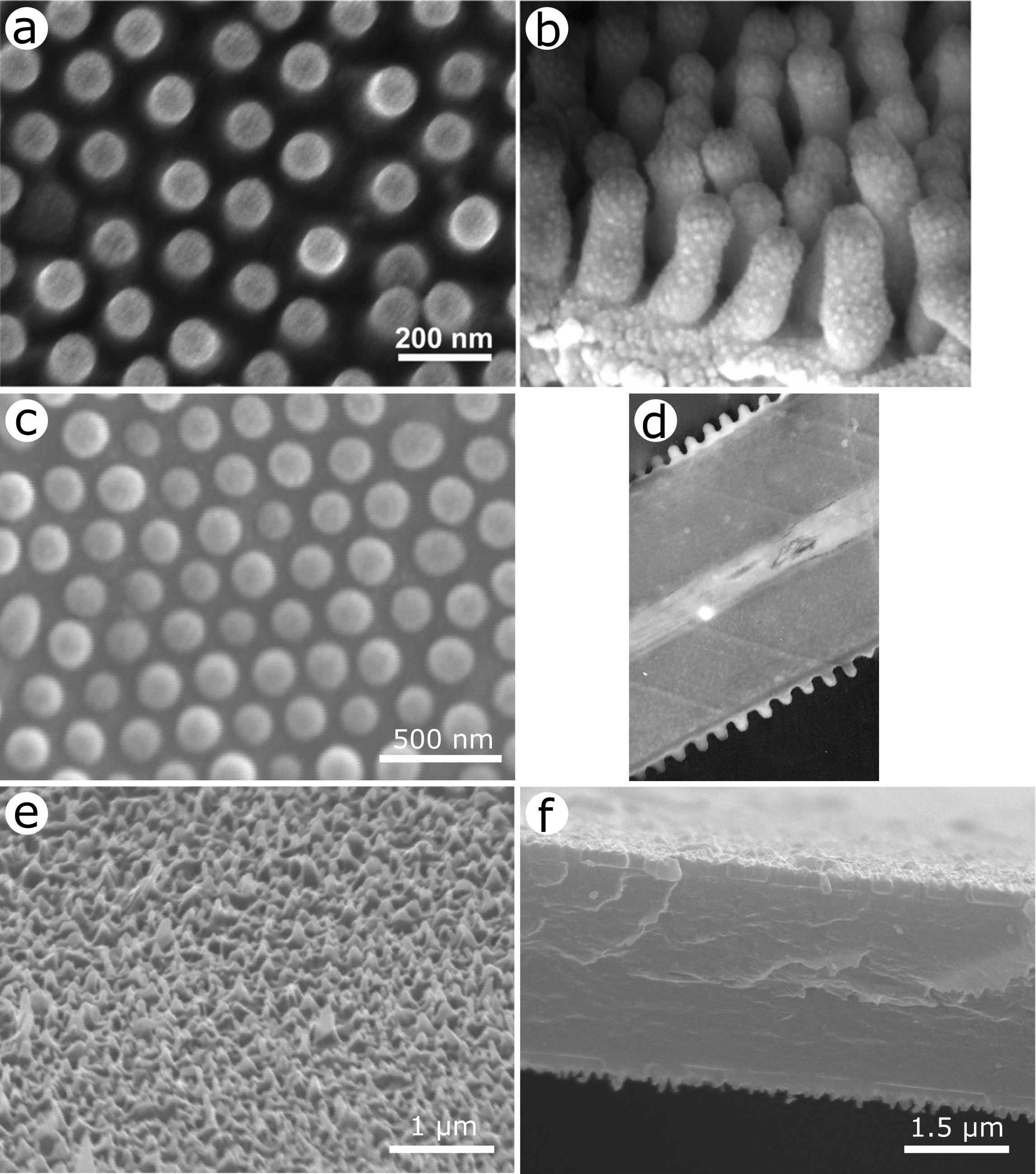
Fluorescence emission takes place in the integuments of several natural organisms including species from the animal classes Insecta, Aves, Amphibia, Reptilia and Mammalia as well as the plant kingdom [1, 2, 3, 4, 5, 6, 7, 8, 9, 10, 11, 12, 13, 14, 15, 16, 17, 18, 19]. This emission of light has been shown to play a role in the visual communication of some organisms, facilitating species recognition, mate selection, prey detection, camouflage and agonistic behaviour [7, 11]. Fluorescence results from the presence of fluorophores, such as papiliochrome II, biopterin, psittacofulvin, green fluorescent protein (GFP) within the biological integuments. Following the absorption of incident light (typically in the UV or the shorter-wavelength part of the visible range), these molecules emit light at a longer wavelength (usually in the visible), resulting from transitions between their electron states. In spite of the crucial role it is believed to play in nature, fluorescence in natural organisms remains under-investigated from optical, chemical and biological perspectives. One example is the transparent wings of insects from the order Hemiptera. For example, in both males and females of many cicadas, both pairs of wings are known for their anti-reflective properties [20, 21, 22, 23, 24] arising from electromagnetic impedance matching between the air and the wing material covered by quasi-periodic arrays of hexagonally close-packed protrusions (Fig. 1a,b). Similar structures also cover parts of the integument in other organisms such as the wings of several moths and butterflies (Order Lepidoptera), dragonflies and damselflies (Order Odonata) (Fig. 1c-f) [25, 26, 27, 28, 29, 30, 23, 31, 32, 33] and the corneas of some insect eyes [34, 35]. In all these examples, the basic building material of the insect tissues is chitin, a polysaccharide, overlaid by a thin epicuticle of wax. In cicadas, transmittance spectra of their wings have been measured to reach more than 90% and to peak at up to 98% over the range from 500 to 2500 nm [20, 21]. These anti-reflective properties are thought to be involved as part of the insects’ cryptic strategy and the nanostructured protrusions have been mimicked for the development of anti-reflective coatings for a full range of applications such as solar cells, screens, anti-glare glasses, light-sensitive detectors, telescopes and camera lenses [36, 37, 38]. In addition to this optical property, the transparent wings of insects are known to be hydrophobic [39, 40, 21, 41, 22, 23, 42] and some exhibit anti-bacterial behaviour [42, 43, 44, 45, 46]. In the case of cicadas, the surface morphology was found to influence jointly the hydrophobic and anti-reflective properties (Fig. 1) [22, 23]. The protrusions on the wing surface can be modelled as truncated cones covered by hemispheres. The cone shape affects the transparency and the hemispheres enhance the wing’s hydrophobicity. Despite these properties, which are relevant in many scientific fields ranging from physics and material science to chemistry and biology, fluorescence emission has so far not been reported in any of the 3,000 described species of the superfamily Cicadoidea. The phenomenon has however been investigated in several butterfly and moth species, in which the coloured wing scales are known to embed fluorophores such as papiliochrome II [47, 48, 3, 49, 6, 50]. Photonic structures present in these scales may mediate the associated light emission [51, 52, 53]. Fluorescence has also been described in the vein joints and the membrane of the transparent wings of damselflies and dragonflies [54, 55, 56, 57]. This emission was attributed to the presence of autofluorescent proteins such as resilin. Resilin proteins emit blue light (at about 400 nm) under incident UV light and were reported to decrease in concentration toward the distal regions of the wings (on both sides) [54, 55]. They give rise to low stiffness and high strain in the biological tissues, allowing passive wing deformations [58, 59]. We note that resilin fluorescence may often be incidental with little or no semiotic function. However, in the case of males of the red-winged damselfly Mnesarete pudica (Hagen in Selys, 1853), fluorescence emission from resilin-containing wings was demonstrated to have a role in signalling (e.g., in courtship and territorial behaviour) in combination with pigments and UV reflection, by modulating the visual appearance of the insect according to sex and age [60].
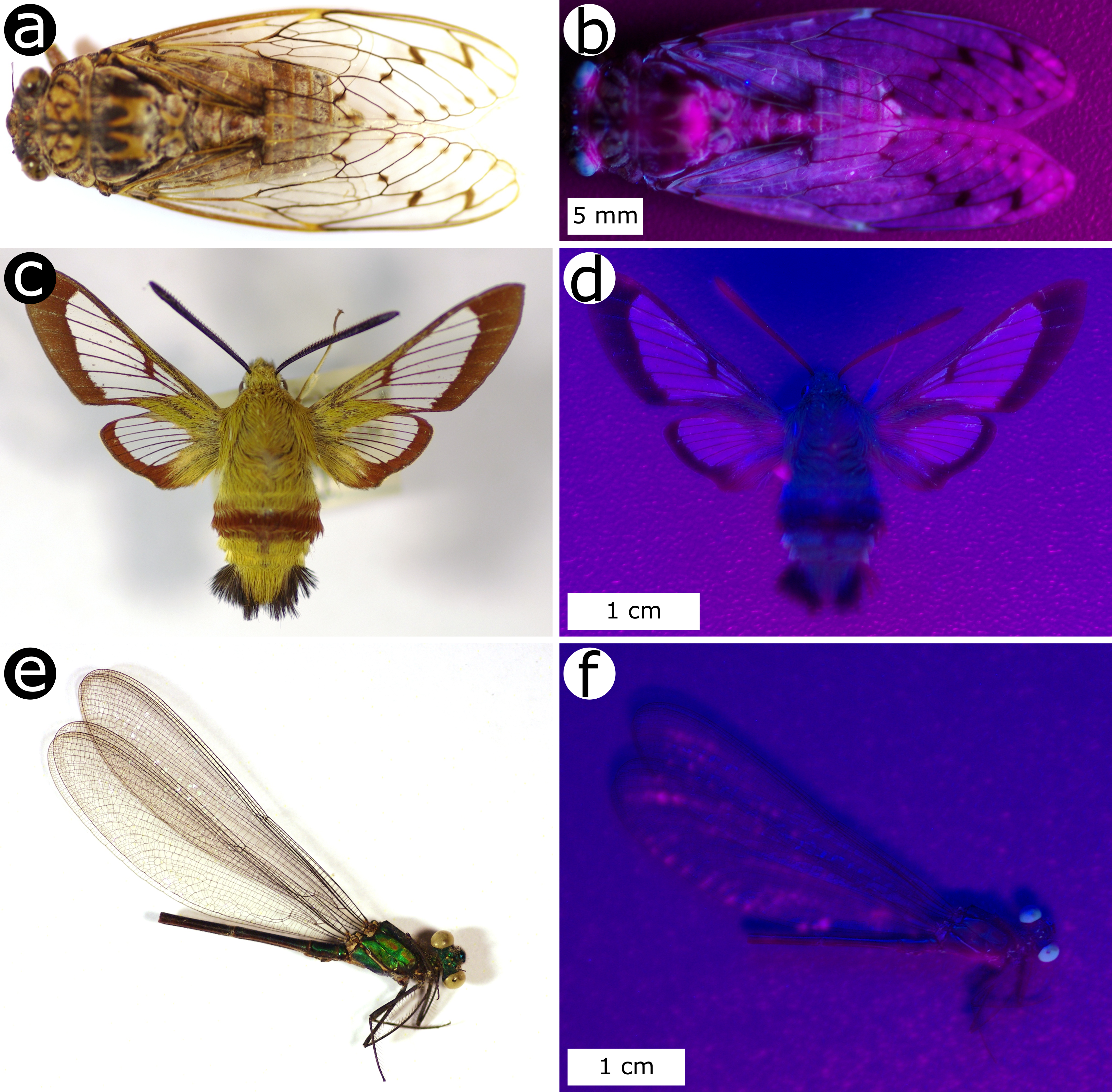
In this article, we characterised the fluorescence emission from the transparent wings of two species of cicadas, the grey cicada Cicada orni (Linnaeus, 1758) (Fig. 2a,b) and the common cicada Lyristes (Tibicen) plebejus (Scopoli, 1763), using both linear and nonlinear optical methods. We compared their properties with the fluorescence emission from two other insects exhibiting transparent wings, namely, Hemaris fuciformis (Linnaeus, 1758) commonly known as the broad-bordered bee hawk-moth (Fig. 2c,d) and the Bornean damselfly Vestalis amabilis Lieftinck, 1965 (Fig. 2e,f). These fluorescent behaviours were optically characterised by one- and multi-photon microscopy and spectrometry, allowing us to infer the symmetry of the material as well as the biological role of the fluorophores in the visual perception of insects and to explain the previously measured high absorption in the range 300-400 nm [22].
2 Materials and Methods
2.1 Sample collection
Adult specimens of C. orni and L. plebejus were collected in Fournès in the Gard Department (France) on the 22nd of June 2014 (by S.R. Mouchet and L. Dellieu), and V. amabilis was collected in swamp forest in Belait Division (Brunei Darussalam) in 1994 (by A.G. Orr). Adult specimens of H. fuciformis were bought from licensed vendors. V. amabilis was immersed for 24 hours in with acetone and air-dried. Other specimens were air-dried. No further sample preparation was necessary for the microscopy and spectrofluorimetry observations. All analyses were performed on the insects’ wings.
2.2 Optical and fluorescence microscopy
The sample wings were analysed using two different configurations of the microscope: a reflection mode (illumination with visible light and observation in the visible range) and a fluorescence mode (illumination with ultraviolet light and observation in the visible range). Optical microscopy analysis was performed using an Olympus BX61 (Tokyo, Japan) microscope, an Olympus XC50 camera and an Olympus BX-UCB visible light source (in reflection mode) or a Lumen Dynamics X-cite Series 120PCQ (Mississauga, Ontario, Canada) UV-lamp (in fluorescence mode).
2.3 Spectrofluorimetry
One photon fluorescence (1PF) emission spectra were recorded using a 450 W xenon lamp as steady state excitation source. The incident light formed a 45° angle with the normal direction to the sample surface and emitted light was detected at a 45° angle on the other side of the normal direction. The sample was excited at different excitation wavelengths: 320 nm, 340 nm, 360 nm, 380 nm, 400 nm, 420 nm and 440 nm. Time-resolved measurements of fluoresence emission were performed using a microF920H xenon Flashlamp light source operating at a frequency of 100 Hz with a pulse width of 4 µs and excitation wavelengths equal to 340 nm and 360 nm. These time-resolved measurements were performed at the wavelengths of the maximum of emission peak intensity. They were fitted to double exponential functions. These best fits allowed us to assess the decay time of the fluorescence emission.
2.4 Multiphoton microscopy
Multiphoton microscopy experiments were carried out with an Olympus (Münster, Germany) BX61 WI-FV1200-M system. The laser was a Spectra-Physics (Santa Clara, CA, USA) InSight DS+ laser (82-MHz repetition rate, 120-fs pulse width, p-polarised) at 800 nm, 900 nm, 1000 nm and 1100 nm fundamental wavelengths. The incident laser power was controlled by an achromatic half-wave plate and a s-oriented polariser directly after the laser. The laser beam was focused on the sample via either a 15X LMV objective (NA 0.3) or a 50X SLMPlan N objective (NA 0.35). The resulting signal was detected non-descanned in backwards reflection via a Hamamatsu R3896 photomultiplier tube. Different detection optics were used, depending on the excitation wavelength, in order to separate the two-photon fluorescence (2PF) and the Second Harmonic Generation (SHG) signal. At 1000 nm, a 525 LPXR dichroic mirror was used to remove the SHG from the 2PF, and an additional 500/10 bandpass filter cleared the SHG signal further. Similar filters were used for the other three fundamental wavelengths. The signal at each pixel was depicted in the micrographs as different intensities in false green (SHG) or false red (2PF) colours. A routine providing an extended depth field allowed us to obtain in-focus micrographs from multiple images recorded at different depths in steps of 4 µm. Samples were observed over several minutes of time, in order to assess the photostability.
2.5 Electron microscopy
Apart from two scanning electron microscopy (SEM) images taken from the literature [22], SEM observations were performed with a FEI (Hillsboro, OR) Nova 600 NanoLab Dual-Beam FIB/SEM microscope on small transparent regions of the wing membranes of H. fuciformis and A. cyanea. These samples were mounted onto SEM stubs with electrically conducting epoxy resin and sputter-coated with ca. 8 nm of gold palladium.
Transmission electron microscopy (TEM) imaging was carried on with cross-sections of transparent regions of H. fuciformis’s wings using a JEOL (Tokyo, Japan) 100S TEM instrument. The samples were fixed in 3% glutaraldehyde at 21°C for 2 hours and consequently rinsed in sodium cacodylate buffer before being fixed in 1% osmic acid in buffer for 1 hour. The samples were then block-stained in 2% aqueous uranyl acetate for 1 hour, dehydrated through an acetone series (ending in 100% acetone) and embedded in Spurr resin [61]. After microtoming, cross-sections were stained with lead citrate.
3 Results and Discussion
Both C. orni and L. plebejus cicada species have brown-grey bodies and transparent wings (Fig. 2) with very low light reflection [20, 21, 22, 23, 24]. The bodies and wings are approximately 25 mm and 30-35 mm long, respectively. Like most insects including moths and damselflies, the wings of cicadas are divided by veins (Fig. 3a-d) appearing as darker structures forming the structurally flexible frame supporting of the wing membranes, while the transparent parts are membranes that form the main aerodynamic surface of the wing [58, 62].
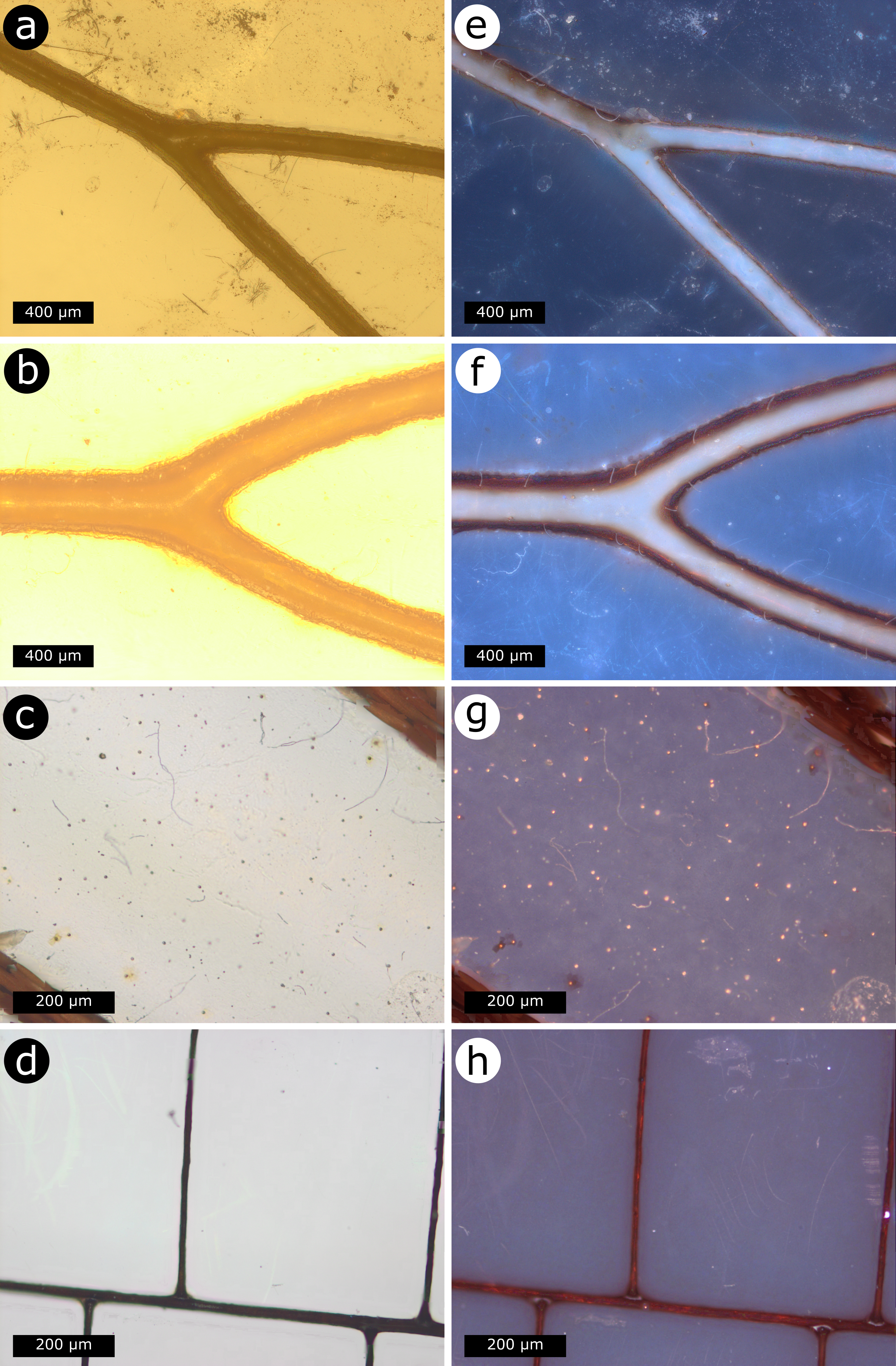
Upon illumination with UV light (Fig. 2), the wings still appear transparent with a bluish hue. Fluorescent microscopy observations indicate that both the transparent membrane and the veins of the wings fluoresce (Fig. 3). The latter are clearly brighter in the case of the two investigated cicada species (Fig. 3e,f), indicating that they might contain many more fluorescent molecules than the membrane. Chitin is known to give rise to autofluorescence, with a very low quantum yield [63]. In addition, autofluorescent proteins such as resilin are known to be at the origin of blue hue fluorescence emission from damselfly and dragonfly species [54, 55, 56, 60]. Hence both chitin and resilin are likely to play a role in the fluorescence emission observed from investigated cicada and moth species (Fig. 3e-g). It was previously reported that the extinction coefficient -namely, the imaginary part of the complex refractive index- of the wing material of C. orni exhibits a double peak in the UV range 200-300 nm, reaching ca. and , with a non-negligible component in the range 300-400 nm [22]. Similarly, Azofeifa et al. measured peaks at 280 nm and 325 nm in the extinction coefficient for chitin samples from the exoskeleton of fresh Pacific white shrimps Litopenaeus (formerly Penaeus) vannamei (Boone, 1931) [64]. Our results show that light absorption in the wings of insects such as cicadas within the range 300-400 nm gives rise to fluorescence emission.
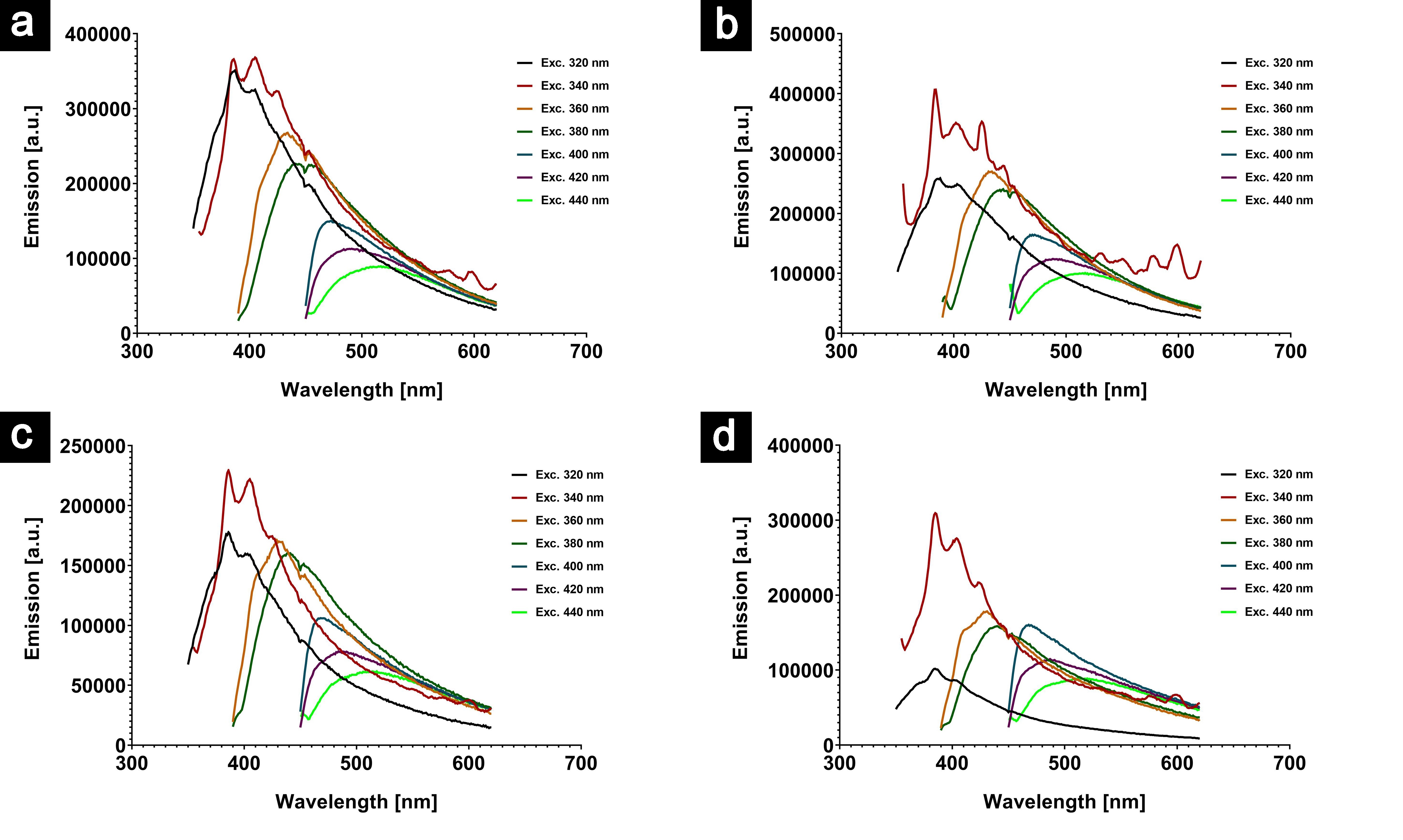
In all four investigated insect wings, fluorescence emission single peaks were measured upon excitation with wavelengths ranging from 320 nm to 440 nm (Fig. 3). A 340-nm excitation wavelength appears to give rise to the most intense emission. For instance, the measured emission spectra display peaks at about 410-420 nm with a 360-nm excitation wavelength. The presence of a single peak in the emission spectra suggests that there is likely only a single type of fluorophore within the insect wings. The presence of mixtures of several fluorophores is less likely as this would imply emission spectra with multiple peaks. The measured emission peaks are similar to the fluorescence emission measured by Chuang et al. from the wings of the damselfly Ischnura senegalensis (Rambur, 1842) [57]. Despite the fact that our observations are significantly different from the fluorescence response from M. pudica male neotropical damselfly (i.e., emission at ca. 650 nm upon excitation at 405 nm), the wings of which are known to contain resilin, the emission peaks we measured are in agreement with the narrow emission band of resilin located at ca. 415 nm [56, 65].
In addition, the emission peak widths and positions strongly depend on the excitation wavelength (Fig. 4). The peak position shifts from ca. 380 nm to ca. 490 nm as the excitation wavelength is increased from 320 nm to 440 nm. This dependence is similar to the previously reported one for males of the beetle Hoplia coerulea (Drury, 1773) [14]. It suggests that the fluorophores exhibit complex dynamics with several excited states. In complex organic fluorophores, several excited states are likely related to exciton resonances (i.e., energy transfers between different donor and acceptor groups within the same molecule) [66, 67, 14].
In order to understand better the radiative response in the linear regime, we performed time-resolved measurement of the emitted fluorescence intensity (Fig. 5).
| Species | Excitation | Emission | Decay time |
|---|---|---|---|
| Wavelength [nm] | Wavelength [nm] | [ps] | |
| Cicada orni | 340 | 385 | = 1271.23 |
| = 8933.32 | |||
| 340 | 403 | = 1309.57 | |
| = 9272.42 | |||
| 360 | 388 | = 1192.96 | |
| = 8508.55 | |||
| 360 | 406 | = 1237.31 | |
| = 8831.80 | |||
| Lyristes (Tibicen) plebejus | 340 | 384 | = 1372.48 |
| = 8288.30 | |||
| 340 | 402 | = 1184.63 | |
| = 7688.22 | |||
| 360 | 387 | = 1107.87 | |
| = 7708.26 | |||
| 360 | 406 | = 1328.55 | |
| = 8299.55 | |||
| Hemaris fuciformis | 340 | 384 | = 1322.55 |
| = 8249.07 | |||
| 340 | 401 | = 1278.86 | |
| = 8149.58 | |||
| 340 | 425 | = 1313.99 | |
| = 8491.21 | |||
| 360 | 406 | = 1298.20 | |
| = 7997.02 | |||
| 360 | 424 | = 1284.24 | |
| = 8262.70 | |||
| 360 | 451 | = 1245.23 | |
| = 8126.60 | |||
| Vestalis amabilis | 340 | 384 | = 1235.50 |
| = 7578.67 | |||
| 340 | 402 | = 1374.48 | |
| = 8060.57 | |||
| 340 | 425 | = 1338.49 | |
| = 8220.30 | |||
| 360 | 387 | = 1326.02 | |
| = 8226.46 | |||
| 360 | 406 | = 1203.75 | |
| = 7596.25 | |||
| 360 | 450 | = 1119.60 | |
| = 7841.73 |
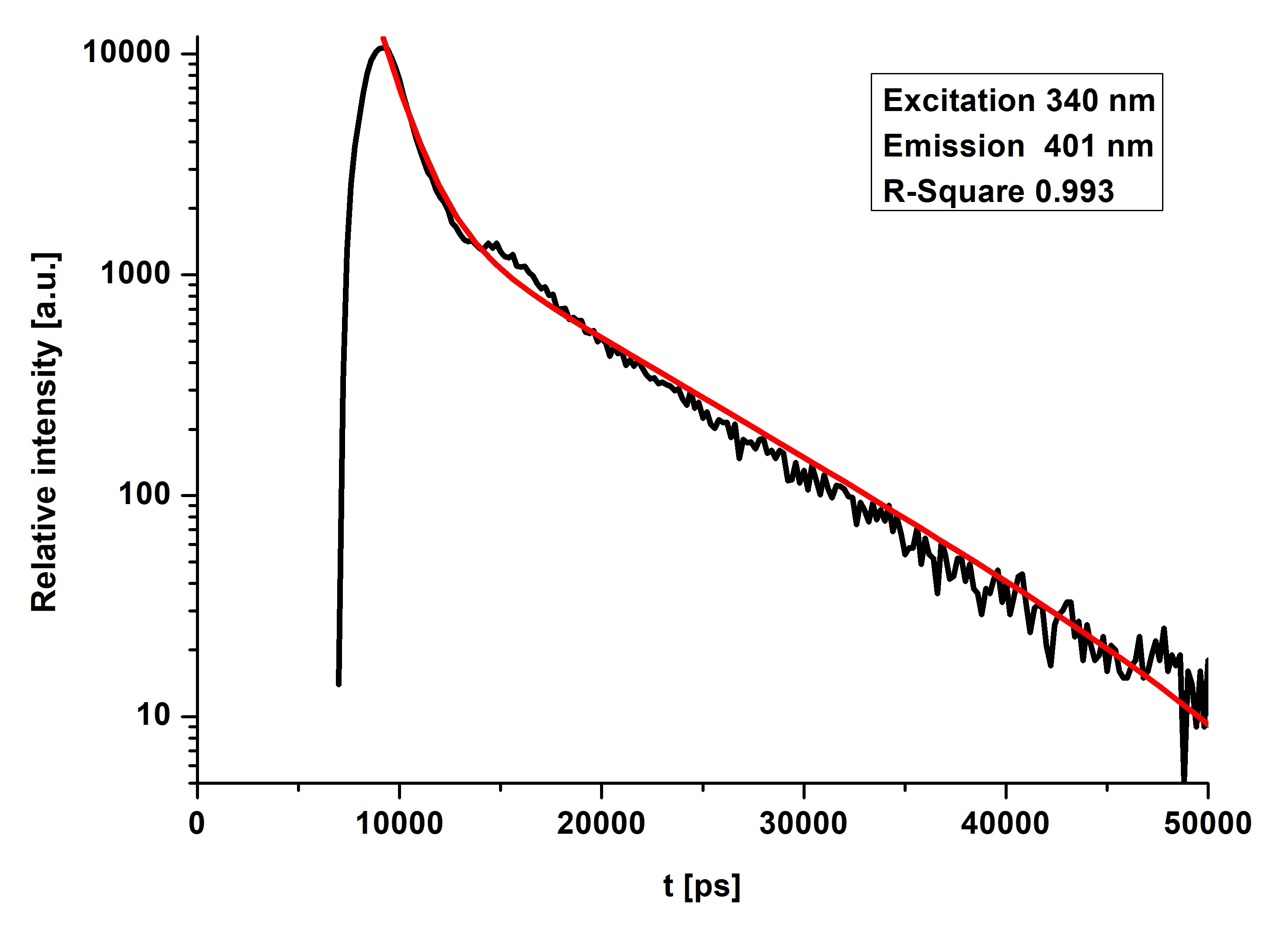
In all cases (Table 1), the best fits to double exponential functions gave rise to a long decay time (of ca. 7-8 ns) and a short decay time (of ca. 1.1-1.3 ns), whatever the excitation and detection wavelengths were. This also suggests that only one type of fluorophores is present in the wings of each insect. The long decay time is related to the radiative decay of the fluorophores, whereas the short one is related to nonradiative decay [68]. If we assume that measured fluorescent signals originate from the same molecule (namely, resilin), the discrepancy observed in the emission spectra and decay time among the investigated samples could be attributed to differences in the chemical environment from one species to another.
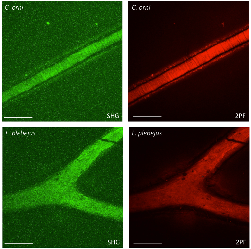
In addition, both C. orni and L. plebejus were probed at fundamental wavelengths ranging from 800 nm to 1100 nm, and showed significant SHG and 2PF signal response over all this wavelength range (see Fig. 6 at a 1000-nm wavelength). At any of these selected fundamental wavelengths, the veins clearly appear in both SHG and 2PF observations. The difference in signals between the veins and the membrane (in 1PF, 2PF and SHG) is likely due to the significant difference in composition [54, 55, 62]: the veins of cicadas would show a much higher concentration in chitin and resilin. The former is known to give rise to an SHG signal [69]. Additionally, the veins are much thicker than the membrane, leading to an increased amount of material. Optical second-order behaviours such as SHG are known to take place exclusively in non-centrosymmetric media [70]. The detected SHG signal implies that the veins are materially organised in a non-centrosymmetric way.
From a visual point of view, the biological significance of fluorescence emission in the transparent wings of insects depends on the total number of photons emitted or scattered from the wings and transmitted through the wings from their backgrounds: many cicadas are known to be mainly active during the hottest hours of summer days, when sunlight is very intense but also at twilight and during the night. Although the measured fluorescence emission ranges match the range of both scotopic and photopic photoreceptors of many insects [71, 72, 73, 74], it is hard to conclude that the measured 1PF signal plays any role in cicadas’ behaviour since the quantum yield of natural fluorophores are generally low [4, 5, 12], specifically the one of chitin [63], and the ratio of UV light in both sun- and moonlight is very limited with respect to the one of visible light (for instance, ca. 6% of sunlight at sea level lies in the UV range, whereas about 50% is visible) [75]. The role of this fluorescence emission may possibly be a by-product of another biological function such as photoprotection due to the increased absorption of UV by the fluorophores (for example, when the wings are at rest over the insect’s body in the case of cicadas) and incidental property of resilin that enhances wing flexibility.
4 Conclusion
Transparent wings of two cicada species, namely C. orni, L. plebejus, as well as the broad-bordered bee hawk-moth H. fuciformis, were demonstrated to emit light under incident UV light by fluorescence decay. All of them showed similar 1PF properties, as their wings were excited in the 320-440 nm range and emitted in the range 380-490 nm. They were compared to the transparent wings of the damselfly V. amabilis, which showed similar fluorescent behaviours. Dameselflies fluoresecence emission is reported to arise from substances such as resilin embedded in their tissues. Additionally, the wings of both cicada species and more specifically their veins showed strong SHG and 2PF signals, which are possibly due to the higher concentration of resilin or chitin in the veins with respect to the membrane that makes up most of the wing. The nonlinear optical characteristics of the cicadas’ wings gave us more insight into the wing structure, indicating that multiphoton microscopy add valuable information while characterising insect integuments.
Acknowledgments SRM was supported by the Belgian National Fund for Scientific Research (FRS-FNRS) (91400/1.B.309.18F), the Maturation Fund of the Walloon Region, and a BEWARE Fellowship of the Walloon Region (Convention n°2110034), as a Postdoctoral Researcher. DM acknowledges KU Leuven Postdoctoral Mandate Internal Funds (PDM) for a Postdoctoral felowship (PDM/20/092). TV acknowledges financial support from the Hercules Foundation. BK acknowledges financial support from the "Action de Recherche Concertée" (BIOSTRUCT project‚ No.10/15-033) of UNamur, from Nanoscale Quantum Optics COST-MP1403 action and from FRS-FNRS; Interuniversity Attraction Pole: Photonics@be (P7-35, Belgian Science Policy Office).
References
- [1] M. Pavan and M. Vachon. Sur l’existence d’une substance fluorescente dans les téguments des Scorpions (Arachnides). C. R. Acad. Sci, 239:1700–1702, 1954.
- [2] K. Tani, F. Watari, M. Uo, and M. Morita. Fluorescent properties of porcelain-restored teeth and their discrimination. Mater. Trans., 45:1010–1014, 2004.
- [3] P. Vukusic and I. Hooper. Directionally controlled fluorescence emission in butterflies. Science, 310:1151, 2005.
- [4] Analía Iriel and María Gabriela Lagorio. Is the flower fluorescence relevant in biocommunication? Naturwissenschaften, 97(10):915–924, Oct 2010.
- [5] Analía Iriel and María Gabriela Lagorio. Implications of reflectance and fluorescence of rhododendron indicum flowers in biosignaling. Photochem. Photobiol. Sci., 9:342–348, 2010.
- [6] Victoria L. Welch, Eloise Van Hooijdonk, Nurit Intrater, and Jean-Pol Vigneron. Fluorescence in insects. Proc. SPIE, 8480:848004, 2012.
- [7] M. Gabriela Lagorio, Gabriela. B. Cordon, and Analia Iriel. Reviewing the relevance of fluorescence in biological systems. Photochem. Photobiol. Sci., 14:1538–1559, 2015.
- [8] David F. Gruber, Jean P. Gaffney, Shaadi Mehr, Rob DeSalle, John S. Sparks, Jelena Platisa, and Vincent A. Pieribone. Adaptive evolution of eel fluorescent proteins from fatty acid binding proteins produces bright fluorescence in the marine environment. PLoS ONE, 10(11):e0140972, 2015.
- [9] David F. Gruber, Ellis R. Loew, Dimitri D. Deheyn, Derya Akkaynak, Jean P. Gaffney, W. Leo Smith, Matthew P. Davis, Jennifer H. Stern, Vincent A. Pieribone, and John S. Sparks. Biofluorescence in catsharks (Scyliorhinidae): Fundamental description and relevance for elasmobranch visual ecology. Scientific Reports, 6(24751):1–16, 2016.
- [10] Sébastien R. Mouchet, Michaël Lobet, Branko Kolaric, Anna M. Kaczmarek, Rik Van Deun, Peter Vukusic, Olivier Deparis, and Eloise Van Hooijdonk. Controlled fluorescence in a beetle’s photonic structure and its sensitivity to environmentally induced changes. Proceedings of the Royal Society of London B: Biological Sciences, 283(1845), 2016.
- [11] Justin Marshall and Sonke Johnsen. Fluorescence as a means of colour signal enhancement. Philosophical Transactions of the Royal Society of London B: Biological Sciences, 372(1724), 2017.
- [12] C. Taboada, A. E. Brunetti, F. N. Pedron, F. Carnevale Neto, D. A. Estrin, S. E. Bari, L. B. Chemes, N. Peporine Lopes, M. G. Lagorio, and J. Faivovich. Naturally occurring fluorescence in frogs. Proc. Natl Acad. Sci. USA, 114(14):3672–3677, 2017.
- [13] Pablo Deschepper, Bert Jonckheere, and Jasper Matthys. A light in the dark: The discovery of another fluorescent frog in the Costa Rican rainforests. Wilderness & Environmental Medicine, 29(3):421–422, 2018.
- [14] Sébastien R. Mouchet, Anna M. Kaczmarek, Dimitrije Mara, Rik Van Deun, and Pete Vukusic. Colour and fluorescence emission of Euchroea auripigmenta beetle. Proceedings of SPIE, 10965:72 – 82, 2019.
- [15] Mathieu Ladouce, Tarek Barakat, Bao-Lian Su, Olivier Deparis, and Sébastien R. Mouchet. Scattering of ultraviolet light by avian eggshells. Faraday Discuss., 223:63–80, 2020.
- [16] Marina Mohd Top, Chong Leong Puan, Ming-Feng Chuang, Siti N. Othman, and Amaël Borzée. First record of ultraviolet fluorescence in the bent-toed gecko Cyrtodactylus quadrivirgatus taylor, 1962 (Gekkonidae: Sauria). Herpetology Notes, 13:211–212, 2020.
- [17] Anna C. Croce. Light and autofluorescence, multitasking features in living organisms. Photochem, 1(2):67–124, 2021.
- [18] Linda Reinhold. Mammals with fluorescent fur: Observations from the wet tropics. North Queensland Naturalist, 51:1–8, 2021.
- [19] Séverine Toussaint, Jasper Ponstein, Mathieu Thoury, Rémi Métivier, Daniela Kalthoff, Benoît Habermeyer, Roger Guilard, Steffen Bock, Peter Mortensen, Sverre Sandberg, Pierre Gueriau, and Eli Amson. Fur glowing under uv: a widespread consequence of porphyrin accumulation in mammals. Research Square, 2021.
- [20] P R Stoddart, P J Cadusch, T M Boyce, R M Erasmus, and J D Comins. Optical properties of chitin: surface-enhanced raman scattering substrates based on antireflection structures on cicada wings. Nanotechnology, 17(3):680, 2006.
- [21] Mingxia Sun, Aiping Liang, Yongmei Zheng, Gregory S Watson, and Jolanta A Watson. A study of the anti-reflection efficiency of natural nano-arrays of varying sizes. Bioinsp. Biomim., 6(2):026003, 2011.
- [22] Louis Dellieu, Michaël Sarrazin, Priscilla Simonis, Olivier Deparis, and Jean-Pol Vigneron. A two-in-one superhydrophobic and anti-reflective nanodevice in the grey cicada Cicada orni (Hemiptera). Journal of Applied Physics, 116(2):024701, 2014.
- [23] Olivier Deparis, Sébastien R. Mouchet, Louis Dellieu, Jean-François Colomer, and Michaël Sarrazin. Nanostructured surfaces: Bioinspiration for transparency, coloration and wettability. Materials Today: Proceedings, 1S:122–129, 2014.
- [24] Charlotte Verstraete, Sébastien R Mouchet, Thierry Verbiest, and Branko Kolaric. Linear and nonlinear optical effects in biophotonic structures using classical and nonclassical light. Journal of biophotonics, page e201800262, 2019.
- [25] Akihiro Yoshida, Mayumi Motoyama, Akinori Kosaku, and Kiyoshi Miyamoto. Nanoprotuberance array in the transparent wing of a hawkmoth, Cephonodes hylas. Zoological science, 13(4):525–526, 1996.
- [26] Akihiro Yoshida, Mayumi Motoyama, Akinori Kosaku, and Kiyoshi Miyamoto. Antireflective nanoprotuberance array in the transparent wing of a hawkmoth, Cephonodes hylas. Zoological science, 14(5):737–741, 1997.
- [27] Akihiro Yoshida. Antireflection of the butterfly and moth wings through microstructure. Forma, 17(2):75–89, 2002.
- [28] P. Vukusic and J. R. Sambles. Photonic structures in biology. Nature, 424(6950):852–855, 2003.
- [29] I. R. Hooper, P. Vukusic, and R. J. Wootton. Detailed optical study of the transparent wing membranes of the dragonfly Aeshna cyanea. Opt. Express, 14(11):4891–4897, 2006.
- [30] Olivier Deparis, Nadia Khuzayim, Andrew Parker, and Jean-Pol Vigneron. Assessment of the antireflection property of moth wings by three-dimensional transfer-matrix optical simulations. Phys. Rev. E, 79:041910, Apr 2009.
- [31] D.G. Stavenga. Thin film and multilayer optics cause structural colors of many insects and birds. Mater. Today Proc., 1S:109–121, 2014.
- [32] R.H. Siddique, G. Gomard, and H. Hölscher. The role of random nanostructures for the omnidirectional anti-reflection properties of the glasswing butterfly. Nat. Commun., 6:6909, 2015.
- [33] Sébastien R. Mouchet, Charlotte Verstraete, Dimitrije Mara, Stijn Van Cleuvenbergen, Ewan D. Finlayson, Rik Van Deun, Olivier Deparis, Thierry Verbiest, Bjorn Maes, Pete Vukusic, and Branko Kolaric. Nonlinear optical spectroscopy and two-photon excited fluorescence spectroscopy reveal the excited states of fluorophores embedded in a beetle’s elytra. Interface Focus, 9(1):20180052, 2019.
- [34] Carl Gustaf Bernhard. The insect corneal nipple array. a biological, broad-band impedance transformer that acts as an antireflection coating. Acta physiol. scand., 63(243):1–79, 1965.
- [35] D.G. Stavenga, S. Foletti, G. Palasantzas, and K. Arikawa. Light on the moth-eye corneal nipple array of butterflies. Proceedings of the Royal Society of London B: Biological Sciences, 273(1587):661–667, 2006.
- [36] Guoyong Xie, Guoming Zhang, Feng Lin, Jin Zhang, Zhongfan Liu, and Shichen Mu. The fabrication of subwavelength anti-reflective nanostructures using a bio-template. Nanotechnology, 19(9):095605, 2008.
- [37] Z.W. Han, Z. Wang, X.M. Feng, B. Li, Z.Z. Mu, J.Q. Zhang, S.C. Niun, and L.Q. Ren. Antireflective surface inspired from biology: A review. Biosurface and Biotribology, 2:137–150, 2016.
- [38] Imran Zada, Wang Zhang, Peng Sun, Muhammad Imtiaz, Waseem Abbas, and Di Zhang. Multifunctional, angle dependent antireflection, and hydrophilic properties of SiO2 inspired by nano-scale structures of cicada wings. Applied Physics Letters, 111(15):153701, 2017.
- [39] Guoming Zhang, Jin Zhang, Guoyong Xie, Zhongfan Liu, and Huibo Shao. Cicada wings: A stamp from nature for nanoimprint lithography. Small, 2(12):1440–1443, 2006.
- [40] Mingxia Sun, Gregory S. Watson, Yongmei Zheng, Jolanta A. Watson, and Aiping Liang. Wetting properties on nanostructured surfaces of cicada wings. Journal of Experimental Biology, 212(19):3148–3155, 2009.
- [41] Katrina M. Wisdom, Jolanta A. Watson, Xiaopeng Qu, Fangjie Liu, Gregory S. Watson, and Chuan-Hua Chen. Self-cleaning of superhydrophobic surfaces by self-propelled jumping condensate. Proceedings of the National Academy of Sciences, 110(20):7992–7997, 2013.
- [42] Jessica Román-Kustas, Jacob B. Hoffman, Julian H. Reed, Andrew E. Gonsalves, Junho Oh, Longnan Li, Sungmin Hong, Kyoo D. Jo, Catherine E. Dana, Nenad Miljkovic, Donald M. Cropek, and Marianne Alleyne. Molecular and topographical organization: Influence on cicada wing wettability and bactericidal properties. Advanced Materials Interfaces, 7(10):2000112, 2020.
- [43] Elena P Ivanova, Jafar Hasan, Hayden K Webb, Vi Khanh Truong, Gregory S Watson, Jolanta A Watson, Vladimir A Baulin, Sergey Pogodin, James Y Wang, Mark J Tobin, Christian Löbbe, and Russell J. Crawford. Natural bactericidal surfaces: mechanical rupture of Pseudomonas aeruginosa cells by cicada wings. Small, 8(16):2489–2494, 2012.
- [44] Elena P. Ivanova, Jafar Hasan, Hayden K. Webb, Gediminas Gervinskas, Saulius Juodkazis, Vi Khanh Truong, Alex H.F. Wu, Robert N. Lamb, Vladimir A. Baulin, Gregory S. Watson, Jolanta A. Watson, David E. Mainwaring, and Russell J. Crawford. Bactericidal activity of black silicon. Nature Communications, 4:2838, nov 2013.
- [45] Ting Diu, Nilofar Faruqui, Terje Sjoöström, Baptiste Lamarre, Howard F. Jenkinson, Bo Su, and Maxim G. Ryadnov. Cicada-inspired cell-instructive nanopatterned arrays. Scientific Reports, 4:7122, 2014.
- [46] S. M. Kelleher, O. Habimana, J. Lawler, B. O’Reilly, S. Daniels, E. Casey, and A. Cowley. Cicada wing surface topography: An investigation into the bactericidal properties of nanostructural features. ACS Applied Materials & Interfaces, 8(24):14966–14974, 2016.
- [47] EA Cockayne. I. The distribution of fluorescent pigments in Lepidoptera. Transactions of the Royal Entomological Society of London, 72(1-2):1–19, 1924.
- [48] Kinya Kumazawa, Shingo Tanaka, Keishi Negita, and Hiroshi Tabata. Fluorescence from wing of Morpho sulkowskyi butterfly. Jpn J. Appl. Phys., 33(1;4A):2119–2122, 1994.
- [49] T. M. Trzeciak, B. D. Wilts, D. G. Stavenga, and P. Vukusic. Variable multilayer reflection together with long-pass filtering pigment determines the wing coloration of papilionid butterflies of the nireus group. Opt. Express, 20(8):8877–8890, 2012.
- [50] B. D. Wilts, T. M. Trzeciak, P. Vukusic, and D. G. Stavenga. Papiliochrome II pigment reduces the angle dependency of structural wing colouration in nireus group papilionids. J. Exp. Biol., 215(5):796––805, 2012.
- [51] Jean-Pol Vigneron, Krisztián Kertész, Zofia Vértesy, Marie Rassart, Virginie Lousse, Zsolt Bálint, and László P. Biró. Correlated diffraction and fluorescence in the backscattering iridescence of the male butterfly Troides magellanus (papilionidae). Phys. Rev. E, 78:021903, 2008.
- [52] Eloise Van Hooijdonk, Carlos Barthou, Jean-Pol Vigneron, and Serge Berthier. Detailed experimental analysis of the structural fluorescence in the butterfly Morpho sulkowskyi (Nymphalidae). J. Nanophotonics, 5(1):053525, 2011.
- [53] Eloise Van Hooijdonk, Carlos Barthou, Jean-Pol Vigneron, and Serge Berthier. Angular dependence of structural fluorescent emission from the scales of the male butterfly Troïdes magellanus (Papilionidae). Journal of the Optical Society of America B, 29(5):1104–1111, 2012.
- [54] Stanislav N. Gorb. Serial elastic elements in the damselfly wing: Mobile vein joints contain resilin. Naturwissenschaften, 86(11):552–555, Nov 1999.
- [55] Esther Appel and Stanislav N Gorb. Resilin-bearing wing vein joints in the dragonfly Epiophlebia superstes. Bioinspiration & Biomimetics, 6(4):046006, 2011.
- [56] Esther Appel, Lars Heepe, Chung-Ping Lin, and Stanislav N. Gorb. Ultrastructure of dragonfly wing veins: composite structure of fibrous material supplemented by resilin. Journal of Anatomy, 227(4):561–582, 2015.
- [57] Chin-Jung Chuang, Cheng-Der Liu, Ranjit A. Patil, Chi-Chung Wu, Yao-Chih Chang, Chih-Wen Peng, Ting-Kwuan Chao, Je-Wen Liou, Yung Liou, and Yuan-Ron Ma. Impact of cuticle photoluminescence on the color morphism of a male damselfly Ischnura senegalensis (Rambur, 1842). Scientific Reports, 6:38051, 2016.
- [58] Christopher M. Elvin, Andrew G. Carr, Mickey G. Huson, Jane M. Maxwell, Roger D. Pearson, Tony Vuocolo, Nancy E. Liyou, Darren C. C. Wong, David J. Merritt, and Nicholas E Dixon. Synthesis and properties of crosslinked recombinant pro-resilin. Nature, 437:999–1002, 2005.
- [59] Malcolm Burrows, Stephen R. Shaw, and Gregory P. Sutton. Resilin and chitinous cuticle form a composite structure for energy storage in jumping by froghopper insects. BMC Biology, 6(41):1–16, 2008.
- [60] Rhainer Guillermo-Ferreira, Eralci M. Therézio, Marcelo H. Gehlen, Pitágoras C. Bispo, and Alexandre Marletta. The role of wing pigmentation, UV and fluorescence as signals in a neotropical damselfly. Journal of Insect Behavior, 27(1):67–80, Jan 2014.
- [61] Arthur R. Spurr. A low-viscosity epoxy resin embedding medium for electron microscopy. Journal of Ultrastructure Research, 26(1):31–43, 1969.
- [62] John D Gullion and Terry Gullion. Solid-state NMR study of the cicada wing. The Journal of Physical Chemistry B, 121(32):7646–7651, 2017.
- [63] Thien An Phung Hai and Ryuichi Sugimoto. Fluorescence control of chitin and chitosan fabricated via surface functionalization using direct oxidative polymerization. RSC Adv., 8:7005–7013, 2018.
- [64] Daniel E. Azofeifa, Humberto J. Arguedas, and William E. Vargas. Optical properties of chitin and chitosan biopolymers with application to structural color analysis. Optical Materials, 35:175–183, 2012.
- [65] Svend Olav Andersen. Characterization of a new type of cross-linkage in resilin, a rubber-like protein. Biochimica et Biophysica Acta, 69:249 – 262, 1963.
- [66] J.R. Lakowicz. Principles of Fluorescence Spectroscopy. Kluwer Academic/Plenum Publishers, New York, NY, 1999.
- [67] C. Ramanan, C. Hoon Kim, T. J. Marks, and Wasielewski Mr. Excitation energy transfer within covalent tetrahedral perylenediimide tetramers and their intermolecular aggregates. J. Phys. Chem. C, 118:16941–16950, 2014.
- [68] B. Kolaric and R. A. L. Vallée. Dynamics and stability of dna mechano-nanostructures: Energy-transfer investigations. Journal of Physical Chemistry C, 114(3):1430–1435, 2010.
- [69] Mihailo D Rabasović, Dejan V Pantelić, Branislav M Jelenković, Srećko B Ćurčić, Maja S Rabasović, Maja D Vrbica, Vladimir M Lazović, Božidar PM Ćurčić, and Aleksandar J Krmpot. Nonlinear microscopy of chitin and chitinous structures: a case study of two cave-dwelling insects. Journal of Biomedical Optics, 20(1):016010, 2015.
- [70] Thierry Verbiest, Koen Clays, and Vincent Rodriguez. Second-order nonlinear optical characterization techniques: an introduction. CRC press, 2009.
- [71] Dagmar Peitsch, Andrea Fietz, Horst Hertel, John de Souza, Dora Fix Ventura, and Randolf Menzel. The spectral input systems of hymenopteran insects and their receptor-based colour vision. Journal of Comparative Physiology A, 170(1):23–40, Jan 1992.
- [72] Doekele G. Stavenga. Colour in the eyes of insects. Journal of Comparative Physiology A, 188(5):337–348, Jun 2002.
- [73] Doekele G. Stavenga. Surface colors of insects: Wings and eyes. In Functional Surfaces in Biology: Little Structures with Big Effects Volume 1, pages 285–306, Dordrecht, 2009. Springer Netherlands.
- [74] Klaus Lunau. Visual ecology of flies with particular reference to colour vision and colour preferences. Journal of Comparative Physiology A, 200(6):497–512, Jun 2014.
- [75] J. Moan. Visible light and uv radiation. In A. Brune, R. Hellborg, B.R.R. Persson, and R. Pääkkönen, editors, Radiation at home, outdoors and in the workplace, pages 69–85, Oslo, 2001. Scandinavian Science Publisher.